Embed Size (px)
DESCRIPTION
The Respiratory System. Why do we need oxygen?. Answer: We need it to make energy through aerobic respiration!. Organs of the Respiratory System. Nose Pharynx Larynx Trachea Bronchi Lungs – alveoli. Functions of the Respiratory System. - PowerPoint PPT Presentation
Citation preview
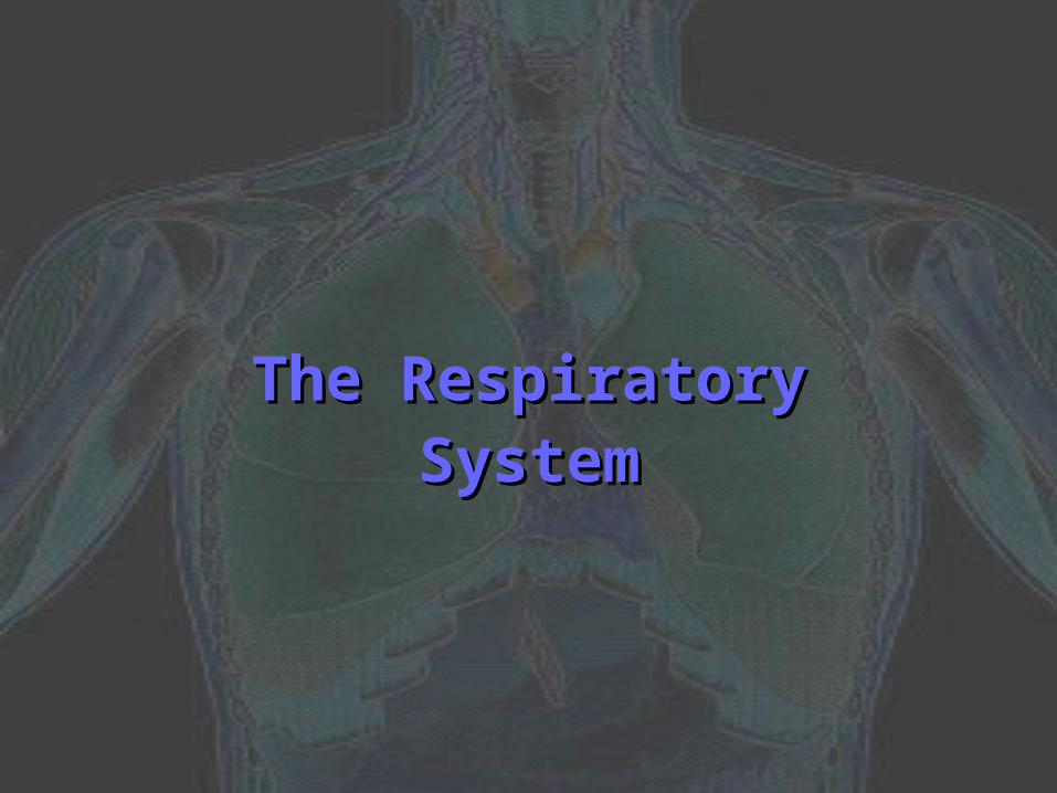
The Respiratory SystemThe Respiratory System
Why do we need oxygen?Why do we need oxygen?
Answer: We need it to make energy Answer: We need it to make energy through aerobic respiration!through aerobic respiration!
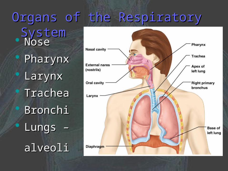
Organs of the Respiratory SystemOrgans of the Respiratory System
NoseNose
PharynxPharynx
LarynxLarynx
TracheaTrachea
BronchiBronchi
Lungs – Lungs – alveolialveoli

Functions of the Respiratory SystemFunctions of the Respiratory System
1.1. Gas exchange between the blood and Gas exchange between the blood and external environmentexternal environment
2.2. Purification, warming, and humidifying Purification, warming, and humidifying of incoming airof incoming air
3.3. Provides olfactory sensations to the Provides olfactory sensations to the brain for sense of smellbrain for sense of smell
4.4. Produces sounds for communicationProduces sounds for communication
Divisions of the Respiratory TractDivisions of the Respiratory Tract
1.1. Conduction portion: nasal cavity Conduction portion: nasal cavity larger bronchioles (cleans and warms larger bronchioles (cleans and warms the air)the air)
2.2. Respiratory portion: smallest Respiratory portion: smallest bronchioles bronchioles alveoli (permits gas alveoli (permits gas exchange)exchange)

Upper Respiratory TractUpper Respiratory Tract
NoseNose Olfactory receptors are located on the Olfactory receptors are located on the
superior surfacesuperior surface
Mucosa: lines the nasal cavity; moistens Mucosa: lines the nasal cavity; moistens air and traps incoming foreign particlesair and traps incoming foreign particles
Conchae: projections of the nasal cavity; Conchae: projections of the nasal cavity; increases air turbulence within the nasal increases air turbulence within the nasal cavitycavity
Sinuses: cavities within bones surrounding Sinuses: cavities within bones surrounding the nasal cavity; lighten the skull, resonate the nasal cavity; lighten the skull, resonate sounds for speech, and produce mucussounds for speech, and produce mucus
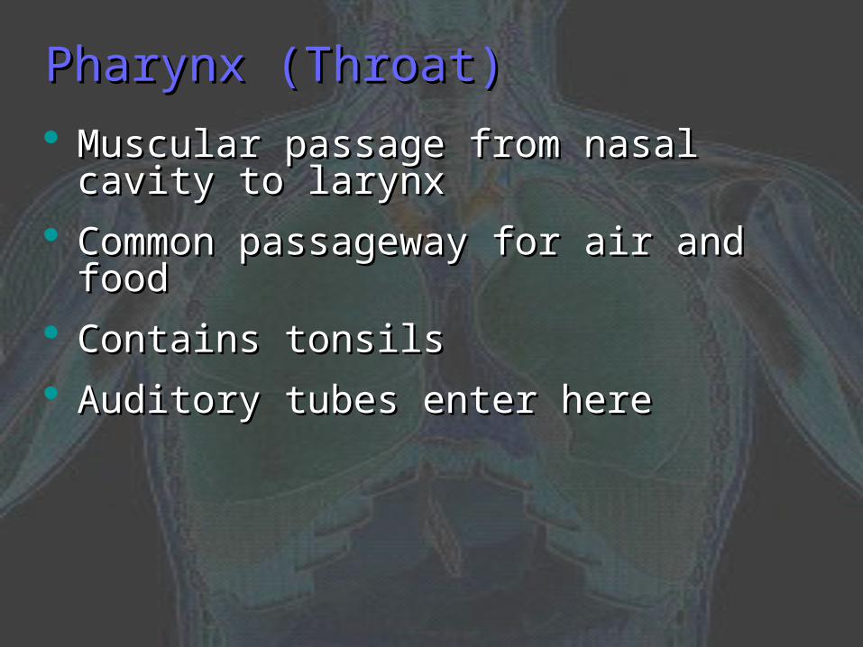
Pharynx (Throat)Pharynx (Throat)
Muscular passage from nasal cavity to Muscular passage from nasal cavity to larynxlarynx
Common passageway for air and foodCommon passageway for air and food
Contains tonsilsContains tonsils
Auditory tubes enter hereAuditory tubes enter here
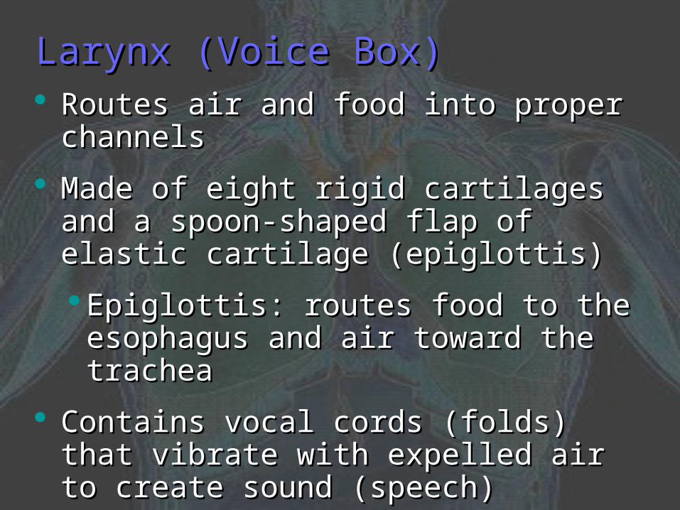
Larynx (Voice Box)Larynx (Voice Box) Routes air and food into proper channelsRoutes air and food into proper channels
Made of eight rigid cartilages and a spoon-Made of eight rigid cartilages and a spoon-shaped flap of elastic cartilage (epiglottis) shaped flap of elastic cartilage (epiglottis)
Epiglottis: routes food to the esophagus Epiglottis: routes food to the esophagus and air toward the tracheaand air toward the trachea
Contains vocal cords (folds) that vibrate Contains vocal cords (folds) that vibrate with expelled air to create sound (speech)with expelled air to create sound (speech)
Glottis: opening between vocal cordsGlottis: opening between vocal cords
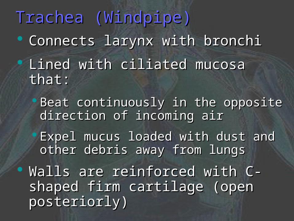
Trachea (Windpipe)Trachea (Windpipe) Connects larynx with bronchiConnects larynx with bronchi
Lined with ciliated mucosa that:Lined with ciliated mucosa that:
Beat continuously in the opposite direction of Beat continuously in the opposite direction of incoming airincoming air
Expel mucus loaded with dust and other Expel mucus loaded with dust and other debris away from lungsdebris away from lungs
Walls are reinforced with C-shaped firm Walls are reinforced with C-shaped firm cartilage (open posteriorly)cartilage (open posteriorly)
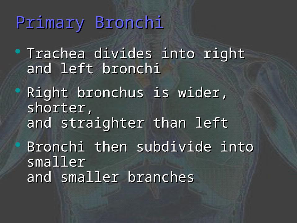
Primary BronchiPrimary Bronchi
Trachea divides into right and left Trachea divides into right and left bronchibronchi
Right bronchus is wider, shorter, Right bronchus is wider, shorter, and straighter than leftand straighter than left
Bronchi then subdivide into smaller Bronchi then subdivide into smaller and smaller branchesand smaller branches

LungsLungs Occupy most of the thoracic cavityOccupy most of the thoracic cavity
Apex is near the clavicle (collar bone) and Apex is near the clavicle (collar bone) and base rests on the diaphragmbase rests on the diaphragm
Each lung is divided into lobes by fissures:Each lung is divided into lobes by fissures:
a.a. Left lung – two lobesLeft lung – two lobes
b.b. Right lung – three lobesRight lung – three lobes
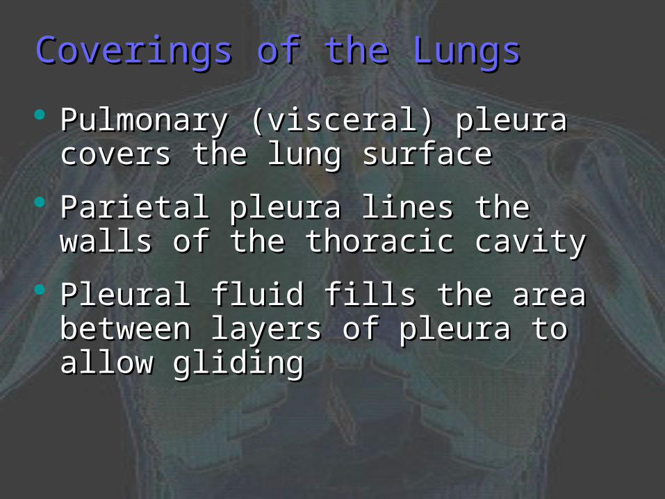
Coverings of the LungsCoverings of the Lungs
Pulmonary (visceral) pleura covers the Pulmonary (visceral) pleura covers the lung surfacelung surface
Parietal pleura lines the walls of the Parietal pleura lines the walls of the thoracic cavitythoracic cavity
Pleural fluid fills the area between layers Pleural fluid fills the area between layers of pleura to allow glidingof pleura to allow gliding
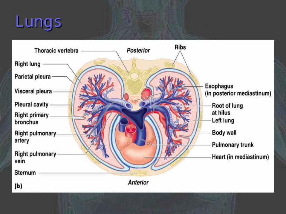
LungsLungs
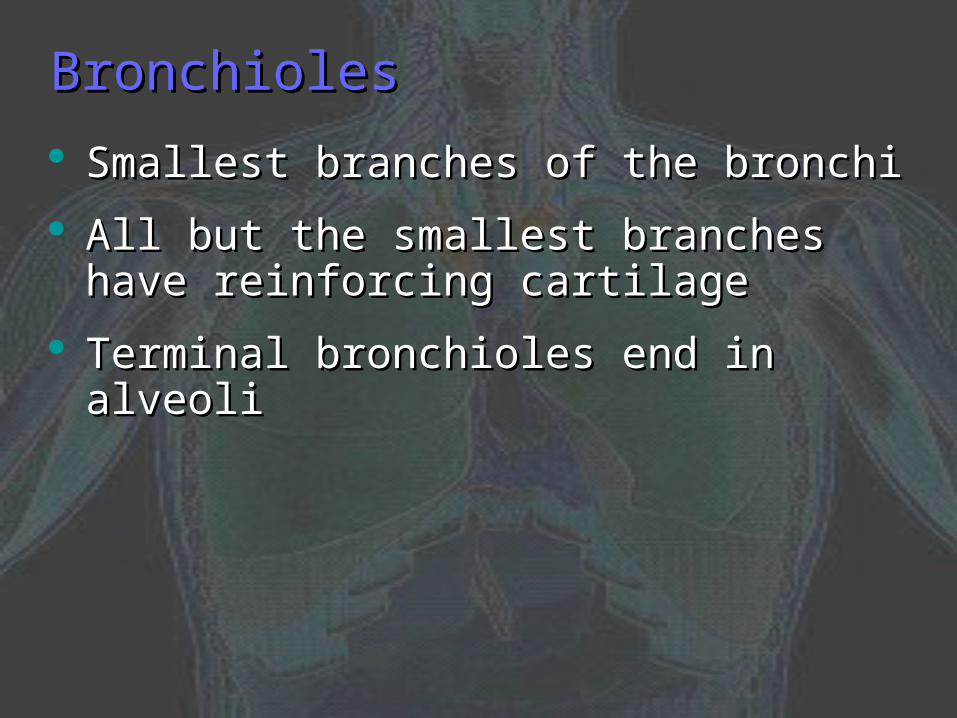
BronchiolesBronchioles
Smallest branches of the bronchiSmallest branches of the bronchi
All but the smallest branches have All but the smallest branches have reinforcing cartilagereinforcing cartilage
Terminal bronchioles end in alveoliTerminal bronchioles end in alveoli

AlveoliAlveoli Consist of a duct, a sac, and the alveolusConsist of a duct, a sac, and the alveolus
Gas exchange takes place within the Gas exchange takes place within the alveoli in the respiratory membranealveoli in the respiratory membrane
Respiratory membrane (air-blood barrier)Respiratory membrane (air-blood barrier)
Thin epithelium lines alveolar wallsThin epithelium lines alveolar walls
Pulmonary capillaries cover external Pulmonary capillaries cover external surfaces of alveolisurfaces of alveoli

Bronchioles and AlveoliBronchioles and Alveoli

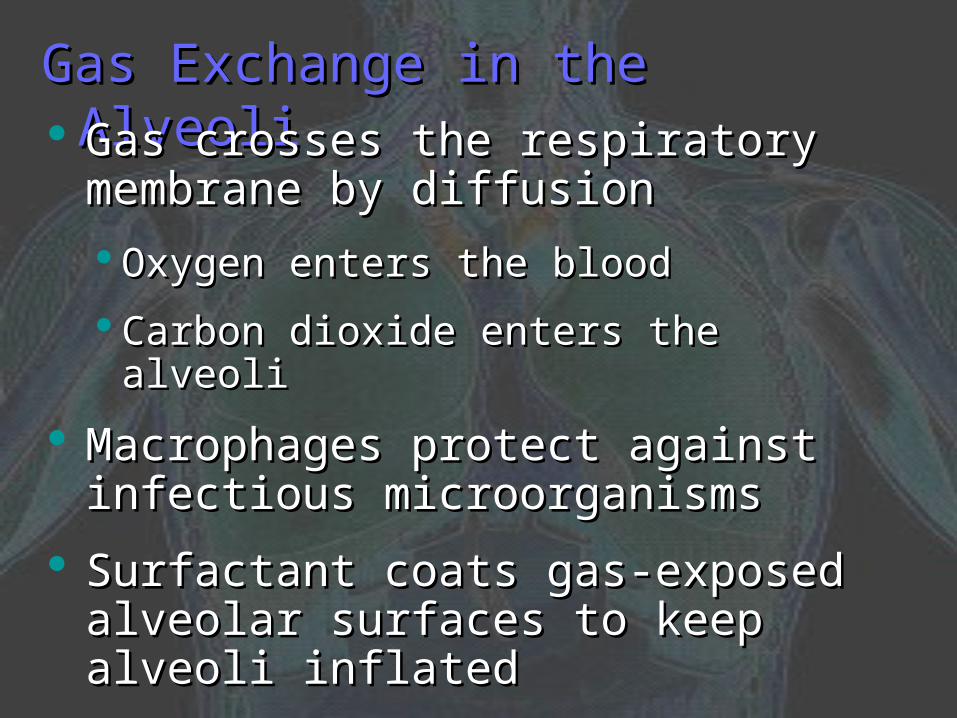
Gas Exchange in the AlveoliGas Exchange in the Alveoli Gas crosses the respiratory membrane Gas crosses the respiratory membrane
by diffusionby diffusion
Oxygen enters the bloodOxygen enters the blood
Carbon dioxide enters the alveoliCarbon dioxide enters the alveoli
Macrophages protect against infectious Macrophages protect against infectious microorganismsmicroorganisms
Surfactant coats gas-exposed alveolar Surfactant coats gas-exposed alveolar surfaces to keep alveoli inflatedsurfaces to keep alveoli inflated
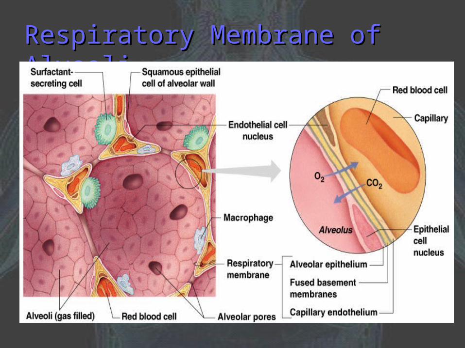
Respiratory Membrane of AlveoliRespiratory Membrane of Alveoli

Events of RespirationEvents of Respiration Pulmonary ventilation: moving air in and out Pulmonary ventilation: moving air in and out
of the lungsof the lungs
External respiration: gas exchange between External respiration: gas exchange between pulmonary capillaries and alveolipulmonary capillaries and alveoli
Internal respiration: gas exchange between Internal respiration: gas exchange between blood and tissue cells in systemic capillariesblood and tissue cells in systemic capillaries
Mechanics of Breathing Mechanics of Breathing (Pulmonary Ventilation)(Pulmonary Ventilation)
Two phases:Two phases:
1.1. Inspiration/inhalation – flow of air into lungInspiration/inhalation – flow of air into lung
2.2. Expiration/exhalation – air leaving lungExpiration/exhalation – air leaving lung
Inspiration/inhalationInspiration/inhalation
Diaphragm contracts (flattens), external Diaphragm contracts (flattens), external intercostals contract raising the rib cageintercostals contract raising the rib cage
Volume of the thoracic cavity increasesVolume of the thoracic cavity increases
Pressure in thoracic cavity decreasesPressure in thoracic cavity decreases
Air is pulled into the lungs (from high to Air is pulled into the lungs (from high to low pressure)low pressure)

Inspiration/inhalationInspiration/inhalation

Expiration/exhalationExpiration/exhalation Largely a passive process which depends Largely a passive process which depends
on natural lung elasticityon natural lung elasticity
Diaphragm relaxes (pushes up), external Diaphragm relaxes (pushes up), external intercostals relax depressing the rib cageintercostals relax depressing the rib cage
Air is pushed out of the lungsAir is pushed out of the lungs
Forced expiration can occur mostly by Forced expiration can occur mostly by contracting internal intercostal muscles to contracting internal intercostal muscles to depress the rib cage furtherdepress the rib cage further

Expiration/exhalationExpiration/exhalation

Respiratory Volumes and CapacitiesRespiratory Volumes and Capacities Tidal volume (TV) is normal breathing and Tidal volume (TV) is normal breathing and
moves about 500 mL of air with each breath moves about 500 mL of air with each breath
Many factors affect respiratory capacity:Many factors affect respiratory capacity:
1.1. A person’s sizeA person’s size
2.2. SexSex
3.3. AgeAge
4.4. Physical conditionPhysical condition
Residual volume (RV) of air: amount of air Residual volume (RV) of air: amount of air remaining in the lungs after normal remaining in the lungs after normal exhalation (~1200 mL)exhalation (~1200 mL)
Respiratory Volumes and CapacitiesRespiratory Volumes and Capacities
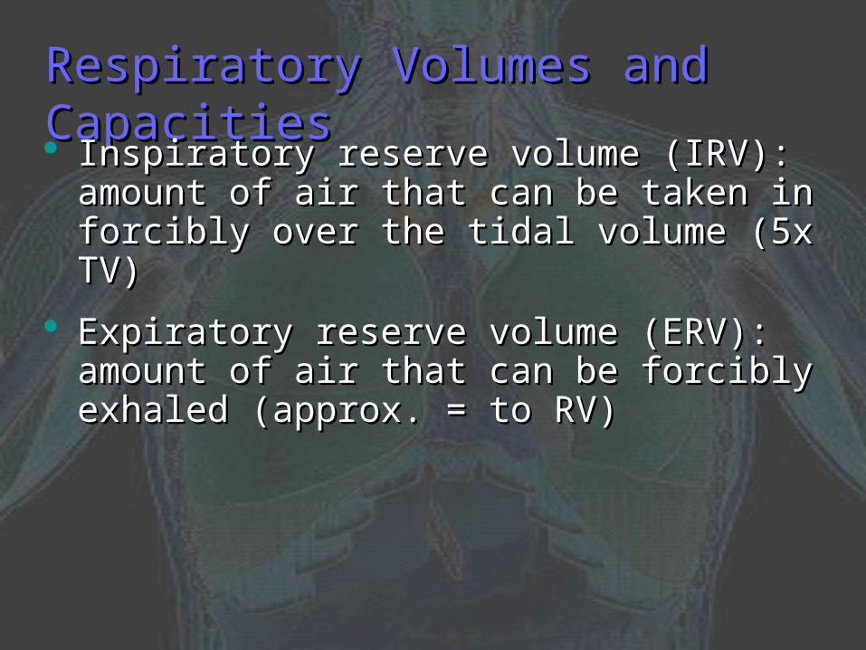
Inspiratory reserve volume (IRV): amount of air Inspiratory reserve volume (IRV): amount of air that can be taken in forcibly over the tidal that can be taken in forcibly over the tidal volume (5x TV)volume (5x TV)
Expiratory reserve volume (ERV): amount of air Expiratory reserve volume (ERV): amount of air that can be forcibly exhaled (approx. = to RV)that can be forcibly exhaled (approx. = to RV)

Respiratory Volumes and CapacitiesRespiratory Volumes and Capacities Vital capacity (VC): the total amount of Vital capacity (VC): the total amount of
exchangeable airexchangeable air
Vital capacity = TV + IRV + ERV Vital capacity = TV + IRV + ERV
Dead space volume: air that remains in Dead space volume: air that remains in conducting zone and never reaches alveoli conducting zone and never reaches alveoli (150 mL)(150 mL)
Functional volume: air that actually Functional volume: air that actually reaches the respiratory zone (350 mL)reaches the respiratory zone (350 mL)

Respiratory Volumes and CapacitiesRespiratory Volumes and Capacities

Gas Transport in the BloodGas Transport in the Blood1.1. Oxygen transport in the bloodOxygen transport in the blood
Inside red blood cells attached to hemoglobin Inside red blood cells attached to hemoglobin
A small amount is carried dissolved in the A small amount is carried dissolved in the plasmaplasma
2.2. Carbon dioxide transport in the bloodCarbon dioxide transport in the blood
Most is transported in the plasma as bicarbonate Most is transported in the plasma as bicarbonate ion (HCOion (HCO33
––))
A small amount is carried inside red blood cells A small amount is carried inside red blood cells on hemoglobin, but at different binding sites than on hemoglobin, but at different binding sites than those of oxygenthose of oxygen
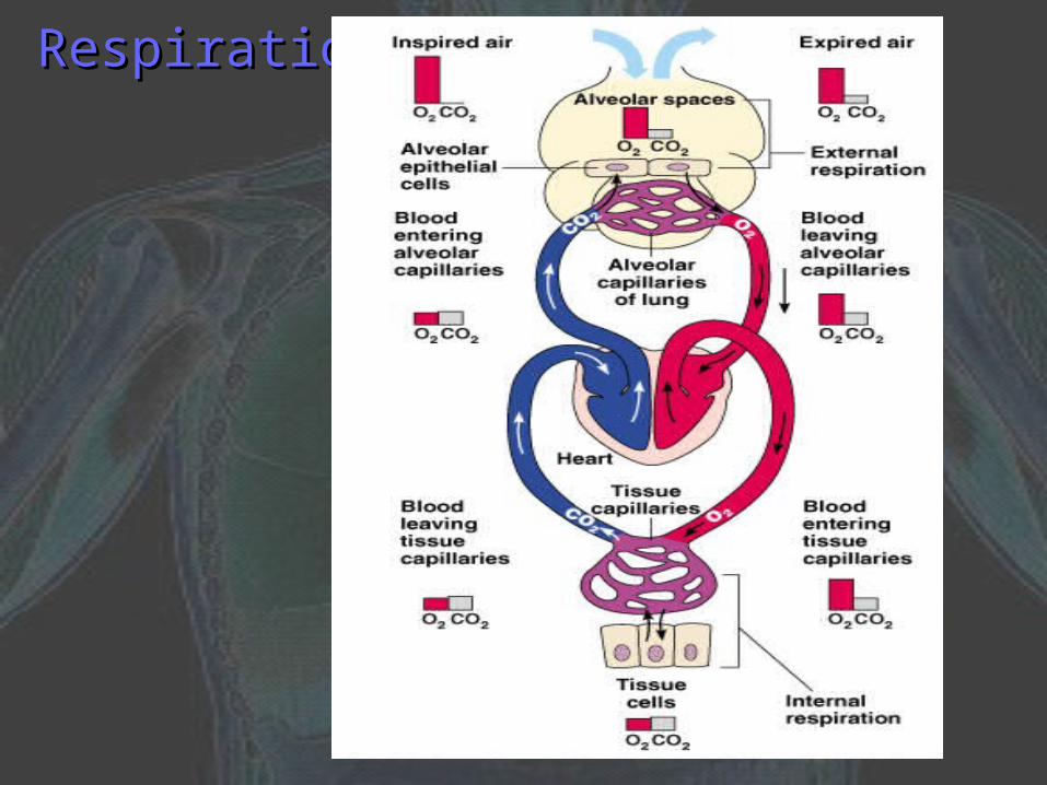
RespirationRespiration

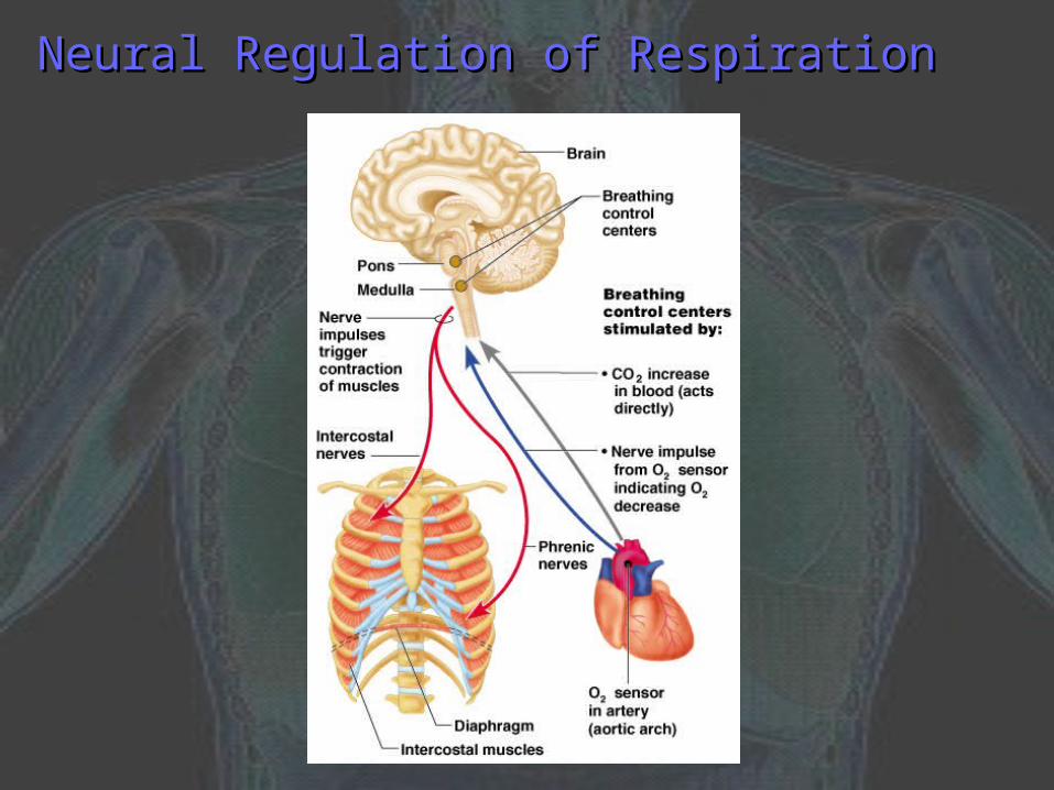
Neural Regulation of RespirationNeural Regulation of Respiration
Activity of respiratory muscles is transmitted Activity of respiratory muscles is transmitted to the brain by the phrenic and intercostal to the brain by the phrenic and intercostal nervesnerves
Phrenic nerve controls the diaphragmPhrenic nerve controls the diaphragm
Neural centers that control rate and depth are Neural centers that control rate and depth are located in the medullalocated in the medulla
The pons appears to smooth out respiratory The pons appears to smooth out respiratory raterate
Normal respiratory rate is 12–15 respirations Normal respiratory rate is 12–15 respirations per minuteper minute
Neural Regulation of RespirationNeural Regulation of Respiration
Chemical Regulation of RespirationChemical Regulation of Respiration1.1. Carbon dioxide levelsCarbon dioxide levels
Level of carbon dioxide in the blood is the main Level of carbon dioxide in the blood is the main regulatory chemical for respirationregulatory chemical for respiration
Increased carbon dioxide increases respirationIncreased carbon dioxide increases respiration
Changes in carbon dioxide act directly on the Changes in carbon dioxide act directly on the medulla oblongatamedulla oblongata
2.2. Oxygen levelsOxygen levels
Changes in oxygen concentration in the blood are Changes in oxygen concentration in the blood are detected by chemoreceptors in the aorta and detected by chemoreceptors in the aorta and carotid arterycarotid artery
Information is sent to the medulla oblongataInformation is sent to the medulla oblongata
Other Factors Influencing Other Factors Influencing Respiratory Rate and DepthRespiratory Rate and Depth
Physical factors:Physical factors:
1.1. Increased body temperatureIncreased body temperature
2.2. ExerciseExercise
3.3. TalkingTalking
4.4. CoughingCoughing
Volition (conscious control)Volition (conscious control)
Emotional factorsEmotional factors
Effects of Aging on the Respiratory SystemEffects of Aging on the Respiratory System
Elasticity of lungs decreasesElasticity of lungs decreases
Vital capacity decreasesVital capacity decreases
Blood oxygen levels decreaseBlood oxygen levels decrease
Respiratory rate often increasesRespiratory rate often increases
Stimulating effects of carbon dioxide Stimulating effects of carbon dioxide decreasesdecreases
More risks of respiratory tract infectionMore risks of respiratory tract infection

Diagnostic Tests for the Respiratory SystemDiagnostic Tests for the Respiratory System
Spirometry: measures how much and how Spirometry: measures how much and how quickly a person can move air out of the lungsquickly a person can move air out of the lungs
Pulse-oximetry: non-invasive measure of the Pulse-oximetry: non-invasive measure of the oxygen saturation of the bloodoxygen saturation of the blood
Arterial blood gas: measures the amount of Arterial blood gas: measures the amount of oxygen and carbon dioxide in a blood sampleoxygen and carbon dioxide in a blood sample
Stress test: lung function is measured during Stress test: lung function is measured during exerciseexercise
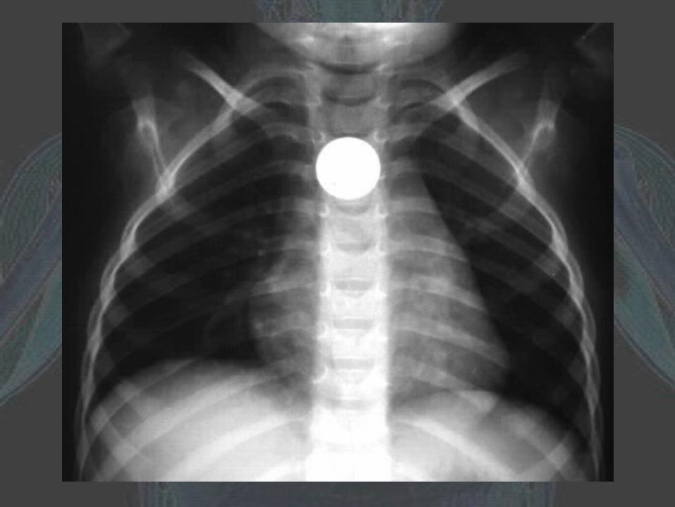
Chest x-ray: x-ray of the thorax while holding Chest x-ray: x-ray of the thorax while holding breathbreath


![Respiratory system roadmap.pptx [Repaired] - Loginanatomical-sciences.health.wits.ac.za/roadmaps/Respiratory system... · DIVISION OF THE RESPIRATORY SYSTEM CONDUCTING PORTION Nasal](https://img.pdfslide.us/doc/110x75/5a78c3d87f8b9ae6228c9db0/respiratory-system-repaired-loginanatomical-scienceshealthwitsaczaroadmapsrespiratory.jpg)