Embed Size (px)
Citation preview

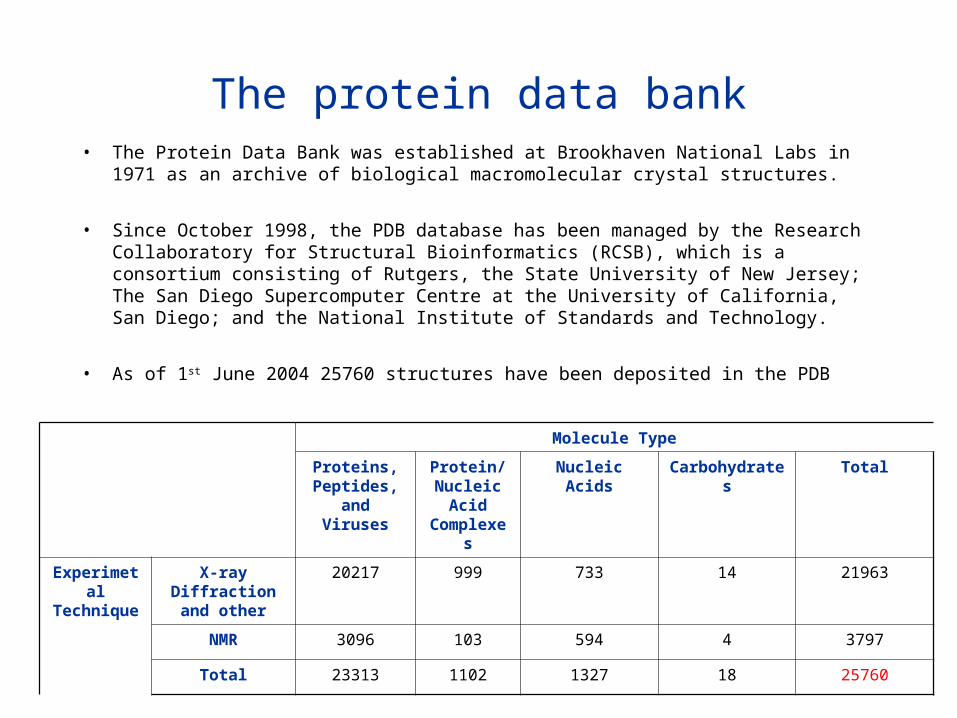
The protein data bank• The Protein Data Bank was established at Brookhaven National Labs in 1971 as an
archive of biological macromolecular crystal structures.
• Since October 1998, the PDB database has been managed by the Research Collaboratory for Structural Bioinformatics (RCSB), which is a consortium consisting of Rutgers, the State University of New Jersey; The San Diego Supercomputer Centre at the University of California, San Diego; and the National Institute of Standards and Technology.
• As of 1st June 2004 25760 structures have been deposited in the PDB
Molecule Type
Proteins, Peptides, and
Viruses
Protein/Nucleic
Acid Complexes
Nucleic Acids Carbohydrates Total
Experimetal Technique
X-ray Diffraction and other
20217 999 733 14 21963
NMR 3096 103 594 4 3797
Total 23313 1102 1327 18 25760
PDB (http://www.pdb.org/)
• The PDB archive contains macromolecular structure data on proteins, nucleic acids, protein-nucleic acid complexes, and viruses. Files in its holdings are deposited by the international user community and maintained by the RCSB PDB staff. Approximately 50-100 new structures are deposited each week. They are annotated by RCSB and released upon the depositor's specifications. PDB data is freely available worldwide.
• A variety of information associated with each structure is available, including sequence details, atomic coordinates, crystallization conditions, 3-D structure neighbours computed using various methods, derived geometric data, structure factors, 3-D images, and a variety of links to other resources.
• Information on structures can be retrieved from the main PDB Web site at http://www.pdb.org/, or one of its mirror sites. Structure files can also be obtained through the main FTP site at ftp://ftp.rcsb.org/ or one of its mirrors.
Theoretical Models
• The PDB separated theoretical model coordinate files from the main archive beginning July 1, 2002. Since that date, the main archive has consisted of structures determined using experimental methods only. Theoretical models are only available for download from the PDB FTP site as follows:
– All theoretical models (current and obsolete) are kept in a separate location in the FTP archive (pub/pdb/data/structures/models/current, pub/pdb/data/structures/models/obsolete)
– Model index files (authors.idx and titles.idx) and a FASTA file (model_seqres.txt) are available at pub/pdb/data/structures/models/index.
– A simple search interface for theoretical models is available http://www.rcsb.org/pdb/cgi/models.cgi. Queries from any other search interface do not return model entries (except for direct lookups by PDB ID).
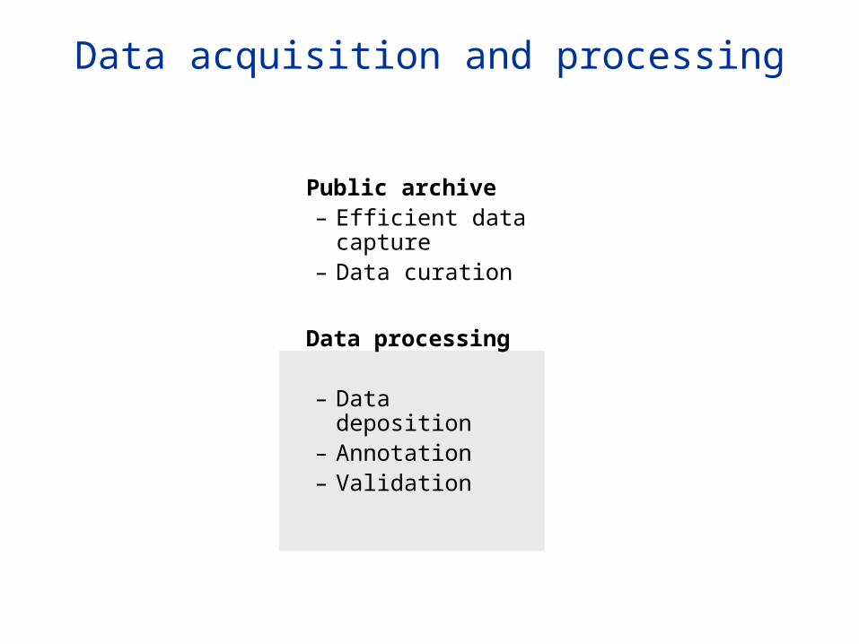
Data acquisition and processing
Public archive– Efficient data
capture– Data curation
Data processing
– Data deposition– Annotation– Validation
PDB ID
DistributionSite
Depositor
ArchivalData
Core DB
PDB Entry
Deposit Annotate Validate
Depositor Approval
Validation Report
Corrections
Step 2
Step 3
Step 4
Step 1
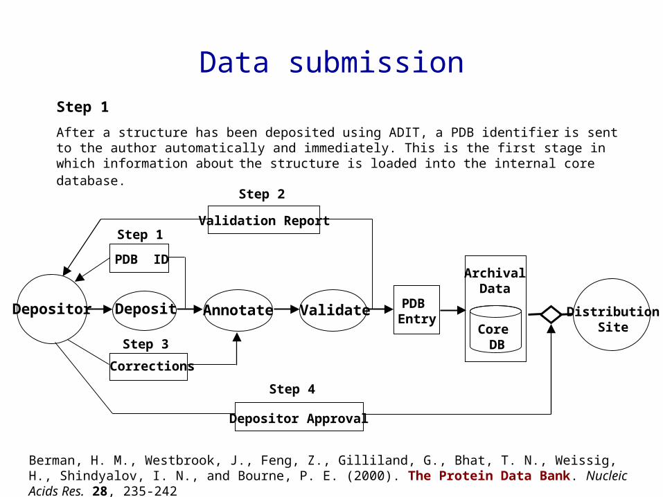
Step 1
After a structure has been deposited using ADIT, a PDB identifier is sent to the author automatically and immediately. This is the first stage in which information about the structure is loaded into the internal core database.
Data submission
Berman, H. M., Westbrook, J., Feng, Z., Gilliland, G., Bhat, T. N., Weissig, H., Shindyalov, I. N., and Bourne, P. E. (2000). The Protein Data Bank. Nucleic Acids Res. 28, 235-242
PDB ID
DistributionSite
Depositor
ArchivalData
Core DB
PDB Entry
Deposit Annotate Validate
Depositor Approval
Validation Report
Corrections
Step 2
Step 3
Step 4
Step 1
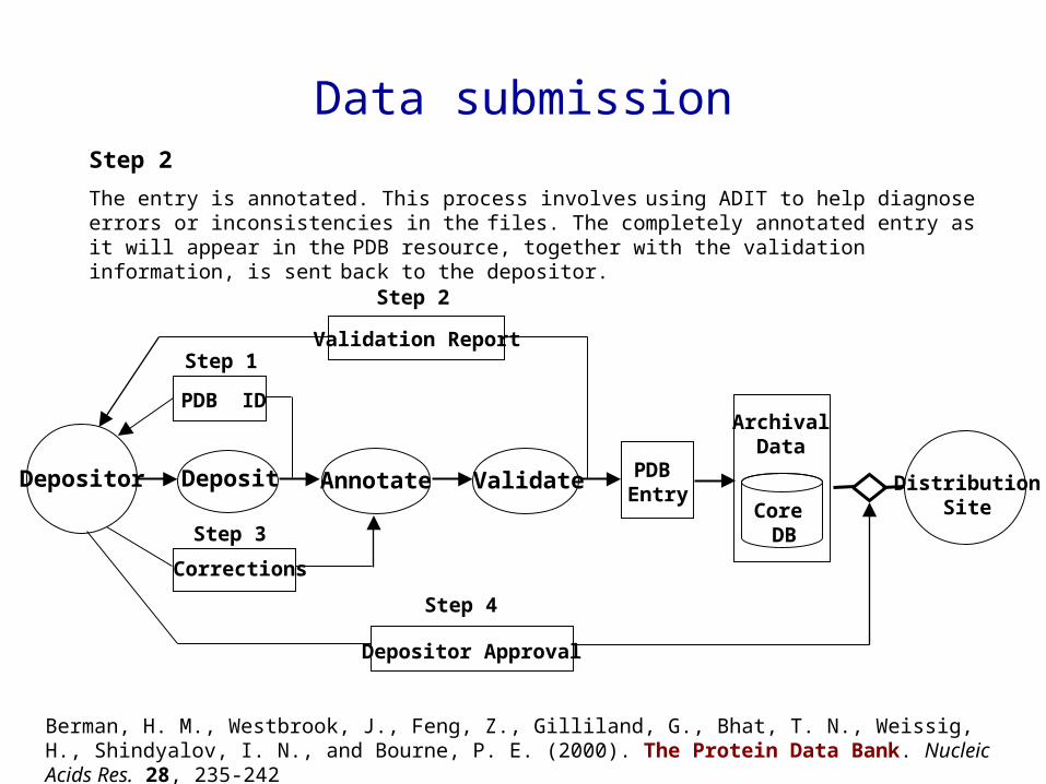
Step 2
The entry is annotated. This process involves using ADIT to help diagnose errors or inconsistencies in the
files. The completely annotated entry as it will appear in the PDB resource, together with the validation information, is sent back to the depositor.
Data submission
Berman, H. M., Westbrook, J., Feng, Z., Gilliland, G., Bhat, T. N., Weissig, H., Shindyalov, I. N., and Bourne, P. E. (2000). The Protein Data Bank. Nucleic Acids Res. 28, 235-242
PDB ID
DistributionSite
Depositor
ArchivalData
Core DB
PDB Entry
Deposit Annotate Validate
Depositor Approval
Validation Report
Corrections
Step 2
Step 3
Step 4
Step 1
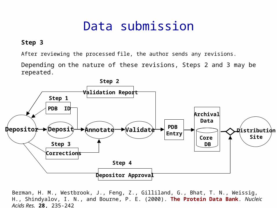
Step 3
After reviewing the processed file, the author sends any revisions.
Depending on the nature of these revisions, Steps 2 and 3 may be repeated.
Data submission
Berman, H. M., Westbrook, J., Feng, Z., Gilliland, G., Bhat, T. N., Weissig, H., Shindyalov, I. N., and Bourne, P. E. (2000). The Protein Data Bank. Nucleic Acids Res. 28, 235-242

PDB ID
DistributionSite
Depositor
ArchivalData
Core DB
PDB Entry
Deposit Annotate Validate
Depositor Approval
Validation Report
Corrections
Step 2
Step 3
Step 4
Step 1
Step 4
Once approval is received from the author, the entry and the tables in the internal core database are ready for distribution. The schema of this core database is a subset of the conceptual schema specified by the mmCIF dictionary. All aspects of data processing, including communications with the author, are recorded and stored in the correspondence archive. This makes it possible for the PDB staff to retrieve information about any aspect of the deposition process and to closely monitor the efficiency of PDB operations.
Data submission
Berman, H. M., Westbrook, J., Feng, Z., Gilliland, G., Bhat, T. N., Weissig, H., Shindyalov, I. N., and Bourne, P. E. (2000). The Protein Data Bank. Nucleic Acids Res. 28, 235-242
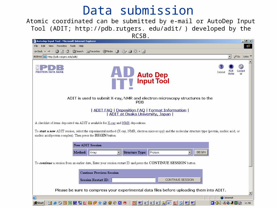
Data submission Atomic coordinated can be submitted by e-mail or AutoDep Input Tool (ADIT;
http://pdb.rutgers. edu/adit/ ) developed by the RCSB.

Data submissionADIT, which is also used to process the entries, is built on top of the mmCIF dictionary which is an ontology of 1700 terms that define the macro molecular structure and the crystallographic experiment, and a data processing program called MAXIT (MAcromolecular EXchange Input Tool). This integrated system helps to ensure that the data submitted are consistent with the mmCIF dictionary which defines data types, enumerates ranges of allowable values where possible and describes allowable relationships between data values.
Berman, H. M., Westbrook, J., Feng, Z., Gilliland, G., Bhat, T. N., Weissig, H., Shindyalov, I. N., and Bourne, P. E. (2000). The Protein Data Bank. Nucleic Acids Res. 28, 235-242
Crystallographic Information File CIF & Self Defining Text Archive and Retrieval (STAR)
Crystallographic Information File (CIF) is a data representation used by several disciplines (predominantly crystallography) concerned with molecular structure. The basis for this data representation is the Self Defining Text Archive and Retrieval (STAR) definition.
STAR is nothing more than a set of syntax rules. Associated with STAR is a Dictionary Definition Language (DDL) from which STAR compliant dictionaries have been developed by several discipline. From the dictionaries it is possible to define data files which use data items referenced in the dictionaries. The STAR DDL and associated dictionaries is considered as example of metadata - data describing how to represent other data.
•Westbrook, J. D. and Bourne, P. E. (2000). STAR/mmCIF: an ontology for macromolecular structure. Bioinformatics. 16, 159-168.

Dictionary Description Languagehttp://ndbserver.rutgers.edu/mmcif/ddl/index.html
The DDL is a dictionary of definitions which describes a language for specifying data definitions. DDL defines the data model that provides the foundation for the description of knowledge about an application domain.
The application knowledge is collected in a dictionary of definitions which describes the domain. DDL provides the framework on which this dictionary is organized by defining the levels of abstraction that are available to hold the data description. The DDL defines both the properties that may be associated with each level of abstraction and the relationships that may exist between levels. This DDL defines a relatively simple set of abstractions which include data blocks, categories, category groups, subcategories, and items.
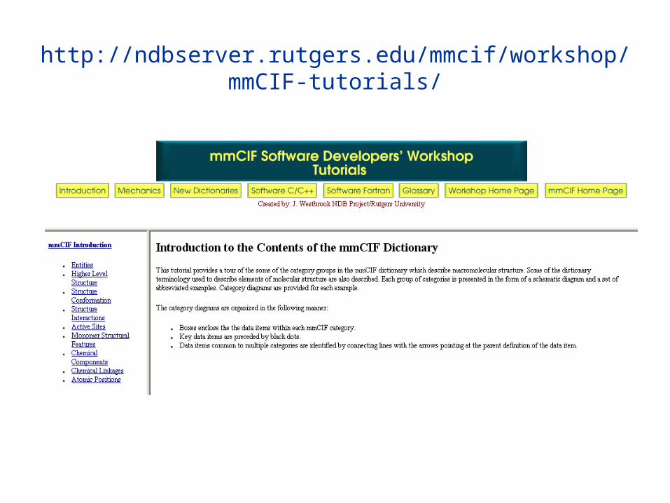
http://ndbserver.rutgers.edu/mmcif/workshop/mmCIF-tutorials/
ValidationValidation refers to the procedure for assessing the quality of deposited atomic models (structure validation) and for assessing how well these models fit the experimental data (experimental validation). The PDB validates structures using accepted community standards as part of ADIT’s integrated data processing system.
Covalent bond distances and angles. Proteins are compared against standard values from Engh and Huber; nucleic acid bases are compared against standard values from Clowney et al; sugar and phosphates are compared against standard values from Gelbin et al.
Stereochemical validation. All chiral centers of proteins and nucleic acids are checked for correct stereochemistry.
Atom nomenclature. The nomenclature of all atoms is checked for compliance with IUPAC standards and is adjusted if necessary.
Close contacts. The distances between all atoms within the asymmetric unit of crystal structures and the unique molecule of NMR structures are calculated. For crystal structures, contacts between symmetry-related molecules are checked as well.
Ligand and atom nomenclature. Residue and atom nomen clature is compared against the PDB dictionary (ftp://ftp.rcsb. org/pub/pdb/data/monomers/het_dictionary.txt ) for all ligands as well as standard residues and bases. Unrecognised ligand groups are flagged and any discrepancies in known ligands are listed as extra or missing atoms.
Sequence comparison. The sequence given in the PDB SEQRES records is compared against the sequence derived from the coordinate records. This information is displayed in a table where any differences or missing residues are marked. During structure processing, the sequence database references given by DBREF and SEQADV are checked for accuracy. If no reference is given, a BLAST search is used to find the best match. Any conflict between the PDB SEQRES records and the sequence derived from the coordinate records is resolved by comparison with various
sequence databases.
Distant waters. The distances between all water oxygen atoms and all polar atoms (oxygen and nitrogen) of the macromolecules, ligands and solvent in the asymmetric unit are calculated. Distant solvent atoms are repositioned using crystallographic symmetry such that they fall within the solvation sphere of the macromolecule.
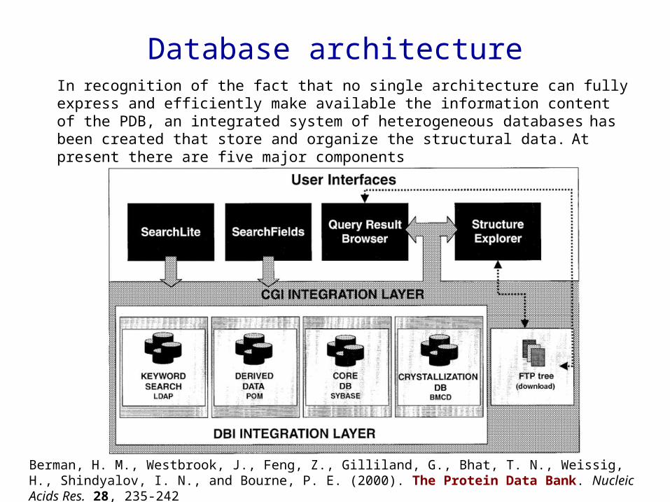
Database architectureIn recognition of the fact that no single architecture can fully express and efficiently make available the information content of the PDB, an integrated system of heterogeneous databases
has been created that store and organize the structural data. At present there are five major components
Berman, H. M., Westbrook, J., Feng, Z., Gilliland, G., Bhat, T. N., Weissig, H., Shindyalov, I. N., and Bourne, P. E. (2000). The Protein Data Bank. Nucleic Acids Res. 28, 235-242

The core relational database managed by Sybase (Sybase SQL server release 11.0, Emeryville, CA) provides the central physical storage for the primary experimental and coordinate data
The final curated data files (in PDB and mmCIF formats) and data dictionaries are the archival data and are present as ASCII files in the ftp archive.
• The POM (Property Object Model)-based databases, which consist of indexed objects containing native (e.g., atomic coordinates) and derived properties (e.g., calculated secondary structure assignments and property profiles). Some properties require no derivation, for example, B factors; others must be derived, for example, exposure of each amino acid residue or C contact maps. Properties requiring significant computation time, such as structure neighbours, are pre-calculated when the database is incremented to save considerable user access time.
• The Biological Macromolecule Crystallization Database (BMCD;) is organized as a relational database within Sybase and contains three general categories of literature derived
information: macromolecular, crystal and summary data.
• The Netscape LDAP server is used to index the textual content of the PDB in a structured format and provides support for keyword searches.
Database architecture
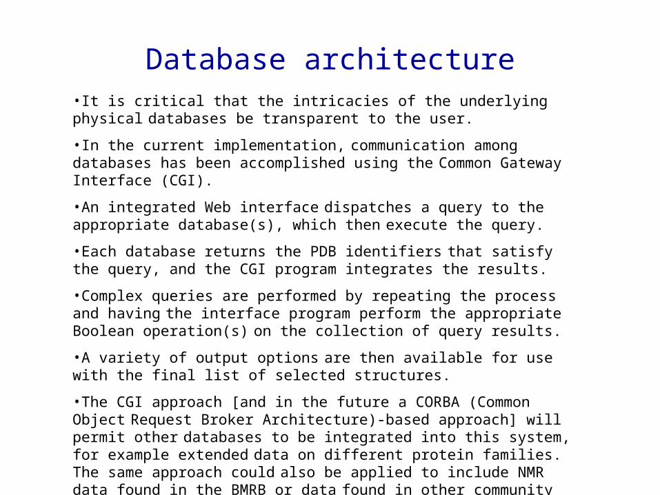
Database architecture•It is critical that the intricacies of the underlying physical databases be transparent to the user.
•In the current implementation, communication among databases has been accomplished using the Common Gateway Interface (CGI).
•An integrated Web interface dispatches a query to the appropriate database(s), which then execute the query.
•Each database returns the PDB identifiers that satisfy the query, and the CGI program integrates the results.
•Complex queries are performed by repeating the process and having the interface program perform the appropriate Boolean operation(s) on the collection of query results.
•A variety of output options are then available for use with the final list of selected structures.
•The CGI approach [and in the future a CORBA (Common Object Request Broker Architecture)-based approach] will permit other databases to be integrated into this system, for example extended data on different protein families. The same approach could also be applied to include NMR data found in the BMRB or data found in other community databases.
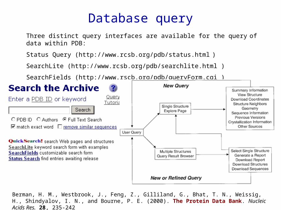
Database queryThree distinct query interfaces are available for the query of data within PDB:
Status Query (http://www.rcsb.org/pdb/status.html )
SearchLite (http://www.rcsb.org/pdb/searchlite.html )
Search Fields (http://www.rscb.org/pdb/queryForm.cgi )
Berman, H. M., Westbrook, J., Feng, Z., Gilliland, G., Bhat, T. N., Weissig, H., Shindyalov, I. N., and Bourne, P. E. (2000). The Protein Data Bank. Nucleic Acids Res. 28, 235-242

RCSB Partner Sites
I NSTI TUTI ON WEB SI TE FTP SI TE
SDSC/UCSD La Jolla, CA United States
http://www.rcsb.org/ http://www.pdb.org/
ftp://ftp.rcsb.org/pub/pdb/
Rutgers University Piscataway, NJ United States
http://rutgers.rcsb.org/ ftp://rutgers.rcsb.org/PDB/pub/pdb/
CARB/NIST Rockville, MD United States
http://nist.rcsb.org/ ftp://pdb.nist.gov/pub/pdb/
Other RCSB PDB Mirrors
I NSTI TUTI ON WEB SI TE FTP SI TE
Cambridge Crystallographic Data Centre United Kingdom
http://pdb.ccdc.cam.ac.uk/ ftp://pdb.ccdc.cam.ac.uk/rcsb/
National University of Singapore Singapore
http://pdb.bic.nus.edu.sg/ ftp://pdb.bic.nus.edu.sg/pub/pdb/
Osaka University Japan
http://pdb.protein.osaka-u.ac.jp/ ftp://pdb.protein.osaka-u.ac.jp/
Max Delbrück Center for Molecular Medicine Germany
http://www.pdb.mdc-berlin.de/ ftp://ftp.pdb.mdc-berlin.de/pub/pdb/

PDB (http://www.pdb.org/)• A search requires that at least one search field is filled. Case is ignored. The
search is then executed by pressing the search button.
• A search can return a single structure or multiple structures.
• Iterative searches can be performed, using the output from one search as input for the next.
• NOTE: The PDB is a historical archive. Its contents are not uniform, but reflect the knowledge of the time as well as the data management practices. This may produce incomplete query results.
8th June 2004
HIV 1 2 result
HIV –1 178 results
HIV I 1 result
HIV-I 1 result
10th October 2000
HIV 1 2 result
HIV –1 118 results
HIV I 1 result
HIV-I 1 result
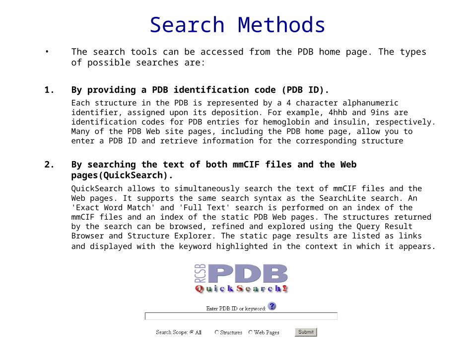
Search Methods• The search tools can be accessed from the PDB home page. The types of possible
searches are:
1. By providing a PDB identification code (PDB ID).
Each structure in the PDB is represented by a 4 character alphanumeric identifier, assigned upon its deposition. For example, 4hhb and 9ins are identification codes for PDB entries for hemoglobin and insulin, respectively. Many of the PDB Web site pages, including the PDB home page, allow you to enter a PDB ID and retrieve information for the corresponding structure
2. By searching the text of both mmCIF files and the Web pages(QuickSearch).
QuickSearch allows to simultaneously search the text of mmCIF files and the Web pages. It supports the same search syntax as the SearchLite search. An 'Exact Word Match' and 'Full Text' search is performed on an index of the mmCIF files and an index of the static PDB Web pages. The structures returned by the search can be browsed, refined and explored using the Query Result Browser and Structure Explorer. The static page results are listed as links and displayed with the keyword highlighted in the context in which it appears.

3. By searching the text found in mmCIF files (SearchLite).SearchLite searches the text of each mmCIF file as follows:Queries locate literal text phrases. A search for protein kinase will locate the phrase protein kinase, NOT protein and kinase separately.
– Partial word searches will retrieve all words they are included in, unless the match exact wordbox is checked. A search for hend will locate both hendrickson and henderson when the box is not checked, but will only retrieve hend when the box is checked.
– A second checkbox allows a user to remove sequence homologs from a search.
– Compound searches can be performed using and, or, not clauses. A search for protein and kinase will locate all structures that contain both protein AND kinase, not just the structures that contain the phrase protein kinase.
– SearchLite will locate entries with an "on hold" status by querying their title records. For queries on unreleased entries specifically, a Status Search is most optimal.
Search Methods
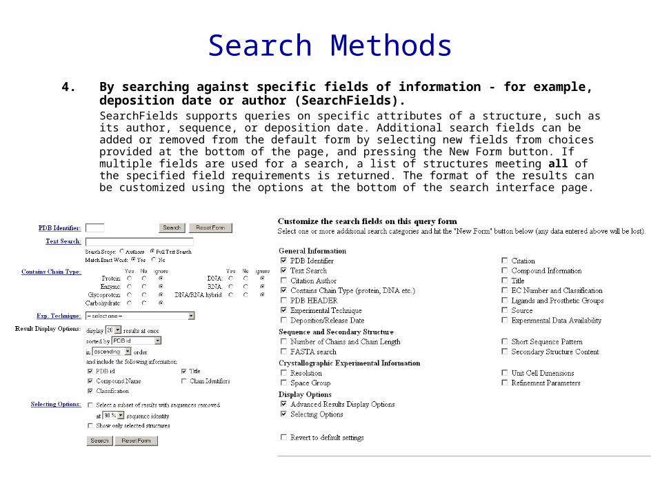
4. By searching against specific fields of information - for example, deposition date or author (SearchFields). SearchFields supports queries on specific attributes of a structure, such as its author, sequence, or deposition date. Additional search fields can be added or removed from the default form by selecting new fields from choices provided at the bottom of the page, and pressing the New Form button. If multiple fields are used for a search, a list of structures meeting all of the specified field requirements is returned. The format of the results can be customized using the options at the bottom of the search interface page.
Search Methods
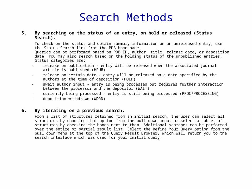
5. By searching on the status of an entry, on hold or released (Status Search). To check on the status and obtain summary information on an unreleased entry, use the Status Search link from the PDB home page.Queries can be performed based on PDB ID, author, title, release date, or deposition date. You may also search based on the holding status of the unpublished entries. Status categories are:
– release on publication - entry will be released when the associated journal article is published (HPUB)
– release on certain date - entry will be released on a date specified by the authors at the time of deposition (HOLD)
– await author input - entry is being processed but requires further interaction between the processor and the depositor (WAIT)
– currently being processed - entry is still being processed (PROC/PROCESSING) – deposition withdrawn (WDRN)
6. By iterating on a previous search.From a list of structures returned from an initial search, the user can select all structures by choosing that option from the pull-down menu, or select a subset of structures by checking the boxes next to them. Additional searches can be performed over the entire or partial result list. Select the Refine Your Query option from the pull down menu at the top of the Query Result Browser, which will return you to the search interface which was used for your initial query.
Search Methods

Results (Papillomavirus)
Results

ResultsView StructureOffers static images and several interactive displays: VRML (uses Molscript from P. Kraulis), RasMol, FirstGlance (simple Chime display), Protein Explorer (advanced Chime display), MICE (uses Java plug-in) STING Millennium (uses Chime), Swiss-Pdb Viewer, and QuickPDB (Java applet)
Download/Display FileDownload the PDB or mmCIF file to your local computer as plain text or in one of 3 common compression formats: Unix compressed, GNU zip, or ZIP.Display the PDB file or mmCIF file which includes links to relevant format documents
Structural NeighboursProvides access to the most common methods for finding and analysing structures which have 3-D structure homology to the protein currently being explored. There is currently no exact solution to finding 3-D structure homologs. All methods require making assumptions to be computationally tractable. These assumptions lead to somewhat different results, particularly when the homology is weak. Difference in detected homology leads to differences in alignment. Resources included are CATH, CE, FSSP, SCOP and MMDB (part of Entrez).
GeometryA tabular listing of bond lengths, bond angles and dihedral angles (phi, psi, omega, and chi) can be displayed, color coded to highlight significant deviations from ideality according to the criteria of Engh and Huber; a fold deviation score (FDS) provides a snapshot of the overall geometry of the selected structure. Ramachandran plots and links to related resources are also available here
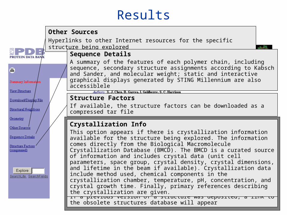
ResultsOther SourcesHyperlinks to other Internet resources for the specific structure being explored
Sequence DetailsA summary of the features of each polymer chain, including sequence, secondary structure assignments according to Kabsch and Sander, and molecular weight; static and interactive graphical displays generated by STING Millennium are also accessiblele
Previous VersionsIf a previous version of a structure was deposited, a link to the obsolete structures database will appear
Structure FactorsIf available, the structure factors can be downloaded as a compressed tar file
Crystallization InfoThis option appears if there is crystallization information available for the structure being explored. The information comes directly from the Biological Macromolecule Crystallization Database (BMCD). The BMCD is a curated source of information and includes crystal data (unit cell parameters, space group, crystal density, crystal dimensions, and lifetime in the beam if available). Crystallization data include method used, chemical components in the crystallization chamber, temperature, pH, concentration, and crystal growth time. Finally, primary references describing the crystallization are given.
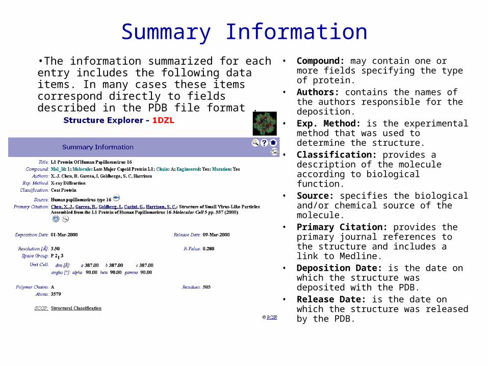
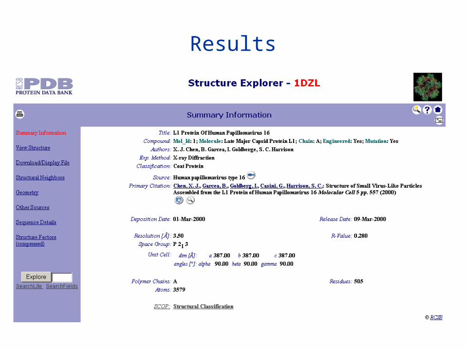
Summary Information• Compound: may contain one or more
fields specifying the type of protein. • Authors: contains the names of the
authors responsible for the deposition. • Exp. Method: is the experimental
method that was used to determine the structure.
• Classification: provides a description of the molecule according to biological function.
• Source: specifies the biological and/or chemical source of the molecule.
• Primary Citation: provides the primary journal references to the structure and includes a link to Medline.
• Deposition Date: is the date on which the structure was deposited with the PDB.
• Release Date: is the date on which the structure was released by the PDB.
•The information summarized for each entry includes the following data items. In many cases these items correspond directly to fields described in the PDB file format .
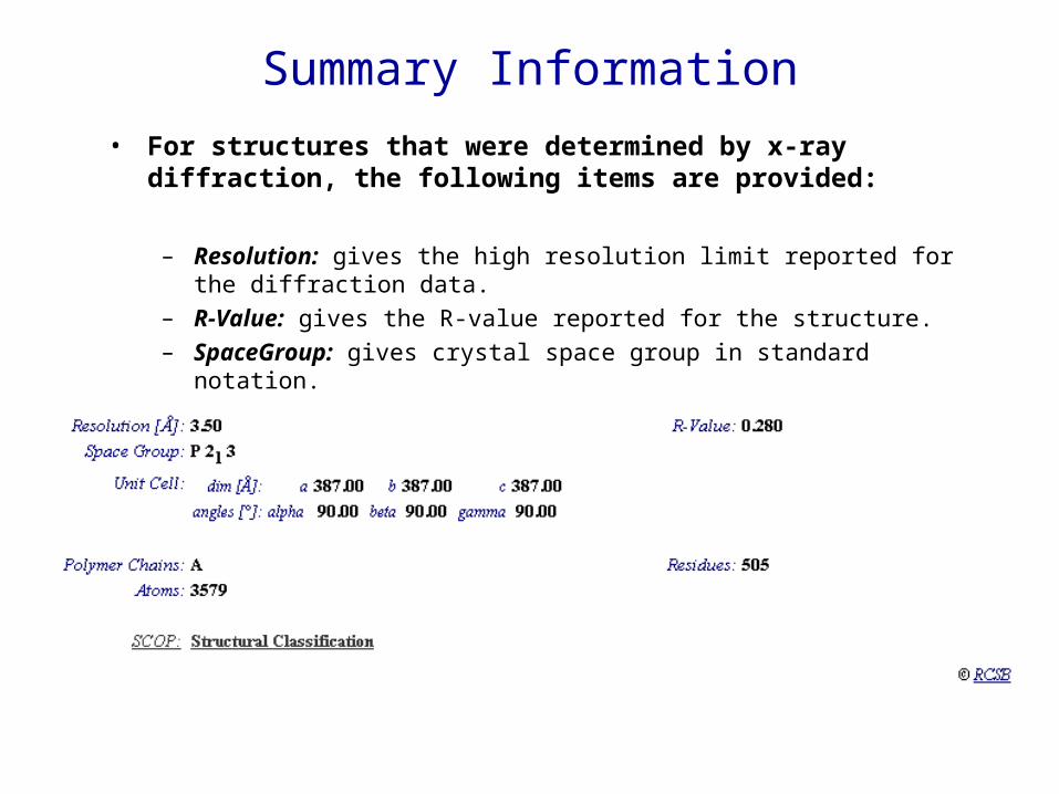
• For structures that were determined by x-ray diffraction, the following items are provided:
– Resolution: gives the high resolution limit reported for the diffraction data.
– R-Value: gives the R-value reported for the structure.
– SpaceGroup: gives crystal space group in standard notation.
– Unit Cell: gives the crystal cell lengths and angles.
Summary Information

• For structures that were determined by NMR spectroscopy, the following items are provided:
– Minimized Mean: links to the PDB ID for the file that contains the minimized mean structure if this structure was provided.
– Regularized Mean: links to the PDB ID for the file that contains the regularized mean structure if this structure was provided.
– Representative: links to the PDB ID for the file that contains the representative structure from the ensemble of structure solutions if this structure was provided.
– Ensemble Members: links to the PDB IDs for the files that contain the ensemble of structure solutions if these files were provided.
Summary Information
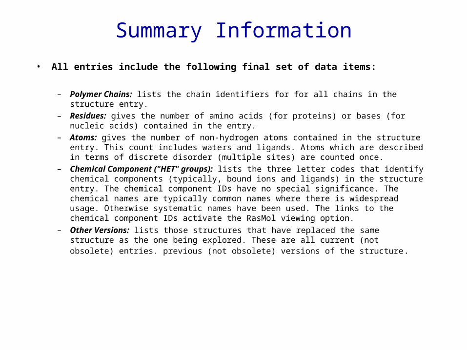
• All entries include the following final set of data items:
– Polymer Chains: lists the chain identifiers for for all chains in the structure entry.
– Residues: gives the number of amino acids (for proteins) or bases (for nucleic acids) contained in the entry.
– Atoms: gives the number of non-hydrogen atoms contained in the structure entry. This count includes waters and ligands. Atoms which are described in terms of discrete disorder (multiple sites) are counted once.
– Chemical Component ("HET" groups): lists the three letter codes that identify chemical components (typically, bound ions and ligands) in the structure entry. The chemical component IDs have no special significance. The chemical names are typically common names where there is widespread usage. Otherwise systematic names have been used. The links to the chemical component IDs activate the RasMol viewing option.
– Other Versions: lists those structures that have replaced the same structure as the one being explored. These are all current (not obsolete) entries. previous (not obsolete) versions of the structure.
Summary Information

Interactive 3D Display
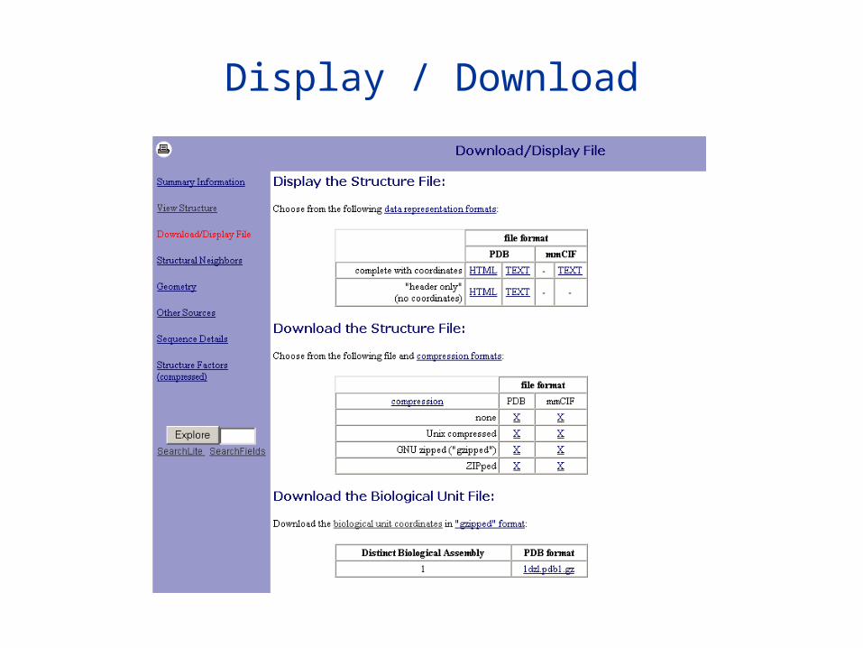
Display / Download
Other databases
3D_ali database of aligned protein structures and related sequenceshttp://www.embl-heidelberg.de/argos/ali/ali_info.html
EMBL-EMI THE MACROMOLECULAR STRUCTURE DATABASE http://www.ebi.ac.uk/msd/
BioMagResBank - Database of NMR-derived Protein Structures - BIMAS-NIH (US)
http://bimas.dcrt.nih.gov/sql/BMRBgate.html
CATH - Protein Structure Classification at the - U College London (UK)
http://www.biochem.ucl.ac.uk/bsm/cath/
ENTREZ Structure - Biomolecule 3D Structure Search - NCBI (US) http://www.ncbi.nlm.nih.gov/entrez/query.fcgi?db=Structure

MMDB, Molecular Modelling DataBase (NCBI) http://www.ncbi.nlm.nih.gov/Structure/MMDB/mmdb.shtml
Enzyme Structure Database - UCL (UK) http://www.biochem.ucl.ac.uk/bsm/enzymes/index.html
Nucleic Acid DatabaseA repository of three-dimensional structural information about nucleic acids at Rutgers
http://ndbserver.rutgers.edu/
BioMolQuesthttp://bioinformatics.buffalo.edu/new_buffalo/people/wli7/
public/home.html
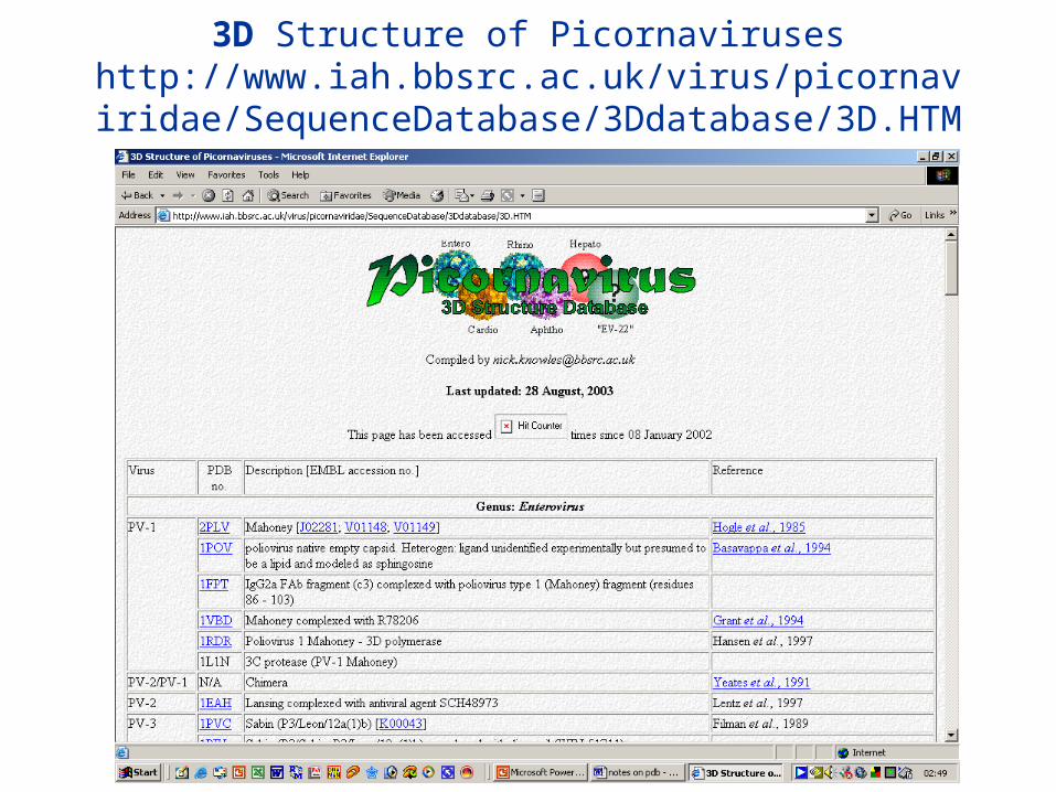
3D Structure of Picornaviruseshttp://www.iah.bbsrc.ac.uk/virus/picornaviridae/SequenceDat
abase/3Ddatabase/3D.HTM
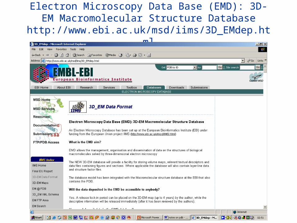
Electron Microscopy Data Base (EMD): 3D-EM Macromolecular Structure Database
http://www.ebi.ac.uk/msd/iims/3D_EMdep.html
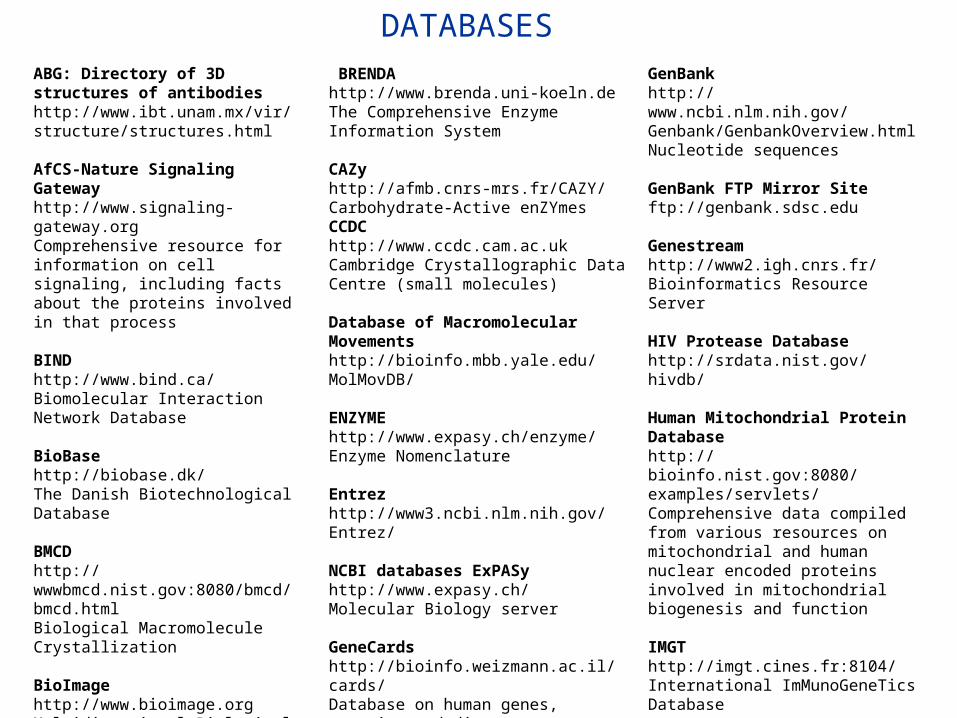
ABG: Directory of 3D structures of antibodieshttp://www.ibt.unam.mx/vir/structure/structures.html AfCS-Nature Signaling Gatewayhttp://www.signaling-gateway.orgComprehensive resource for information on cell signaling, including facts about the proteins involved in that process BINDhttp://www.bind.ca/Biomolecular Interaction Network Database BioBasehttp://biobase.dk/The Danish Biotechnological Database BMCDhttp://wwwbmcd.nist.gov:8080/bmcd/bmcd.htmlBiological Macromolecule Crystallization BioImagehttp://www.bioimage.orgMultidimensional Biological Images (EM) BMRBhttp://www.bmrb.wisc.eduBioMagResBank (NMR)
BRENDAhttp://www.brenda.uni-koeln.deThe Comprehensive Enzyme Information System CAZyhttp://afmb.cnrs-mrs.fr/CAZY/Carbohydrate-Active enZYmes CCDChttp://www.ccdc.cam.ac.ukCambridge Crystallographic Data Centre (small molecules) Database of Macromolecular Movementshttp://bioinfo.mbb.yale.edu/MolMovDB/ ENZYMEhttp://www.expasy.ch/enzyme/Enzyme Nomenclature Entrezhttp://www3.ncbi.nlm.nih.gov/Entrez/ NCBI databases ExPASyhttp://www.expasy.ch/Molecular Biology server GeneCardshttp://bioinfo.weizmann.ac.il/cards/Database on human genes, proteins and diseases GDBhttp://www.gdb.org/Genome Data Base
GenBankhttp://www.ncbi.nlm.nih.gov/Genbank/GenbankOverview.htmlNucleotide sequences GenBank FTP Mirror Siteftp://genbank.sdsc.edu Genestreamhttp://www2.igh.cnrs.fr/Bioinformatics Resource Server HIV Protease Databasehttp://srdata.nist.gov/hivdb/ Human Mitochondrial Protein Databasehttp://bioinfo.nist.gov:8080/examples/servlets/Comprehensive data compiled from various resources on mitochondrial and human nuclear encoded proteins involved in mitochondrial biogenesis and function IMGThttp://imgt.cines.fr:8104/International ImMunoGeneTics Database Klothohttp://www.biocheminfo.org/klotho/Biochemical Compounds Declarative Database
DATABASES
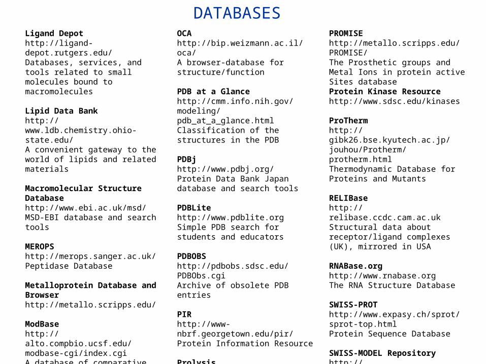
Ligand Depothttp://ligand-depot.rutgers.edu/Databases, services, and tools related to small molecules bound to macromolecules Lipid Data Bankhttp://www.ldb.chemistry.ohio-state.edu/A convenient gateway to the world of lipids and related materials Macromolecular Structure Databasehttp://www.ebi.ac.uk/msd/MSD-EBI database and search tools MEROPShttp://merops.sanger.ac.uk/Peptidase Database Metalloprotein Database and Browserhttp://metallo.scripps.edu/ ModBasehttp://alto.compbio.ucsf.edu/modbase-cgi/index.cgiA database of comparative protein structure models NDBhttp://ndbserver.rutgers.edu:80/Nucleic Acid Database
OCAhttp://bip.weizmann.ac.il/oca/A browser-database for structure/function
PDB at a Glancehttp://cmm.info.nih.gov/modeling/pdb_at_a_glance.htmlClassification of the structures in the PDB PDBjhttp://www.pdbj.org/Protein Data Bank Japan database and search tools PDBLitehttp://www.pdblite.orgSimple PDB search for students and educators PDBOBShttp://pdbobs.sdsc.edu/PDBObs.cgiArchive of obsolete PDB entries PIRhttp://www-nbrf.georgetown.edu/pir/Protein Information Resource Prolysishttp://delphi.phys.univ-tours.fr/ProlysisProteases and protease inhibitors
PROMISEhttp://metallo.scripps.edu/PROMISE/The Prosthetic groups and Metal Ions in protein active Sites database Protein Kinase Resourcehttp://www.sdsc.edu/kinases ProThermhttp://gibk26.bse.kyutech.ac.jp/jouhou/Protherm/protherm.htmlThermodynamic Database for Proteins and Mutants RELIBasehttp://relibase.ccdc.cam.ac.ukStructural data about receptor/ligand complexes (UK), mirrored in USA RNABase.orghttp://www.rnabase.orgThe RNA Structure Database SWISS-PROThttp://www.expasy.ch/sprot/sprot-top.htmlProtein Sequence Database SWISS-MODEL Repositoryhttp://swissmodel.expasy.org/repository/A database of annotated protein structure homology models Vitamin D Nuclear Receptor Sitehttp://VDR.bu.edu/
DATABASES
References/reading
• Bourne, P. E., Addess, K. J., Bluhm, W. F., Chen, L., Deshpande, N., Feng, Z., Fleri, W., Green, R., Merino-Ott, J. C., Townsend-Merino, W., Weissig, H., Westbrook, J., and Berman, H. M. (2004). The distribution and query systems of the RCSB Protein Data Bank. Nucleic Acids Res. 32 Database issue, D223-D225.
• Bhat, T. N., Bourne, P., Feng, Z., Gilliland, G., Jain, S., Ravichandran, V., Schneider, B., Schneider, K., Thanki, N., Weissig, H., Westbrook, J., and Berman, H. M. (2001). The PDB data uniformity project. Nucleic Acids Res. 29, 214-218.
• Berman, H. M., Westbrook, J., Feng, Z., Gilliland, G., Bhat, T. N., Weissig, H., Shindyalov, I. N., and Bourne, P. E. (2000). The Protein Data Bank. Nucleic Acids Res. 28, 235-242.
• Greer, D. S., Westbrook, J. D., and Bourne, P. E. (2002). An ontology driven architecture for derived representations of macromolecular structure. Bioinformatics. 18, 1280-1281.
• Westbrook, J. D. and Bourne, P. E. (2000). STAR/mmCIF: an ontology for macromolecular structure. Bioinformatics. 16, 159-168.
• Westbrook, J., Feng, Z., Jain, S., Bhat, T. N., Thanki, N., Ravichandran, V., Gilliland, G. L., Bluhm, W., Weissig, H., Greer, D. S., Bourne, P. E., and Berman, H. M. (2002). The Protein Data Bank: unifying the archive. Nucleic Acids Res. 30, 245-248.
• Westbrook, J., Feng, Z., Chen, L., Yang, H., and Berman, H. M. (2003). The Protein Data Bank and structural genomics. Nucleic Acids Res. 31, 489-491.