Embed Size (px)
Citation preview
Effects of Macromolecular Crowding on Protein Folding
- in-vitro equilibrium and kinetic studies on selected model systems
Alexander Christiansen
Kemiska Institutionen
Umeå 2013-11-20

Responsible publisher under swedish law: the Dean of the Faculty of Science and Technology This work is protected by the Swedish Copyright Legislation (Act 1960:729) ISBN: 978-91-7459-764-6 Elektronisk version tillgänglig på http://umu.diva-portal.org/ Tryck/Printed by: Service Center KBC Umeå Sweden 2013

众鸟高飞去
孤云独去闲
相看两不厌
只有敬停山
—李白
The birds have vanished down the sky.
Now the last cloud drains away.
We sit together, the mountain and I,
Until only the mountain remains.
-Li Bai
(Translated by Xiaowei Song)
i
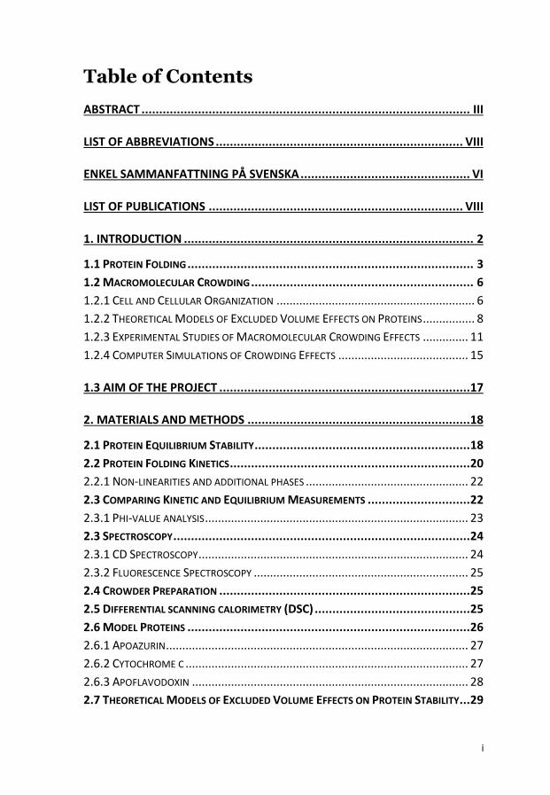
Table of Contents
ABSTRACT ............................................................................................. III
LIST OF ABBREVIATIONS ...................................................................... VIII
ENKEL SAMMANFATTNING PÅ SVENSKA ................................................ VI
LIST OF PUBLICATIONS ........................................................................ VIII
1. INTRODUCTION .................................................................................. 2
1.1 PROTEIN FOLDING ................................................................................. 3
1.2 MACROMOLECULAR CROWDING ............................................................... 6
1.2.1 CELL AND CELLULAR ORGANIZATION ............................................................. 6
1.2.2 THEORETICAL MODELS OF EXCLUDED VOLUME EFFECTS ON PROTEINS ................ 8
1.2.3 EXPERIMENTAL STUDIES OF MACROMOLECULAR CROWDING EFFECTS .............. 11
1.2.4 COMPUTER SIMULATIONS OF CROWDING EFFECTS ........................................ 15
1.3 AIM OF THE PROJECT ....................................................................... 17
2. MATERIALS AND METHODS ............................................................... 18
2.1 PROTEIN EQUILIBRIUM STABILITY ............................................................. 18
2.2 PROTEIN FOLDING KINETICS .................................................................... 20
2.2.1 NON-LINEARITIES AND ADDITIONAL PHASES .................................................. 22
2.3 COMPARING KINETIC AND EQUILIBRIUM MEASUREMENTS ............................. 22
2.3.1 PHI-VALUE ANALYSIS ................................................................................. 23
2.3 SPECTROSCOPY .................................................................................... 24
2.3.1 CD SPECTROSCOPY ................................................................................... 24
2.3.2 FLUORESCENCE SPECTROSCOPY .................................................................. 25
2.4 CROWDER PREPARATION ....................................................................... 25
2.5 DIFFERENTIAL SCANNING CALORIMETRY (DSC) ............................................ 25
2.6 MODEL PROTEINS ................................................................................ 26
2.6.1 APOAZURIN ............................................................................................. 27
2.6.2 CYTOCHROME C ....................................................................................... 27
2.6.3 APOFLAVODOXIN ..................................................................................... 28
2.7 THEORETICAL MODELS OF EXCLUDED VOLUME EFFECTS ON PROTEIN STABILITY ... 29
ii
3. RESULTS ............................................................................................ 33
3.1 EFFECT OF CROWDING ON EQUILIBRIUM .................................................... 33
3.1.1 CYTOCHROME C ....................................................................................... 34
3.1.2 APOAZURIN ............................................................................................. 38
3.1.3 SUMMARY OF THE EQUILIBRIUM DATA ........................................................ 43
3.2 EFFECTS OF CROWDING ON FOLDING KINETICS ............................................ 44
3.2.1 APOAZURIN FOLDING KINETICS ................................................................... 44
3.2.2 APOFLAVODOXIN FOLDING KINETICS ........................................................... 45
3.2.3 FAST FOLDING KINETICS OF CYTOCHROME C ................................................. 48
3.2.4 EFFECT OF VISCOSITY ON FOLDING KINETICS .................................................. 49
3.2.5 SUMMARY OF THE EFFECTS OF CROWDING ON FOLDING KINETICS .................... 50
4. DISCUSSION....................................................................................... 52
4.1 ATTRACTIVE INTERACTIONS? ................................................................... 55
4.2 CROWDING EFFECTS ON KINETICS ............................................................ 58
4.3 IN-VITRO VS IN-VIVO CONDITIONS ............................................................ 59
5. CONCLUSION AND SUMMARY ............................................................ 62
6. OUTLOOK .......................................................................................... 63
ACKNOWLEDGEMENTS .......................................................................... 65
REFERENCES .......................................................................................... 67
iii
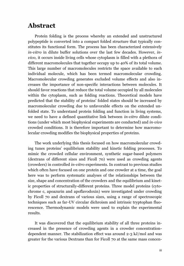
Abstract
Protein folding is the process whereby an extended and unstructured
polypeptide is converted into a compact folded structure that typically con-
stitutes its functional form. The process has been characterized extensively
in-vitro in dilute buffer solutions over the last few decades. However, in-
vivo, it occurs inside living cells whose cytoplasm is filled with a plethora of
different macromolecules that together occupy up to 40% of its total volume.
This large number of macromolecules restricts the space available to each
individual molecule, which has been termed macromolecular crowding.
Macromolecular crowding generates excluded volume effects and also in-
creases the importance of non-specific interactions between molecules. It
should favor reactions that reduce the total volume occupied by all molecules
within the cytoplasm, such as folding reactions. Theoretical models have
predicted that the stability of proteins’ folded states should be increased by
macromolecular crowding due to unfavorable effects on the extended un-
folded state. To understand protein folding and function in living systems,
we need to have a defined quantitative link between in-vitro dilute condi-
tions (under which most biophysical experiments are conducted) and in-vivo
crowded conditions. It is therefore important to determine how macromo-
lecular crowding modifies the biophysical properties of proteins.
The work underlying this thesis focused on how macromolecular crowd-
ing tunes proteins’ equilibrium stability and kinetic folding processes. To
mimic the crowded cellular environment, synthetic sugar-based polymers
(dextrans of different sizes and Ficoll 70) were used as crowding agents
(crowders) in controlled in-vitro experiments. In contrast to previous studies
which often have focused on one protein and one crowder at a time, the goal
here was to perform systematic analyses of the relationships between the
size, shape and concentration of the crowders and the equilibrium and kinet-
ic properties of structurally-different proteins. Three model proteins (cyto-
chrome c, apoazurin and apoflavodoxin) were investigated under crowding
by Ficoll 70 and dextrans of various sizes, using a range of spectroscopic
techniques such as far-UV circular dichroism and intrinsic tryptophan fluo-
rescence. Thermodynamic models were used to explain the experimental
results.
It was discovered that the equilibrium stability of all three proteins in-
creased in the presence of crowding agents in a crowder concentration-
dependent manner. The stabilization effect was around 2-3 kJ/mol and was
greater for the various Dextrans than for Ficoll 70 at the same mass concen-
iv
tration but independent of dextran size (for dextrans ranging from 20 to 70
kDa). A theoretical crowding model was used to investigate the origins of this
stabilization. In this model, Dextran and Ficoll were modeled as elongated
rods and the protein was represented as a sphere, with the folded sphere
representation being smaller than the unfolded sphere representation. Nota-
bly, this model was able to reproduce the observed stability changes while
only accounting for steric interactions. This correlation showed that when
using sugar-based crowding agents, excluded volume effects can be studied
in isolation with no contributions from nonspecific interactions.
Time-resolved experiments using apoazurin and apoflavodoxin revealed
an increase in the folding rate constants while the unfolding rates were un-
changed by the presence of crowding agents. For apoflavodoxin and cyto-
chrome c, the presence of crowding agents also altered the folding pathway
such that it became more homogeneous (cytochrome c) and gave less mis-
folding (apoflavodoxin). These results showed that macromolecular crowd-
ing restricts the conformational space of the unfolded polypeptide chain,
making its conformations more compact. This in turn eliminates access to
certain folding/misfolding pathways.
The results of the kinetic and equilibrium measurements on three model
proteins, together with available data from the literature, demonstrate that
macromolecular crowding effects due to volume exclusion are on the order of
a few kJ/mol. Considering the numerous concentration balances and cross-
dependent reactions of the cellular machinery, small changes in energet-
ics/kinetics of the magnitudes found here can have dramatic consequences
for cellular fitness. In fact, local and transient changes in macromolecular
crowding levels may be one way of tuning cellular biochemical processes
without invoking gene expression.
v
List of Abbreviations
CD Circular Dichroism
DLS Dynamic Light Scattering
DSC Differential Scanning Calorimetry
FCS Fluorescence Correlation Spectroscopy
FRAP Fluorescence Recovery after Photobleaching
FRET Fluorescence Resonance Energy Transfer
GuHCl Guanidine Hydrochloride
IR Infrared Spectroscopy
MD Molecular Dynamics
NMR Nuclear Magnetic Resonance
PEG Polyethylene glycol
PGK Phosphoglycerate Kinase
PVP Polyvinylpyrrolidone
Rg Radius of Gyration
SANS Small angle neutron Scattering
SAXS Small angle x-ray Scattering
SOD Superoxide Dismutase
SPT Scaled Particle Theory
SPR Single Particle Tracking
vi
Enkel sammanfattning på svenska
Proteiner verkar i en trång miljö
Proteiner utgör en av biologins viktigaste molekyler. De fungerar som
byggmaterial, strukturelement, transportmedel och katalysatorer inne i
cellerna. Att undersöka proteiners egenskaper i detalj kan ge ökad förståelse
för hur celler, och därmed levande organismer, fungerar. Proteiner tillverkas
inne i cellerna i form av långa aminosyrakedjor. Dessa kedjor genomgår
sedan en spontan process som kallas proteinveckning för att nå sin
slutgiltiga kompakta och funktionella form. Det finns många sjukdomar,
t.ex. Alzheimers och Parkisons, som beror på fel i veckningsprocesserna.
Proteiners veckningsprocesser brukar undersökas i
laboratorieexperiment i utspädda vattenlösningar. I motsats till denna
artificiella miljö är en levande cell fylld med en stor mängd olika molekyler
som tillsammans tar upp 40 procent av den totala volymen. En viktig fråga
är om proteiners egenskaper är desamma i den trånga cellmiljö som i en
utspädd in-vitro-lösning? I den trånga cellmiljön uppkommer effekter
såsom ospecifika växelverkningar mellan molekyler, ändrad viskositet och så
kallade 'excluded volume‘-effekter. Excluded volume-effekten är en sterisk
effekt som beror på att två molekyler inte kan uppta samma plats samtidigt.
Är det trångt i lösningen leder excluded volume-effekten till att
molekylformer som upptar mindre plats prioriteras över sådana former som
tar upp mycket plats. Eftersom uppveckade proteiner tar upp mer plats än de
kompakta aktiva formerna bör veckade proteiner stabiliseras i cellmiljö.
Steriska effekter av den trånga cellmiljön kan också påverka stabiliteten av
protein-protein-komplex och enzymatisk aktivitet jämfört med in vitro.
Olika teoretiska modeller har tagits fram som förutspår hur excluded
volume-effekten kan påverka proteiners stabilitet.
I arbetet som ligger till grund för denna avhandling har effekterna av
cellliknande miljö på proteiners stabilitet (jämvikt) och veckningsreaktioner
(kinetik) undersökts med hjälp av spektroskopiska metoder. Tre
modellproteiner har studerats: cytokrom c, apoazurin och apoflavodoxin.
För att skapa en miljö som liknar situationen i en cell har långa socker-
baserade polymerer (dextraner av olika storlekar och Ficoll 70) använts som
‘crowding-agenter‘. Dessa molekyler tar upp plats men växelverkar ej med de
undersökta proteinerna.
vii
Jämviktsmätningar för apoazurin och cytokrom c visade att dessa
proteiner stabiliseras i närvaro av crowding-agenter och effekten på
stabiliteten berodde på koncentrationen av crowding-agent och på
polymerens form. Ökningen i proteinstabilitet är i storleksordningen 2-3
kJ/mol. Även om denna effekt kan anses liten, kan den ha betydelse i
levande celler där små förändringar kan påverkar många olika jämvikter som
beror av varandra. En teoretisk modell som bara tar hänsyn till steriska
interaktioner och modellerar crowding-agenterna som långa stavar kan
reproducera de experimentella resultaten.
Tidsupplösta experiment visade att veckningshastigheten för apoazurin
och apoflavodxin blir snabbare i närvaro av en crowding-agent. Också här är
ökningen större om den tillsatta mängden crowding-agent ökades.
Cytokrom c och apoflavodoxin veckas i reaktioner som innefattar felveckade
temporära strukturer. För dessa proteiner upptäcktes att i närvaro av
crowding-agent ändrades veckningsvägen så att det blev mindre felveckning
och mer homogena reaktioner än i vattenlösning.
Experimenten som presenteras i denna avhandling visar på ett
systematiskt sätt hur några olika proteiners stabilitet och veckning påverkas
av cellliknande miljö. Från resultaten kan slutsatsen dras att socker-
baserade polymerer är bra redskap för isolerade studier av ‘excluded
volume‘-effekter utan bidrag från ospecifika interaktioner mellan polymer
och protein.
viii
List of Publications
I) Alexander Christiansen, Qian Wang, Antonios Samiotakis,
Margaret S. Cheung, and Pernilla Wittung-Stafshede. 2010. Fac-
tors Defining Effects of Macromolecular Crowding on Protein
Stability: An in Vitro/in Silico Case Study Using Cytochrome c.
Biochemistry 49 (31), 6519-6530
Reprinted with permission from Biochemistry 49 (31), 6519-6530,
2013. Copyright 2013 American Chemical Society.
II) Alexander Christiansen, Pernilla Wittung-Stafshede. 2013.
Quantification of Excluded Volume Effects on the Folding Land-
scape of Pseudomonas aeruginosa Apoazurin In Vitro, Biophysi-
cal Journal, Volume 105, Issue 7, 1689-1699
Reprinted with permission from Biophysical Journal 105 (7):1689-
1699, 2013. Copyright © 2013, Elsevier
III) Loren Stagg, Alexander Christiansen, and Pernilla Wittung-
Stafshede. 2011. Macromolecular Crowding Tunes Folding Land-
scape of Parallel / Protein, Apoflavodoxin. Journal of the
American Chemical Society 133 (4), 646-648
Reprinted with permission from Journal of the American Chemical
Society 133 (4): 646-648, 2010. Copyright 2010 American Chemical
Society.
IV) Eefei Chen, Alexander Christiansen, Qian Wang, Margaret S.
Cheung, David S. Kliger, and Pernilla Wittung-Stafshede. 2012.
Effects of Macromolecular Crowding on Burst Phase Kinetics of
Cytochrome c Folding. Biochemistry 51 (49), 9836-9845
Reprinted with permission from Biochemistry 51 (59): 9836-9845,
2012. Copyright 2012 American Chemical Society.
ix
Publications not covered in the thesis
V) Alexander Christiansen, Qian Wang, Margaret S. Cheung and
Pernilla Wittung-Stafshede. 2013. Effects of macromolecular
crowding agents on protein folding in vitro and in silico. Biophys-
ical Reviews 5 (2), 137-145
VI) Qian Wang, Alexander Christiansen, Antonios Samiotakis,
Pernilla Wittung-Stafshede, and Margaret S. Cheung. 2011. Com-
parison of chemical and thermal protein denaturation by combi-
nation of computational and experimental approaches. II. Jour-
nal Chemical Physics 135, 175102-1 – 175102-12
VII) Jörgen Ådén, Marcus Wallgren, Patrik Storm, Christoph F. Weise,
Alexander Christiansen, Wolfgang P. Schröder, Christiane
Funk, Magnus Wolf-Watz. 2011. Extraordinary μs–ms backbone
dynamics in Arabidopsis thaliana peroxiredoxin Q. Biochimica et
Biophysica Acta (BBA) - Proteins and Proteomics, Volume 1814,
Issue 12, P1880-1890
VIII) Alexander Christiansen, Pernilla Wittung-Stafshede. 2013.
Synthetic crowding agent causes excluded volume interactions ex-
clusively in tracer protein solution. Submitted.
Alexander Christiansen’s Contributions:
Paper I: designed, performed, and analyzed in-vitro experiments. Assisted
in writing the manuscript.
Paper II: designed, performed, and analyzed all experiments. Wrote the
manuscript together with the co-author.
Paper III: performed and analyzed kinetic and equilibrium data for some
protein variants. Helped with the revision version of the manuscript.
Paper IV: designed, performed and analyzed the chemical equilibrium ex-
periments. Assisted in writing the manuscript.
2
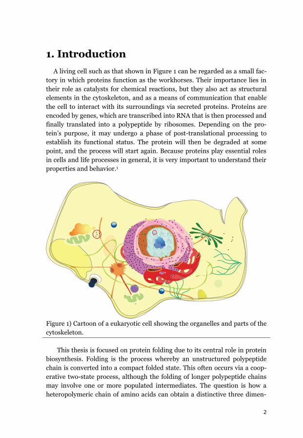
1. Introduction
A living cell such as that shown in Figure 1 can be regarded as a small fac-
tory in which proteins function as the workhorses. Their importance lies in
their role as catalysts for chemical reactions, but they also act as structural
elements in the cytoskeleton, and as a means of communication that enable
the cell to interact with its surroundings via secreted proteins. Proteins are
encoded by genes, which are transcribed into RNA that is then processed and
finally translated into a polypeptide by ribosomes. Depending on the pro-
tein’s purpose, it may undergo a phase of post-translational processing to
establish its functional status. The protein will then be degraded at some
point, and the process will start again. Because proteins play essential roles
in cells and life processes in general, it is very important to understand their
properties and behavior.1
Figure 1) Cartoon of a eukaryotic cell showing the organelles and parts of the
cytoskeleton.
This thesis is focused on protein folding due to its central role in protein
biosynthesis. Folding is the process whereby an unstructured polypeptide
chain is converted into a compact folded state. This often occurs via a coop-
erative two-state process, although the folding of longer polypeptide chains
may involve one or more populated intermediates. The question is how a
heteropolymeric chain of amino acids can obtain a distinctive three dimen-
3
sional structure. In particular, it is not clear why a polypeptide chain with a
given sequence should usually adopt its final structure and in addition in
most cases that process proceeds spontaneously without any help from other
proteins, although larger protein might be dependent on chaperones as fold-
ing helpers.2, 3 The information that determines which folded structure will
be adopted and how it should be established must be somehow encoded in
the polypeptide’s amino acid sequence (and thus the sequence of the corre-
sponding gene); deciphering this code is one of the Holy Grails of protein
science.
1.1 Protein Folding
The structural information encoded within a protein’s sequence can be
investigated in both the folded and unfolded states. The folded state of a
protein is often regarded as a single defined state, although folded proteins
have a degree of flexibility that enables them to “breathe”. The folded state is
held together by hydrogen bonds and van der Waals-, ionic- (between
charged groups) and hydrophobic interactions in the protein core. Covalent
bonds are rare intracellularly and usually confined to disulfide bridges. The
structure of a folded protein can be analyzed in hierarchical terms. Its prima-
ry structure is its amino acid sequence. The secondary structure consists of
defined sub-structural elements such as -helices and -strands. The tertiary
structure refers to the three-dimensional arrangement of the secondary
structural elements. Finally, if the folded protein associates with other folded
proteins to form a multimeric assembly (e.g. a homo- or hetero- dimer or
trimer), it is said to exhibit a quaternary structure. Some proteins also incor-
porate non-protein cofactors such as metal ions that offer otherwise-
unavailable functionality.4 Information on the structure of folded proteins
can be obtained using a plethora of techniques including Nuclear Magnetic
Resonance (NMR)5, cryo-EM6 and X-ray crystallography (which can provide
high resolution structures), as well as Circular dichroism (CD)7, fluores-
cence8, Raman9 and infrared (IR)10 spectroscopy.
The unfolded state is harder to characterize than the folded state. Instead
of one defined structure, it is an ensemble of different chain conformations
separated by small energy barriers, so interconversion between the different
conformations proceeds readily.11, 12 From his studies on chemically dena-
tured proteins in 1972 Tanford concluded that the unfolded state probably
adopts a random coil conformation.13, 14 In general, no residual secondary
structural elements or tertiary structure are apparent in the unfolded state,
but exceptions have been reported.11, 12, 15 Many experimental techniques that

4
can be applied to the folded state do not provide sufficient detail for analysis
of the unfolded state. What can be measured in-vitro is the average exten-
sion of the unfolded state, using techniques such as small angle scattering
(SAXS or SANS)16, NMR17, 18 or fluorescence resonance energy transfer
(FRET)19. A SAXS study by Millett et al. compared results for a range of pro-
teins of varying length in the folded and unfolded states using different
means of denaturation. The authors proposed a power law relating the radi-
us of gyration of the folded and unfolded states to the number of amino acids
in the protein.20 The scaling exponent for the unfolded state was found to be
around 0.61. This is close to the value proposed by Flory (0.6) for the rela-
tionship between the extension of a real random coil polymer chain and the
chain length.21, 22 On the other hand, there are a range of proteins for which
this relationship does not hold. These deviations could potentially be due to
the retention of some residual structure in the unfolded state.23, 24 FRET
experiments have demonstrated that in some cases, the protein’s radius of
gyration increases with the concentration of denaturants such as urea or
GuHCl.25-27 However, no such increase was observed in SAXS studies con-
ducted using the same solvent conditions.28, 29 It is therefore possible that the
observed dependence of the radius of gyration on the denaturant concentra-
tion is an artifact of the technique.29
The unfolded and folded states of a protein represent the start and end
points of the protein folding reaction. Folding is a spontaneous process, so it
decreases the free energy of the system. The free energy of folding for most
proteins is relatively small (about ~20 kJ/mol).30, 31 The overall free energy
change of folding is determined by two large and opposing quantities: en-
thalpy and entropy.30, 31 The ensemble of unfolded chain conformations is
stabilized because it has many more degrees of freedom than the single fold-
ed state. In other words, the entropy of the unfolded ensemble is greater
than that of the folded state. Conversely, the final folded state is enthalpically
stabilized by hydrogen bonds in its secondary structure and hydrophobic
interactions in the protein core. However, the loss in entropy and gain in
enthalpy associated with the polypeptide chain alone are not sufficient to
explain the whole process. The surrounding solvent (which is water for pro-
teins in-vivo) must also be considered. There are two main factors that affect
the entropy of water molecules during protein folding. First, in the unfolded
chain, the hydrophobic side chains are exposed, so water molecules will be
arranged around them in a highly immobile fashion. Additionally, polar
groups and the hydrogen bond donors/acceptors of the amide backbone may
form hydrogen bonds or other enthalpically favorable interactions with wa-
ter molecules. The net effect results in an increase in entropy of water upon
5
folding, which partly compensates the loss in configurational chain entropy.
Another enthalpic consequence of solvation is that unfolding changes the
system’s heat capacity by quite a large amount compared to the changes as-
sociated with reactions of smaller molecules. This is partly due to the large
number of immobilized water molecules that surround the hydrophobic
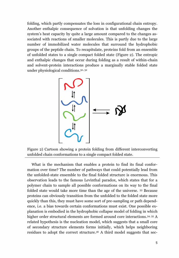
groups of the peptide chain. To recapitulate, proteins fold from an ensemble
of unfolded states to a single compact folded state (Figure 2). The entropic
and enthalpic changes that occur during folding as a result of within-chain
and solvent-protein interactions produce a marginally stable folded state
under physiological conditions.30, 32
Figure 2) Cartoon showing a protein folding from different interconverting
unfolded chain conformations to a single compact folded state.
What is the mechanism that enables a protein to find its final confor-
mation over time? The number of pathways that could potentially lead from
the unfolded-state ensemble to the final folded structure is enormous. This
observation leads to the famous Levinthal paradox, which states that for a
polymer chain to sample all possible conformations on its way to the final
folded state would take more time than the age of the universe. 33 Because
proteins can obviously transition from the unfolded to the folded state more
quickly than this, they must have some sort of pre-sampling or path depend-
ence, i.e. a bias towards certain conformations must exist. One possible ex-
planation is embodied in the hydrophobic collapse model of folding in which
higher order structural elements are formed around core interactions.34, 35 A
related hypothesis is the nucleation model, which suggests that a small core
of secondary structure elements forms initially, which helps neighboring
residues to adopt the correct structure.36 A third model suggests that sec-
6
ondary structures form independently and the final step in the folding pro-
cess involves their rearrangement to give the correct tertiary conformation.37
All of these models resolve the Levinthal paradox by assuming that there is a
bias towards a specific subset of the available conformations. However, it has
yet to be determined which of these frameworks is correct, or whether there
is one single framework that applies to all proteins.
In order to properly describe folding kinetics, there is one more state that
must be probed: the high-energy transition state that connects the unfolded
and folded states. Transition state theory was initially proposed by Eyring to
describe the kinetics of reactions involving small molecules.38 Its key concept
is that there is a high energy barrier between the reactants and products. At
that barrier a few key bonds are broken and formed, pushing the process in
one direction. The application of transition state theory to the kinetics of
protein folding is somewhat challenging because no covalent bonds are bro-
ken or made in the process; instead many weak interactions are broken and
formed.39 However, the assumption of a high energy barrier between the
unfolded and folded states has been helpful in understanding and rationaliz-
ing folding processes. Structural information on folding transition states can
be obtained indirectly through phi-value analysis, which was pioneered by
Alan Fersht.39, 40
In summary, the equilibrium and kinetic properties of protein folding
describe the transition from an ensemble of unfolded chain conformations to
a single folded conformation. The process is complex and not fully under-
stood at present. However, in order to understand how proteins attain their
functional form in-vivo, there is another layer of complexity that must be
considered: the cellular environment.
1.2 Macromolecular Crowding
1.2.1 Cell and Cellular Organization
The cell is the basic working unit of an organism; in the case of prokary-
otes and single-celled eukaryotes, it is the entirety of the organism. In gen-
eral, the cell is organized around its cytosol. In eukaryotic cells, the cytosol
contains a set of membrane-encapsulated organelles such as the mitochon-
dria and Golgi apparatus. Figure 1 shows a cartoon representation of an eu-
karyotic cell, with an outer cell membrane surrounding the cytosol and a set
of distinct cellular compartments. However, this depiction fails to show the
cytosol’s complex composition. The cytosol contains all of the proteins, me-
7
tabolites, and machinery required for protein synthesis as well as the neces-
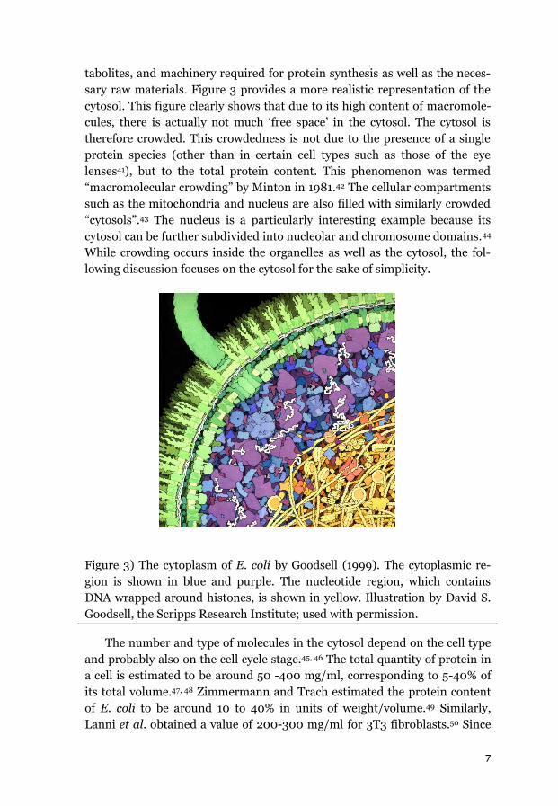
sary raw materials. Figure 3 provides a more realistic representation of the
cytosol. This figure clearly shows that due to its high content of macromole-
cules, there is actually not much ‘free space’ in the cytosol. The cytosol is
therefore crowded. This crowdedness is not due to the presence of a single
protein species (other than in certain cell types such as those of the eye
lenses41), but to the total protein content. This phenomenon was termed
“macromolecular crowding” by Minton in 1981.42 The cellular compartments
such as the mitochondria and nucleus are also filled with similarly crowded
“cytosols”.43 The nucleus is a particularly interesting example because its
cytosol can be further subdivided into nucleolar and chromosome domains.44
While crowding occurs inside the organelles as well as the cytosol, the fol-
lowing discussion focuses on the cytosol for the sake of simplicity.
Figure 3) The cytoplasm of E. coli by Goodsell (1999). The cytoplasmic re-
gion is shown in blue and purple. The nucleotide region, which contains
DNA wrapped around histones, is shown in yellow. Illustration by David S.
Goodsell, the Scripps Research Institute; used with permission.
The number and type of molecules in the cytosol depend on the cell type
and probably also on the cell cycle stage.45, 46 The total quantity of protein in
a cell is estimated to be around 50 -400 mg/ml, corresponding to 5-40% of
its total volume.47, 48 Zimmermann and Trach estimated the protein content
of E. coli to be around 10 to 40% in units of weight/volume.49 Similarly,
Lanni et al. obtained a value of 200-300 mg/ml for 3T3 fibroblasts.50 Since

8
most of the space in the cytosol is already occupied by other macromole-
cules, it is tempting to ask how proteins fold and function in such surround-
ings. This is particularly important because most of our current information
on protein folding was obtained from in-vitro experiments in dilute solu-
tions. In fact, experimentalists often strive to use the most dilute solution
possible in order to avoid non-idealities and to focus on the “pure” protein
properties. However, given the composition of the cytosol, non-idealities are
to be expected. This raises another question: to what extent do inferences
drawn from in-vitro experiments accurately represent the in-vivo situation?
Various non-idealities could arise in the cytosol, such as excluded volume
effects and non-specific interactions. In addition, the cytosol may be much
more viscous than the very dilute solutions used for in-vitro studies.
Even this more “realistic” picture of the cytosol neglects an important
layer of complexity: the spatial and temporal organization of the cytosol. The
cytosol is not homogenous – its composition varies both spatially and tem-
porally.46, 51, 52 Differences in its local composition can cause density fluctua-
tions and changes in the local concentrations of specific proteins. These dif-
ferences can create what are effectively (micro-) compartments based on
local density fluctuations rather than an enclosing membrane.53
1.2.2 Theoretical Models of Excluded Volume Effects on Proteins
The main aim of this thesis was to explore the consequences of excluded
volume effects arising from steric repulsion. Excluded volume effects occur
with all macromolecules and are particularly important in-vivo due to large
number of macromolecules present in the cytosol. The concept of volume
exclusion was first proposed by the polymer chemist Kuhn to explain the
observation that real polymer chains tend to show less compaction than
would expected in the absence of excluded volume effects.54 The description
of non-ideal gases (using the van’t Hoff isobar) also relies on the concept of
an excluded volume.55 The simplest explanation of the phenomenon is that
two molecules cannot occupy the same space at the same time. Consequent-
ly, there is a zone surrounding each molecule that cannot be entered by any
other molecules without provoking a clash. This can be illustrated by consid-
ering a pair of solid spheres (Figure 4) whose centers must always be sepa-
rated by at least the sum of their radii.
9
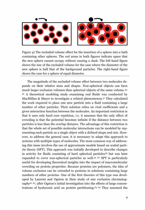
Figure 4) The excluded volume effect for the insertion of a sphere into a bath
containing other spheres. The red areas in both figures indicate space that
the new sphere cannot occupy without causing a clash. The left-hand figure
shows the size of the excluded volume for the case where the diameter of the
new sphere is half that of the background particles. The right-hand figure
shows the case for a sphere of equal diameter.
The magnitude of the excluded volume effect between two molecules de-
pends on their relative sizes and shapes. Non-spherical objects can have
much larger exclusion volumes than spherical objects of the same volume.42,
56 A theoretical modeling study examining real fluids was conducted by
MacMillan & Mayer to investigate a related phenomenon.57 They calculated
the work required to place one new particle into a fluid containing a large
number of other particles. Their solution relies on viral coefficients and a
given interaction function between the molecules. An important restriction is
that it uses only hard core repulsion, i.e. it assumes that the only effect of
crowding is that the potential becomes infinite if the distance between two
particles is less than the overlap distance. The advantage of this restriction is
that the whole set of possible molecular interactions can be modeled by rep-
resenting each particle as a single object with a defined shape and size. How-
ever, to address the general case, it is necessary to adapt this approach to
systems with multiple types of molecules. The most common way of address-
ing this issue involves the use of approximate models based on scaled parti-
cle theory (SPT). This approach was initially developed to describe changes
in activity for fluids consisting of hard spherical particles58 but was later
expanded to cover non-spherical particles as well.59, 60 SPT is particularly
useful for developing theoretical insights into the impact of macromolecular
crowding on protein properties. Because proteins are polymers, the idea of
volume exclusion can be extended to proteins in solutions containing large
numbers of other proteins. One of the first theories of this type was devel-
oped by Laurent and Ogston in their study of size exclusion chromatog-
raphy61, 62, after Ogston’s initial investigation into the effects of large concen-
trations of hyaluronic acid on protein partitioning.63, 64 They assumed the
10
protein to be a sphere and the dextran chromatographic matrix to be an ar-
ray of rods through which the proteins have to migrate. Minton subsequently
built on these results to develop a model of protein activity using a hard-
sphere approximation.65 This approximation was based on the osmotic pres-
sure dependence for concentrated solutions of hemoglobin, which were as-
sumed to behave like collections of hard spheres that only interacted with
one another via hard-core steric exclusion.65-67 Minton later extended his
model to describe protein folding/unfolding and protein association equilib-
ria. In these models, he treated both the folded and unfolded states as ap-
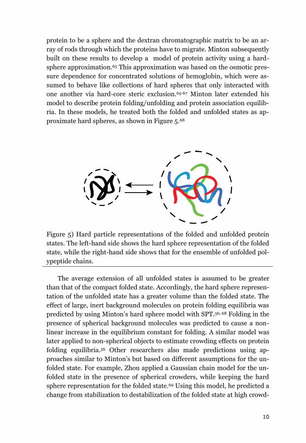
proximate hard spheres, as shown in Figure 5.68
Figure 5) Hard particle representations of the folded and unfolded protein
states. The left-hand side shows the hard sphere representation of the folded
state, while the right-hand side shows that for the ensemble of unfolded pol-
ypeptide chains.
The average extension of all unfolded states is assumed to be greater
than that of the compact folded state. Accordingly, the hard sphere represen-
tation of the unfolded state has a greater volume than the folded state. The
effect of large, inert background molecules on protein folding equilibria was
predicted by using Minton’s hard sphere model with SPT.56, 68 Folding in the
presence of spherical background molecules was predicted to cause a non-
linear increase in the equilibrium constant for folding. A similar model was
later applied to non-spherical objects to estimate crowding effects on protein
folding equilibria.56 Other researchers also made predictions using ap-
proaches similar to Minton’s but based on different assumptions for the un-
folded state. For example, Zhou applied a Gaussian chain model for the un-
folded state in the presence of spherical crowders, while keeping the hard
sphere representation for the folded state.69 Using this model, he predicted a
change from stabilization to destabilization of the folded state at high crowd-
11
er concentrations because the Gaussian chain accommodates voids between
the background molecules. This treatment led to a weaker destabilization of
the unfolded state than the folded state. Minton subsequently proposed a
similar model, assuming the unfolded state to behave like a Gaussian cloud.
The extension of the unfolded ensemble was predicted to decrease, but there
was no obvious change in the overall stabilization effect relative to that ob-
tained using the earlier hard-sphere model.70 All of these models are similar
in that they treat the folded state as a hard sphere, but differ in their repre-
sentation of the unfolded state.
The crowders are also typically modeled as hard particles (usually
spheres) that only interact with the different protein states via hard-core
repulsion. However, new approaches that incorporate attractive interactions
between the crowder particles and proteins have recently been developed.
Minton71 and Zhou72 assumed that the additional attractive interactions
scaled with the exposed surface area, which implies that the unfolded state
will experience more attractive interactions than the folded state. This as-
sumption led to a stabilization of the unfolded state at sufficiently high
crowder concentrations. It was further claimed that the additional attractive
interactions had enthalpic effects whose magnitude should change with the
temperature. When these contributions are considered, the net effect of
crowding may be either stabilizing to destabilizing towards the folded state
depending on the crowder concentration and temperature.
1.2.3 Experimental Studies of Macromolecular Crowding
Effects
To test these theoretical predictions, it is necessary to create controlled
crowded environments in-vitro. An ideal crowder should: 1) be highly solu-
ble; 2) be similar in size to the target protein; 3) have a defined shape; 4)
form no attractive interactions with the protein of interest; 5) not interfere
with the spectroscopic techniques used to study the protein. Crowding with
another protein may seem to be the most straightforward option since that
would most closely represent the situation encountered in a cell. However,
protein crowders usually are not soluble in high enough concentrations and
also form numerous charge-charge interactions because proteins have
charges distributed over their surface. It is therefore necessary to either
screen these charges with either high salt concentrations or to just use low
protein concentrations. Another important concern is that the spectroscopic
techniques used to probe the target protein will be subject to interference
from the protein crowder. Since the protein crowder is present at a much
12
higher concentration, it may dominate the signal and complicate the analy-
sis. Last but not least, the background protein crowder should not undergo
any folding/unfolding transition under the conditions used to induce folding
in the protein of interest.
An alternative option is to use synthetic polymers, so called crowding
agents, to induce the effects of macromolecular crowding. Polymers that
have been used for this purpose include polyethylene glycol (PEG), Dextrans,
Ficoll and Polyvinylpyrrolidone (PVP). PEG, PVP and Dextran offer the ad-
vantage that they can be prepared in different sizes. PEG is a polymer of
ethylene glycol, PVP of N-vinylpyrrolidone, Dextran of glucose, and Ficoll of
sucrose. They are all highly soluble (up to 400 mg/ml or more in water) and
bear no charge at neutral pH. They do not have strong absorption above 210
nm nor are they fluorescent upon excitation at 280 nm. When studying ex-
cluded volume effects, it is desirable to avoid attractive interactions between
the crowding agent and the protein of interest. There is evidence that PEG
forms attractive interactions with proteins in addition to inducing volume
exclusion.73-75 PVP has not been used widely and the only group that had
used it for protein stability studies found that it too forms unwanted attrac-
tive interactions with the protein.76 Another important property of the
crowding agents is their molecular shape. PEG and PVP are likely to be very
flexible polymers.77 In contrast, Ficoll has a more specific, spherical shape.
This is because Ficoll is highly branched copolymer of sucrose and epichlo-
rohydrin, which gives it a relatively compact and often sphere-like
structure.78-81 However, DLS studies have shown that Ficoll 70 adopts a
shape that is intermediate between a sphere and a random coil.82 In another
study, Ficoll was modeled as a sphero-cylinder with a radius of 14 Å.83 Dex-
tran is a polymer of D-glucose with a lower degree of branching than Ficoll
that adopts a more elongated, flexible shape.81, 84, 85 Hydrodynamic radius
values for different Dextrans have been determined by light scattering.85
Excluded volume effects on proteins due to macromolecular crowding
had been investigated for some time, even before Minton coined the term in
1981. For example, it was demonstrated that the addition of PEG or Ficoll
promotes the formation of HIV 86 and bacteriophage 2987 capsids, which
are large macromolecular assemblies. For individual proteins, the volume
changes associated with folding or binding will be smaller than those for
viral capsids, but are still predicted to be sufficiently large to give a macro-
molecular crowding effect. The following section discusses the effects of
macromolecular crowding on phenomena such as association equilibria,
enzymatic activity and the folding equilibria and structure of proteins.
13
In the case of association equilibria, there are two parameters that can
potentially change during the reaction: the volume and shape of the mono-
mer and multimer. Depending on how these parameters change upon asso-
ciation, steric repulsion may stabilize the associated state. The advantage of
using association equilibria to study crowding is that they have well-defined
start and end states. Snoussi and Halle reported a 30-fold increase in the
association equilibrium constant for the formation of bovine trypsin inhibi-
tor decamers based on NMR analyses.88 Similarly, Diaz-Lopez et al. estimat-
ed a 10-fold increase in the equilibrium association constant for a RepA-
DNA complex when using bovine serum albumin (BSA) as crowder.89 In
another study involving protein crowders (Ribonuclease A, RNase A and
human serum albumin), Zorilla et al. used steady state and time-resolved
fluorescence anisotropy to show that the self association of apomyoglobin
increased in the presence of RNase A, but was unaffected by human serum
albumin.90 The free energy change for the conversion of human co-
chaperonin 10 into a heptameric species increased by around 14 kJ/mol in
response to crowding with 300 mg/ml Ficoll 70. It was further shown that
this was primarily caused by effects on the stability of the individual mono-
mers and that effects on the monomer-monomer interfaces were compara-
tively unimportant.91 In 2010, Jiao et al. measured the binding of catalase to
Superoxide dismutase (SOD) using Dextran and Ficoll 70 as crowders and
found that the binding affinity increased by 3.6 kJ/mol in the presence of
either crowder but concluded that the crowders’ steric effects were tempered
by attractive interactions.92
When analyzing enzymatic activity, it is important to understand how
crowding affects the reaction mechanism and whether the reaction is diffu-
sion- or transition state-controlled. Crowded solutions are more viscous than
pure water. This will increase diffusion times, which will reduce the rate of
diffusion-controlled reactions and so would reduce enzymatic activity rather
than increasing it due to any change in volume.93 Especially for reactions
involving small substrates, the changes in volume on going from the free
protein and substrate to the substrate-protein complex to the free protein
and products can be very small. Indeed, Homchaudhuri et al. reported an
increase in the catalytic rate for alkaline phosphatases in the presence of
Dextran and Ficoll.94 Moran-Zorzano et al. also found that high concentra-
tions of PEG increased the rate of the reaction catalyzed by AspP from E.
coli.95 However, Derham and Harding observed a linear decrease in the ac-
tivity of urease in the presence of increasing concentrations of Dextran or
PEG, although the use of protein crowders caused a non-linear increase.96
Similar non-linear crowding effects on enzymatic activity have also been
14
reported by Pozdnyakova and Wittung-Stafshede for multi-copper oxidase
Fet3p. In this case, the addition of Ficoll or Dextran 20 initially increased the
enzyme’s Km and Kcat values, which peaked at crowder concentrations of
~150 mg/ml.97 The effects of crowding on enzyme kinetics have been re-
viewed by Vöpel and Makhadatzde, who reported that the addition of Ficoll
70 did not generally change the Michaelis constant or catalytic turnover
number, but that some exceptions have been presented.98 Overall, no com-
mon effect of macromolecular crowding on enzyme activity has yet been
identified, and more studies in this area are needed.
Macromolecular crowding can also promote structural transformations.
Most crowding theories predict a change in the relative free energies of the
folded and unfolded states, assuming that the structures of the two states do
not also change. The most obvious such change that might occur is that the
expanded unfolded state may become compacted. Minton postulated a com-
paction of the unfolded state in the presence of crowding agents.70 For ade-
nylate kinase, Ittah et al. showed that adding Dextran 40 caused the distance
between two residues in the unfolded chain to decrease but observed no such
effect on the folded state.99 Two other studies also reported similar unfolded
state compaction in the presence of crowders based on two different tech-
niques and two different proteins (CRAB I 100 and ribosomal protein S16 101).
A more striking and unpredicted result was the finding that crowding by
Dextran 70 or Ficoll 70 affected the folded structures of three proteins: apo-
flavodoxin, VlsE and phosphoglycerate kinase (PGK). Far-UV CD spectro-
scopic analyses indicated that crowding increased the secondary structure
content of apoflavodoxin and VlsE in addition to affecting their equilibrium
properties. These results were rationalized with the help of coarse-grained
simulations 78, 102, and are important because they suggest that the folded
structure observed in-vitro is not necessarily that adopted in-vivo. A subse-
quent in-vivo study by Dahr et al. provided some important support for this
idea, showing that PGK also adopted a more compact tertiary structure in-
vivo than had been observed in-vitro.103
Finally, a number of studies have reported crowding effects on protein
folding equilibria and kinetics. There have been around 20 reports of crowd-
ing effects on thermal or chemical protein unfolding reactions based on stud-
ies using synthetic crowding agents. However, it is difficult to compare these
studies directly because they generally used different types and concentra-
tions of crowding agents. In most cases, the crowders increased the tested
protein’s equilibrium stability and resistance to thermal or chemical dena-
turation. However, the magnitude of the increased resistance to thermal
15
denaturation varied significantly from protein to protein. The midpoint of
thermal denaturation increased by around 20 °C for the molten globular
form of apomyoglobin in the presence of 270 mg/ml Dextran 30104 while that
of DNase I rose by around 15 °C in 200 mg/ml PEG105. Much more modest
changes have also been reported: the midpoint for the thermal denaturation
of PGK increased by only ~1.5 °C in 150 mg/ml Ficoll103 while that of maltose
binding protein increased by ~1.0 °C in the presence of 300 mg/ml Ficoll106.
Compared to equilibrium studies, the effects of macromolecular crowding on
protein folding kinetics have received relatively little attention. The refolding
rate constant of carbonic anhydrase increased in the presence of Ficoll 70
but the total amount of refolded protein decreased.107 Similarly, crowding
caused reduced lysozyme to exhibit a slightly increased refolding speed but a
reduced level of correct refolding due to aggregation.108, 109 The refolding rate
constants of VlsE110, apoflavodoxin79, 111 and apocytochrome b562112 in-
creased in the presence of crowders such as Ficoll, Dextran or PEG, but their
unfolding rate constants were unaffected.
1.2.4 Computer Simulations of Crowding Effects
Computer simulations, especially molecular dynamics (MD) simulations
have become important tools in crowding studies. It is difficult to calculate
the excluded volume for non spherical objects, but simulations offer a way of
approximating their effects. Such simulations can be simplified by using
coarse-grained rather than all-atom approaches. In a coarse-grained simula-
tion, individual amino acids (rather than individual atoms) are represented
by balls and springs. This approach greatly reduces the number of interac-
tions that have to be calculated. In both approaches, crowders are modeled
as spheres or rods of a given size that only interact repulsively with the pro-
tein. Their sizes are often chosen to fit experimental data for Ficoll 70 and
Dextrans. The simulations that have been reported correlate well with exper-
imental findings and indicate that crowding destabilizes the unfolded state
relative to the folded state.113-118 A common finding in these studies was that
crowding decreased the radius of gyration for the unfolded state.118-120 A sim-
ilar conclusion was drawn by Goldenberg, who performed a Monte Carlo
simulation of a set of proteins and found that the unfolded state should be
more compact.121 MD simulations have shown that crowding accelerates
peptide folding. Interestingly, however, the rate of folding does not increase
linearly with crowder concentration; instead, it rises at low crowder concen-
trations and then starts to fall when the concentration is further increased.117
16
Elcock and Cheung both built models of the whole cytoplasm of the pro-
karyotic cell. Elcock’s simulation showed that it was necessary to consider
attractive interactions as well as excluded volume effects in order to explain
the difference between computed results and in-vivo observations of the
translational diffusion of green fluorescent protein.122 Cheung et al. also
showed that the melting temperature of a tracer protein (apoazurin) in-
creased by 5 °C at equilibrium in a cytoplasm model.123
To sum up, computer simulations are a useful tool for predicting the ef-
fects of crowding on protein properties that can complement experimental
data and provide key insights into the mechanisms by which various pro-
cesses occur.
17
1.3 Aim of the Project
The cell is filled with macromolecules and so the intracellular environ-
ment differs from the dilute in-vitro conditions that are typically used in
experimental studies on protein folding. Macromolecular crowding causes
excluded volume effects, viscosity changes and changes in non-specific inter-
actions. Theoretical models of volume exclusion predict that there should be
differences between thermodynamic parameters measured in-vitro and
those seen in-vivo. It is therefore important to experimentally characterize
volume exclusion effects on protein biophysics. While many studies have
addressed the stabilizing effects of macromolecular crowding on folded pro-
teins, most of these studies have focused on only one crowding agent and
one protein at a time.
The aim of the thesis is to use a range of “well behaved” model proteins
to systematically investigate the effects of crowding agents with varying
chemical properties and shapes, at different concentrations, on protein fold-
ing in terms of both equilibrium and kinetic effects. These effects were
probed using equilibrium and time-resolved spectroscopy methods. The
findings provide new insights into the underlying determinants of macromo-
lecular crowding effects on protein folding and stability in-vitro, and show
how these effects can be connected to in-silico simulations and theoretical
models.
18
2. Materials and Methods
2.1 Protein Equilibrium Stability
The unfolding of a protein from a compact folded state to an ensemble of
extended unfolded conformations can be described by the equilibrium con-
stant for unfolding (KU), which is defined as the ratio of the concentration of
the unfolded (cu) and the folded (cf) states, or in normalized terms as the
fraction unfolded (fu) over the fraction folded (ff). The corresponding free
energy change of unfolding is ΔGU and is related to KU by the general gas
constant (R) and the absolute temperature (T).
Reaction: Folded State Unfolded State
Unfolded State
KU = cu/cf = fu/ff (1)
ΔGU = -R * T * ln(KU) (2)
To obtain information about the equilibrium constant, the system must
be disturbed using heat or chemical denaturants such as urea or GuHCl, with
the latter being the stronger denaturant.13 Important quantities in these
modes of denaturation are the midpoints of chemical (D1/2) and thermal
denaturation (Tm), defined as the point at which KU=1 or, in a two state pro-
cess, where the fractions of folded and unfolded material are both equal to
0.5.
For chemical denaturation, the change in the free energy of unfolding
can often be estimated by linear approximation.124, 125 The observed changes
in the transition region are then extrapolated to determine the value ex-
pected for a denaturant concentration of 0 M in order to estimate the pro-
tein’s stability in buffer, ΔGU(H2O).
ΔGU(GuHCl) = ΔGU(H2O) + m * [GuHCl] (3)
Pace et al. found that the m-value (i.e. the derivative of equation (3) with
respect to the GuHCl concentration) corresponds to the increase in solvent-
accessible surface area upon denaturation. Accordingly, larger proteins will
often exhibit higher m-value than smaller ones.124, 126
19
The free energy of unfolding for thermal denaturation can be determined
as a function of the changes in enthalpy (ΔHU) and entropy (ΔSU) of unfold-
ing according to
ΔGU = ΔHU – T * ΔSU (4)
ΔHU and ΔSU are themselves temperature dependent.
ΔSU(T2) = ΔSU(T1) + Δcp * ln(T2/T1)
(5)
ΔHU(T2) = ΔHU(T1) + Δcp(T2-T1) (6)
Those expressions can be combined to yield (7), which gives the change
in free energy on going from the starting temperature (T1) to the final tem-
perature (T2).
ΔGU(T2) = ΔHU(T1) + Δcp (T2-T1) – T2 * (ΔSU(T1) + Δcp * ln(T2/T1)) (7)
Like the m-value, the change in heat capacity upon unfolding (Δcp) par-
tially reflects the protein’s solvent-accessible surface area and therefore the
difference in compactness between the folded and unfolded states. The m-
value and Δcp can therefore provide comparable information.127 Δcp can be
determined by directly incorporating it into the fitting procedure as a free
parameter. However, in many cases, the thermal data obtained from spec-
troscopy do not cover a wide enough range to allow reliable determination of
Δcp. DSC can enable the direct determination of Δcp (usually with an error of
about 10%), but can present other problems (see 2.5).
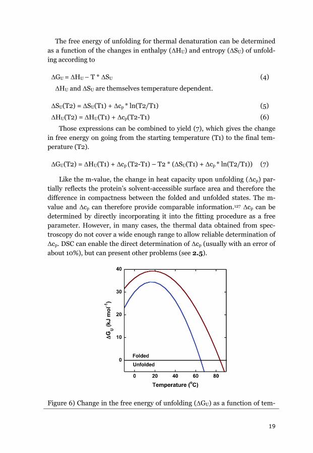
Figure 6) Change in the free energy of unfolding (GU) as a function of tem-
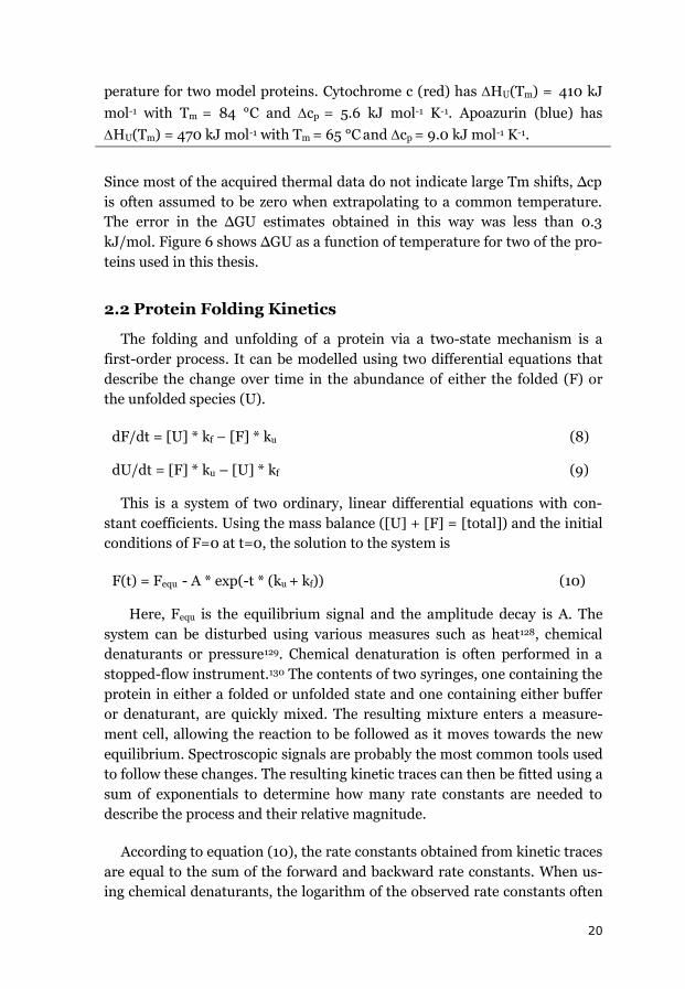
20
perature for two model proteins. Cytochrome c (red) has HU(Tm) = 410 kJ
mol-1 with Tm = 84 °C and cp = 5.6 kJ mol-1 K-1. Apoazurin (blue) has
HU(Tm) = 470 kJ mol-1 with Tm = 65 °C and cp = 9.0 kJ mol-1 K-1.
Since most of the acquired thermal data do not indicate large Tm shifts, Δcp
is often assumed to be zero when extrapolating to a common temperature.
The error in the ΔGU estimates obtained in this way was less than 0.3
kJ/mol. Figure 6 shows ΔGU as a function of temperature for two of the pro-
teins used in this thesis.
2.2 Protein Folding Kinetics
The folding and unfolding of a protein via a two-state mechanism is a
first-order process. It can be modelled using two differential equations that
describe the change over time in the abundance of either the folded (F) or
the unfolded species (U).
dF/dt = [U] * kf – [F] * ku (8)
dU/dt = [F] * ku – [U] * kf (9)
This is a system of two ordinary, linear differential equations with con-
stant coefficients. Using the mass balance ([U] + [F] = [total]) and the initial
conditions of F=0 at t=0, the solution to the system is
F(t) = Fequ - A * exp(-t * (ku + kf)) (10)
Here, Fequ is the equilibrium signal and the amplitude decay is A. The
system can be disturbed using various measures such as heat128, chemical
denaturants or pressure129. Chemical denaturation is often performed in a
stopped-flow instrument.130 The contents of two syringes, one containing the
protein in either a folded or unfolded state and one containing either buffer
or denaturant, are quickly mixed. The resulting mixture enters a measure-
ment cell, allowing the reaction to be followed as it moves towards the new
equilibrium. Spectroscopic signals are probably the most common tools used
to follow these changes. The resulting kinetic traces can then be fitted using a
sum of exponentials to determine how many rate constants are needed to
describe the process and their relative magnitude.
According to equation (10), the rate constants obtained from kinetic traces
are equal to the sum of the forward and backward rate constants. When us-
ing chemical denaturants, the logarithm of the observed rate constants often
21
exhibit the same linear dependence as is seen for the free energy of unfolding
in equilibrium. A plot of the logarithm of the observed rate constants for
folding and refolding against the concentration of denaturant gives a v-
shaped (Chevron) plot. The rate constants for unfolding and folding can then
be obtained by extrapolating the refolding or unfolding limbs of this plot to a
denaturant concentration of 0 M. Additional information can be obtained
from the slopes of the two limbs.
lnku = ln ku(H2O) + mu * [denaturant] (11)
lnkf = ln kf(H2O) + mf * [denaturant] (12)
The m-values of the folding (mf) and unfolding (mu) limbs of the Chevron
plot can be used to calculate the Tanford β (Tβ) value.14, 131 This is a measure
of the relative compaction of the transition state in the direction of the folded
structure relative to the folded (Tβ=1) and unfolded states (Tβ=0).
Tβ = mf / (mu - mf) (13)
Stopped-flow mixing can only be used to study reactions on timescales of
milliseconds or longer. Its main limitation is the so-called dead time. The
samples are mixed before the combined mixture enters the measurement
chamber. Due to this delay, the starting point of a kinetic trace is not t=0.
The dead-time of a stopped-flow instrument is around 2-4 ms, but it de-
pends on the viscosity of the solutions used.
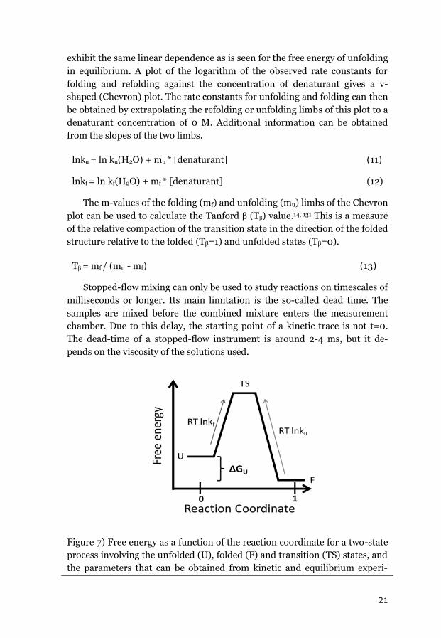
Figure 7) Free energy as a function of the reaction coordinate for a two-state
process involving the unfolded (U), folded (F) and transition (TS) states, and
the parameters that can be obtained from kinetic and equilibrium experi-
22
ments.
An advantage of kinetic over equilibrium measurements is the wider
range of denaturant concentrations that can be studied. Equilibrium meas-
urements are done at around the midpoint of denaturation, within a range of
about 1 M or 10 °C, whereas the range of kinetic measurements extends al-
most all the way down to pure buffer for chemical denaturation.
2.2.1 Non-linearities and additional phases
Proteins (especially small ones132) can fold via a simple two-state pro-
cess. Nevertheless, non-linearities are often observed in the limbs of Chevron
plots, or multiple rate constants may be required to fit the kinetic traces.132,
133 These are sometimes due to cis-trans proline isomerizations. The rate of
these isomerizations can be around 0.1 s–1 – 0.01 s–1 and they are often sepa-
rated from the main folding phase for fast folding proteins.134-136 Various
explanations have been proposed for cases involving roll-overs or curvature
in the limbs of the Chevron plot, such as moving transition states137, 138 and
switches between different transition states.139 140
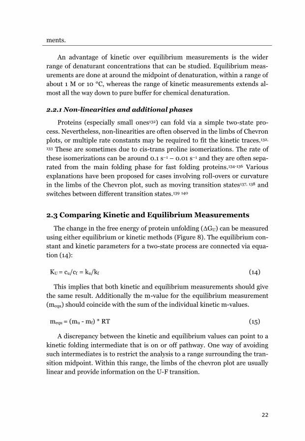
2.3 Comparing Kinetic and Equilibrium Measurements
The change in the free energy of protein unfolding (ΔGU) can be measured
using either equilibrium or kinetic methods (Figure 8). The equilibrium con-
stant and kinetic parameters for a two-state process are connected via equa-
tion (14):
KU = cu/cf = ku/kf (14)
This implies that both kinetic and equilibrium measurements should give
the same result. Additionally the m-value for the equilibrium measurement
(mequ) should coincide with the sum of the individual kinetic m-values.
mequ = (mu - mf) * RT (15)
A discrepancy between the kinetic and equilibrium values can point to a
kinetic folding intermediate that is on or off pathway. One way of avoiding
such intermediates is to restrict the analysis to a range surrounding the tran-
sition midpoint. Within this range, the limbs of the chevron plot are usually
linear and provide information on the U-F transition.
23
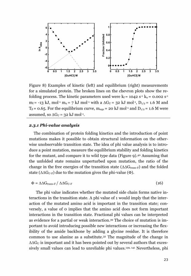
Figure 8) Examples of kinetic (left) and equilibrium (right) measurements
for a simulated protein. The broken lines on the chevron plots show the re-
folding process. The kinetic parameters used were kf = 1042 s-1 ku = 0.002 s-1
mf = -13 kJ, mol-1 mu = 7 kJ mol-1 with a GU = 32 kJ mol-1, D1/2 = 1.6 M and
Tβ = 0.65. For the equilibrium curve, mequ = 20 kJ mol-1 and D1/2 = 1.6 M were
assumed, so GU = 32 kJ mol-1.
2.3.1 Phi-value analysis
The combination of protein folding kinetics and the introduction of point
mutations makes it possible to obtain structural information on the other-
wise unobservable transition state. The idea of phi value analysis is to intro-
duce a point mutation, measure the equilibrium stability and folding kinetics
for the mutant, and compare it to wild type data (Figure 9).40 Assuming that
the unfolded state remains unperturbed upon mutation, the ratio of the
change in the free energies of the transition state (ΔΔGtrans-F) and the folded
state (ΔΔGU-F) due to the mutation gives the phi-value (Φ).
Φ = ΔΔGtrans-F / ΔΔGU-F (16)
The phi value indicates whether the mutated side chain forms native in-
teractions in the transition state. A phi value of 1 would imply that the inter-
action of the mutated amino acid is important in the transition state; con-
versely, a value of 0 implies that the amino acid does not form important
interactions in the transition state. Fractional phi values can be interpreted
as evidence for a partial or weak interaction.39 The choice of mutation is im-
portant to avoid introducing possible new interactions or increasing the flex-
ibility of the amide backbone by adding a glycine residue. It is therefore
common to use alanine as a substitute.32 The magnitude of the change in
ΔΔGU is important and it has been pointed out by several authors that exces-
sively small values can lead to unreliable phi values.139, 141 Nevertheless, phi
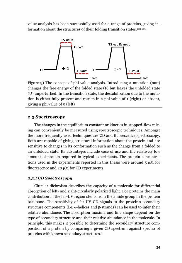
24
value analysis has been successfully used for a range of proteins, giving in-
formation about the structures of their folding transition states.142-145
Figure 9) The concept of phi value analysis. Introducing a mutation (mut)
changes the free energy of the folded state (F) but leaves the unfolded state
(U) unperturbed. In the transition state, the destabilization due to the muta-
tion is either fully present and results in a phi value of 1 (right) or absent,
giving a phi value of 0 (left)
2.3 Spectroscopy
The changes in the equilibrium constant or kinetics in stopped-flow mix-
ing can conveniently be measured using spectroscopic techniques. Amongst
the more frequently used techniques are CD and fluorescence spectroscopy.
Both are capable of giving structural information about the protein and are
sensitive to changes in its conformation such as the change from a folded to
an unfolded state. Its advantages include ease of use and the relatively low
amount of protein required in typical experiments. The protein concentra-
tions used in the experiments reported in this thesis were around 5 μM for
fluorescence and 20 μM for CD experiments.
2.3.1 CD Spectroscopy
Circular dichroism describes the capacity of a molecule for differential
absorption of left- and right-circularly polarized light. For proteins the main
contribution in the far-UV region stems from the amide group in the protein
backbone. The sensitivity of far-UV CD signals to the protein’s secondary
structure components (i.e. α-helices and β-strands) can be used to infer their
relative abundance. The absorption maxima and line shape depend on the
type of secondary structure and their relative abundance in the molecule. In
principle, this makes it possible to determine the secondary structure com-
position of a protein by comparing a given CD spectrum against spectra of
proteins with known secondary structures.7
25
In the case of protein unfolding, the difference in CD spectra between the
folded and unfolded states can be used to determine ΔGU. For a two-state
system, it can be assumed that only folded and unfolded species exist in
equilibrium and that the measured CD signal is a linear combination of the
folded and unfolded signals. Thus, by measuring the CD spectra under dif-
ferent denaturing conditions, an unfolding curve can be obtained by plotting
fu against the concentration of GuHCl or the temperature.
2.3.2 Fluorescence Spectroscopy
Fluorescence is the light emitted by a molecule as it returns to its ground
state after excitation. The three aromatic amino acids tryptophan, phenylal-
anine and tyrosine can be excited by light in the 260 -295 nm region. Since
their excitation and emission spectra are dependent on their respective sur-
roundings, they are good probes for investigating a protein’s integrity. The
probes sense their local environment and therefore provide localized infor-
mation on protein structure. As in the case of CD spectroscopy, the differ-
ence between the emission spectra for the folded and unfolded states can be
used to follow the denaturation of a protein by gradually changing the dena-
turing conditions. The advantage of fluorescence spectroscopy compared to
CD is its greater sensitivity. This makes it possible to use lower protein con-
centrations, avoiding possible side reactions such as aggregation.8
2.4 Crowder Preparation
The Ficoll (Sigma-Aldrich) and Dextrans (Pharmacosmos, Denmark)
powders used in the experimental studies readily dissolved in buffer without
any heating. The concentrations of the crowder solutions were determined
by measuring their optical rotation. The reducing sugar content of the Ficoll
and Dextran solutions was determined to be around 0.1 % using 3,5-
dinitrosalicylic acid, which was considered to be negligible.146
2.5 Differential scanning calorimetry (DSC)
DSC makes it possible to directly measure the heat released during pro-
tein unfolding, in contrast to the indirect inferences obtained using CD or
fluorescence. DSC measures the energy needed to keep the temperature of a
sample cell relative to a buffer cell constant while scanning through a tem-
perature range. Processes that cause heat release or uptake (like protein
unfolding) in the sample cell can thus be detected and quantified. Addition-
ally, Δcp can be obtained from the pre- and post-transitional baselines.
26
Because DSC only measures actual heat transfers, the thermograms are
independent of the background protein concentration as long as the back-
ground species does not undergo any transitions. In principle, this means
that it should be possible to measure the stability of one protein in the pres-
ence of a large background concentration of another protein, i.e. to investi-
gate the effects of crowding one protein with another provided that their
melting points are sufficiently far apart. Unfortunately, in a preliminary trial
using ubiquitin and cytochrome c, it was found that the solubilities of the
proteins were below 100 mg/ml. In addition, large shifts in the baselines for
the crowding agents Ficoll and Dextran were observed during re-scans,
probably due to oxidation. This made the estimated integrated ΔHU values
unreliable. The van’t Hoff ΔHU value that was obtained was comparable to
that determined using CD and fluorescence, but the main advantage of DSC
(i.e. the ability to directly measure heat exchange) was lost. In summary,
while DSC could in principle be very useful for measuring the thermal stabil-
ity of proteins, the stability problems encountered while using sugar crowd-
ers, the high required protein concentrations, and the inability to obtain
results that cannot be obtained using CD or fluorescence spectroscopy pre-
cluded further experiments.
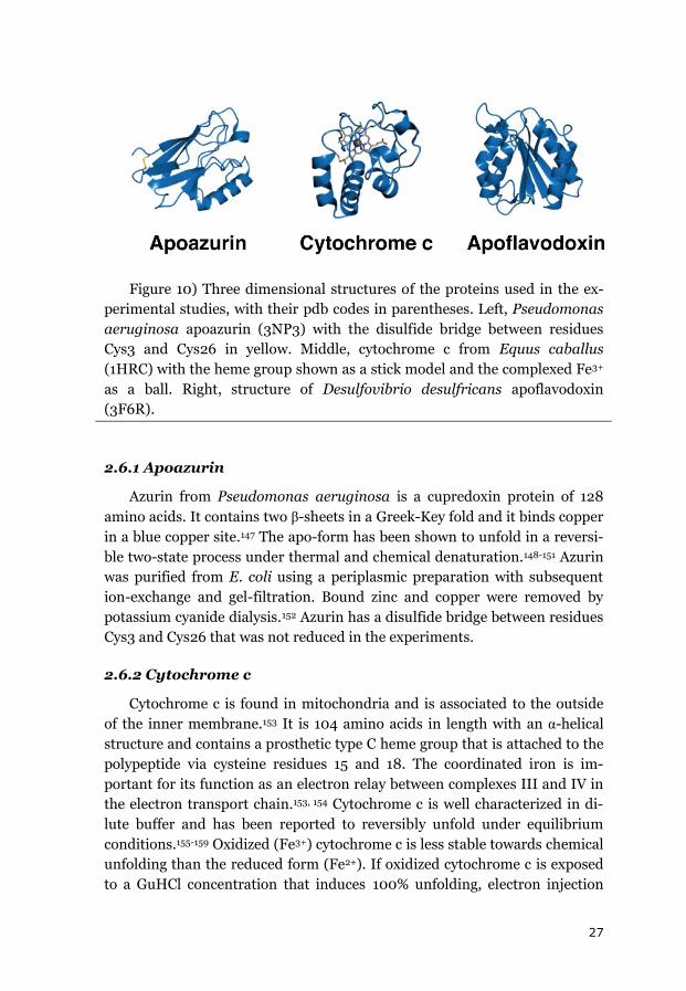
2.6 Model Proteins
The model systems chosen for the investigation of macromolecular
crowding effects on protein folding were apoazurin, cytochrome c and apo-
flavodoxin. These proteins have been studied in dilute buffer systems and
have been reported to fold via equilibrium two-state processes. Azurin and
cytochrome c bind metal cofactors (copper and heme) while flavodoxin binds
flavin mononucleotide (FMN). Table (1) summarizes some of the most inter-
esting properties of the three proteins and figure 10 shows their structures.
In the experiments, cytochrome c was used with the heme group attached
but azurin and flavodoxin were used in their apo forms.
Table 1) General Parameters of the Model Proteins
Protein Length Fold pI Cofactor
Cytochrome c 104 α 9.6 heme
Flavodoxin 148 α/β 4.2 FMN
Azurin 128 β 5.7 Copper
27
Figure 10) Three dimensional structures of the proteins used in the ex-
perimental studies, with their pdb codes in parentheses. Left, Pseudomonas
aeruginosa apoazurin (3NP3) with the disulfide bridge between residues
Cys3 and Cys26 in yellow. Middle, cytochrome c from Equus caballus
(1HRC) with the heme group shown as a stick model and the complexed Fe3+
as a ball. Right, structure of Desulfovibrio desulfricans apoflavodoxin
(3F6R).
2.6.1 Apoazurin
Azurin from Pseudomonas aeruginosa is a cupredoxin protein of 128
amino acids. It contains two β-sheets in a Greek-Key fold and it binds copper
in a blue copper site.147 The apo-form has been shown to unfold in a reversi-
ble two-state process under thermal and chemical denaturation.148-151 Azurin
was purified from E. coli using a periplasmic preparation with subsequent
ion-exchange and gel-filtration. Bound zinc and copper were removed by
potassium cyanide dialysis.152 Azurin has a disulfide bridge between residues
Cys3 and Cys26 that was not reduced in the experiments.
2.6.2 Cytochrome c
Cytochrome c is found in mitochondria and is associated to the outside
of the inner membrane.153 It is 104 amino acids in length with an α-helical
structure and contains a prosthetic type C heme group that is attached to the
polypeptide via cysteine residues 15 and 18. The coordinated iron is im-
portant for its function as an electron relay between complexes III and IV in
the electron transport chain.153, 154 Cytochrome c is well characterized in di-
lute buffer and has been reported to reversibly unfold under equilibrium
conditions.155-159 Oxidized (Fe3+) cytochrome c is less stable towards chemical
unfolding than the reduced form (Fe2+). If oxidized cytochrome c is exposed
to a GuHCl concentration that induces 100% unfolding, electron injection
28
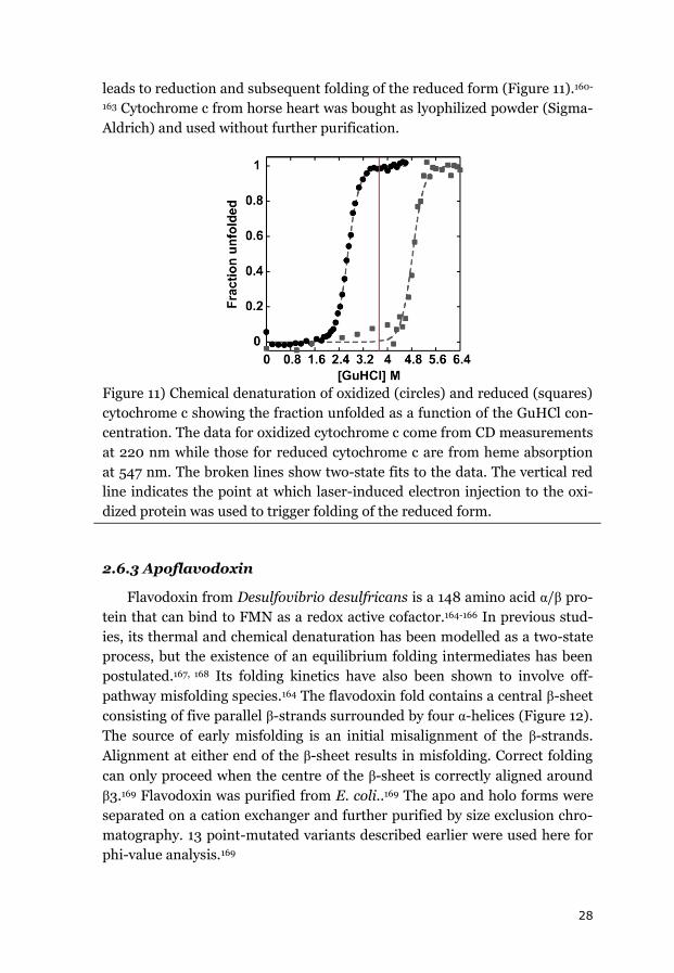
leads to reduction and subsequent folding of the reduced form (Figure 11).160-
163 Cytochrome c from horse heart was bought as lyophilized powder (Sigma-
Aldrich) and used without further purification.
Figure 11) Chemical denaturation of oxidized (circles) and reduced (squares)
cytochrome c showing the fraction unfolded as a function of the GuHCl con-
centration. The data for oxidized cytochrome c come from CD measurements
at 220 nm while those for reduced cytochrome c are from heme absorption
at 547 nm. The broken lines show two-state fits to the data. The vertical red
line indicates the point at which laser-induced electron injection to the oxi-
dized protein was used to trigger folding of the reduced form.
2.6.3 Apoflavodoxin
Flavodoxin from Desulfovibrio desulfricans is a 148 amino acid α/β pro-
tein that can bind to FMN as a redox active cofactor.164-166 In previous stud-
ies, its thermal and chemical denaturation has been modelled as a two-state
process, but the existence of an equilibrium folding intermediates has been
postulated.167, 168 Its folding kinetics have also been shown to involve off-
pathway misfolding species.164 The flavodoxin fold contains a central β-sheet
consisting of five parallel β-strands surrounded by four α-helices (Figure 12).
The source of early misfolding is an initial misalignment of the β-strands.
Alignment at either end of the β-sheet results in misfolding. Correct folding
can only proceed when the centre of the β-sheet is correctly aligned around
β3.169 Flavodoxin was purified from E. coli..169 The apo and holo forms were
separated on a cation exchanger and further purified by size exclusion chro-
matography. 13 point-mutated variants described earlier were used here for
phi-value analysis.169
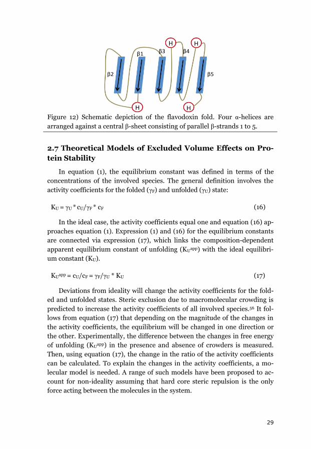
29
Figure 12) Schematic depiction of the flavodoxin fold. Four α-helices are
arranged against a central β-sheet consisting of parallel β-strands 1 to 5.
2.7 Theoretical Models of Excluded Volume Effects on Pro-
tein Stability
In equation (1), the equilibrium constant was defined in terms of the
concentrations of the involved species. The general definition involves the
activity coefficients for the folded (γF) and unfolded (γU) state:
KU = γU * cU/γF * cF
(16)
In the ideal case, the activity coefficients equal one and equation (16) ap-
proaches equation (1). Expression (1) and (16) for the equilibrium constants
are connected via expression (17), which links the composition-dependent
apparent equilibrium constant of unfolding (KUapp) with the ideal equilibri-
um constant (KU).
KUapp = cU/cF = γF/γU * KU
(17)
Deviations from ideality will change the activity coefficients for the fold-
ed and unfolded states. Steric exclusion due to macromolecular crowding is
predicted to increase the activity coefficients of all involved species.56 It fol-
lows from equation (17) that depending on the magnitude of the changes in
the activity coefficients, the equilibrium will be changed in one direction or
the other. Experimentally, the difference between the changes in free energy
of unfolding (KUapp) in the presence and absence of crowders is measured.
Then, using equation (17), the change in the ratio of the activity coefficients
can be calculated. To explain the changes in the activity coefficients, a mo-
lecular model is needed. A range of such models have been proposed to ac-
count for non-ideality assuming that hard core steric repulsion is the only
force acting between the molecules in the system.
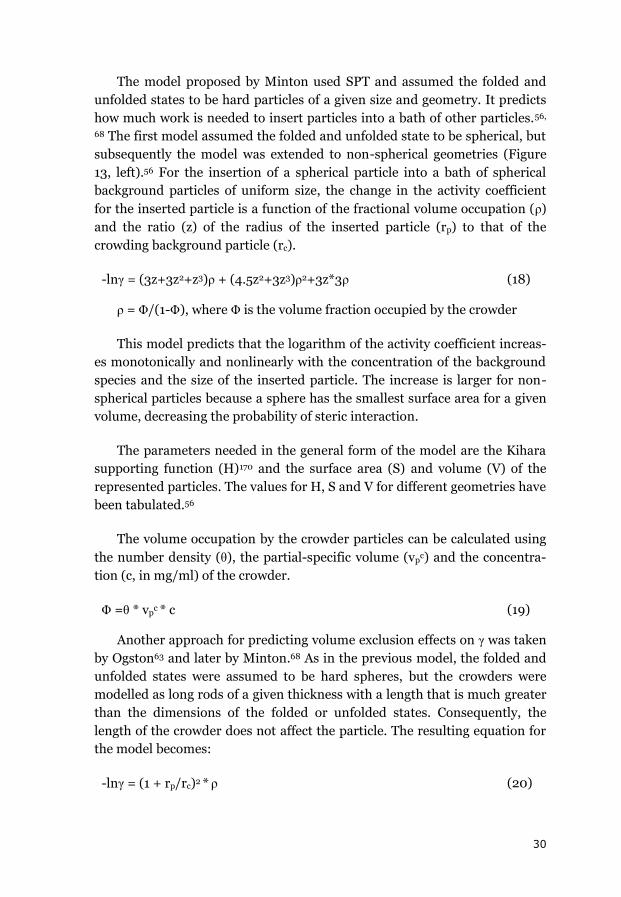
30
The model proposed by Minton used SPT and assumed the folded and
unfolded states to be hard particles of a given size and geometry. It predicts
how much work is needed to insert particles into a bath of other particles.56,
68 The first model assumed the folded and unfolded state to be spherical, but
subsequently the model was extended to non-spherical geometries (Figure
13, left).56 For the insertion of a spherical particle into a bath of spherical
background particles of uniform size, the change in the activity coefficient
for the inserted particle is a function of the fractional volume occupation (ρ)
and the ratio (z) of the radius of the inserted particle (rp) to that of the
crowding background particle (rc).
-lnγ = (3z+3z2+z3)ρ + (4.5z2+3z3)ρ2+3z*3ρ
(18)
ρ = Φ/(1-Φ), where Φ is the volume fraction occupied by the crowder
This model predicts that the logarithm of the activity coefficient increas-
es monotonically and nonlinearly with the concentration of the background
species and the size of the inserted particle. The increase is larger for non-
spherical particles because a sphere has the smallest surface area for a given
volume, decreasing the probability of steric interaction.
The parameters needed in the general form of the model are the Kihara
supporting function (H)170 and the surface area (S) and volume (V) of the
represented particles. The values for H, S and V for different geometries have
been tabulated.56
The volume occupation by the crowder particles can be calculated using
the number density (θ), the partial-specific volume (vpc) and the concentra-
tion (c, in mg/ml) of the crowder.
Φ =θ * vpc * c
(19)
Another approach for predicting volume exclusion effects on γ was taken
by Ogston63 and later by Minton.68 As in the previous model, the folded and
unfolded states were assumed to be hard spheres, but the crowders were
modelled as long rods of a given thickness with a length that is much greater
than the dimensions of the folded or unfolded states. Consequently, the
length of the crowder does not affect the particle. The resulting equation for
the model becomes:
-lnγ = (1 + rp/rc)2 * ρ (20)
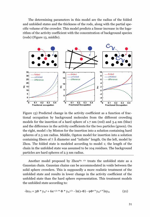
31
The determining parameters in this model are the radius of the folded
and unfolded states and the thickness of the rods, along with the partial spe-
cific volume of the crowder. This model predicts a linear increase in the loga-
rithm of the activity coefficient with the concentration of background species
(rods) (Figure 13, middle).
Figure 13) Predicted change in the activity coefficient as a function of frac-
tional occupation by background molecules from the different crowding
models for the insertion of a hard sphere of 1.7 nm (red) and 3.4 nm (blue)
and the difference in the activity coefficients for the two particles (green). On
the right, model 1 by Minton for the insertion into a solution containing hard
spheres of 2.3 nm radius. Middle, Ogston model for insertion into a solution
containing fibers of 7 Å diameter and “infinite” length. On the left, model by
Zhou. The folded state is modeled according to model 1; the length of the
chain in the unfolded state was assumed to be 104 residues. The background
particles are hard spheres of 2.3 nm radius.
Another model proposed by Zhou69, 171 treats the unfolded state as a
Gaussian chain. Gaussian chains can be accommodated in voids between the
solid sphere crowders. This is supposedly a more realistic treatment of the
unfolded state and results in lower change in the activity coefficient of the
unfolded state than the hard sphere representation. This treatment models
the unfolded state according to:
-lnγU = 3Φ * ym2 + 6π-1/2 * Φ * ym
1/2 - ln(1-Φ) - 9Φ2 * ym2 * lnym
(21)
32
where ym = Rs/61/2 * rc and the root mean square of the end-to-end dis-
tance Rs is a function of the length of the polypeptide chain (N), which was
taken to be equal to172
Rs = 5.31 * N0.6
(22)
The most interesting aspect of this model is the much weaker effect of
the excluded volume on the unfolded state. For high crowder concentrations,
this would result in a net destabilization of the folded state (Figure 13, right).
33
3. Results
The following section presents the main findings of the experimental
studies included in this thesis. The majority of the data have been summa-
rized in the publications that form the thesis’ foundation, but some un-
published results are also presented. The experiments can be divided into
equilibrium and kinetic measurements. The subsection 3.1 discusses the
effects of Dextrans and Ficoll 70 on the equilibrium thermodynamic stability
of cytochrome c and apoazurin; this is followed by a summary of the effect of
macromolecular crowding on protein folding kinetics (section 3.2).
3.1 Effect of Crowding on Equilibrium
The far-UV CD spectral data on the secondary structures of the studied
proteins did not change following the addition of either Ficoll 70 or Dextrans
of any size or at any concentration (Figure 14). This was true for both the
folded and the thermally or chemically unfolded states. The same conclusion
was drawn from the fluorescence emission spectra of the tryptophan envi-
ronment. Visible CD can provide additional information on the integrity of
the folded state of cytochrome c. The CD signal reporting on the heme-group
at 409 nm did not change in the presence of crowding agents. Thus, at this
level of resolution, the crowding agents did not affect the secondary structure
content of any of the target proteins or the environments of the tryptophan
or heme-group in apoazurin and cytochrome c, respectively.
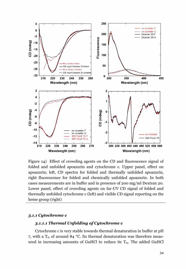
34
Figure 14) Effect of crowding agents on the CD and fluorescence signal of
folded and unfolded apoazurin and cytochrome c. Upper panel, effect on
apoazurin; left, CD spectra for folded and thermally unfolded apoazurin,
right fluorescence for folded and chemically unfolded apoazurin. In both
cases measurements are in buffer and in presence of 200 mg/ml Dextran 20.
Lower panel, effect of crowding agents on far-UV CD signal of folded and
thermally unfolded cytochrome c (left) and visible CD signal reporting on the
heme group (right)
3.1.1 Cytochrome c
3.1.1.1 Thermal Unfolding of Cytochrome c
Cytochrome c is very stable towards thermal denaturation in buffer at pH
7, with a Tm of around 84 °C. Its thermal denaturation was therefore meas-
ured in increasing amounts of GuHCl to reduce its Tm. The added GuHCl
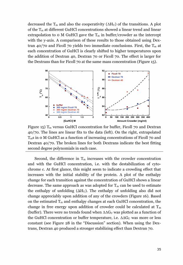
35
decreased the Tm and also the cooperativity (ΔHU) of the transitions. A plot
of the Tm at different GuHCl concentrations showed a linear trend and linear
extrapolation to 0 M GuHCl gave the Tm in buffer/crowder as the intercept
with the y-axis. A comparison of these results to those obtained using Dex-
tran 40/70 and Ficoll 70 yields two immediate conclusions. First, the Tm at
each concentration of GuHCl is clearly shifted to higher temperatures upon
the addition of Dextran 40, Dextran 70 or Ficoll 70. The effect is larger for
the Dextrans than for Ficoll 70 at the same mass concentration (Figure 15).
Figure 15) Tm versus GuHCl concentration for buffer, Ficoll 70 and Dextran
40/70. The lines are linear fits to the data (left). On the right, extrapolated
Tms in 0 M GuHCl as a function of increasing concentrations of Ficoll 70 and
Dextran 40/70. The broken lines for both Dextrans indicate the best fitting
second degree polynomials in each case.
Second, the difference in Tm increases with the crowder concentration
and with the GuHCl concentration, i.e. with the destabilization of cyto-
chrome c. At first glance, this might seem to indicate a crowding effect that
increases with the initial stability of the protein. A plot of the enthalpy
change for each transition against the concentration of GuHCl shows a linear
decrease. The same approach as was adopted for Tm can be used to estimate
the enthalpy of unfolding (ΔHU). The enthalpy of unfolding also did not
change appreciably upon addition of any of the crowders (Figure 16). Based
on the estimated Tm and enthalpy changes at each GuHCl concentration, the
change in free energy upon addition of crowder could be calculated at Tm
(buffer). There were no trends found when ΔΔGU was plotted as a function of
the GuHCl concentration or buffer temperature, i.e. ΔΔGU was more or less
constant (see Figure 28 in the “Discussion” section). When using the Dex-
trans, Dextran 40 produced a stronger stabilizing effect than Dextran 70.
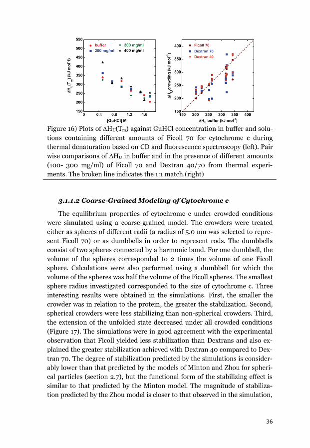
36
Figure 16) Plots of HU(Tm) against GuHCl concentration in buffer and solu-
tions containing different amounts of Ficoll 70 for cytochrome c during
thermal denaturation based on CD and fluorescence spectroscopy (left). Pair
wise comparisons of ΔHU in buffer and in the presence of different amounts
(100- 300 mg/ml) of Ficoll 70 and Dextran 40/70 from thermal experi-
ments. The broken line indicates the 1:1 match.(right)
3.1.1.2 Coarse-Grained Modeling of Cytochrome c
The equilibrium properties of cytochrome c under crowded conditions
were simulated using a coarse-grained model. The crowders were treated
either as spheres of different radii (a radius of 5.0 nm was selected to repre-
sent Ficoll 70) or as dumbbells in order to represent rods. The dumbbells
consist of two spheres connected by a harmonic bond. For one dumbbell, the
volume of the spheres corresponded to 2 times the volume of one Ficoll
sphere. Calculations were also performed using a dumbbell for which the
volume of the spheres was half the volume of the Ficoll spheres. The smallest
sphere radius investigated corresponded to the size of cytochrome c. Three
interesting results were obtained in the simulations. First, the smaller the
crowder was in relation to the protein, the greater the stabilization. Second,
spherical crowders were less stabilizing than non-spherical crowders. Third,
the extension of the unfolded state decreased under all crowded conditions
(Figure 17). The simulations were in good agreement with the experimental
observation that Ficoll yielded less stabilization than Dextrans and also ex-
plained the greater stabilization achieved with Dextran 40 compared to Dex-
tran 70. The degree of stabilization predicted by the simulations is consider-
ably lower than that predicted by the models of Minton and Zhou for spheri-
cal particles (section 2.7), but the functional form of the stabilizing effect is
similar to that predicted by the Minton model. The magnitude of stabiliza-
tion predicted by the Zhou model is closer to that observed in the simulation,
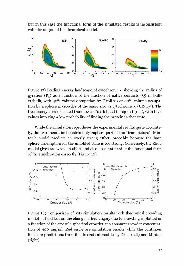
37
but in this case the functional form of the simulated results is inconsistent
with the output of the theoretical model.
Figure 17) Folding energy landscape of cytochrome c showing the radius of
gyration (Rg) as a function of the fraction of native contacts (Q) in buff-
er/bulk, with 40% volume occupation by Ficoll 70 or 40% volume occupa-
tion by a spherical crowder of the same size as cytochrome c (CR-Cyt). The
free energy is color-coded from lowest (dark blue) to highest (red), with high
values implying a low probability of finding the protein in that state
While the simulation reproduces the experimental results quite accurate-
ly, the two theoretical models only capture part of the “true picture”: Min-
ton’s model predicts an overly strong effect, probably because the hard
sphere assumption for the unfolded state is too strong. Conversely, the Zhou
model gives too weak an effect and also does not predict the functional form
of the stabilization correctly (Figure 18).
Figure 18) Comparison of MD simulation results with theoretical crowding
models. The effect on the change in free engery due to crowding is plotted as
a function of the size of a spherical crowder at a constant crowder concentra-
tion of 400 mg/ml. Red circle are simulation results while the continous
lines are predictions from the theoretical models by Zhou (left) and Minton
(right).
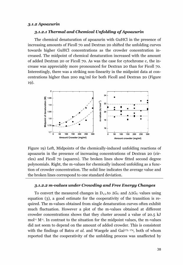
38
3.1.2 Apoazurin
3.1.2.1 Thermal and Chemical Unfolding of Apoazurin
The chemical denaturation of apoazurin with GuHCl in the presence of
increasing amounts of Ficoll 70 and Dextran 20 shifted the unfolding curves
towards higher GuHCl concentrations as the crowder concentration in-
creased. The midpoint of chemical denaturation increased with the amount
of added Dextran 20 or Ficoll 70. As was the case for cytochrome c, the in-
crease was appreciably more pronounced for Dextran 20 than for Ficoll 70.
Interestingly, there was a striking non-linearity in the midpoint data at con-
centrations higher than 200 mg/ml for both Ficoll and Dextran 20 (Figure
19).
Figure 19) Left, Midpoints of the chemically-induced unfolding reactions of
apoazurin in the presence of increasing concentrations of Dextran 20 (cir-
cles) and Ficoll 70 (squares). The broken lines show fitted second degree
polynomials. Right, the m-values for chemically induced unfolding as a func-
tion of crowder concentration. The solid line indicates the average value and
the broken lines correspond to one standard deviation.
3.1.2.2 m-values under Crowding and Free Energy Changes
To convert the measured changes in D1/2 to ΔGU and ΔΔGU values using
equation (3), a good estimate for the cooperativity of the transition is re-
quired. The m-values obtained from single denaturation curves often exhibit
much fluctuation. However a plot of the m-values obtained at different
crowder concentrations shows that they cluster around a value of 20.5 kJ
mol-1 M-1. In contrast to the situation for the midpoint values, the m-values
did not seem to depend on the amount of added crowder. This is consistent
with the findings of Batra et al. and Waegele and Gai173, 174, both of whom
reported that the cooperativity of the unfolding process was unaffected by
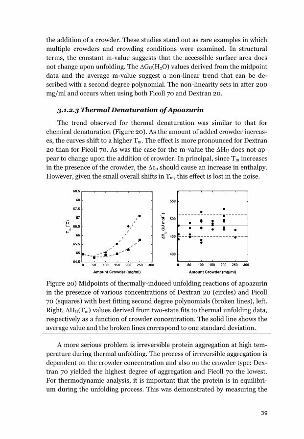
39
the addition of a crowder. These studies stand out as rare examples in which
multiple crowders and crowding conditions were examined. In structural
terms, the constant m-value suggests that the accessible surface area does
not change upon unfolding. The ΔGU(H2O) values derived from the midpoint
data and the average m-value suggest a non-linear trend that can be de-
scribed with a second degree polynomial. The non-linearity sets in after 200
mg/ml and occurs when using both Ficoll 70 and Dextran 20.
3.1.2.3 Thermal Denaturation of Apoazurin
The trend observed for thermal denaturation was similar to that for
chemical denaturation (Figure 20). As the amount of added crowder increas-
es, the curves shift to a higher Tm. The effect is more pronounced for Dextran
20 than for Ficoll 70. As was the case for the m-value the ΔHU does not ap-
pear to change upon the addition of crowder. In principal, since Tm increases
in the presence of the crowder, the Δcp should cause an increase in enthalpy.
However, given the small overall shifts in Tm, this effect is lost in the noise.
Figure 20) Midpoints of thermally-induced unfolding reactions of apoazurin
in the presence of various concentrations of Dextran 20 (circles) and Ficoll
70 (squares) with best fitting second degree polynomials (broken lines), left.
Right, ΔHU(Tm) values derived from two-state fits to thermal unfolding data,
respectively as a function of crowder concentration. The solid line shows the
average value and the broken lines correspond to one standard deviation.
A more serious problem is irreversible protein aggregation at high tem-
perature during thermal unfolding. The process of irreversible aggregation is
dependent on the crowder concentration and also on the crowder type: Dex-
tran 70 yielded the highest degree of aggregation and Ficoll 70 the lowest.
For thermodynamic analysis, it is important that the protein is in equilibri-
um during the unfolding process. This was demonstrated by measuring the
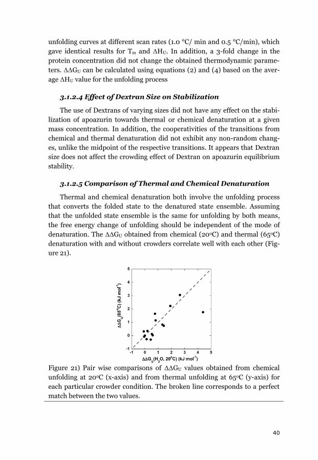
40
unfolding curves at different scan rates (1.0 °C/ min and 0.5 °C/min), which
gave identical results for Tm and ΔHU. In addition, a 3-fold change in the
protein concentration did not change the obtained thermodynamic parame-
ters. ΔΔGU can be calculated using equations (2) and (4) based on the aver-
age ΔHU value for the unfolding process
3.1.2.4 Effect of Dextran Size on Stabilization
The use of Dextrans of varying sizes did not have any effect on the stabi-
lization of apoazurin towards thermal or chemical denaturation at a given
mass concentration. In addition, the cooperativities of the transitions from
chemical and thermal denaturation did not exhibit any non-random chang-
es, unlike the midpoint of the respective transitions. It appears that Dextran
size does not affect the crowding effect of Dextran on apoazurin equilibrium
stability.
3.1.2.5 Comparison of Thermal and Chemical Denaturation
Thermal and chemical denaturation both involve the unfolding process
that converts the folded state to the denatured state ensemble. Assuming
that the unfolded state ensemble is the same for unfolding by both means,
the free energy change of unfolding should be independent of the mode of
denaturation. The ΔΔGU obtained from chemical (20oC) and thermal (65oC)
denaturation with and without crowders correlate well with each other (Fig-
ure 21).
Figure 21) Pair wise comparisons of ΔΔGU values obtained from chemical
unfolding at 20oC (x-axis) and from thermal unfolding at 65oC (y-axis) for
each particular crowder condition. The broken line corresponds to a perfect
match between the two values.
41
The effect of macromolecular crowding on the unfolding of apo-azurin is
therefore the same no matter whether measured using chemical or thermal
denaturation. In addition, two probes (CD and fluorescence) were used for
both perturbations, yielding comparable results. This shows that the unfold-
ing of apo-azurin occurs via a two-state process even under crowding condi-
tions.
3.1.2.6 Modeling of Crowding Effects on Apo-azurin
The equilibrium measurements of the thermal and chemical denatura-
tion of apoazurin showed that its free energy of folding increased in the pres-
ence of the crowding agents Dextran or Ficoll. The degree of stabilization
increased monotonically with the amount of crowder added. Ficoll 70 had a
smaller effect than Dextrans of any size at the same mass concentration, and
the stabilization of apoazurin was independent of Dextran size. Finally, the
stabilizing effect of each crowder followed a linear trend for mass concentra-
tions of up to 200 mg/ml but then became non-linear. Ideally, the theoreti-
cal model used to analyze experimental data should reproduce as many of
the experimental properties as possible. Minton’s hard-particle model uses
fixed geometries for the protein and crowder (sphere and rods) and can ex-
plain the non-linearity but would predict too strong a stabilization for the
Dextrans and assumes a dependence of stabilization on Dextran size. Fur-
thermore, the effect for Ficoll would be non-linear but the non-linearity from
a sphere of 2.3 nm would not be sufficient to explain the observed non-
linearity. The model proposed by Zhou predicts a destabilization at higher
crowder concentrations that does not fit the experimental data. The third
model of Ogston and the equivalent developed by Minton that treats the
crowder molecules as rods (or an array of rods) with a length much greater
than the protein’s radius proved to be the best candidates. Both models pre-
dict a linear response to increases in crowder concentration. An important
input parameter in these models is the size of the equivalent hard sphere for
the folded and unfolded state. For a sphere, this value can be calculated us-
ing the radius of gyration. The radius of gyration of the folded state can be
estimated using the power law relationship proposed by Millett et al.16 The
parameters for the partial specific volume and the thickness of the rods were
determined or taken from literature.175 It was then necessary to decide how
Ficoll should be modeled. In many cases, it has been treated as a hard sphere
with a radius of 5.0 nm. However, some reports indicate that it behaves
much more like an elongated molecule.82, 83 Minton proposed that Ficoll
resembled a sphericocylinder of radius 14 Å with a length to radius ratio of 7.
That would imply that Ficoll is much larger in its extension than the protein

42
in either the folded or unfolded state. Therefore Ficoll was assumed to act
like a rod.
Figure 22) Application of the Ogston model to the Ficoll 70 and Dextran 20
data for the chemical denaturation of apoazurin. The data were fitted exclud-
ing the values for 250 and 300 mg/ml. The radius of gyration of the folded
state was assumed to be 1.6 nm, the diameters of the crowders were 0.7 nm
for Dextran 20 and 1.4 nm for Ficoll 70, and the partial specific volumes of
Dextran 20 and Ficoll 70 were taken to be 0.65 ml/mg.
Using these input parameters, the model was used to fit the ΔΔGU data to
obtain the best fitting size for the unfolded state (Figure 22). The obtained Rg
value was 20 Å for both the Ficoll and the Dextran stability data. This is a
reasonable size because apoazurin contains a disulfide bridge and therefore
the extension of the unfolded state will not be much greater than the folded
state (16 Å). For other proteins of comparable size that also contain disulfide
bridges, the determined unfolded state dimensions match the prediction for
apoazurin. In addition, the fact that the same model gives the same size for
unfolded azurin when used to fit the Ficoll 70 and Dextran data also suggests
that the model and input parameters were well-chosen.
The use of other models is complicated by the fact that only Minton’s model
can describe molecules of different shapes; the models of Ogston and Zhou
always assumes the crowder to be rod-like (Ogston) or spherical (Zhou).
Ficoll 70 is more compact than Dextran and has been modeled as a sphere
before. A comparison of the three models’ output to the Ficoll 70 data (Fig-
ure 23) shows that the Zhou model cannot replicate the observed effects in
magnitude or functional form. One important variable that must be estimat-
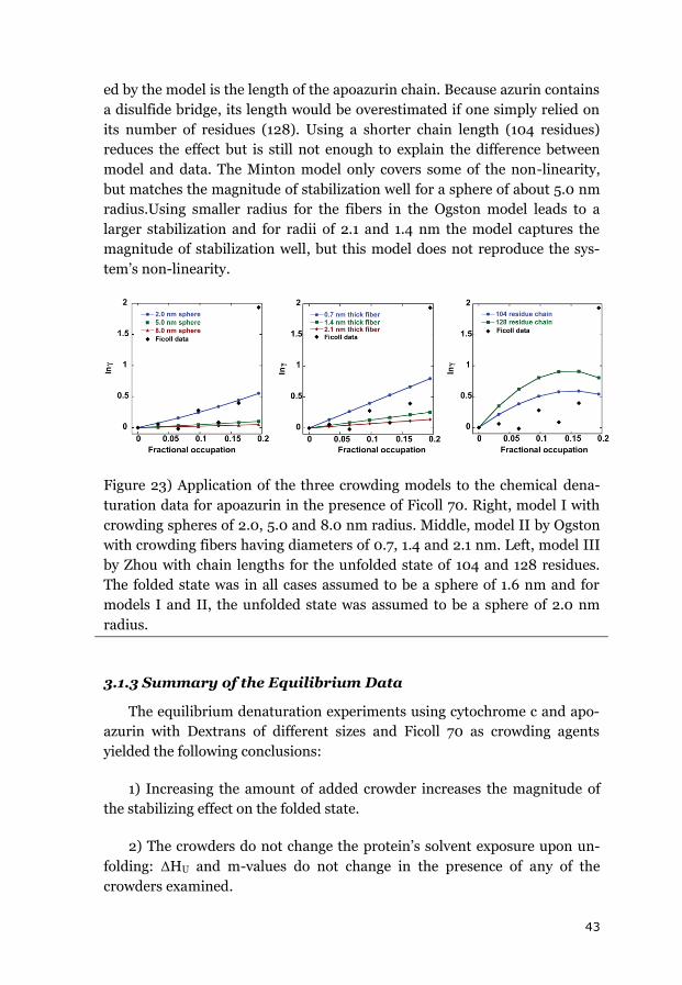
43
ed by the model is the length of the apoazurin chain. Because azurin contains
a disulfide bridge, its length would be overestimated if one simply relied on
its number of residues (128). Using a shorter chain length (104 residues)
reduces the effect but is still not enough to explain the difference between
model and data. The Minton model only covers some of the non-linearity,
but matches the magnitude of stabilization well for a sphere of about 5.0 nm
radius.Using smaller radius for the fibers in the Ogston model leads to a
larger stabilization and for radii of 2.1 and 1.4 nm the model captures the
magnitude of stabilization well, but this model does not reproduce the sys-
tem’s non-linearity.
Figure 23) Application of the three crowding models to the chemical dena-
turation data for apoazurin in the presence of Ficoll 70. Right, model I with
crowding spheres of 2.0, 5.0 and 8.0 nm radius. Middle, model II by Ogston
with crowding fibers having diameters of 0.7, 1.4 and 2.1 nm. Left, model III
by Zhou with chain lengths for the unfolded state of 104 and 128 residues.
The folded state was in all cases assumed to be a sphere of 1.6 nm and for
models I and II, the unfolded state was assumed to be a sphere of 2.0 nm
radius.
3.1.3 Summary of the Equilibrium Data
The equilibrium denaturation experiments using cytochrome c and apo-
azurin with Dextrans of different sizes and Ficoll 70 as crowding agents
yielded the following conclusions:
1) Increasing the amount of added crowder increases the magnitude of
the stabilizing effect on the folded state.
2) The crowders do not change the protein’s solvent exposure upon un-
folding: ΔHU and m-values do not change in the presence of any of the
crowders examined.
44
3) Heat and chemical denaturation give comparable results in terms of
ΔΔGU effects of crowders.
4) The crowding effect is independent or only weakly dependent on Dex-
tran size and always gives a stronger stabilization effect with Dextrans than
Ficoll 70.
5) A simple model using rod-shaped crowders for the excluded volume
can replicate the observed experimental data.
3.2 Effects of Crowding on Folding Kinetics
Folding kinetics were measured for apoazurin, apoflavodoxin and cyto-
chrome c. In the case of apoflavodoxin, a phi value analysis was also per-
formed to determine the effects of crowding on the folding transition state.
3.2.1 Apoazurin Folding Kinetics
Kinetic experiments on the folding of apoazurin with GuHCl as the dena-
turant revealed curvature in the folding and unfolding limbs of the Chevron
plot. To avoid these non-linear regions, the analysis was restricted to the
linear part, which corresponds to the interval where equilibrium denatura-
tion was measured. Nevertheless, it is noteworthy that the curvature did not
disappear upon the addition of the crowder. When using larger dextrans, the
performance of the stopped-flow instrument deteriorated and so Dextran 20
was the only dextran crowder for which large quantities of crowder (up to
200 mg/ml) could be investigated. Since the equilibrium measurements did
not reveal any dependence of the crowding effect on Dextran size, Dextran
20 was taken to be representative of the effects of Dextrans on kinetics. This
assumption was supported by the fact that the chevron plots obtained using
Dextran 40 and 70 at concentrations of up to 100 mg/ml were comparable to
those generated using Dextran 20 data (see Figure 27 in section 3.2.4). Dex-
tran addition caused an increase in the observed refolding rate constants but
left the unfolding rate constants unchanged. The increase in the measured
refolding rate constant at different GuHCl concentrations and increasing
Dextran 20 concentrations was linear. When the shift in rate constants due
to crowding was converted into a ΔΔGU value, the same degree of stabiliza-
tion towards unfolding was obtained as in the equilibrium unfolding experi-
ments. Furthermore the m-values for folding and unfolding were also un-
changed by the addition of Dextran 20 and their sum matched the equilibri-
um m-value well. These results can be best explained by assuming that Dex-
tran has an energetic effect on the unfolded state, leaving the transition and
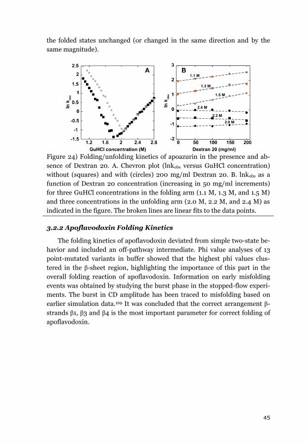
45
the folded states unchanged (or changed in the same direction and by the
same magnitude).
Figure 24) Folding/unfolding kinetics of apoazurin in the presence and ab-
sence of Dextran 20. A. Chevron plot (lnkobs versus GuHCl concentration)
without (squares) and with (circles) 200 mg/ml Dextran 20. B. lnkobs as a
function of Dextran 20 concentration (increasing in 50 mg/ml increments)
for three GuHCl concentrations in the folding arm (1.1 M, 1.3 M, and 1.5 M)
and three concentrations in the unfolding arm (2.0 M, 2.2 M, and 2.4 M) as
indicated in the figure. The broken lines are linear fits to the data points.
3.2.2 Apoflavodoxin Folding Kinetics
The folding kinetics of apoflavodoxin deviated from simple two-state be-
havior and included an off-pathway intermediate. Phi value analyses of 13
point-mutated variants in buffer showed that the highest phi values clus-
tered in the β-sheet region, highlighting the importance of this part in the
overall folding reaction of apoflavodoxin. Information on early misfolding
events was obtained by studying the burst phase in the stopped-flow experi-
ments. The burst in CD amplitude has been traced to misfolding based on
earlier simulation data.169 It was concluded that the correct arrangement β-
strands β1, β3 and β4 is the most important parameter for correct folding of
apoflavodoxin.
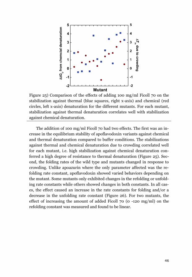
46
Figure 25) Comparison of the effects of adding 100 mg/ml Ficoll 70 on the
stabilization against thermal (blue squares, right x-axis) and chemical (red
circles, left x-axis) denaturation for the different mutants. For each mutant,
stabilization against thermal denaturation correlates well with stabilization
against chemical denaturation.
The addition of 100 mg/ml Ficoll 70 had two effects. The first was an in-
crease in the equilibrium stability of apoflavodoxin variants against chemical
and thermal denaturation compared to buffer conditions. The stabilizations
against thermal and chemical denaturation due to crowding correlated well
for each mutant, i.e. high stabilization against chemical denaturation con-
ferred a high degree of resistance to thermal denaturation (Figure 25). Sec-
ond, the folding rates of the wild type and mutants changed in response to
crowding. Unlike apoazurin where the only parameter affected was the re-
folding rate constant, apoflavodoxin showed varied behaviors depending on
the mutant. Some mutants only exhibited changes in the refolding or unfold-
ing rate constants while others showed changes in both constants. In all cas-
es, the effect caused an increase in the rate constants for folding and/or a
decrease in the unfolding rate constant (Figure 26). For two mutants, the
effect of increasing the amount of added Ficoll 70 (0 -120 mg/ml) on the
refolding constant was measured and found to be linear.
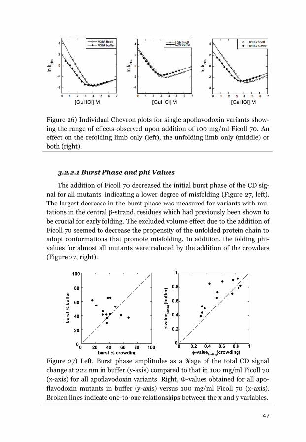
47
Figure 26) Individual Chevron plots for single apoflavodoxin variants show-
ing the range of effects observed upon addition of 100 mg/ml Ficoll 70. An
effect on the refolding limb only (left), the unfolding limb only (middle) or
both (right).
3.2.2.1 Burst Phase and phi Values
The addition of Ficoll 70 decreased the initial burst phase of the CD sig-
nal for all mutants, indicating a lower degree of misfolding (Figure 27, left).
The largest decrease in the burst phase was measured for variants with mu-
tations in the central β-strand, residues which had previously been shown to
be crucial for early folding. The excluded volume effect due to the addition of
Ficoll 70 seemed to decrease the propensity of the unfolded protein chain to
adopt conformations that promote misfolding. In addition, the folding phi-
values for almost all mutants were reduced by the addition of the crowders
(Figure 27, right).
Figure 27) Left, Burst phase amplitudes as a %age of the total CD signal
change at 222 nm in buffer (y-axis) compared to that in 100 mg/ml Ficoll 70
(x-axis) for all apoflavodoxin variants. Right, -values obtained for all apo-
flavodoxin mutants in buffer (y-axis) versus 100 mg/ml Ficoll 70 (x-axis).
Broken lines indicate one-to-one relationships between the x and y variables.
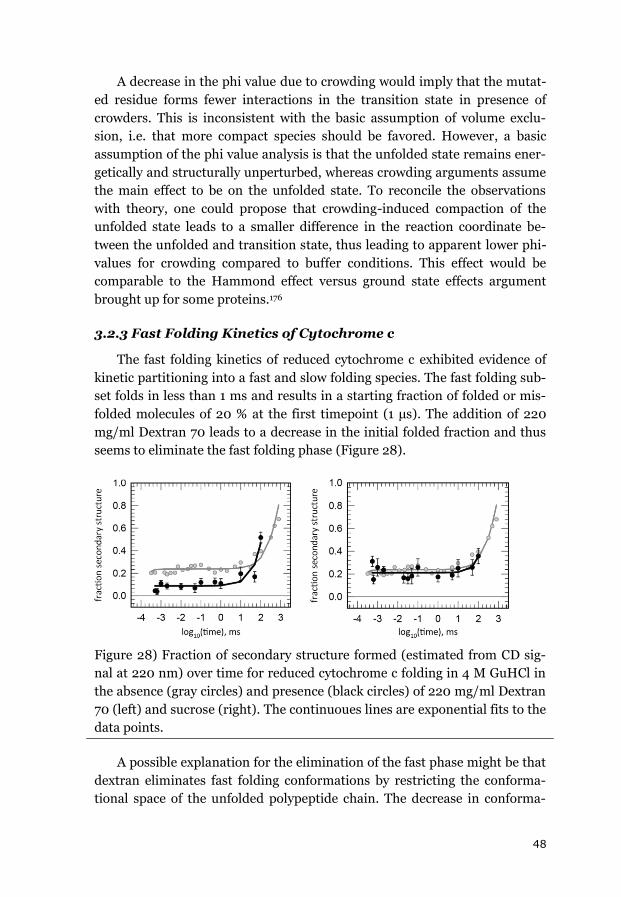
48
A decrease in the phi value due to crowding would imply that the mutat-
ed residue forms fewer interactions in the transition state in presence of
crowders. This is inconsistent with the basic assumption of volume exclu-
sion, i.e. that more compact species should be favored. However, a basic
assumption of the phi value analysis is that the unfolded state remains ener-
getically and structurally unperturbed, whereas crowding arguments assume
the main effect to be on the unfolded state. To reconcile the observations
with theory, one could propose that crowding-induced compaction of the
unfolded state leads to a smaller difference in the reaction coordinate be-
tween the unfolded and transition state, thus leading to apparent lower phi-
values for crowding compared to buffer conditions. This effect would be
comparable to the Hammond effect versus ground state effects argument
brought up for some proteins.176
3.2.3 Fast Folding Kinetics of Cytochrome c
The fast folding kinetics of reduced cytochrome c exhibited evidence of
kinetic partitioning into a fast and slow folding species. The fast folding sub-
set folds in less than 1 ms and results in a starting fraction of folded or mis-
folded molecules of 20 % at the first timepoint (1 μs). The addition of 220
mg/ml Dextran 70 leads to a decrease in the initial folded fraction and thus
seems to eliminate the fast folding phase (Figure 28).
Figure 28) Fraction of secondary structure formed (estimated from CD sig-
nal at 220 nm) over time for reduced cytochrome c folding in 4 M GuHCl in
the absence (gray circles) and presence (black circles) of 220 mg/ml Dextran
70 (left) and sucrose (right). The continuoues lines are exponential fits to the
data points.
A possible explanation for the elimination of the fast phase might be that
dextran eliminates fast folding conformations by restricting the conforma-
tional space of the unfolded polypeptide chain. The decrease in conforma-
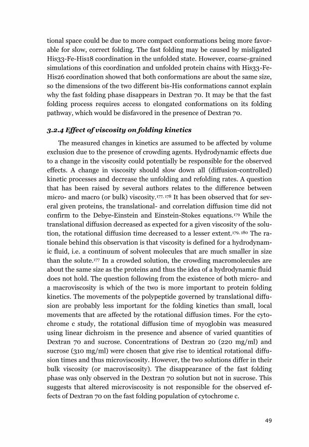
49
tional space could be due to more compact conformations being more favor-
able for slow, correct folding. The fast folding may be caused by misligated
His33-Fe-His18 coordination in the unfolded state. However, coarse-grained
simulations of this coordination and unfolded protein chains with His33-Fe-
His26 coordination showed that both conformations are about the same size,
so the dimensions of the two different bis-His conformations cannot explain
why the fast folding phase disappears in Dextran 70. It may be that the fast
folding process requires access to elongated conformations on its folding
pathway, which would be disfavored in the presence of Dextran 70.
3.2.4 Effect of viscosity on folding kinetics
The measured changes in kinetics are assumed to be affected by volume
exclusion due to the presence of crowding agents. Hydrodynamic effects due
to a change in the viscosity could potentially be responsible for the observed
effects. A change in viscosity should slow down all (diffusion-controlled)
kinetic processes and decrease the unfolding and refolding rates. A question
that has been raised by several authors relates to the difference between
micro- and macro (or bulk) viscosity.177, 178 It has been observed that for sev-
eral given proteins, the translational- and correlation diffusion time did not
confirm to the Debye-Einstein and Einstein-Stokes equations.179 While the
translational diffusion decreased as expected for a given viscosity of the solu-
tion, the rotational diffusion time decreased to a lesser extent.179, 180 The ra-
tionale behind this observation is that viscosity is defined for a hydrodynam-
ic fluid, i.e. a continuum of solvent molecules that are much smaller in size
than the solute.177 In a crowded solution, the crowding macromolecules are
about the same size as the proteins and thus the idea of a hydrodynamic fluid
does not hold. The question following from the existence of both micro- and
a macroviscosity is which of the two is more important to protein folding
kinetics. The movements of the polypeptide governed by translational diffu-
sion are probably less important for the folding kinetics than small, local
movements that are affected by the rotational diffusion times. For the cyto-
chrome c study, the rotational diffusion time of myoglobin was measured
using linear dichroism in the presence and absence of varied quantities of
Dextran 70 and sucrose. Concentrations of Dextran 20 (220 mg/ml) and
sucrose (310 mg/ml) were chosen that give rise to identical rotational diffu-
sion times and thus microviscosity. However, the two solutions differ in their
bulk viscosity (or macroviscosity). The disappearance of the fast folding
phase was only observed in the Dextran 70 solution but not in sucrose. This
suggests that altered microviscosity is not responsible for the observed ef-
fects of Dextran 70 on the fast folding population of cytochrome c.
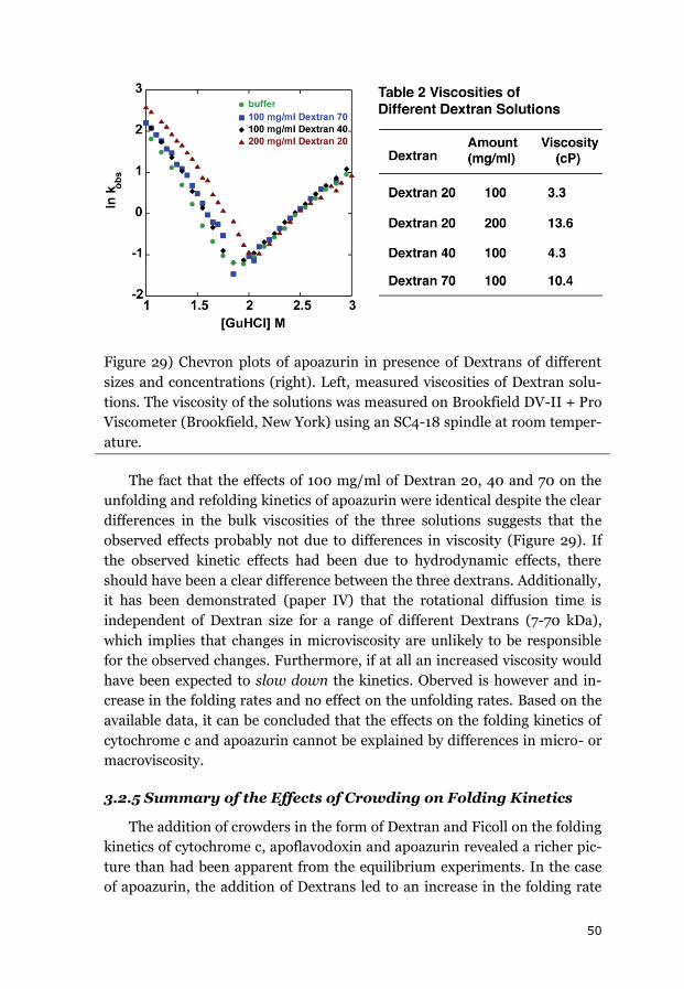
50
Figure 29) Chevron plots of apoazurin in presence of Dextrans of different
sizes and concentrations (right). Left, measured viscosities of Dextran solu-
tions. The viscosity of the solutions was measured on Brookfield DV-II + Pro
Viscometer (Brookfield, New York) using an SC4-18 spindle at room temper-
ature.
The fact that the effects of 100 mg/ml of Dextran 20, 40 and 70 on the
unfolding and refolding kinetics of apoazurin were identical despite the clear
differences in the bulk viscosities of the three solutions suggests that the
observed effects probably not due to differences in viscosity (Figure 29). If
the observed kinetic effects had been due to hydrodynamic effects, there
should have been a clear difference between the three dextrans. Additionally,
it has been demonstrated (paper IV) that the rotational diffusion time is
independent of Dextran size for a range of different Dextrans (7-70 kDa),
which implies that changes in microviscosity are unlikely to be responsible
for the observed changes. Furthermore, if at all an increased viscosity would
have been expected to slow down the kinetics. Oberved is however and in-
crease in the folding rates and no effect on the unfolding rates. Based on the
available data, it can be concluded that the effects on the folding kinetics of
cytochrome c and apoazurin cannot be explained by differences in micro- or
macroviscosity.
3.2.5 Summary of the Effects of Crowding on Folding Kinetics
The addition of crowders in the form of Dextran and Ficoll on the folding
kinetics of cytochrome c, apoflavodoxin and apoazurin revealed a richer pic-
ture than had been apparent from the equilibrium experiments. In the case
of apoazurin, the addition of Dextrans led to an increase in the folding rate
51
constants but left the unfolding rate unchanged. The rate constants for the
apoflavodoxin mutants exhibited diverse behavior depending on the muta-
tion in question, but the presence of the crowder always provided at least
some degree of increased stabilization against unfolding. Furthermore, the
misfolded intermediate was destabilized in the presence of Ficoll 70. For
cytochrome c, the addition of Dextran caused the disappearance of the fast
folding phase. In all cases, the observed effects could not be explained by
differences in viscosity. Instead, the data suggest that excluded volume ef-
fects are responsible for these intriguing observations.
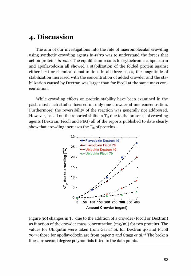
52
4. Discussion
The aim of our investigations into the role of macromolecular crowding
using synthetic crowding agents in-vitro was to understand the forces that
act on proteins in-vivo. The equilibrium results for cytochrome c, apoazurin
and apoflavodoxin all showed a stabilization of the folded protein against
either heat or chemical denaturation. In all three cases, the magnitude of
stabilization increased with the concentration of added crowder and the sta-
bilization caused by Dextran was larger than for Ficoll at the same mass con-
centration.
While crowding effects on protein stability have been examined in the
past, most such studies focused on only one crowder at one concentration.
Furthermore, the reversibility of the reaction was generally not addressed.
However, based on the reported shifts in Tm due to the presence of crowding
agents (Dextran, Ficoll and PEG) all of the reports published to date clearly
show that crowding increases the Tm of proteins.
Figure 30) changes in Tm due to the addition of a crowder (Ficoll or Dextran)
as function of the crowder mass concentration (mg/ml) for two proteins. The
values for Ubiquitin were taken from Gai et al. for Dextran 40 and Ficoll
70173; those for apoflavodoxin are from paper 2 and Stagg et al.78 The broken
lines are second degree polynomials fitted to the data points.
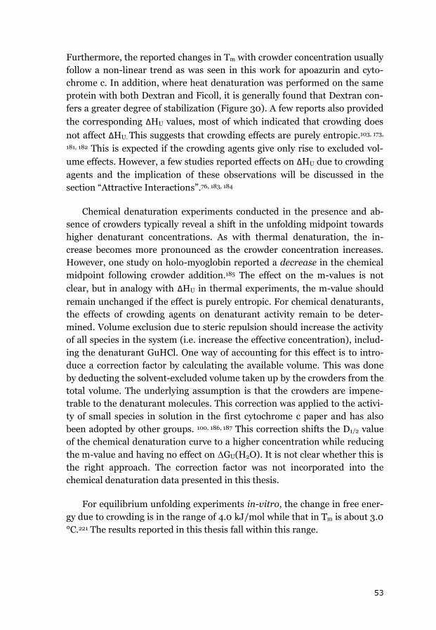
53
Furthermore, the reported changes in Tm with crowder concentration usually
follow a non-linear trend as was seen in this work for apoazurin and cyto-
chrome c. In addition, where heat denaturation was performed on the same
protein with both Dextran and Ficoll, it is generally found that Dextran con-
fers a greater degree of stabilization (Figure 30). A few reports also provided
the corresponding ΔHU values, most of which indicated that crowding does
not affect ΔHU. This suggests that crowding effects are purely entropic.103, 173,
181, 182 This is expected if the crowding agents give only rise to excluded vol-
ume effects. However, a few studies reported effects on ΔHU due to crowding
agents and the implication of these observations will be discussed in the
section “Attractive Interactions”.76, 183, 184
Chemical denaturation experiments conducted in the presence and ab-
sence of crowders typically reveal a shift in the unfolding midpoint towards
higher denaturant concentrations. As with thermal denaturation, the in-
crease becomes more pronounced as the crowder concentration increases.
However, one study on holo-myoglobin reported a decrease in the chemical
midpoint following crowder addition.185 The effect on the m-values is not
clear, but in analogy with ΔHU in thermal experiments, the m-value should
remain unchanged if the effect is purely entropic. For chemical denaturants,
the effects of crowding agents on denaturant activity remain to be deter-
mined. Volume exclusion due to steric repulsion should increase the activity
of all species in the system (i.e. increase the effective concentration), includ-
ing the denaturant GuHCl. One way of accounting for this effect is to intro-
duce a correction factor by calculating the available volume. This was done
by deducting the solvent-excluded volume taken up by the crowders from the
total volume. The underlying assumption is that the crowders are impene-
trable to the denaturant molecules. This correction was applied to the activi-
ty of small species in solution in the first cytochrome c paper and has also
been adopted by other groups. 100, 186, 187 This correction shifts the D1/2 value
of the chemical denaturation curve to a higher concentration while reducing
the m-value and having no effect on ΔGU(H2O). It is not clear whether this is
the right approach. The correction factor was not incorporated into the
chemical denaturation data presented in this thesis.
For equilibrium unfolding experiments in-vitro, the change in free ener-
gy due to crowding is in the range of 4.0 kJ/mol while that in Tm is about 3.0
°C.221 The results reported in this thesis fall within this range.
54
Figure 31) Collection of reported effects on protein folding equilibria as func-
tion of the amount of Ficoll 70 (A) or Dextran of any size (B). See table (x) for
values and sources. Effects of stabilization as a function of protein size (in
number of residues) for 100 mg/ml Ficoll 70 (C) and Dextran (D). The bro-
ken lines in C and D are linear fits to the data points.
Theoretical considerations suggest that in addition to the size and geom-
etry of the crowders, the relative sizes of the folded and unfolded protein
states and the ratio of protein to crowder size should have important impacts
on the crowding effect. Larger proteins have a larger folded state but a com-
paratively larger unfolded state, so the effect of crowding should increase
with protein size. The paucity of studies using the same crowder (or crowd-
ers) and concentrations make comparisons difficult. However, analysis of the
literature data for all of the proteins that have been used in denaturation
experiments in the presence of Ficoll or Dextran (on the assumption that the
effects of Dextrans are independent of their size) gives Figure 31. It is clear
that the crowding effect scales with the concentrations of Dextran and Ficoll,
and that the effect is somewhat non-linear. For a crowder mass concentra-
tion of 100 mg/ml, there are data for more than 2 proteins in solution with
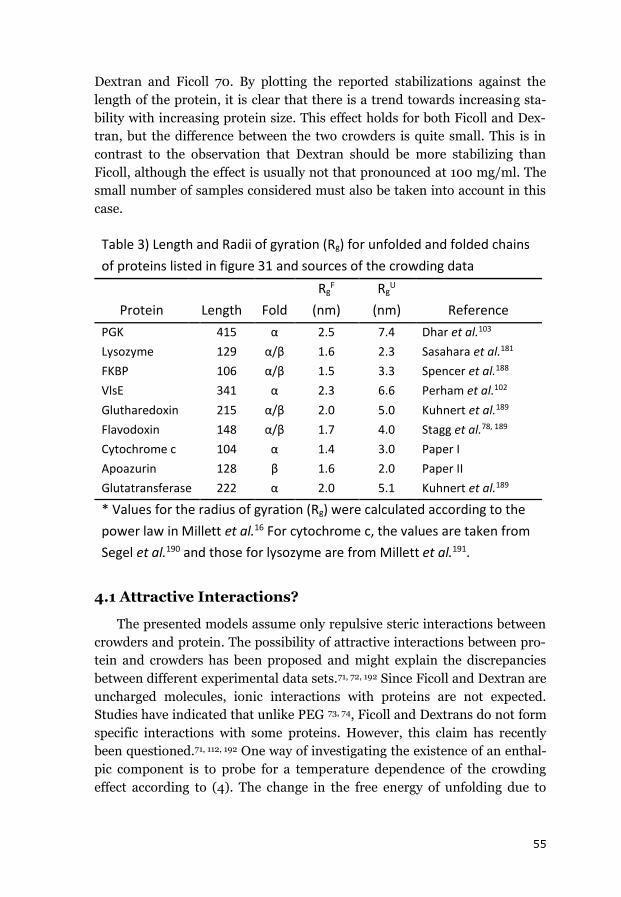
55
Dextran and Ficoll 70. By plotting the reported stabilizations against the
length of the protein, it is clear that there is a trend towards increasing sta-
bility with increasing protein size. This effect holds for both Ficoll and Dex-
tran, but the difference between the two crowders is quite small. This is in
contrast to the observation that Dextran should be more stabilizing than
Ficoll, although the effect is usually not that pronounced at 100 mg/ml. The
small number of samples considered must also be taken into account in this
case.
Table 3) Length and Radii of gyration (Rg) for unfolded and folded chains
of proteins listed in figure 31 and sources of the crowding data
Protein Length Fold
RgF
(nm)
RgU
(nm) Reference
PGK 415 α 2.5 7.4 Dhar et al.103
Lysozyme 129 α/β 1.6 2.3 Sasahara et al.181
FKBP 106 α/β 1.5 3.3 Spencer et al.188
VlsE 341 α 2.3 6.6 Perham et al.102
Glutharedoxin 215 α/β 2.0 5.0 Kuhnert et al.189
Flavodoxin 148 α/β 1.7 4.0 Stagg et al.78, 189
Cytochrome c 104 α 1.4 3.0 Paper I
Apoazurin 128 β 1.6 2.0 Paper II
Glutatransferase 222 α 2.0 5.1 Kuhnert et al.189
* Values for the radius of gyration (Rg) were calculated according to the
power law in Millett et al.16 For cytochrome c, the values are taken from
Segel et al.190 and those for lysozyme are from Millett et al.191.
4.1 Attractive Interactions?
The presented models assume only repulsive steric interactions between
crowders and protein. The possibility of attractive interactions between pro-
tein and crowders has been proposed and might explain the discrepancies
between different experimental data sets.71, 72, 192 Since Ficoll and Dextran are
uncharged molecules, ionic interactions with proteins are not expected.
Studies have indicated that unlike PEG 73, 74, Ficoll and Dextrans do not form
specific interactions with some proteins. However, this claim has recently
been questioned.71, 112, 192 One way of investigating the existence of an enthal-
pic component is to probe for a temperature dependence of the crowding
effect according to (4). The change in the free energy of unfolding due to
56
crowding can be split into enthalpic and entropic contributions according to
(23).
ΔΔGU=-RT ln(Kcrowded/Kbuffer)
(23)
This expression can be substituted to yield (24):
ln(Kcrowded/Kbuffer)=-ΔΔHU/RT+ΔΔSU/R
(24)
According to equation (24), enthalpic contributions due to attractive in-
teractions that favor the unfolded state should cause ln(Kcrowded/Kbuffer) to
decrease with decreasing temperature.
Jiao et al. reported that the crowding effects of Ficoll and PEG on the as-
sociation between SOD and catalase depends on the temperature and con-
cluded that an enthalpic component exists in that system92. However in a
study on the interaction between bovine serum albumin (BSA) and Ficoll
using equilibrium sedimentation, no temperature dependence of the ob-
served changes in buoyant mass was observed and so it was concluded that
only entropic effects were relevant in this case 83. However, a temperature
dependence was observed for protein crowders.193 The Pielak group reported
that the HU values for CI2 and ubiquitin changed following the addition of
crowders (PVP, Ficoll) and interpreted that as a consequence of non-specific
attractive crowder-protein interactions.76, 183, 184
For the proteins studied in this thesis work, cytochrome c and apoazurin,
the available data do not indicate that the crowding effect is temperature
dependent (Figure 32). The chemical denaturation of apoazurin was per-
formed at 20 °C while the thermal denaturation was measured at around 65
°C. The changes in the KU values for these two methods following the addi-
tion of Ficoll or Dextran were very similar. If attractive interactions were
present, the two values should have been different, with a more pronounced
stabilizing effect at 65 °C than at 20 °C. For cytochrome c, conversion of the
measured Tm and ΔHU values to ΔGU, ΔΔGU (crowding-buffer) and KU also
did not reveal any change in the ratio of lnKU with temperature. Based on
these results, we conclude that the temperature did not measurably affect the
stabilization provided by crowding agents. This suggests that there are no
attractive crowder-protein interactions in the studied systems and supports
the conclusion of purely steric repulsions between the crowding agents and
protein systems considered.
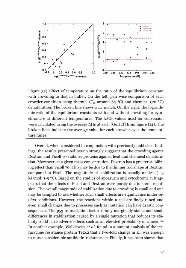
57
Figure 32) Effect of temperature on the ratio of the equilibrium constant
with crowding to that in buffer. On the left: pair wise comparison of each
crowder condition using thermal (Tm around 65 °C) and chemical (20 °C)
denaturation. The broken line shows a 1:1 match. On the right: the logarith-
mic ratio of the equilibrium constants with and without crowding for cyto-
chrome c at different temperatures. The GU values used for conversion
were calculated using the average HU at each [GuHCl] from figure (14). The
broken lines indicate the average value for each crowder over the tempera-
ture range.
Overall, when considered in conjunction with previously published find-
ings, the results presented herein strongly suggest that the crowding agents
Dextran and Ficoll 70 stabilize proteins against heat and chemical denatura-
tion. Moreover, at a given mass concentration, Dextran has a greater stabiliz-
ing effect than Ficoll 70. This may be due to the thinner rod shape of Dextran
compared to Ficoll. The magnitude of stabilization is usually modest (1-3
kJ/mol; 1-5 °C). Based on the studies of apoazurin and cytochrome c, it ap-
pears that the effects of Ficoll and Dextran were purely due to steric repul-
sion. The overall magnitude of stabilization due to crowding is small and one
may be tempted to ask whether such small effects are significance under in-
vivo conditions. However, the reactions within a cell are finely tuned and
even small changes due to processes such as mutation can have drastic con-
sequences. The p53 transcription factor is only marginally stable and small
differences in stabilization caused by a single mutation that reduces its sta-
bility could have adverse effects such as an elevated probability of cancer.194
In another example, Walkiewicz et al. found in a mutant analysis of the tet-
racycline resistance protein TetX2 that a two-fold change in Km was enough
to cause considerable antibiotic resistance.195 Finally, it has been shown that
58
very small changes in affinity of hemoglobin for O2 can have significant ef-
fects on activity.196 It should also be noted that to date, only effects on single
proteins have been considered. Reactions within a cell usually occur as parts
of a larger network, and small effects in one link could have important up-
stream consequences. Minton et al. have argued that, for example, activity
changes due to slight changes in cellular volume could lead to a strong re-
sponse in the transport flux over a membrane in a hypothetical transport
system.197
These examples illustrate how narrowly stabilities and activities are
tuned and how even small perturbations, such as those due to crowding, can
have strong effects.
4.2 Crowding Effects on Kinetics
Only a few reports have addressed the effects of crowding agents on pro-
tein folding kinetics. For simple two-state kinetic reactions, the effects of
crowding on the folded, unfolded and transition states need to be consid-
ered. For more complex folding reactions, intermediate structures and/or
parallel pathways must also be taken into account. Excluded volume effects
should be most unfavorable for the more expanded unfolded state, followed
by the transition and finally the folded state. This should result in faster re-
folding rates and invariant or slower unfolding rates. The effects on unfold-
ing rates depend on how the compaction of folded and transition states dif-
fer. For apoazurin and apoflavodoxin, the Tβ is ~0.65, implying that the tran-
sition state is close to the folded state in terms of compaction/interactions.
In keeping with this conclusion, crowding has a noticeable effect on the re-
folding of apoazurin but the unfolding rate is unaffected. Reports from other
groups have shown that the folding rate constant for apocytochrome b562112
is increased in the presence of 100 mg/ml PEG while the unfolding rate con-
stant is unaffected, and similar results have been reported for VlsE in the
presence of Ficoll 70 at mass concentrations of up t0 100 mg/ml110. Howev-
er, for the ribosomal protein S16, both the folding and unfolding rate con-
stants changed upon the addition of 200 mg/ml Dextran 20. 101
Our time-resolved studies indicated that folding is not a two-state pro-
cess for apoflavodoxin and cytochrome c. There is a misfolded burst inter-
mediate of apoflavodoxin that becomes less populated under crowded condi-
tions. It was suggested that this happens because the unfolded state is more
compact in crowded conditions and that this biases the reaction towards
productive folding. In keeping with this hypothesis, coarse-grained simula-
59
tions showed that unfolded apoflavodoxin is more compact and forms more
native interactions in the presence of crowders. For cytochrome c, less fast
folding is observed on the μs timescale in the presence of crowders. Instead,
cytochrome c molecules all fold homogenously to the folded state via a slow-
er reaction under crowded conditions. The proposed explanation is similar to
that for apoflavodoxin: it is suggested that the unfolded state has a compac-
tion bias that pushes the reaction in a certain direction. It is still unclear
whether the fast μs-scale phase of cytochrome c corresponds to correct fold-
ing or misfolding. While the observed effects could be due to heme mis-
ligation in the unfolded state, in-silico data suggest that this is not the case.
Two reports by Monterroso and Minton as well as van den Berg et al.
reached a conclusion similar to that presented here. In the former study, the
refolding speed (measured in terms of the gain in catalytic activity) of car-
bonic anhydrase was probed in the presence of up to 150 mg/ml Ficoll 70.
An increase in the refolding rate was observed, but at the same time the total
amount of refolded protein decreased under crowding. The authors proposed
that a non-productive protein conformation formed to a larger extent under
crowded conditions.107 Van der Berg et al. found that the refolding speed of
reduced lysozyme in 80 mg/ml Ficoll 70 increased, but at the same time they
observed a lower yield of correctly folded lysozyme. Their explanation for the
lower yield was a higher propensity for aggregation of reduced lysozyme in
the presence of Ficoll 70.108, 109
In summary, the effects of crowding agents on the folding kinetics of pro-
teins are (1) increased rate constants of folding (due to destabilization of the
unfolded state) and (2) mechanistic changes and/or bias towards certain
folding pathways due to the altered ensemble of unfolded chain confor-
mations.
4.3 In-vitro vs In-vivo Conditions
The discussion so far has centered on how excluded volume effects can
be mimicked in-vitro using crowding agents such as Ficoll or Dextran. What
does this tell us about in-vivo conditions? In cells, more factors affect pro-
teins, such as viscosity and non-specific interactions. Moreover, in-vivo, the
protein ensemble has a large distribution of different sizes and shapes, which
contribute to the excluded volume effects. In E. coli, the average protein
length is estimated to be around 325 amino acids.198 This is larger than the
proteins used in most studies of in-vitro model systems to date but still
smaller than most crowding agents. Dill et al. have concluded that the major-

60
ity of all proteins in the E. coli proteome have a stability (ΔGU) of about 28
kJ/mol at 37 oC. Thus even a modest increase in stability due to crowding
may have a significant effect on the survival of the proteome during stress
conditions.199
It is difficult to perform experiments inside cells, partly because all of the
factors present in the cell can contribute to the results.200 This challenge has
nevertheless been taken up by some groups. Ghaemmaghami and Oas meas-
ured the stability of suppressor both in-vitro and inside E. coli cells, using
urea as denaturant and monitoring the process via hydrogen exchange
measured by Matrix-assisted laser desorption/ionization (MALDI). They
reported that the stability measured in-vivo did not deviate form the-in vitro
values.201 In contrast, Gierasch measured the stability of retinoic acid-
binding protein (CRAB) in E. coli cells against urea denaturation using
FRET. They found that the protein is destabilized in-vivo and that both its
midpoint and m-values are smaller than the corresponding in-vitro
values.202, 203 Using temperature jumps, Dhar et al. measured the folding
kinetics and stability of FRET-labeled PGK in different cell compartments
(ER, cytosol, nucleus). They found that PGK was slightly stabilized against
heat denaturation and the folding kinetic effects depended on the cell com-
partment in which PGK was situated.103, 204 High resolution structure deter-
mination in cells is difficult, but progress in NMR has yielded preliminary
results as discussed in two review articles.205, 206 Sakakibara et al. succeeded
in determining the structure of TTHAI1718, a metal-binding protein, in E.
coli. The resulting structure was very similar to the in-vitro structure, but
showed differences in a few loop regions.207 A completely different result was
observed for FlgM, an intrinsically disordered protein that becomes struc-
tured in E. coli. The same was observed in-vitro upon addition of 400 mg/ml
bovine serum albumin or glucose.208 For eukaryotic systems, one way of
performing such studies involves microinjection into oocytes of Xenopus
laevis. This approach has been used by Bodart et al. for the human neuronal
protein Tau, which binds and stabilizes microtubules of the cytoskeleton,
and is also intrinsically disordered. The conformation of Tau in Xenopus
oocytes did not differ detectably from that seen in-vitro.209 For mammalian
cells, the challenge was even greater, but early attempts yielded NMR spectra
that were not too different from in-vitro recordings. While a few residues
appeared to have changed, this may have been due to modifications of the
amino-acid residues in many cases.210-212 The measurement of in-vivo pro-
tein structures is still in its early stages, but so far it seems that the overall
folded structure in-vivo is similar to that in-vitro.
61
Viscosity differences between in-vivo and in-vitro environments are an-
other factor that must be considered. The studies on folding kinetics for cy-
tochrome c and apoazurin highlighted the potential viscosity effects caused
by crowding. However, the results obtained suggested that changes in viscos-
ity were not responsible for the measured effects. The viscosity of Dextran
and Ficoll solutions is considerably higher than that of pure water. However,
measurements of the viscosity of the cytoplasm using a range of different
techniques such as single particle tracking (SPR)213, fluorescence recovery
after photobleaching (FRAP)214, 215, and fluorescence correlation spectrosco-
py (FCS)216, 217 all suggested that the diffusion speed of molecules in the cyto-
plasm is only a few times slower than in pure water, and much lower than in
the crowder solutions.177 A larger difference that has been reported is hin-
dered diffusion, i.e. fractions of molecules have been observed to diffuse
differently to others. This was related to membranes or sieving effects of the
cytoskeleton.218 Based on viscosity measurements in the cytoplasm, it ap-
peared that the viscosity is only about 3-times higher than that of pure water
and was less restrictive than one might expect given the large number of
macromolecules present.43, 177, 219, 220 Given that solutions containing crowd-
ing agents have much higher viscosities, it seems likely that the observation
of folding kinetics acquired using crowding agents at high viscosities will also
be valid in the less viscous cytoplasm.
Thus, in-vivo experiments definitely provide information about how pro-
teins fold in the context of the cytoplasm, but it is difficult to discern what
factors influence the observed behaviors or the magnitude of their effects.
The approach presented here involves studying one effect at a time (here,
volume exclusion due to steric repulsion) by careful and systematic experi-
mentation in-vitro.
62
5. Conclusion and Summary
This study sought to elucidate the effects of the macromolecular crowd-
ing agents Ficoll and Dextran on the folding equilibria and kinetic folding
reactions of three proteins. The addition of crowding agents led to a concen-
tration-dependent stabilization of all three proteins against heat and chemi-
cal denaturation. In all cases, the protein stabilization due to Ficoll was
smaller than that for Dextran at the same mass concentration. The stabiliza-
tion effects were shown to be independent of temperature, ruling out enthal-
pic contributions, and simple excluded volume theory could reproduce the
effects. The role of the crowders’ geometry remains unclear: for apoazurin,
modeling that treated Ficoll and Dextran as rods explained the observed
stabilization well while for cytochrome c, modeling Ficoll as a sphere was
sufficient. The studies show that Ficoll and Dextran can be used as crowding
agents to mimic volume exclusion exclusively.
The folding kinetics for apoazurin were faster in the presence of crowd-
ing agents but its unfolding rate was unchanged. The two-state folding
mechanism remained unchanged by crowding. Apoflavodoxin and cyto-
chrome c did not fold via a two-state kinetic process, and crowding also in-
fluenced their folding mechanisms and their channeling between pathways.
For cytochrome c and apoflavodoxin, it was shown that the kinetic effects
were not due to changes in the viscosity of the solution. The kinetic experi-
ments showed that macromolecular crowding speeds up folding by reducing
the unfolded state entropy and that these unfolded state effects reduce the
population of mis-folded species.
63
6. Outlook
The results of this thesis and related investigations have shown that Fi-
coll and Dextrans can be used as crowding agents. Their effects can be de-
scribed in terms of volume exclusion alone, without needing to consider any
specific attractive interactions. However, Ficoll and Dextrans needed to be
modeled as long rods, with the only difference between them being that one
is thicker than the other. This implies that some of the most widely cited and
discussed models of crowding do not capture the effects of Dextran and Fi-
coll. Future experiments in this area should arguably focus on two lines of
investigation, one dealing exclusively with the Ficoll/Dextran systems and
the other looking at crowding in a more general sense.
Attractive crowder-protein interactions have been proposed to occur in
Ficoll and Dextran systems, although many cases exist where pure steric
repulsion was sufficient to explain the observed effects. The nature and
origin of these attractive interactions remains unclear, especially because the
basic chemistry of all proteins is identical. One way of investigating these
interactions would be to measure crowding effects over wide temperature
ranges as opposed to the usual short temperature intervals. Additionally, the
effect should scale with the size of the protein.
Considering the experimental techniques used in this work, it might be in-
teresting to conduct classical colligative experiments, i.e. ultracentrifugation
and osmotic or vapor pressure analyses. Ultracentrifugation has been used to
measure activity coefficients for association equilibria, but unfolding equilib-
ria have not been investigated. It could potentially enable measurement of
the activity coefficients of the unfolded and folded state in the presence of
crowders.
In principal, colligative techniques could be applied to protein crowders
as well. In-vivo studies have shown that proteins are either not affected at all
by such techniques and may even be slightly destabilized. Sometimes this is
seen as proof that crowding has less of an effect or is unimportant in the in-
vivo than expected. Still, these experiments do not explain why the stability
is as observed. On the contrary, it just raises another question, what destabi-
lizes the protein so much as to cancel out these repulsive effects? By bearing
the findings from in-vitro crowding studies with crowding agents in mind,
64
protein crowding systems could be studied with a much stronger focus on
non-specific attractive interactions.
Another experimental approach that has been only used to a small de-
gree in this field to date is NMR. At present, only one group has used this
technique to elucidate crowding effects. NMR too could allow the study of
systems with protein crowders, since labeling of proteins could render the
background protein basically invisible. The residue-specific resolution to-
gether with colligative techniques might give new insights into the thermo-
dynamic parameters underlying protein properties in crowded environ-
ments.
65
Acknowledgements
So, after almost 5 years it is nearly done. Altough the thesis has to be de-
fended by me alone there are still a whole bunch of persons who more or less
directly took part in it. Be it by being involved in the experiments or by help-
ing to make the time more pleasant.
At first place I would like to thank my supervisor Pernilla, afterall thanks
to you I actullay could do my work in macromolecular crowding. After you
recruitment talk in Umea I was already interested in crowding and I was
quite happy the moment in your office when we talked about a project for my
then masterthesis that you directly suggested crowding. I want to thank for
your help, input and advice on my research and experiments. We for sure
had to crack here and there some tough nuts with the crowding stuff. Also
besides research it was always pleasent talking to you and you were always
willing to share a lot of information about how science, funding and publish-
ing works. I admire your way of writing articles and also your determination
as soon as the critical mass for publication has been reached. While others
might then need another year before something more has happened, in your
case that is focused and done!
Next is my second supervisor Magnus. My interest on protein folding was
awakended during your ‘Protein Structure and Function’ course, plus you
gave extra points on the exam if we visited Pernillas’ lecuture. Also, my
thanks for your input and ideas during all the years.
I would also like to thanks the rest of my committee Dan and Ove for your
patience and discussions during the sometimes somewhat lengthy yearly
evaluations. A vague guess from my side: the actual defence will be shorter
than the last evaluation.
Of course I would like to mention and thank my research group, past and
present members. Especially I would like to thank the “original” three Maria,
Moritz and Ximena. Maria besides working together and later as office mates
we had a lot of funny and sometimes not so funny conversations about work,
life and ,of course, the news on “People Magazine”. During the years you
were a great help, a good listener and a good friend! I wish you all the best
for your future and hope we stay in touch! Ximenita, muchas gracias por el
tiempo como compadre en nuestra oficina i nuestras conversaciónes más o
66
menos científicas i que tengas suerte con la defensa tuya! Also thanks to our
post-docs Jörgen and Istavan (egészségedre!).
Yo Mo, danke für die super Zeit. Hat ja schon gleich beim ersten Treffen
„gefunkt“ und jemand der Alpha Centauri kennt, kann ja nur ein guter
Mensch sein, oder? Vielen Dank und Grüße auch an deine Frau Hanna und
die kleine Herja und den weiteren Zuwachs (Kevin oder David?). Grüße auch
an den Rest der deutschen Mafia: Constantin und Familie, Mark, Lena und
Familie und Manuela und seit kurzem dazugestossen Dana und Michael
(hättet mal früher kommen sollen).
Tack Johan för en introduktion till ölbryggning och dem tillfällen när du
och Karin tog hand om en ensam kille pa nyårsafton! Marcus… tack för att
lära mig vara ödmjuk… 5 år och inte en enda gång har jag vunnit i våra
squash matcher. Samuel som jag tackar för att ha varit tandem partner,
squah motståndare och också för fika och alla samtal genom nin tid i Umeå!
Also I would like to acknowledge the rest of the biophyscial corridor. Here
especially P.O.for all conversations about science, actual research and ethics.
Also Tobias, who has always a great help in scientific questions, and Lars
Backman, I liked your course about protein purification and that was one of
the reasons that I decided to stay in Umeå in the first place, even before fi-
nally starting my PhD.
Last but not least thank you Xiaowei for all your help, especially during
the last part of the thesis and for the translation of the poem. I just hope you
did not by “accident” introcduce any “funny” characters; it might still take
some time before I will be able to spot that.
All in all thank you all for the time and all the best for the future!
67
References
1. Alberts, B., Johnson, A., Lewis, J., Raff, M., Roberts, K., and Walter, P. 2002. Molecular Biology of the Cell, 4th edition. Garland Science, New York.
2. Feldman, D. E., and J. Frydman. 2000. Protein folding in vivo: the importance of molecular chaperones. Current Opinion in Structural Biology 10:26-33.
3. Ulrich Hartl, F., and J. Martin. 1995. Molecular chaperones in cellular protein folding. Current Opinion in Structural Biology 5:92-102.
4. Gomes, C. M., Wittung-Stafshede P. (eds.). 2011. Protein Folding and Metal Ions- Mechanisms, Biology and Disease. Boca Raton, FL : CRC Press.
5. Marion, D. 2013. An introduction to biological NMR spectroscopy. Mol Cell Proteomics.
6. van Heel, M., B. Gowen, R. Matadeen, E. V. Orlova, R. Finn, T. Pape, D. Cohen, H. Stark, R. Schmidt, M. Schatz, and A. Patwardhan. 2000. Single-particle electron cryo-microscopy: towards atomic resolution. Q Rev Biophys 33:307-369.
7. Berova N., N. K., Woody R.W. Circualar Dichrosim Principles and Applications. Wiley-VCH, Canada.
8. Royer, C. A. 2006. Probing protein folding and conformational transitions with fluorescence. Chem Rev 106:1769-1784.
9. Nemecek, D., J. Stepanek, and G. J. Thomas. 2001. Raman Spectroscopy of Proteins and Nucleoproteins. In Current Protocols in Protein Science. John Wiley & Sons, Inc.
10. Barth, A., and C. Zscherp. 2002. What vibrations tell us about proteins. Q Rev Biophys 35:369-430.
11. Rose, P. J. F. a. G. D. 2005. Conformational Properties of Unfolded Proteins. Protein Folding Handbook. Wiley-VCH, Weinheim.
12. Dill, K. A., and D. Shortle. 1991. Denatured states of proteins. Annu Rev Biochem 60:795-825.
13. Greene, R. F., and C. N. Pace. 1974. Urea and Guanidine Hydrochloride Denaturation of Ribonuclease, Lysozyme, α-Chymotrypsin, and β-Lactoglobulin. Journal of Biological Chemistry 249:5388-5393.
68
14. C., T. 1968. Protein Denaturation. Advances in Protein Chemistry 23:121-282.
15. Dyson, H. J., and P. E. Wright. 2004. Unfolded Proteins and Protein Folding Studied by NMR. Chemical Reviews 104:3607-3622.
16. Millett, I. S., S. Doniach, and K. W. Plaxco. 2002. Toward a taxonomy of the denatured state: small angle scattering studies of unfolded proteins. Adv Protein Chem 62:241-262.
17. Meier, S., M. Blackledge, and S. Grzesiek. 2008. Conformational distributions of unfolded polypeptides from novel NMR techniques. The Journal of Chemical Physics 128:-.
18. Nodet, G., L. c. Salmon, V. r. Ozenne, S. Meier, M. R. Jensen, and M. Blackledge. 2009. Quantitative Description of Backbone Conformational Sampling of Unfolded Proteins at Amino Acid Resolution from NMR Residual Dipolar Couplings. Journal of the American Chemical Society 131:17908-17918.
19. Haran, G. 2012. How, when and why proteins collapse: the relation to folding. Curr Opin Struct Biol 22:14-20.
20. Millet, I. S., S. Doniach, and K. W. Plaxco. 2002. Toward a taxonomy of the denatured state: Small angle scattering studies of unfolded proteins. Unfolded Proteins 62:241-262.
21. Flory, P. J. 1953. Principles of Polymer Chemistry. Configuration of Polymer Chains:399-425.
22. Flory P. J., F. S. 1965. Effect of Volume Exclusion on the Dimensions of Polymer Phains. Journal of Chemical Physics 44:2243-2247.
23. Garcia, P., L. Serrano, D. Durand, M. Rico, and M. Bruix. 2001. NMR and SAXS characterization of the denatured state of the chemotactic protein CheY: implications for protein folding initiation. Protein Sci 10:1100-1112.
24. Kazmirski, S. L., K. B. Wong, S. M. Freund, Y. J. Tan, A. R. Fersht, and V. Daggett. 2001. Protein folding from a highly disordered denatured state: the folding pathway of chymotrypsin inhibitor 2 at atomic resolution. Proc Natl Acad Sci U S A 98:4349-4354.
25. Sherman, E., and G. Haran. 2006. Coil–globule transition in the denatured state of a small protein. Proceedings of the National Academy of Sciences 103:11539-11543.
26. Merchant, K. A., R. B. Best, J. M. Louis, I. V. Gopich, and W. A. Eaton. 2007. Characterizing the unfolded states of proteins using
69
single-molecule FRET spectroscopy and molecular simulations. Proceedings of the National Academy of Sciences 104:1528-1533.
27. Tezuka-Kawakami, T., C. Gell, D. J. Brockwell, S. E. Radford, and D. A. Smith. 2006. Urea-Induced Unfolding of the Immunity Protein Im9 Monitored by spFRET. Biophysical Journal 91:L42-L44.
28. Kohn, J. E., I. S. Millett, J. Jacob, B. Zagrovic, T. M. Dillon, N. Cingel, R. S. Dothager, S. Seifert, P. Thiyagarajan, T. R. Sosnick, M. Z. Hasan, V. S. Pande, I. Ruczinski, S. Doniach, and K. W. Plaxco. 2004. Random-coil behavior and the dimensions of chemically unfolded proteins. Proceedings of the National Academy of Sciences of the United States of America 101:12491-12496.
29. Yoo, T. Y., S. P. Meisburger, J. Hinshaw, L. Pollack, G. Haran, T. R. Sosnick, and K. Plaxco. 2012. Small-Angle X-ray Scattering and Single-Molecule FRET Spectroscopy Produce Highly Divergent Views of the Low-Denaturant Unfolded State. Journal of Molecular Biology 418:226-236.
30. Dill, K. A. 1990. Dominant forces in protein folding. Biochemistry 29:7133-7155.
31. Privalov, P. L., and S. J. Gill. 1988. Stability of protein structure and hydrophobic interaction. Adv Protein Chem 39:191-234.
32. Fersht, A. 1999. Structure and mechanism in protein science. A guide to enzyme catalysis and protein folding. Freeman Co, New York.
33. Levinthal, C. 1969. How to fold Graciously. Mossbauer Spectroscopy in Biological Systems: Proceedings of a meeting held at Allerton House, Monticello, Illinois:22-24.
34. Dill, K. A., S. Bromberg, K. Yue, K. M. Fiebig, D. P. Yee, P. D. Thomas, and H. S. Chan. 1995. Principles of protein folding--a perspective from simple exact models. Protein Sci 4:561-602.
35. Kuwajima, K. 1989. The molten globule state as a clue for understanding the folding and cooperativity of globular-protein structure. Proteins: Structure, Function, and Bioinformatics 6:87-103.
36. Wetlaufer, D. B. 1973. Nucleation, Rapid Folding, and Globular Intrachain Regions in Proteins. Proceedings of the National Academy of Sciences 70:697-701.
37. Kim, P. S., and R. L. Baldwin. 1982. Specific intermediates in the folding reactions of small proteins and the mechanism of protein folding. Annu Rev Biochem 51:459-489.
70
38. Eyring, H. 1935. The Activated Complex in Chemical Reactions. Journal of Chemical Physics 3:107-115.
39. Fersht, A. R. 1995. Characterizing transition states in protein folding: an essential step in the puzzle. Current Opinion in Structural Biology 5:79-84.
40. Fersht, A. R., A. Matouschek, and L. Serrano. 1992. The folding of an enzyme: I. Theory of protein engineering analysis of stability and pathway of protein folding. Journal of Molecular Biology 224:771-782.
41. Jester, J. V. 2008. Corneal crystallins and the development of cellular transparency. Seminars in Cell & Developmental Biology 19:82-93.
42. Minton, A. P. 1981. Excluded volume as a determinant of macromolecular structure and reactivity. Biopolymers 20:2093-2120.
43. Partikian, A., B. Olveczky, R. Swaminathan, Y. Li, and A. S. Verkman. 1998. Rapid diffusion of green fluorescent protein in the mitochondrial matrix. J Cell Biol 140:821-829.
44. Cremer, T., M. Cremer, S. Dietzel, S. Müller, I. Solovei, and S. Fakan. 2006. Chromosome territories--a functional nuclear landscape. Curr Opin Cell Biol 18:307-316.
45. Luby-Phelps, K. 2000. Cytoarchitecture and physical properties of cytoplasm: volume, viscosity, diffusion, intracellular surface area. Int Rev Cytol 192:189-221.
46. Fuller, B. G. 2010. Self-organization of intracellular gradients during mitosis. Cell Div 5:5.
47. Fulton, A. B. 1982. How crowded is the cytoplasm? Cell 30:345-347.
48. Ellis, R. J., and A. P. Minton. 2003. Cell biology: join the crowd. Nature 425:27-28.
49. Zimmerman, S. B., and S. O. Trach. 1991. Estimation of macromolecule concentrations and excluded volume effects for the cytoplasm of Escherichia coli. Journal of Molecular Biology 222:599-620.
50. Lanni F, W. A., Taylor DL. 1985. Structural organization of interphase 3T3 fibroblasts studied by total internal reflection fluorescence microscopy. Journal of Cell Biology 100:1091-1102.
71
51. Luby-Phelps, K. 2013. The physical chemistry of cytoplasm and its influence on cell function: an update. Mol Biol Cell 24:2593-2596.
52. de Heredia, M. L., and R.-P. Jansen. 2004. mRNA localization and the cytoskeleton. Current Opinion in Cell Biology 16:80-85.
53. Holthuis Joost, C. M., and C. Ungermann. 2013. Cellular microcompartments constitute general suborganellar functional units in cells. In Biological Chemistry. 151.
54. Kuhn, W. 1934. Über die Gestalt fadenförmiger Moleküle in Lösungen. Kolloid-Zeitschrift 68:2-15.
55. Wedler, G. 2004. Lehrbuch der Physikalischen Chemie, 5. edition. Wiley-VCH Verlag GmbH & Co. KGaA, Darmstadt.
56. Minton, A. P. 1998. Molecular crowding: analysis of effects of high concentrations of inert cosolutes on biochemical equilibria and rates in terms of volume exclusion. Methods Enzymol 295:127-149.
57. McMillan, W. G., and J. E. Mayer. 1945. The Statistical Thermodynamics of Multicomponent Systems. J Chem Phys 13:276-305.
58. Reiss, H., H. L. Frisch, and J. L. Lebowitz. 1959. Statistical Mechanics of Rigid Spheres. The Journal of Chemical Physics 31:369-380.
59. Boublik, T. 1974. Statistical thermodynamics of convex molecule fluids. Mol Phys 27:1415-1427.
60. Gibbons, R. M. 1969. The scaled particle theory for particles of arbitrary shape. Molecular Physics 17:81-86.
61. Ogston, A. G., and P. Silpananta. 1970. The thermodynamics of interaction between Sephadex and penetrating solutes. Biochem J 116:171-175.
62. Ogston, A. G. 1970. On the interaction of solute molecules with porous networks. J Phys Chem 74:668-669.
63. Ogston, A. G. 1958. The spaces in a uniform random suspension of fibres. Transactions of the Faraday Society 54:1754-1757.
64. Ogston A.G., P. C. F. 1960. The Partition of Solutes between Buffer Solutions and Solutions Containing Hyaluronic Acid. Biochemical Journal 78:827-833.
65. Ross, P. D., and A. P. Minton. 1977. Analysis of non-ideal behavior in concentrated hemoglobin solutions. J Mol Biol 112:437-452.
72
66. Ross, P. D., and A. P. Minton. 1979. The effect of non-aggregating proteins upon the gelation of sickle cell hemoglobin: Model calculations and data analysis. Biochemical and Biophysical Research Communications 88:1308-1314.
67. Guttman, H. J., C. F. Anderson, and M. T. Record Jr. 1995. Analyses of thermodynamic data for concentrated hemoglobin solutions using scaled particle theory: implications for a simple two-state model of water in thermodynamic analyses of crowding in vitro and in vivo. Biophysical Journal 68:835-846.
68. Minton, A. P. 1983. The effect of volume occupancy upon the thermodynamic activity of proteins: some biochemical consequences. Mol Cell Biochem 55:119-140.
69. Zhou, H.-X. 2008. Protein folding in confined and crowded environments. Archives of Biochemistry and Biophysics 469:76-82.
70. Minton, A. P. 2005. Models for excluded volume interaction between an unfolded protein and rigid macromolecular cosolutes: macromolecular crowding and protein stability revisited. Biophys J 88:971-985.
71. Minton, A. P. 2013. Quantitative assessment of the relative contributions of steric repulsion and chemical interactions to macromolecular crowding. Biopolymers 99:239-244.
72. Zhou, H. X. 2013. Influence of crowded cellular environments on protein folding, binding, and oligomerization: biological consequences and potentials of atomistic modeling. FEBS Lett 587:1053-1061.
73. Bloustine, J., T. Virmani, G. M. Thurston, and S. Fraden. 2006. Light Scattering and Phase Behavior of Lysozyme-Poly(Ethylene Glycol) Mixtures. Physical Review Letters 96:087803.
74. Tubio, G., B. Nerli, and G. Picó. 2004. Relationship between the protein surface hydrophobicity and its partitioning behaviour in aqueous two-phase systems of polyethyleneglycol–dextran. Journal of Chromatography B 799:293-301.
75. Winzor, D. J., and P. R. Wills. 2006. Molecular crowding effects of linear polymers in protein solutions. Biophysical Chemistry 119:186-195.
76. Miklos, A. C., M. Sarkar, Y. Wang, and G. J. Pielak. 2011. Protein Crowding Tunes Protein Stability. Journal of the American Chemical Society 133:7116-7120.
73
77. Armstrong, J. K., R. B. Wenby, H. J. Meiselman, and T. C. Fisher. 2004. The hydrodynamic radii of macromolecules and their effect on red blood cell aggregation. Biophys J 87:4259-4270.
78. Stagg, L., S. Q. Zhang, M. S. Cheung, and P. Wittung-Stafshede. 2007. Molecular crowding enhances native structure and stability of alpha/beta protein flavodoxin. Proc Natl Acad Sci U S A 104:18976-18981.
79. Homouz, D., L. Stagg, P. Wittung-Stafshede, and M. S. Cheung. 2009. Macromolecular Crowding Modulates Folding Mechanism of α/β Protein Apoflavodoxin. Biophysical Journal 96:671-680.
80. Homouz, D., H. Sanabria, M. N. Waxham, and M. S. Cheung. 2009. Modulation of Calmodulin Plasticity by the Effect of Macromolecular Crowding. Journal of Molecular Biology 391:933-943.
81. Bohrer, M. P., G. D. Patterson, and P. J. Carroll. 1984. Hindered diffusion of dextran and ficoll in microporous membranes. Macromolecules 17:1170-1173.
82. Venturoli, D., and B. Rippe. 2005. Ficoll and dextran vs. globular proteins as probes for testing glomerular permselectivity: effects of molecular size, shape, charge, and deformability. Am J Physiol Renal Physiol 288:F605-613.
83. Fodeke, A. A., and A. P. Minton. 2010. Quantitative characterization of polymer-polymer, protein-protein, and polymer-protein interaction via tracer sedimentation equilibrium. J Phys Chem B 114:10876-10880.
84. Davidson, M. G., and W. M. Deen. 1988. Hindered diffusion of water-soluble macromolecules in membranes. Macromolecules 21:3474-3481.
85. Granath, K. A. 1958. Solution properties of branched dextrans. Journal of Colloid Science 13:308-328.
86. del Álamo, M., G. Rivas, and M. G. Mateu. 2005. Effect of Macromolecular Crowding Agents on Human Immunodeficiency Virus Type 1 Capsid Protein Assembly In Vitro. Journal of Virology 79:14271-14281.
87. Fu, C.-y., M. C. Morais, A. J. Battisti, M. G. Rossmann, and P. E. Prevelige Jr. 2007. Molecular Dissection of Ø29 Scaffolding Protein Function in an in Vitro Assembly System. Journal of Molecular Biology 366:1161-1173.
74
88. Snoussi, K., and B. Halle. 2005. Protein Self-Association Induced by Macromolecular Crowding: A Quantitative Analysis by Magnetic Relaxation Dispersion. Biophysical Journal 88:2855-2866.
89. Díaz-López, T., C. Dávila-Fajardo, F. Blaesing, M. P. Lillo, and R. Giraldo. 2006. Early Events in the Binding of the pPS10 Replication Protein RepA to Single Iteron and Operator DNA Sequences. Journal of Molecular Biology 364:909-920.
90. Zorrilla, S., G. Rivas, A. U. Acuña, and M. P. Lillo. 2004. Protein self-association in crowded protein solutions: A time-resolved fluorescence polarization study. Protein Science 13:2960-2969.
91. Aguilar, X., C. F. Weise, T. Sparrman, M. Wolf-Watz, and P. Wittung-Stafshede. 2011. Macromolecular Crowding Extended to a Heptameric System: The Co-chaperonin Protein 10. Biochemistry 50:3034-3044.
92. Jiao, M., H.-T. Li, J. Chen, A. P. Minton, and Y. Liang. 2010. Attractive Protein-Polymer Interactions Markedly Alter the Effect of Macromolecular Crowding on Protein Association Equilibria. Biophysical Journal 99:914-923.
93. Zhou, H. X., G. Rivas, and A. P. Minton. 2008. Macromolecular crowding and confinement: biochemical, biophysical, and potential physiological consequences. Annu Rev Biophys 37:375-397.
94. Homchaudhuri, L., N. Sarma, and R. Swaminathan. 2006. Effect of crowding by dextrans and Ficolls on the rate of alkaline phosphatase–catalyzed hydrolysis: A size-dependent investigation. Biopolymers 83:477-486.
95. Morán-Zorzano, M. T., A. M. Viale, F. J. Muñoz, N. Alonso-Casajús, G. G. Eydallín, B. Zugasti, E. Baroja-Fernández, and J. Pozueta-Romero. 2007. Escherichia coli AspP activity is enhanced by macromolecular crowding and by both glucose-1,6-bisphosphate and nucleotide-sugars. FEBS Letters 581:1035-1040.
96. Derham, B. K., and J. J. Harding. 2006. The effect of the presence of globular proteins and elongated polymers on enzyme activity. Biochimica et Biophysica Acta (BBA) - Proteins and Proteomics 1764:1000-1006.
97. Pozdnyakova, I., and P. Wittung-Stafshede. 2010. Non-linear effects of macromolecular crowding on enzymatic activity of multi-copper oxidase. Biochimica et Biophysica Acta (BBA) - Proteins and Proteomics 1804:740-744.
98. Vöpel, T., and G. I. Makhatadze. 2012. Enzyme activity in the crowded milieu. PLoS One 7:e39418.
75
99. Ittah, V., E. Kahana, D. Amir, and E. Haas. 2004. Applications of time-resolved resonance energy transfer measurements in studies of the molecular crowding effect. Journal of Molecular Recognition 17:448-455.
100. Hong, J., and L. M. Gierasch. 2010. Macromolecular crowding remodels the energy landscape of a protein by favoring a more compact unfolded state. J Am Chem Soc 132:10445-10452.
101. Mikaelsson, T., J. Ådén, Lennart B. Å. Johansson, and P. Wittung-Stafshede. 2013. Direct Observation of Protein Unfolded State Compaction in the Presence of Macromolecular Crowding. Biophysical Journal 104:694-704.
102. Perham, M., L. Stagg, and P. Wittung-Stafshede. 2007. Macromolecular crowding increases structural content of folded proteins. FEBS Letters 581:5065-5069.
103. Dhar, A., A. Samiotakis, S. Ebbinghaus, L. Nienhaus, D. Homouz, M. Gruebele, and M. S. Cheung. 2010. Structure, function, and folding of phosphoglycerate kinase are strongly perturbed by macromolecular crowding. Proc Natl Acad Sci U S A 107:17586-17591.
104. McPhie, P., Y.-s. Ni, and A. P. Minton. 2006. Macromolecular Crowding Stabilizes the Molten Globule Form of Apomyoglobin with Respect to Both Cold and Heat Unfolding. Journal of Molecular Biology 361:7-10.
105. Sasaki, Y., D. Miyoshi, and N. Sugimoto. 2007. Regulation of DNA nucleases by molecular crowding. Nucleic Acids Research 35:4086-4093.
106. Kulothungan, S. R., M. Das, M. Johnson, C. Ganesh, and R. Varadarajan. 2009. Effect of Crowding Agents, Signal Peptide, and Chaperone SecB on the Folding and Aggregation of E. coli Maltose Binding Protein. Langmuir 25:6637-6648.
107. Monterroso, B., and A. P. Minton. 2007. Effect of high concentration of inert cosolutes on the refolding of an enzyme: carbonic anhydrase B in sucrose and ficoll 70. J Biol Chem 282:33452-33458.
108. van den Berg, B., R. Wain, C. M. Dobson, and R. J. Ellis. 2000. Macromolecular crowding perturbs protein refolding kinetics: implications for folding inside the cell. EMBO J 19:3870-3875.
109. van den Berg, B., R. J. Ellis, and C. M. Dobson. 1999. Effects of macromolecular crowding on protein folding and aggregation. EMBO J 18:6927-6933.
76
110. Homouz, D., M. Perham, A. Samiotakis, M. S. Cheung, and P. Wittung-Stafshede. 2008. Crowded, cell-like environment induces shape changes in aspherical protein. Proceedings of the National Academy of Sciences 105:11754-11759.
111. Stagg, L., A. Christiansen, and P. Wittung-Stafshede. 2010. Macromolecular Crowding Tunes Folding Landscape of Parallel α/β Protein, Apoflavodoxin. Journal of the American Chemical Society 133:646-648.
112. Ai, X., Z. Zhou, Y. Bai, and W.-Y. Choy. 2006. 15N NMR Spin Relaxation Dispersion Study of the Molecular Crowding Effects on Protein Folding under Native Conditions. Journal of the American Chemical Society 128:3916-3917.
113. Qin, S., and H.-X. Zhou. 2009. Atomistic Modeling of Macromolecular Crowding Predicts Modest Increases in Protein Folding and Binding Stability. Biophysical Journal 97:12-19.
114. Sanbo Qin and Lu Cai and Huan-Xiang, Z. 2012. A method for computing association rate constants of atomistically represented proteins under macromolecular crowding. Physical Biology 9:066008.
115. Sanbo Qin and Jeetain Mittal and Huan-Xiang, Z. 2013. Folding free energy surfaces of three small proteins under crowding: validation of the postprocessing method by direct simulation. Physical Biology 10:045001.
116. Cheung, J. K., and T. M. Truskett. 2005. Coarse-Grained Strategy for Modeling Protein Stability in Concentrated Solutions. Biophysical Journal 89:2372-2384.
117. Cheung, M. S., and D. Thirumalai. 2007. Effects of Crowding and Confinement on the Structures of the Transition State Ensemble in Proteins. The Journal of Physical Chemistry B 111:8250-8257.
118. Mittal, J., and R. B. Best. 2010. Dependence of Protein Folding Stability and Dynamics on the Density and Composition of Macromolecular Crowders. Biophysical Journal 98:315-320.
119. Batra, J., K. Xu, S. Qin, and H.-X. Zhou. 2009. Effect of Macromolecular Crowding on Protein Binding Stability: Modest Stabilization and Significant Biological Consequences. Biophysical Journal 97:906-911.
120. Tsao, D., and N. V. Dokholyan. 2010. Macromolecular crowding induces polypeptide compaction and decreases folding cooperativity. Phys Chem Chem Phys 12:3491-3500.
77
121. Goldenberg, D. P. 2003. Computational simulation of the statistical properties of unfolded proteins. J Mol Biol 326:1615-1633.
122. McGuffee, S. R., and A. H. Elcock. 2010. Diffusion, Crowding & Protein Stability in a Dynamic Molecular Model of the Bacterial Cytoplasm. PLoS Comput Biol 6:e1000694.
123. Wang, Q., and Margaret S. Cheung. 2012. A Physics-Based Approach of Coarse-Graining the Cytoplasm of Escherichia coli (CGCYTO). Biophysical Journal 102:2353-2361.
124. Pace, C. N., and K. L. Shaw. 2000. Linear extrapolation method of analyzing solvent denaturation curves. Proteins Suppl 4:1-7.
125. Pace, C. N. 1986. Determination and analysis of urea and guanidine hydrochloride denaturation curves. Methods Enzymol 131:266-280.
126. Scholtz, J. M., G. R. Grimsley, and C. N. Pace. 2009. Solvent denaturation of proteins and interpretations of the m value. Methods Enzymol 466:549-565.
127. Myers, J. K., C. Nick Pace, and J. Martin Scholtz. 1995. Denaturant m values and heat capacity changes: Relation to changes in accessible surface areas of protein unfolding. Protein Science 4:2138-2148.
128. Meuzelaar, H., K. A. Marino, A. Huerta-Viga, M. R. Panman, L. E. J. Smeenk, A. J. Kettelarij, J. H. van Maarseveen, P. Timmerman, P. G. Bolhuis, and S. Woutersen. 2013. Folding Dynamics of the Trp-Cage Miniprotein: Evidence for a Native-Like Intermediate from Combined Time-Resolved Vibrational Spectroscopy and Molecular Dynamics Simulations. The Journal of Physical Chemistry B 117:11490-11501.
129. Prigozhin, M. B., Y. Liu, A. J. Wirth, S. Kapoor, R. Winter, K. Schulten, and M. Gruebele. 2013. Misplaced helix slows down ultrafast pressure-jump protein folding. Proceedings of the National Academy of Sciences 110:8087-8092.
130. Chance, B. 1940. The accelerated flow method for rapid reactions. Journal of the Franklin Institute 229:737-766.
131. Tanford, C. 1970. Protein denaturation. C. Theoretical models for the mechanism of denaturation. Adv Protein Chem 24:1-95.
132. Jackson, S. E. 1998. How do small single-domain proteins fold? Fold Des 3:R81-91.
133. Oliveberg, M., Y. J. Tan, M. Silow, and A. R. Fersht. 1998. The changing nature of the protein folding transition state: implications
78
for the shape of the free-energy profile for folding. J Mol Biol 277:933-943.
134. Brandts, J. F., H. R. Halvorson, and M. Brennan. 1975. Consideration of the Possibility that the slow step in protein denaturation reactions is due to cis-trans isomerism of proline residues. Biochemistry 14:4953-4963.
135. Schmid, F. X., and R. L. Baldwin. 1978. Acid catalysis of the formation of the slow-folding species of RNase A: evidence that the reaction is proline isomerization. Proc Natl Acad Sci U S A 75:4764-4768.
136. Schmid, F. X., R. Grafl, A. Wrba, and J. J. Beintema. 1986. Role of proline peptide bond isomerization in unfolding and refolding of ribonuclease. Proc Natl Acad Sci U S A 83:872-876.
137. Wilson, C. J., and P. Wittung-Stafshede. 2005. Snapshots of a Dynamic Folding Nucleus in Zinc-Substituted Pseudomonas aeruginosa Azurin. Biochemistry 44:10054-10062.
138. Ternström, T., U. Mayor, M. Akke, and M. Oliveberg. 1999. From snapshot to movie: φ analysis of protein folding transition states taken one step further. Proceedings of the National Academy of Sciences 96:14854-14859.
139. Sánchez, I. E., and T. Kiefhaber. 2003. Origin of Unusual φ-values in Protein Folding: Evidence Against Specific Nucleation Sites. Journal of Molecular Biology 334:1077-1085.
140. Sánchez, I. E., and T. Kiefhaber. 2003. Evidence for Sequential Barriers and Obligatory Intermediates in Apparent Two-state Protein Folding. Journal of Molecular Biology 325:367-376.
141. Naganathan, A. N., and V. Muñoz. 2010. Insights into protein folding mechanisms from large scale analysis of mutational effects. Proceedings of the National Academy of Sciences 107:8611-8616.
142. Capaldi, A. P., C. Kleanthous, and S. E. Radford. 2002. Im7 folding mechanism: misfolding on a path to the native state. Nat Struct Biol 9:209-216.
143. Chiti, F., N. Taddei, P. M. White, M. Bucciantini, F. Magherini, M. Stefani, and C. M. Dobson. 1999. Mutational analysis of acylphosphatase suggests the importance of topology and contact order in protein folding. Nat Struct Biol 6:1005-1009.
144. Grantcharova, V. P., D. S. Riddle, J. V. Santiago, and D. Baker. 1998. Important role of hydrogen bonds in the structurally polarized
79
transition state for folding of the src SH3 domain. Nat Struct Biol 5:714-720.
145. Jackson, S. E., N. elMasry, and A. R. Fersht. 1993. Structure of the hydrophobic core in the transition state for folding of chymotrypsin inhibitor 2: a critical test of the protein engineering method of analysis. Biochemistry 32:11270-11278.
146. Miller, G. L. 1959. Use of Dinitrosalicylic Acid Reagent for Determination of Reducing Sugar. Analytical Chemistry 31:426-428.
147. Adman, E. T. 1991. Copper protein structures. Adv Protein Chem 42:145-197.
148. Engman, K. C., A. Sandberg, J. Leckner, and B. G. Karlsson. 2004. Probing the influence on folding behavior of structurally conserved core residues in P. aeruginosa apo-azurin. Protein Sci 13:2706-2715.
149. Wilson, C. J., D. Apiyo, and P. Wittung-Stafshede. 2006. Solvation of the folding-transition state in Pseudomonas aeruginosa azurin is modulated by metal: Solvation of azurin's folding nucleus. Protein Sci 15:843-852.
150. Pozdnyakova, I., J. Guidry, and P. Wittung-Stafshede. 2002. Studies of Pseudomonas aeruginosa azurin mutants: cavities in beta-barrel do not affect refolding speed. Biophysical Journal 82:2645-2651.
151. Pozdnyakova, I., and P. Wittung-Stafshede. 2003. Approaching the speed limit for Greek Key beta-barrel formation: transition-state movement tunes folding rate of zinc-substituted azurin. Biochim Biophys Acta 1651:1-4.
152. Pozdnyakova, I., and P. Wittung-Stafshede. 2002. If space is provided, bulky modification on the rim of azurin's beta-barrel results in folded protein. FEBS Lett 531:209-214.
153. Neupert, W. 1997. Protein Import into Mitochondria. Annual Review of Biochemistry 66:863-917.
154. Kadenbach, B. 1992. Book Review: Cytochromes C. Evolutionary, Structural and Physicochemical Aspects. (Springer Series in Molecular Biology). By G. R. Moore And G. W. Pettigrew. Angewandte Chemie International Edition in English 31:1098-1098.
155. Hamada, D., Y. Kuroda, M. Kataoka, S. Aimoto, T. Yoshimura, and Y. Goto. 1996. Role of heme axial ligands in the conformational stability of the native and molten globule states of horse cytochrome c. J Mol Biol 256:172-186.
80
156. Brems, D. N., and E. Stellwagen. 1983. Manipulation of the observed kinetic phases in the refolding of denatured ferricytochromes c. J Biol Chem 258:3655-3660.
157. Segel, D. J., A. L. Fink, K. O. Hodgson, and S. Doniach. 1998. Protein Denaturation: A Small-Angle X-ray Scattering Study of the Ensemble of Unfolded States of Cytochrome c. Biochemistry 37:12443-12451.
158. Tsong, T. Y. 1974. The Trp-59 fluorescence of ferricytochrome c as a sensitive measure of the over-all protein conformation. J Biol Chem 249:1988-1990.
159. Tsong, T. Y. 1975. An acid induced conformational transition of denatured cytochrome c in urea and guanidine hydrochloride solutions. Biochemistry 14:1542-1547.
160. Pascher, T., J. P. Chesick, J. R. Winkler, and H. B. Gray. 1996. Protein Folding Triggered by Electron Transfer. Science 271:1558-1560.
161. Goldbeck, R. A., Y. G. Thomas, E. Chen, R. M. Esquerra, and D. S. Kliger. 1999. Multiple pathways on a protein-folding energy landscape: Kinetic evidence. Proceedings of the National Academy of Sciences 96:2782-2787.
162. Goldbeck, R., E. Chen, and D. Kliger. 2009. Early Events, Kinetic Intermediates and the Mechanism of Protein Folding in Cytochrome c. International Journal of Molecular Sciences 10:1476-1499.
163. Chen, E., M. J. Wood, A. L. Fink, and D. S. Kliger. 1998. Time-Resolved Circular Dichroism Studies of Protein Folding Intermediates of Cytochrome c†. Biochemistry 37:5589-5598.
164. Muralidhara, B. K., and P. Wittung-Stafshede. 2004. Thermal Unfolding of Apo and Holo Desulfovibrio desulfuricans Flavodoxin: Cofactor Stabilizes Folded and Intermediate States†. Biochemistry 43:12855-12864.
165. Muralidhara, B. K., R. Rathinakumar, and P. Wittung-Stafshede. 2006. Folding of Desulfovibrio desulfuricans flavodoxin is accelerated by cofactor fly-casting. Archives of Biochemistry and Biophysics 451:51-58.
166. Caldeira, J., P. N. Palma, M. Regalla, J. Lampreia, J. Calvete, W. Schäfer, J. Legall, I. Moura, and J. J. Moura. 1994. Primary sequence, oxidation-reduction potentials and tertiary-structure prediction of Desulfovibrio desulfuricans ATCC 27774 flavodoxin. Eur J Biochem 220:987-995.
81
167. Apiyo, D., J. Guidry, and P. Wittung-Stafshede. 2000. No cofactor effect on equilibrium unfolding of Desulfovibrio desulfuricans flavodoxin. Biochim Biophys Acta 1479:214-224.
168. Muralidhara, B. K., and P. Wittung-Stafshede. 2004. Thermal unfolding of Apo and Holo Desulfovibrio desulfuricans flavodoxin: cofactor stabilizes folded and intermediate states. Biochemistry 43:12855-12864.
169. Stagg, L., A. Samiotakis, D. Homouz, M. S. Cheung, and P. Wittung-Stafshede. 2010. Residue-Specific Analysis of Frustration in the Folding Landscape of Repeat β/α Protein Apoflavodoxin. Journal of Molecular Biology 396:75-89.
170. Kihara, T. 1953. Virial Coefficients and Models of Molecules in Gases. Reviews of Modern Physics 25:831-843.
171. Zhou, H.-X. 2004. Protein folding and binding in confined spaces and in crowded solutions. Journal of Molecular Recognition 17:368-375.
172. Zhou, H.-X. 2002. Dimensions of Denatured Protein Chains from Hydrodynamic Data. The Journal of Physical Chemistry B 106:5769-5775.
173. Waegele, M. M., and F. Gai. 2011. Power-law dependence of the melting temperature of ubiquitin on the volume fraction of macromolecular crowders. The Journal of Chemical Physics 134:095104-095106.
174. Batra, J., K. Xu, and H. X. Zhou. 2009. Nonadditive effects of mixed crowding on protein stability. Proteins 77:133-138.
175. Laurent, T. C., and J. Killander. 1964. Theory of gel filtration and its experimental verification. Journal of Chromatography 14:317-330.
176. Sánchez, I. E., and T. Kiefhaber. 2003. Hammond Behavior versus Ground State Effects in Protein Folding: Evidence for Narrow Free Energy Barriers and Residual Structure in Unfolded States. Journal of Molecular Biology 327:867-884.
177. Dix, J. A., and A. S. Verkman. 2008. Crowding effects on diffusion in solutions and cells. Annu Rev Biophys 37:247-263.
178. Zeeb, M., M. H. Jacob, T. Schindler, and J. Balbach. 2003. 15N relaxation study of the cold shock protein CspB at various solvent viscosities. J Biomol NMR 27:221-234.
82
179. Li, C., Y. Wang, and G. J. Pielak. 2009. Translational and Rotational Diffusion of a Small Globular Protein under Crowded Conditions. The Journal of Physical Chemistry B 113:13390-13392.
180. Wang, Y., C. Li, and G. J. Pielak. 2010. Effects of proteins on protein diffusion. J Am Chem Soc 132:9392-9397.
181. Sasahara, K., P. McPhie, and A. P. Minton. 2003. Effect of dextran on protein stability and conformation attributed to macromolecular crowding. J Mol Biol 326:1227-1237.
182. Wang, Y., H. He, and S. Li. 2010. Effect of Ficoll 70 on thermal stability and structure of creatine kinase. Biochemistry (Mosc) 75:648-654.
183. Benton, L. A., A. E. Smith, G. B. Young, and G. J. Pielak. 2012. Unexpected effects of macromolecular crowding on protein stability. Biochemistry 51:9773-9775.
184. Wang, Y., M. Sarkar, A. E. Smith, A. S. Krois, and G. J. Pielak. 2012. Macromolecular Crowding and Protein Stability. Journal of the American Chemical Society 134:16614-16618.
185. Malik, A., J. Kundu, S. K. Mukherjee, and P. K. Chowdhury. 2012. Myoglobin Unfolding in Crowding and Confinement. The Journal of Physical Chemistry B 116:12895-12904.
186. Sotomayor-Pérez, A.-C., O. Subrini, A. Hessel, D. Ladant, and A. Chenal. 2013. Molecular Crowding Stabilizes Both the Intrinsically Disordered Calcium-Free State and the Folded Calcium-Bound State of a Repeat in Toxin (RTX) Protein. Journal of the American Chemical Society 135:11929-11934.
187. Liu, Z., W. Weng, R. M. Bookchin, V. L. Lew, and F. A. Ferrone. 2008. Free energy of sickle hemoglobin polymerization: a scaled-particle treatment for use with dextran as a crowding agent. Biophys J 94:3629-3634.
188. Spencer, D. S., K. Xu, T. M. Logan, and H. X. Zhou. 2005. Effects of pH, salt, and macromolecular crowding on the stability of FK506-binding protein: an integrated experimental and theoretical study. J Mol Biol 351:219-232.
189. Kuhnert, D. C., S. Gildenhuys, and H. W. Dirr. 2008. Effect of macromolecular crowding on the stability of monomeric glutaredoxin 2 and dimeric glutathione transferase A1-1. South African Journal of Science 104:76-80.
83
190. Segel, D. J., A. L. Fink, K. O. Hodgson, and S. Doniach. 1998. Protein denaturation: a small-angle X-ray scattering study of the ensemble of unfolded states of cytochrome c. Biochemistry 37:12443-12451.
191. Millet, I. S., L. E. Townsley, F. Chiti, S. Doniach, and K. W. Plaxco. 2001. Equilibrium Collapse and the Kinetic ‘Foldability' of Proteins. Biochemistry 41:321-325.
192. Sarkar, M., C. Li, and G. Pielak. 2013. Soft interactions and crowding. Biophysical Reviews 5:187-194.
193. Fodeke, A. A., and A. P. Minton. 2011. Quantitative Characterization of Temperature-Independent and Temperature-Dependent Protein–Protein Interactions in Highly Nonideal Solutions. The Journal of Physical Chemistry B 115:11261-11268.
194. Brandt, T., J. L. Kaar, A. R. Fersht, and D. B. Veprintsev. 2012. Stability of p53 homologs. PLoS One 7:e47889.
195. Walkiewicz, K., A. S. Benitez Cardenas, C. Sun, C. Bacorn, G. Saxer, and Y. Shamoo. 2012. Small changes in enzyme function can lead to surprisingly large fitness effects during adaptive evolution of antibiotic resistance. Proc Natl Acad Sci U S A 109:21408-21413.
196. Metcalfe, J., D. Dhindsa, M. Edwards, and M. A. 1969. Decreased affinity of blood for oxygen in patients with low-output heart failure. Circ. Res. 25:47-51.
197. Minton, A. P., G. C. Colclasure, and J. C. Parker. 1992. Model for the role of macromolecular crowding in regulation of cellular volume. Proc Natl Acad Sci U S A 89:10504-10506.
198. Zhang, J. 2000. Protein-length distributions for the three domains of life. Trends in Genetics 16:107-109.
199. Dill, K. A., K. Ghosh, and J. D. Schmit. 2011. Physical limits of cells and proteomes. Proceedings of the National Academy of Sciences 108:17876-17882.
200. Gershenson, A., and L. M. Gierasch. 2011. Protein folding in the cell: challenges and progress. Current Opinion in Structural Biology 21:32-41.
201. Ghaemmaghami, S., and T. G. Oas. 2001. Quantitative protein stability measurement in vivo. Nat Struct Biol 8:879-882.
202. Ignatova, Z., and L. M. Gierasch. 2004. Monitoring protein stability and aggregation in vivo by real-time fluorescent labeling. Proceedings of the National Academy of Sciences of the United States of America 101:523-528.
84
203. Ignatova, Z., and L. Gierasch. 2009. A Method for Direct Measurement of Protein Stability In Vivo. In Protein Structure, Stability, and Interactions. J. W. Shriver, editor. Humana Press. 165-178.
204. Dhar, A., K. Girdhar, D. Singh, H. Gelman, S. Ebbinghaus, and M. Gruebele. 2011. Protein stability and folding kinetics in the nucleus and endoplasmic reticulum of eucaryotic cells. Biophys J 101:421-430.
205. Ito, Y., and P. Selenko. 2010. Cellular structural biology. Current Opinion in Structural Biology 20:640-648.
206. Pielak, G. J., C. Li, A. C. Miklos, A. P. Schlesinger, K. M. Slade, G.-F. Wang, and I. G. Zigoneanu. 2008. Protein Nuclear Magnetic Resonance under Physiological Conditions†. Biochemistry 48:226-234.
207. Sakakibara, D., A. Sasaki, T. Ikeya, J. Hamatsu, T. Hanashima, M. Mishima, M. Yoshimasu, N. Hayashi, T. Mikawa, M. Wälchli, B. O. Smith, M. Shirakawa, P. Güntert, and Y. Ito. 2009. Protein structure determination in living cells by in-cell NMR spectroscopy. Nature 458:102-105.
208. Dedmon, M. M., C. N. Patel, G. B. Young, and G. J. Pielak. 2002. FlgM gains structure in living cells. Proceedings of the National Academy of Sciences 99:12681-12684.
209. Bodart, J. F., J. M. Wieruszeski, L. Amniai, A. Leroy, I. Landrieu, A. Rousseau-Lescuyer, J. P. Vilain, and G. Lippens. 2008. NMR observation of Tau in Xenopus oocytes. J Magn Reson 192:252-257.
210. Inomata, K., A. Ohno, H. Tochio, S. Isogai, T. Tenno, I. Nakase, T. Takeuchi, S. Futaki, Y. Ito, H. Hiroaki, and M. Shirakawa. 2009. High-resolution multi-dimensional NMR spectroscopy of proteins in human cells. Nature 458:106-109.
211. Lippens, G., I. Landrieu, and X. Hanoulle. 2008. Studying posttranslational modifications by in-cell NMR. Chem Biol 15:311-312.
212. Selenko, P., D. P. Frueh, S. J. Elsaesser, W. Haas, S. P. Gygi, and G. Wagner. 2008. In situ observation of protein phosphorylation by high-resolution NMR spectroscopy. Nat Struct Mol Biol 15:321-329.
213. Jin, S., and A. S. Verkman. 2007. Single particle tracking of complex diffusion in membranes: simulation and detection of barrier, raft, and interaction phenomena. J Phys Chem B 111:3625-3632.
85
214. Seksek, O., J. Biwersi, and A. S. Verkman. 1997. Translational diffusion of macromolecule-sized solutes in cytoplasm and nucleus. J Cell Biol 138:131-142.
215. Swaminathan, R., C. P. Hoang, and A. S. Verkman. 1997. Photobleaching recovery and anisotropy decay of green fluorescent protein GFP-S65T in solution and cells: cytoplasmic viscosity probed by green fluorescent protein translational and rotational diffusion. Biophys J 72:1900-1907.
216. Weiss, M., M. Elsner, F. Kartberg, and T. Nilsson. 2004. Anomalous Subdiffusion Is a Measure for Cytoplasmic Crowding in Living Cells. Biophysical Journal 87:3518-3524.
217. Bacia, K., S. A. Kim, and P. Schwille. 2006. Fluorescence cross-correlation spectroscopy in living cells. Nat Methods 3:83-89.
218. Dauty, E., and A. S. Verkman. 2005. Actin cytoskeleton as the principal determinant of size-dependent DNA mobility in cytoplasm: a new barrier for non-viral gene delivery. J Biol Chem 280:7823-7828.
219. Dayel, M. J., E. F. Hom, and A. S. Verkman. 1999. Diffusion of green fluorescent protein in the aqueous-phase lumen of endoplasmic reticulum. Biophys J 76:2843-2851.
220. Verkman, A. S. 2002. Solute and macromolecule diffusion in cellular aqueous compartments. Trends in Biochemical Sciences 27:27-33.
221. Alexander Christiansen, Qian Wang, Margaret S. Cheung and Pernilla Wittung-Stafshede. 2013. Effects of macromolecular crowding agents on protein folding in vitro and in silico. Biophysical Reviews 5 (2), 137-145


![Spatiotemporal Measurement of Osmotic Pressures by FRET ......[29c, 32] In fact, ratiometric FRET sensors have previously been used to quantify macromolecular crowding in living cells,[33]](https://img.pdfslide.us/doc/110x75/60a1ef668908c0375c6607bc/spatiotemporal-measurement-of-osmotic-pressures-by-fret-29c-32-in-fact.jpg)