Embed Size (px)
Citation preview
The inside view of cranium is known as
cranial cavityThe cranial cavity contains the brain and its surrounding meninges, portions of the cranial nerves, arteries, veins, and venous sinuses
Cranial cavity is contained by the frontal, parietal, sphenoid, temporal and occipital bones, and in part the ethmoid, all covered by endosteal layer of dura mater
Bones that make up the cranial cavity
The cranial cavity is divided into three cranial fossa1. Anterior cranial fossa which accommodates the anterior lobe
of brain.
2. Middle cranial fossa, much wider than the anterior cranial fossa contain the 2 temporal lobes of brain.
3. Posterior cranial fossa is much shallower and wider than the middle cranial fossa and it accommodates the occipital lobes of the brain.
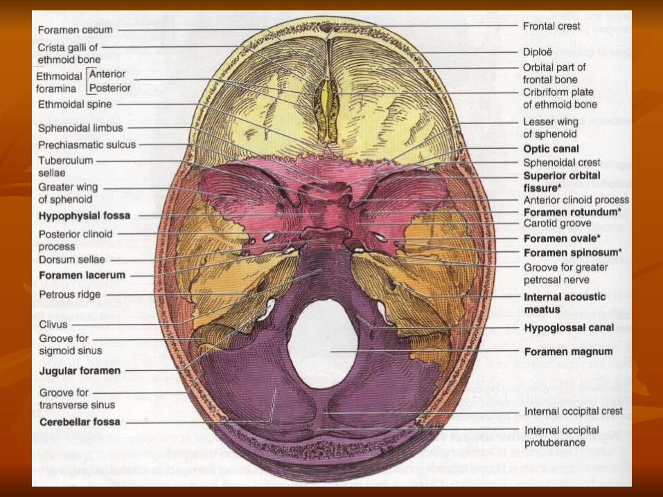
Boundaries Anteriorly and laterally Frontal boneFloor:Orbital plate of frontal bone, ethmiod cribriform plate , anterior border of sphenoid’s lesser wings and crista galli Posteriorly:Posterior border of lesser wing of sphenoid, anterior clinoid process and sulcus chiasmaticus.NBCrista galliIt is a sharp upward projection of ethmoid bone in the midline, for the attachment of falx cerebri.Foramen cecumSmall aperture between the crista galli and the crest of the frontal bone
Anterior cranial fossa
Middle cranial fossaBoundaries Anteriorly Post border of the lesser wings of sphenoid, anterior clinoid processes and sulcus chiasmaticus.Posteriorly Superior borders of petrous part of temporal and sphenoids dorsum sella.Laterally Squamous part of temporal and some part if parietal and greater wings of sphenoid.Floor Greater wing of sphenoid and petrous and squamous parts of the temporal bone.In the centre, floor is formed by the sella tursica of body of sphenoid.
Posterior cranial fossa Boundaries Anteriorly Superior border of the petrous part of temporal bone and dorsum sallae.Posteriorly Internal surface of squamous part of the occipital bone.Floor Basilar, squamous & condylor parts of the occipital bone & mastoid part foramen magnum forms the central part of the floor.
The Dura mater
The Arachnoid mater
The Pia mater
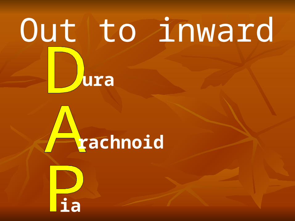
Out to inwardura
rachnoid
ia
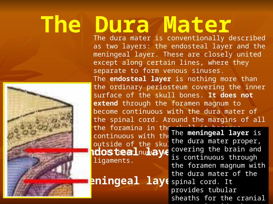
The Dura Mater
endosteal layer
meningeal layer
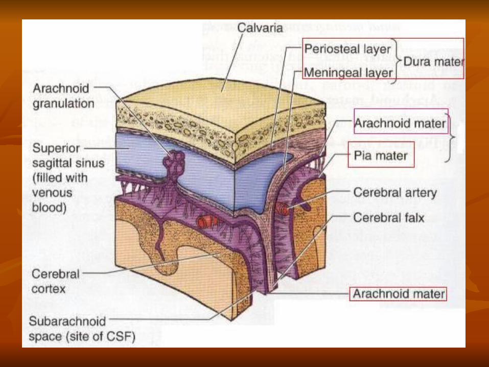
The dura mater is conventionally described as two layers: the endosteal layer and the meningeal layer. These are closely united except along certain lines, where they separate to form venous sinuses.The endosteal layer is nothing more than the ordinary periosteum covering the inner surface of the skull bones. It does not extend through the foramen magnum to become continuous with the dura mater of the spinal cord. Around the margins of all the foramina in the skull it becomes continuous with the periosteum on the outside of the skull bones. At the sutures it is continuous with the sutural ligaments.
The meningeal layer is the dura mater proper, covering the brain and is continuous through the foramen magnum with the dura mater of the spinal cord. It provides tubular sheaths for the cranial nerves. Outside the skull the sheaths fuse with the epineurium of the nerves.
Dural infolding
Falx cerebri
Tentorium cerebelli
Falx cerebelli
Diaphragma sellae
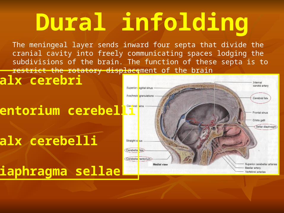
The meningeal layer sends inward four septa that divide the cranial cavity into freely communicating spaces lodging the subdivisions of the brain. The function of these septa is to restrict the rotatory displacement of the brain


Falx CerebriThe falx cerebri is a sickle-shaped fold of dura mater that lies in the midline between the two cerebral hemispheres. Its narrow end in front is attached to the internal frontal crest and the crista galli. Its broad posterior part blends in the midline with the upper surface of the tentorium cerebelli. The superior sagittal sinus runs in its upper fixed margin, the inferior sagittal sinus runs in its lower concave free margin, and the straight sinus runs along its attachment to the tentorium cerebelli.
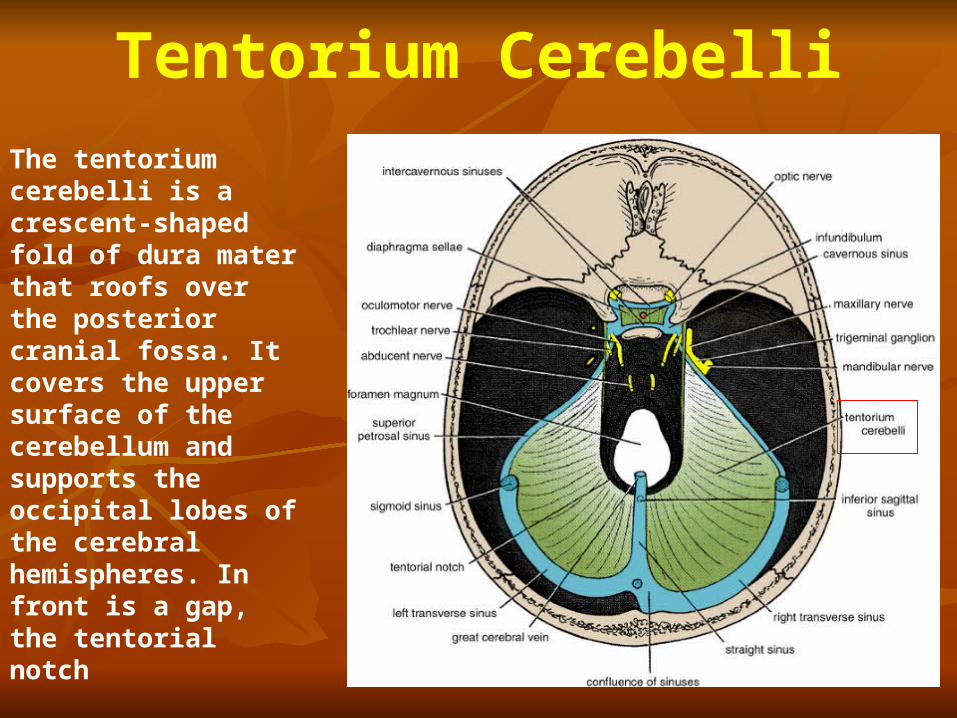
Tentorium Cerebelli
The tentorium cerebelli is a crescent-shaped fold of dura mater that roofs over the posterior cranial fossa. It covers the upper surface of the cerebellum and supports the occipital lobes of the cerebral hemispheres. In front is a gap, the tentorial notch
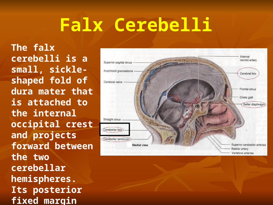
Falx CerebelliThe falx cerebelli is a small, sickle-shaped fold of dura mater that is attached to the internal occipital crest and projects forward between the two cerebellar hemispheres. Its posterior fixed margin contains the occipital sinus.
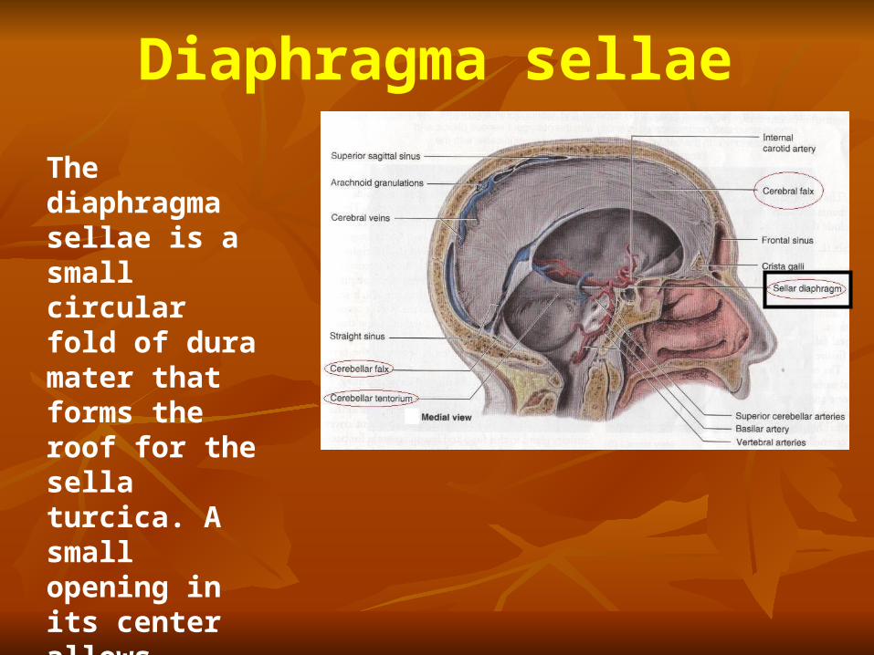
Diaphragma sellae
The diaphragma sellae is a small circular fold of dura mater that forms the roof for the sella turcica. A small opening in its center allows passage of the stalk of the pituitary gland.
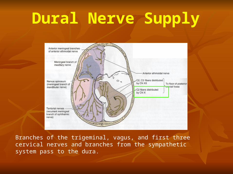
Dural Nerve Supply
Branches of the trigeminal, vagus, and first three cervical nerves and branches from the sympathetic system pass to the dura.
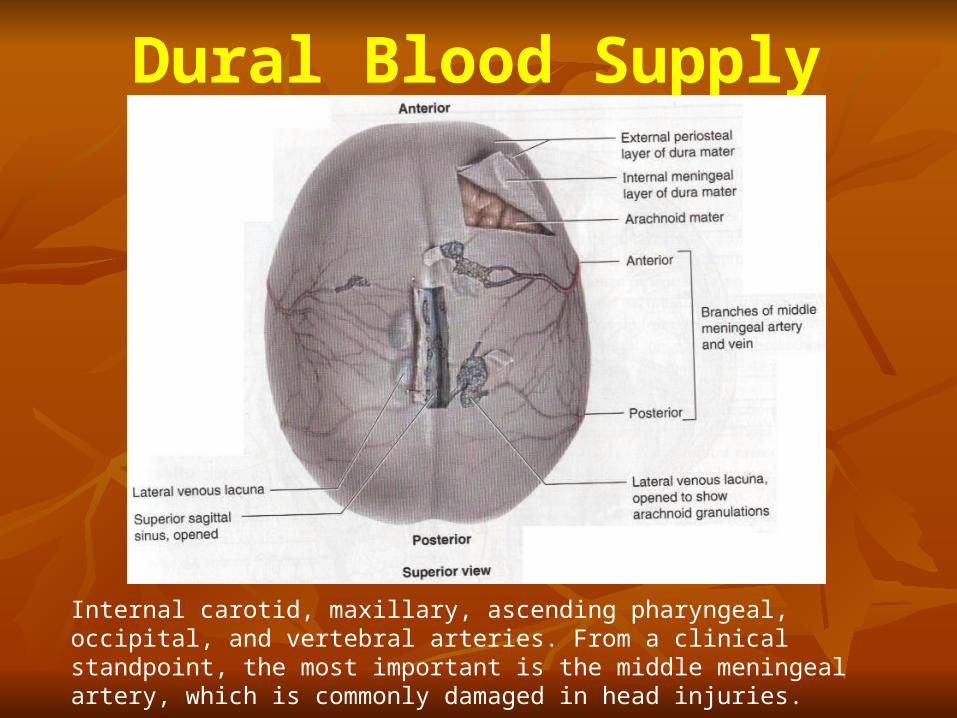
Dural Blood Supply
Internal carotid, maxillary, ascending pharyngeal, occipital, and vertebral arteries. From a clinical standpoint, the most important is the middle meningeal artery, which is commonly damaged in head injuries.
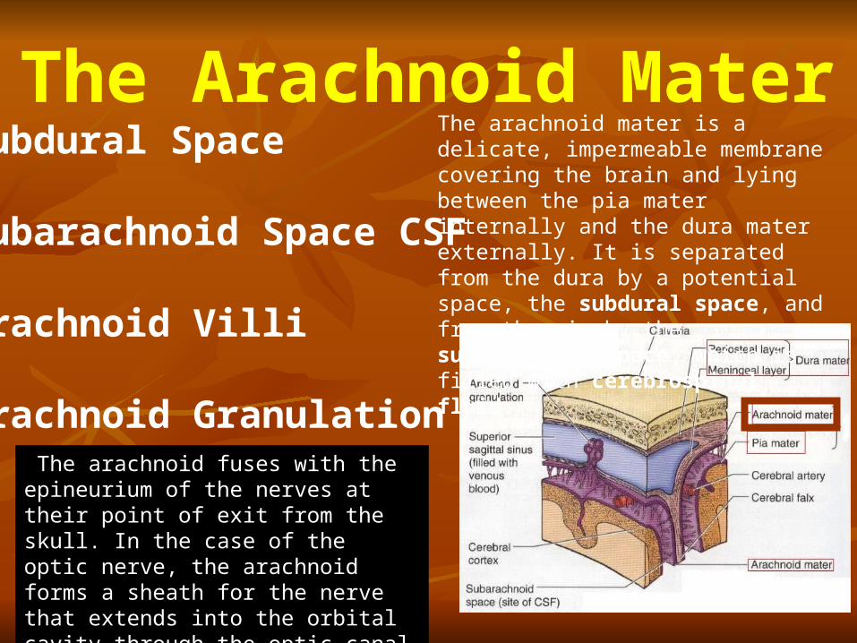
The Arachnoid MaterSubdural Space
Subarachnoid Space CSF
Arachnoid Villi
Arachnoid Granulation
The arachnoid mater is a delicate, impermeable membrane covering the brain and lying between the pia mater internally and the dura mater externally. It is separated from the dura by a potential space, the subdural space, and from the pia by the subarachnoid space, which is filled with cerebrospinal fluid.
The arachnoid fuses with the epineurium of the nerves at their point of exit from the skull. In the case of the optic nerve, the arachnoid forms a sheath for the nerve that extends into the orbital cavity through the optic canal and fuses with the sclera of the eyeball.
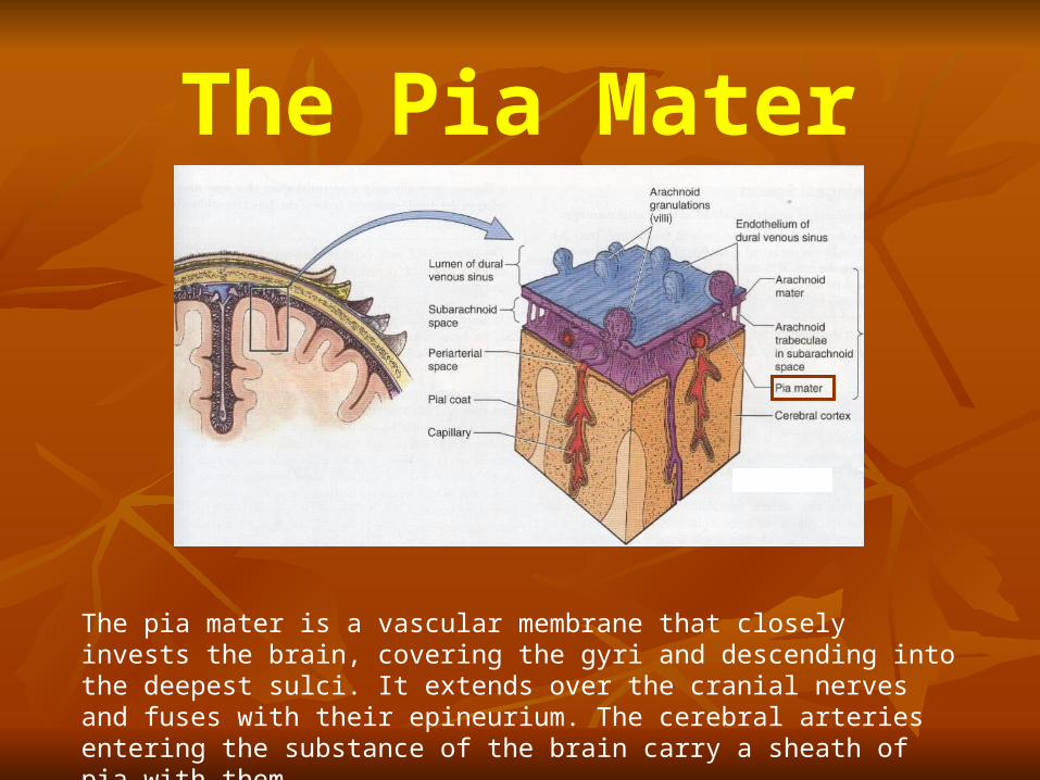
The Pia Mater
The pia mater is a vascular membrane that closely invests the brain, covering the gyri and descending into the deepest sulci. It extends over the cranial nerves and fuses with their epineurium. The cerebral arteries entering the substance of the brain carry a sheath of pia with them.
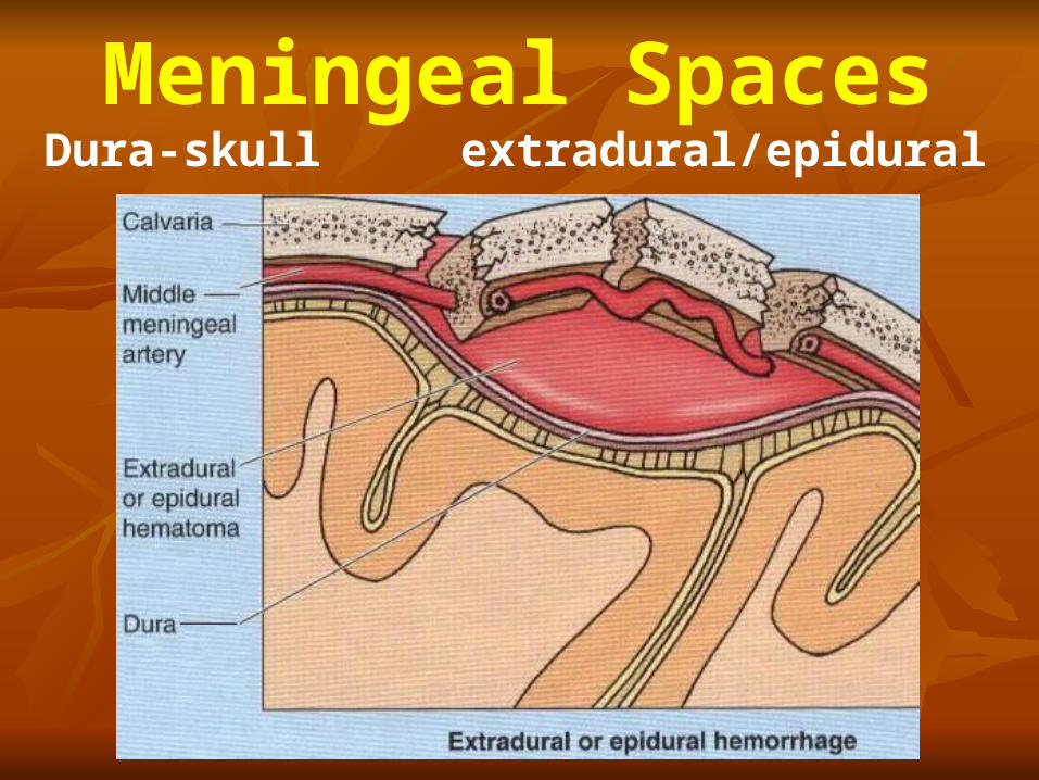
Meningeal SpacesDura-skull extradural/epidural
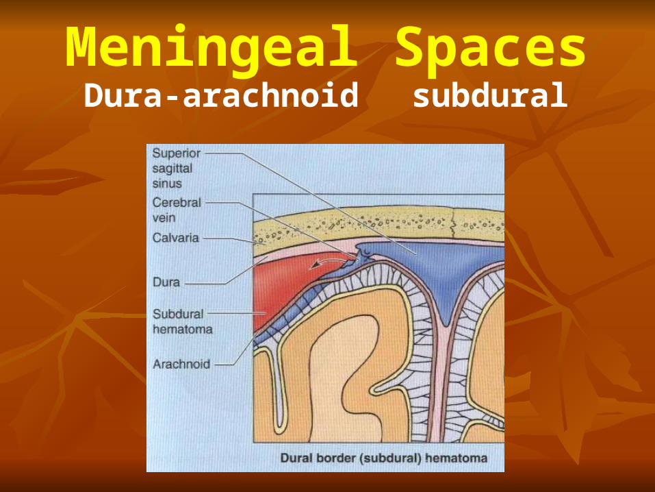
Meningeal SpacesDura-arachnoid subdural
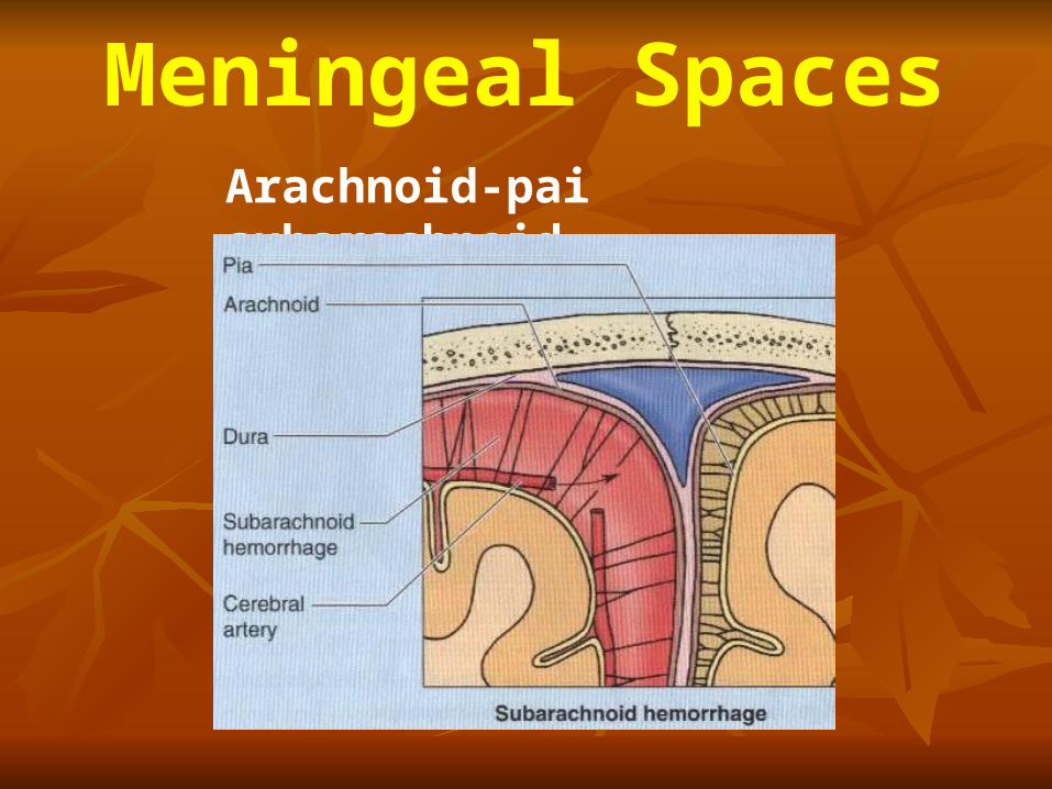
Arachnoid-pai subarachnoid
Meningeal Spaces