Embed Size (px)
Citation preview
Cranial cavity , meninges , dural venous sinuses and pituitary gland
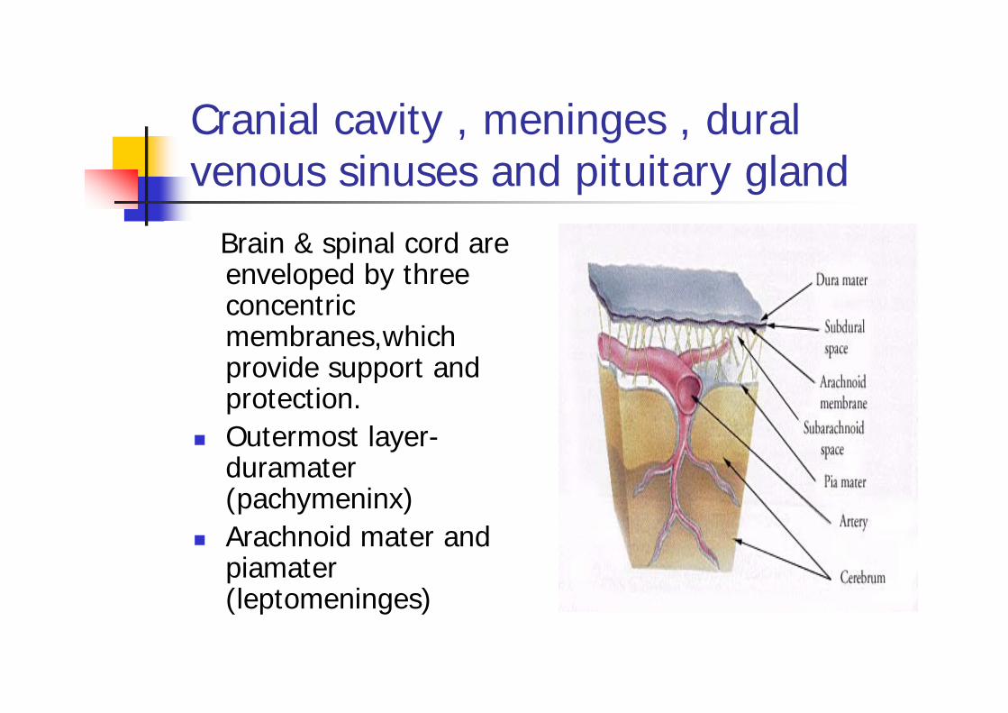
Brain & spinal cord are enveloped by three concentric membranes,which provide support and protection.
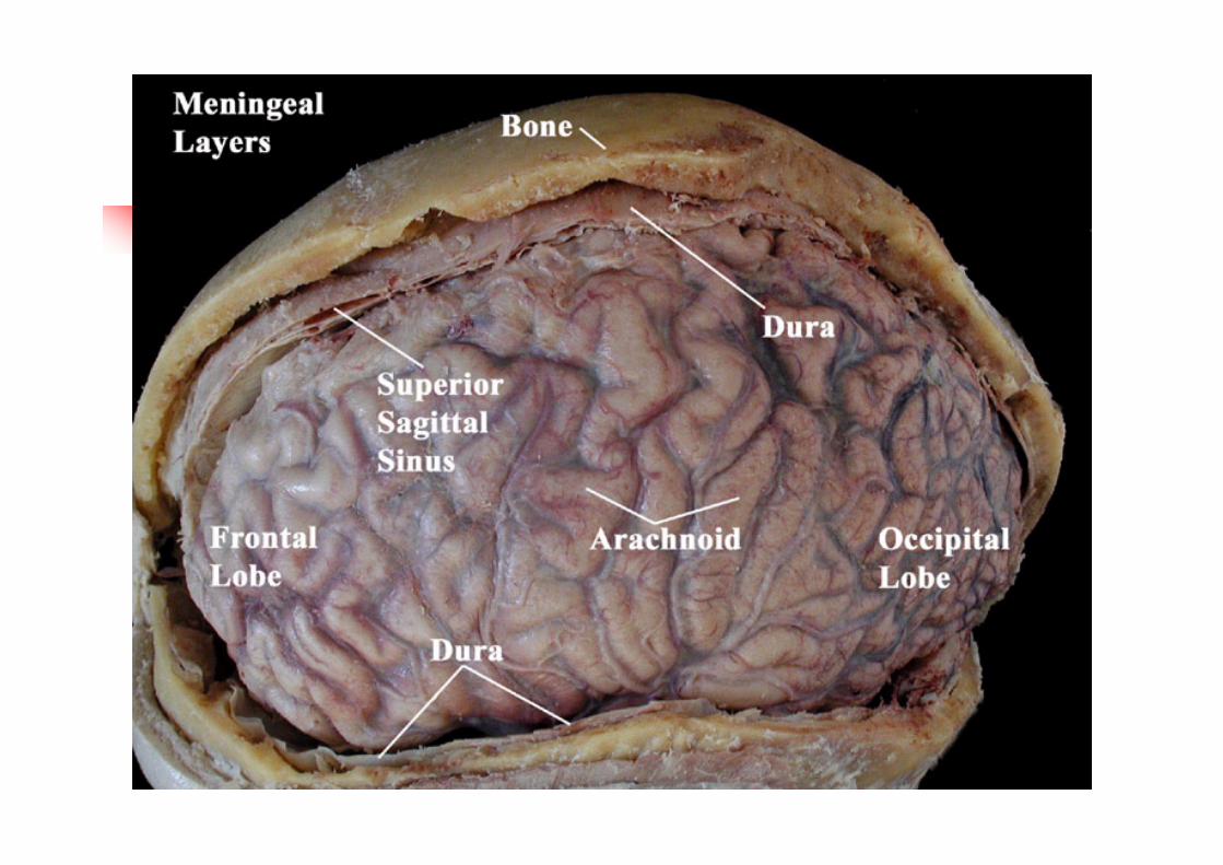
Outermost layer-duramater (pachymeninx)
Arachnoid mater and piamater (leptomeninges)
Durater –opaque,tough,fibrous,thick ,dense composed of densely packed fasicles of collagen fibres arraanged in laminae.
Cranial duramater is composed of two layers outer endosteal layer Inner meningeal layer Characteristic features histologically two are different Duramater-acellular but it contains fibroblasts Meningeal layer-osteoblasts are present in
this layer
Endosteal layer Is continuous throughout the cranial sutures and foramina with pericranium .
the inner aspect of the duramater is closely applied to arachnoid mater over the surface of brain however physically joined at the site where either vein pass from brain into venous sinuses
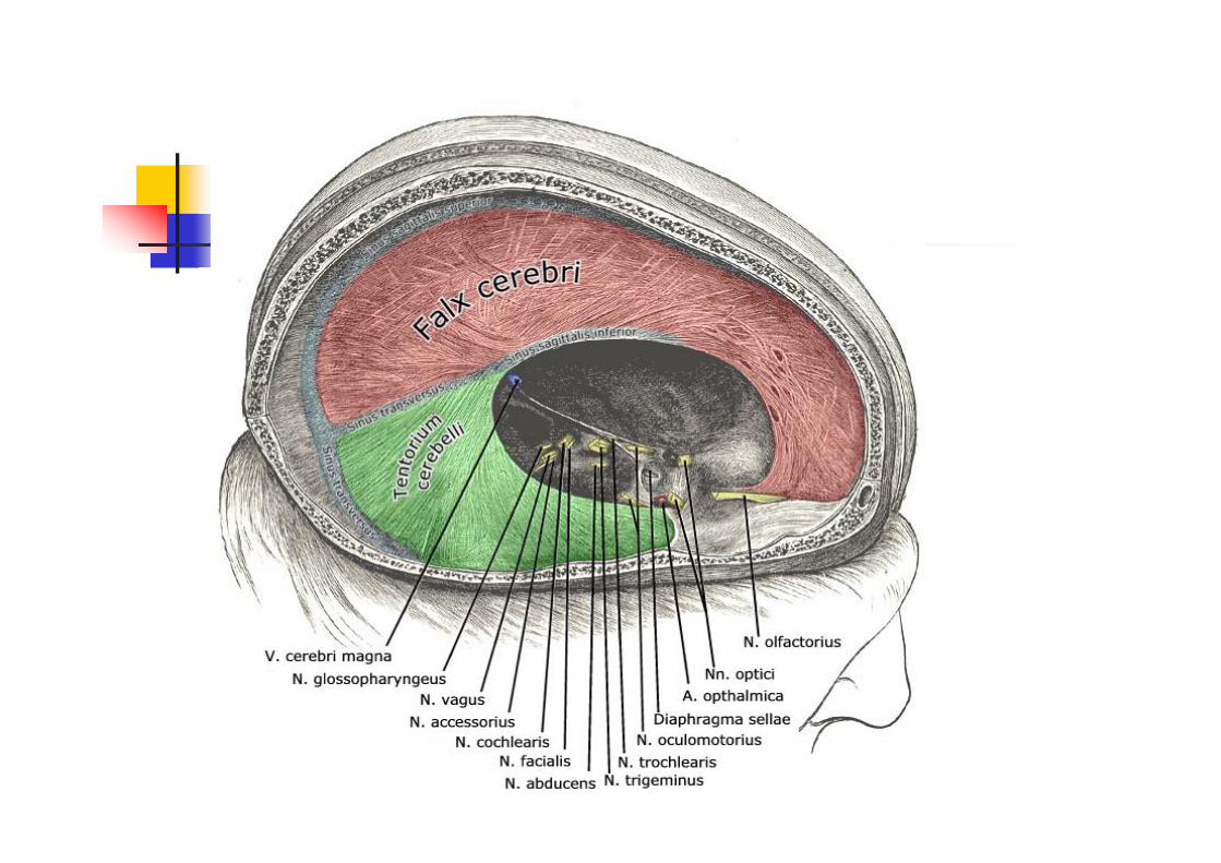
Meningeal layer Is reflected inward to form four folds
Falx cerebri Tentorium cerebelli Falx cerebelli Diaphragma sellae
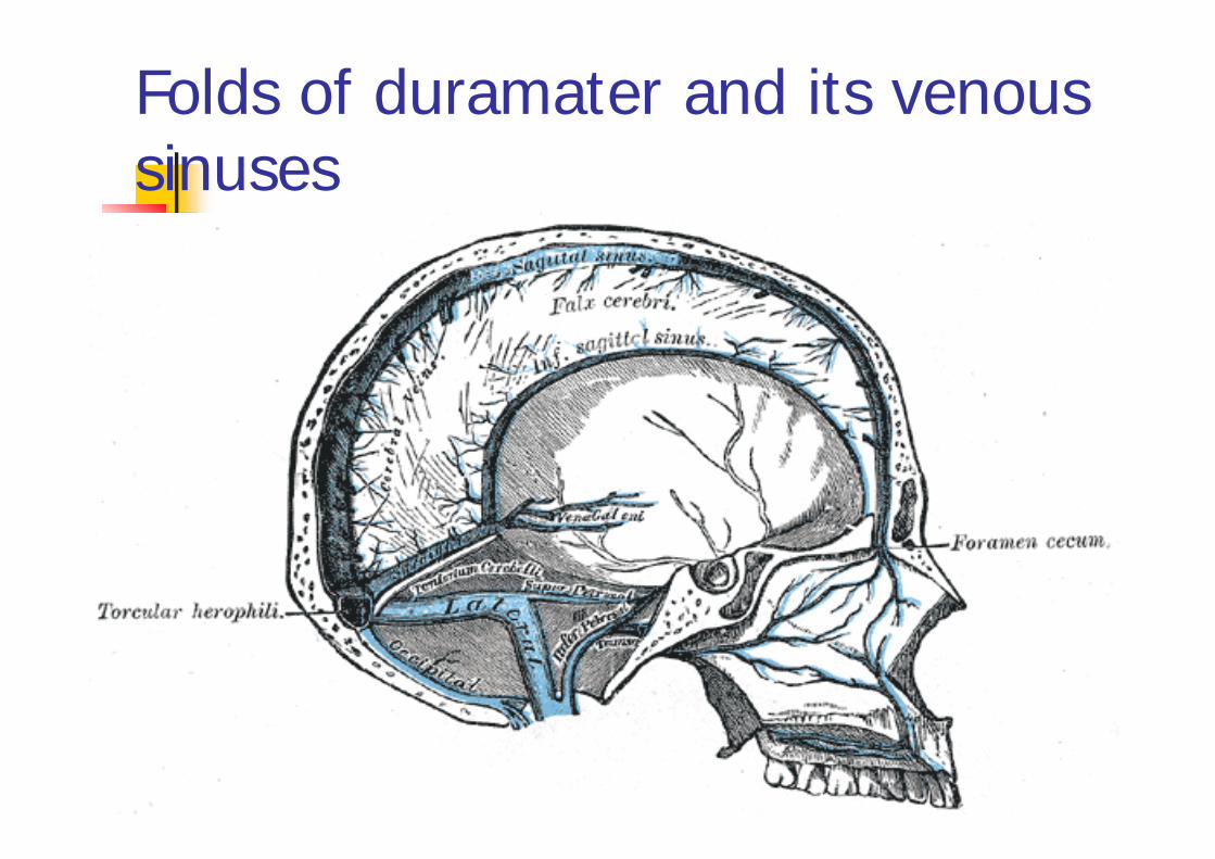
Folds of duramater and its venous sinuses
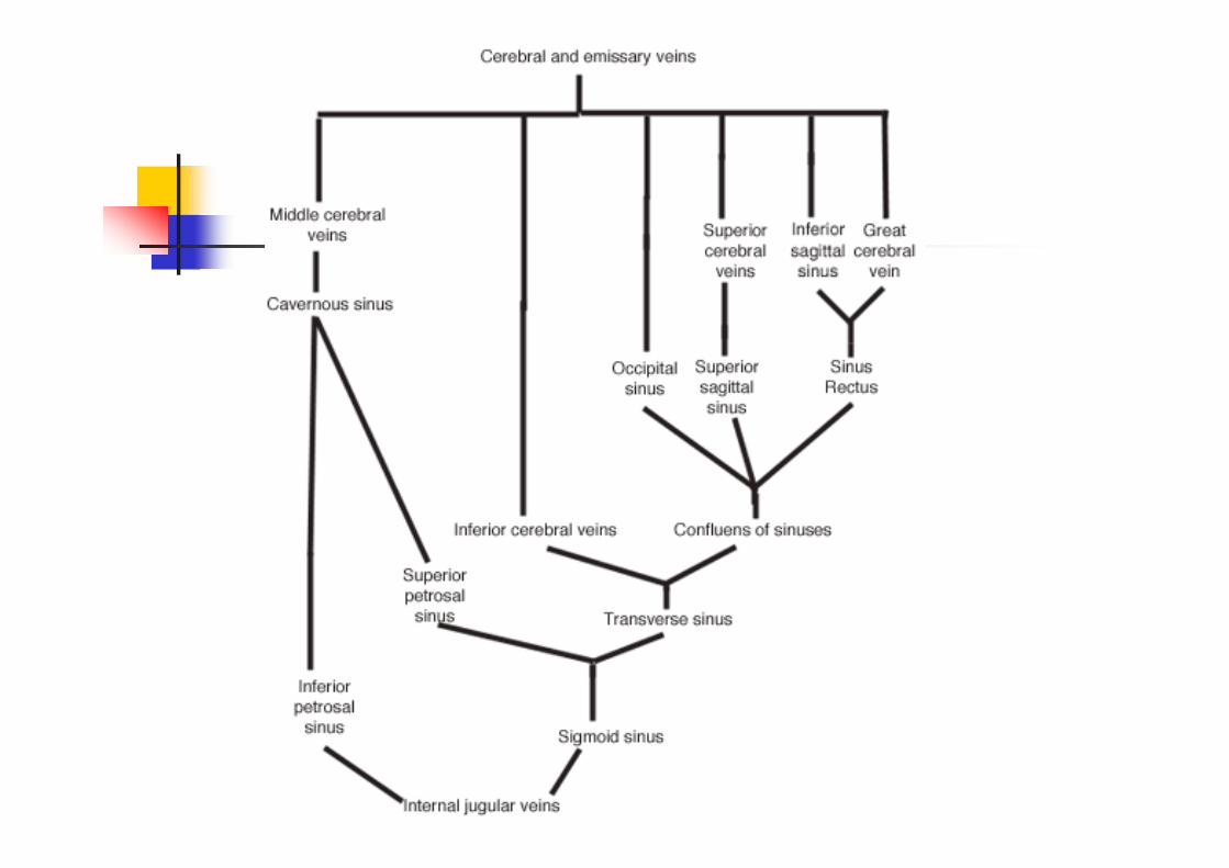
Dural venous sinuses
Complex of venous channels lie between two layers of dura mater
lined by endothelium no valves . walls are devoid of muscular tissue.
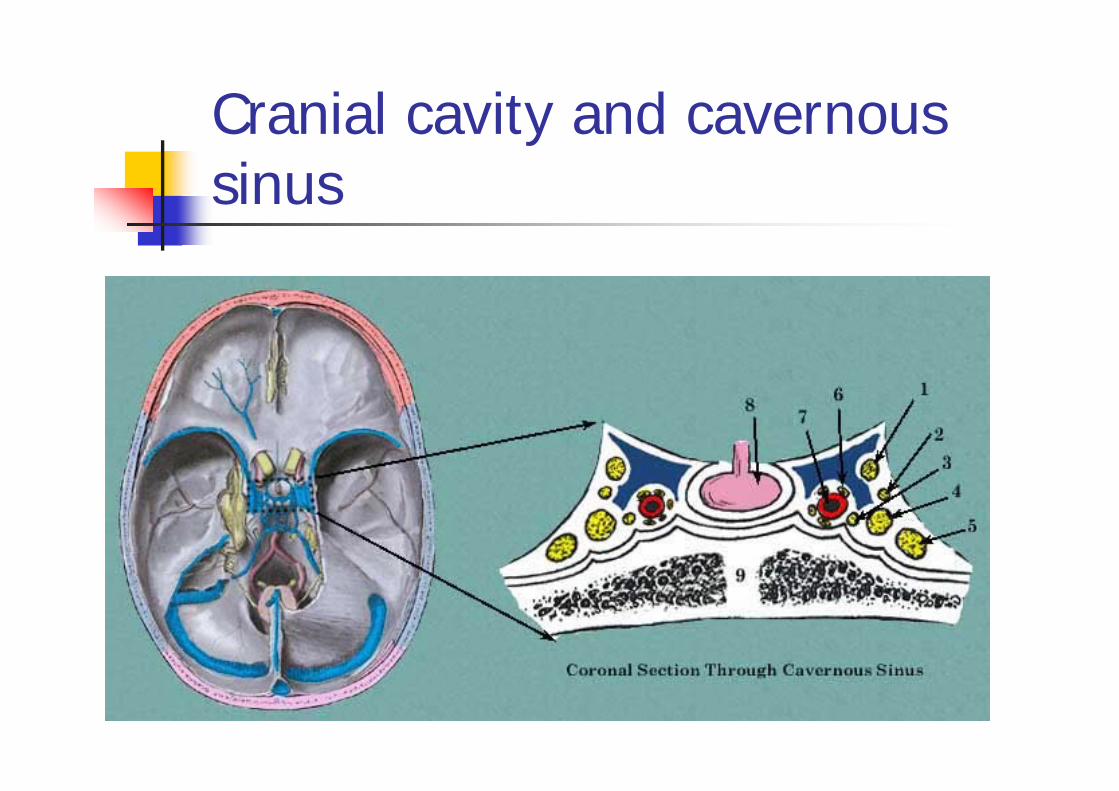
Cranial cavity and cavernous sinus
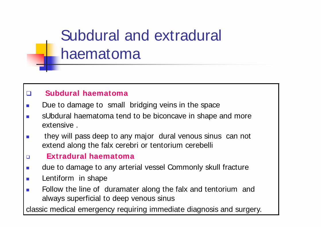
Subdural and extradural haematoma
Subdural haematoma Due to damage to small bridging veins in the space sUbdural haematoma tend to be biconcave in shape and more
extensive . they will pass deep to any major dural venous sinus can not
extend along the falx cerebri or tentorium cerebelli Extradural haematoma due to damage to any arterial vessel Commonly skull fracture Lentiform in shape Follow the line of duramater along the falx and tentorium and
always superficial to deep venous sinus classic medical emergency requiring immediate diagnosis and surgery.
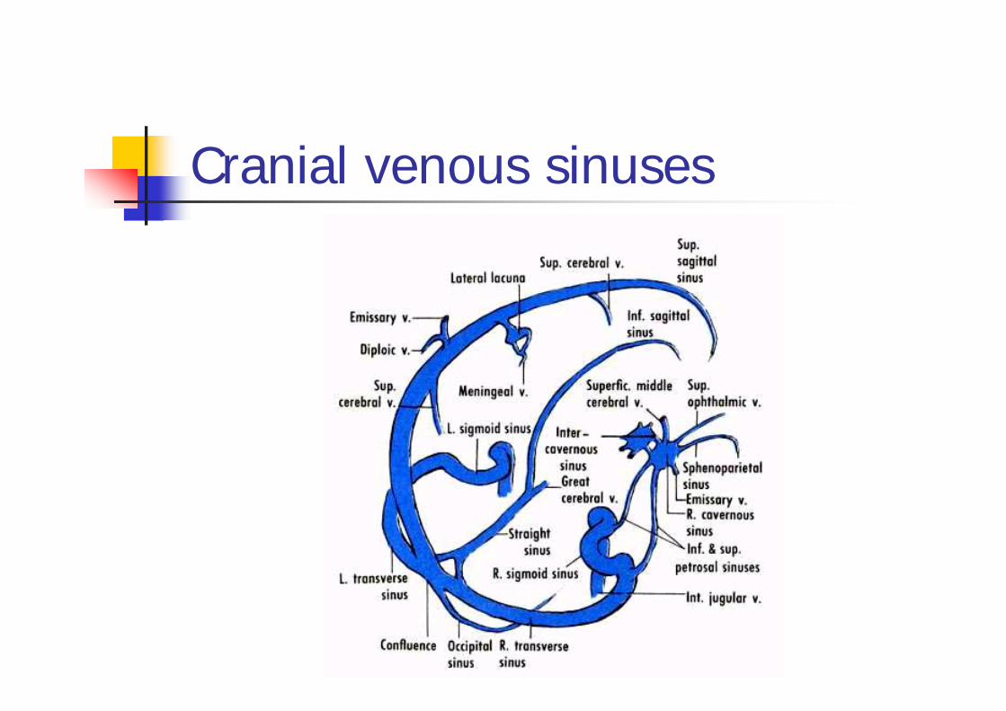
Cranial venous sinuses
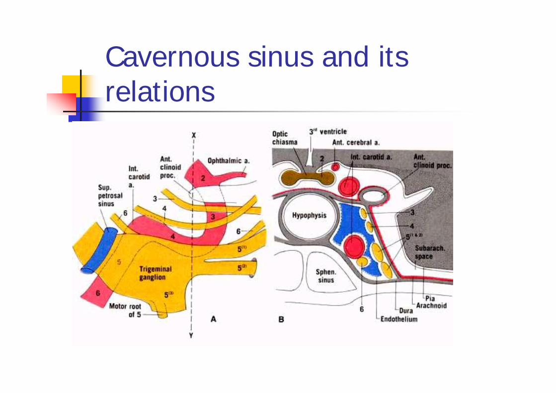
Cavernous sinus and its relations