Embed Size (px)
Citation preview
RESEARCH ARTICLE
The influence of AKT isoforms on radiation sensitivityand DNA repair in colon cancer cell lines
Sara Häggblad Sahlberg & Ann-Sofie Gustafsson &
Prathyusha N. Pendekanti & Bengt Glimelius &
Bo Stenerlöw
Received: 1 October 2013 /Accepted: 22 November 2013 /Published online: 14 December 2013# The Author(s) 2013. This article is published with open access at Springerlink.com
Abstract In response to ionizing radiation, several signalingcascades in the cell are activated to repair the DNA breaks,prevent apoptosis, and keep the cells proliferating. AKT isimportant for survival and proliferation and may also be anactivating factor for DNA-PKcs and MRE11, which are es-sential proteins in the DNA repair process. AKT (PKB) ishyperactivated in several cancers and is associated with resis-tance to radiotherapy and chemotherapy. There are three AKTisoforms (AKT1, AKT2, and AKT3) with different expres-sion patterns and functions in several cancer tumors. The roleof AKT isoforms has been investigated in relation to radiationresponse and their effects on DNA repair proteins (DNA-PKcs and MRE11) in colon cancer cell lines. The knockoutof AKT1 and/or AKT2 affected the radiation sensitivity, and adeficiency of both isoforms impaired the rejoining ofradiation-induced DNA double strand breaks. Importantly,the active/phosphorylated forms of AKT and DNA-PKcs
associate and exposure to ionizing radiation causes an increasein this interaction. Moreover, an increased expression of bothDNA-PKcs and MRE11 was observed when AKT expressionwas ablated, yet only DNA-PKcs expression influenced AKTphosphorylation. Taken together, these results demonstrate arole for both AKT1 and AKT2 in radiotherapy response incolon cancer cells involving DNA repair capacity through thenonhomologous end joining pathway, thus suggesting thatAKT in combination with DNA-PKcs inhibition may be usedfor radiotherapy sensitizing strategies in colon cancer.
Keywords AKT1 . AKT2 . DNA-PKcs .MRE11 .
Radiation . Colorectal cancer
Background
Colorectal cancer is the third most frequent cancer form in theworld and also the third most common reason for cancerdeath. Although surgery is the primary treatment, radiotherapyand/or chemotherapy are used preoperatively or postopera-tively to reduce tumor burden and to diminish recurrence risk[1]. Since not all patients will benefit from chemoradiationtherapy [2], there is a great need to find new drugs withradiosensitizing properties. Understanding the molecularmechanisms of this radiosensitivity is essential for developingmore effective radiotherapy treatments. Most colorectal can-cers initially respond to chemotherapy, although there is a highdevelopment of drug resistance that can be linked tomutationsin the DNA repair mechanism [3, 4].
AKT (also known as Protein Kinase B, PKB) is an impor-tant serine/threonine kinase in the cell signaling downstreamof several growth factors, cytokines, and in response to expo-sure of drugs and ionizing radiation. It is involved in survival,growth, proliferation, glucose uptake, metabolism, and angio-genesis [5]. There are three isoforms of AKT (AKT1, AKT2,
Electronic supplementary material The online version of this article(doi:10.1007/s13277-013-1465-9) contains supplementary material,which is available to authorized users.
S. H. Sahlberg (*) :A.<S. Gustafsson : P. N. Pendekanti :B. StenerlöwBiomedical Radiation Sciences, Uppsala University, 75185 Uppsala,Swedene-mail: [email protected]
A.<S. Gustafssone-mail: [email protected]
P. N. Pendekantie-mail: [email protected]
B. Stenerlöwe-mail: [email protected]
B. GlimeliusSection of Oncology, Department of Radiology, Oncology andRadiation Science, Rudbeck Laboratory, Uppsala University,75185 Uppsala, Swedene-mail: [email protected]
Tumor Biol. (2014) 35:3525–3534DOI 10.1007/s13277-013-1465-9
and AKT3) that are located on separate chromosomes and arebelieved to have different physiological functions, properties,and expression patterns [6, 7]. AKT isoform knockout micehave shown that suppression of AKT1 induces a reduction ofbody and cell size, AKT2 knockouts show diabetes mellitus-like syndrome, and AKT3 deletion causes smaller brain sizeand corpus callosum disorganization [8, 9]. Variations inAKT expression patterns, mutations, and roles of differentisoforms have been observed in various cancer cell lines[10]. AKT1 may function as an oncogene and AKT3 as atumor suppressor [11], and AKT mutations have been detect-ed in human colorectal cancer (AKT2) and lung tumors(AKT1 and AKT3). AKT is also hyperactivated in severalcancer forms and is associated with resistance to radiotherapyand chemotherapy [12].
Cells exposed to ionizing radiation acquire DNA damagesuch as DNA double strand breaks (DSBs), which stimulatethe cells to induce signaling responses including cell cyclearrest, DNA repair, or apoptosis. The main DNA DSB repairpathways are nonhomologous end joining (NHEJ) and ho-mologous recombination (HR) repair. The NHEJ pathwayligates the DNA ends without a long homologous DNAtemplate. HR repair requires a homologous DNA templateto be able to repair the DSBs and is therefore most active inlate S/G2 phase. Both these processes are complex and requireseveral proteins functioning at different stages in the DNArepair and radiation response [13, 14].
The catalytic subunit of nuclear DNA-dependent proteinkinase (DNA-PKcs) is involved in the NHEJ pathway ofDNA repair [15]. Previous studies have shown that there areimportant interactions between AKT and DNA-PKcs. AKT1has been suggested to act downstream of DNA-PKcs in theDNA damage response signaling cascade, independent ofATM (ataxia telangiectasia mutated), where it provides aprosurvival signal by affecting transcriptional p21 regulation[16]. On the other hand, it has been shown that suppressionofAKT1 by siRNA reduced the phosphorylation of DNA-PKcs (Thr2609), which indicates that DNA-PKcs is insteaddownstream of AKT1 [17]. Furthermore, recent findings sug-gest that meiotic recombination 11 (MRE11), a DSB sensorprotein, promotes AKT phosphorylation in response toradiation-induced DSB [18, 19]. Thus, AKT seems to interactwith proteins with distinct functions in DSB recognition andrepair, but knowledge of the role of individual AKT isoformsin the DNA damage response is limited.
The interactions between AKT and DNA-PKcs andMRE11 are probably dependent on a number of factors, suchas celltype, genotype, and microenvironment. Previous stud-ies have used AKT inhibitors, which are somewhat unspecific,or siRNA against AKT, which does not deplete the expressioncompletely. In this study, two colorectal cancer cell lines,HCT116 and DLD-1, were used in which the AKT isoforms,AKT1 and AKT2, have been knocked out with no residual
protein expression, which enables the analyses of the differentAKT isoforms to be more reliable. The two colon cancer celllines, HCT116 and DLD-1, have mutated PI3KCA and KRASgenes. These mutations are also common in colorectal cancerpatients [20, 21]. Further, the DLD-1 cell line has a p53 muta-tion, and the HCT116 cell line has a MRE11 mutation. Muta-tions in MRE11 are common in microsatellite-unstable colorec-tal cancer and cause a higher sensitivity to radiation. HCT116cells have defectiveMRE11 protein that lacks exons 5-7, leadingto defective 3′-5′ exonuclease activity. However, it still possessesthe ability to bind to DNA. These mutations are known to causeabnormal cell signaling and have to be considered when study-ing protein interactions and evaluating future therapies.
This study investigated how the AKT isoforms influenceradiation sensitivity and affect the DSB repair rate as well astheir interaction with MRE11 and DNA-PKcs. In addition, itexplored how the interaction between EGFR and DNA-PKcsis affected by AKT depletion after exposure to ionizing radi-ation. Since the microenvironment may also play an importantrole in therapy response, both high and low concentrations ofserum were used in the cell culture media.
Material and methods
Cell culture The colon cancer cell lines DLD-1 and HCT116X-MAN™ isogenic cell lines were obtained from HorizonDiscovery Ltd. with the different AKT isoforms geneticallyknocked out (parental, AKT1 KO, AKT2 KO, and AKT1 andAKT2 double KO). DLD-1 parental and HCT116 parentalexpress both AKT1 and AKT2, however not AKT3. The cellswere cultured in 75 cm2 culture flasks (Nunclon Surface,Roskilde, Denmark) in McCoy’s 5A medium (Flow Irvine,UK) with 10 % fetal bovine serum (Sigma Aldrich), 2 mML-glutamine, 100 IU/ml penicillin, and 10 μg/ml streptomycinall from Biochrom Kg, Berlin, Germany. All cells were cul-tured in a humidified incubator with 5 % CO2 at 37 °C andtrypsinized with trypsin-EDTA (0.25 % trypsin and 0.02 %EDTA, Biochrom Kg).
Irradiation Cells were irradiated with γ-radiation 137Cssource (Gammacell® 40 Exactor,BestTheratronics, Ottawa,Canada) at a dose rate of 1 Gy/min. The radiation dose wasoptimized for the assay performed.
siRNA transfection The cells were seeded in antibiotic-freecell culture media and incubated overnight at 37 °C with 5 %CO2. Transfection was made according to Thermo ScientificDharmaFECT siRNA transfection protocol with siRNAagainst DNA-PKcs (ON-Target SMART pool, PRKDC withDharmaFECT1) or against MRE11 (ON-TARGET SMARTpool, MRE11A, with DharmaFECT2). The mock treatmentswere made with ON-TARGET plus Non-targeting Pool and
3526 Tumor Biol. (2014) 35:3525–3534
the corresponding DharmaFECT solution. Three days aftertransfection, the cells were used for analysis.
Western blotting for cell signaling Cells were cultivated in3 cm petridishes for at least three doubling times prior toexposure to radiation. Lysates were prepared in posttreatmentby washing the cells with ice-cold PBS followed by additionof 10, 000, 000 cells/ml lysis buffer containing 1% Tween-20,20 mM Tris (pH 8.0), 137 mM NaCl, 10 % glycerol, 2 mMEDTA, 1 mM activated sodium orthovanadate (Sigma), andprotease inhibitor cocktail (P8340, Sigma) and incubation onice for 30 min. Lysates were centrifuged for 10 min in 4 °C.The supernatant was transferred to new tubes, and the pelletwas discarded. The protein concentration of the lysate wasdetermined by BCA protein assay (Pierce). Equal amounts ofprotein were loaded on an SDS PAGE and afterward trans-ferred to a nitrocellulose membrane by wet blotting. Thenitrocellulose membrane was blocked for 1 h in 5 % BSA,PBS and then incubated with the primary antibody overnightat 4 °C. Antibody specific for DNA-PKcs (ab1832), phospho-Ser2056-DNA-PKcs(ab18192), and phospho-Thr2609-DNA-PKcs (ab18356) were from AbCam (Cambridge, UK). Anti-body against MRE11 (PC388) was from Calbiochem (EMDMillipore Corporation, Billerica, MA, USA). AKT1(sc55523and AKT2 sc5270) were purchased from Santa Cruz Biotech-nology (Santa Cruz, CA, USA), and antibodies recognizingthe phosphorylated forms of AKT phospho-Ser-473/474AKT(9271) and AKT phospho-Thr308/309 (9275) were from CellSignaling Technology (Beverly, MS, USA). Antibody againstβ-actin (A5441) was from Sigma-Aldrich (St. Louis, USA).After washing in PBS with 1 % Tween-20, the membrane wasincubated with horseradish peroxidase-labeled secondary an-tibody (626520 and 656120) (Invitrogen) for 1 h at roomtemperature. Immunoreactive bands were visualized in aCCD camera (SuperCCD HR, Fujifilm, Japan) after treatmentwith electrochemiluminescent solution (Immobilon) for5 min.
Immunohistochemistry with PLA Proximity ligation assay(PLA) detects proteins that are in close proximity/interactingwith each other. Primary antibodies (from different species)recognize the proteins of interest and species-specific second-ary antibodies, so called PLA probes, with a unique shortDNA strand attached to it, bind to the primary antibodies.When the proteins are interacting, the PLA probes are in closeproximity, and the DNA strands can, when hybridized withconnector oligos, form circled formed DNA oligonucleotides.The circular DNA is amplified via rolling circle amplificationto hundredfold replication of the DNA circle andfluorochrome-labeled complementary oligonucleotide probeshighlight the product [22]. Cells were seeded on 8-well cham-ber slides (Nunc) in PEST-free cell culture media (McCoy’s,Sigma) and treated with siRNA ormock after 24 h. Three days
after transfection, the cells were exposed to radiation (10 Gy),fixated in ice-cold ethanol 1 h postirradiation, and finally,dipped in acetone. Complexes of EGFR and DNA-PKcs orphospho-Thr2609-DNA-PKcs with phospho-S473-AKTwere detected using the Duolink Proximity Ligation kit (OlinkBiosciences). Cells were incubated with antibodyEGFR(1005):sc-03 (SantaCruzBiotechnology) together withDNA-PKcs (1832) (AbCam) or phosphor-Thr2609-DNA-PKcs (10B1) (AbCam) together with phospho-Ser473474-AKT (Cell Signaling). Cells were incubated with complemen-tary oligonucleotide-conjugated anti-rabbit and anti-mousesecondary antibodies followed by ligation and rolling circleamplifications in the presence of Texas Red conjugated nu-cleotide. The fluorescent amplicons manifest as red fluores-cent dots, with each dot representing an interaction betweenthe two specific proteins. Cells were costained with DAPI, andimages were acquired using a Zeiss Axiophot fluorescencemicroscope. Cell profiler image software was used to measure600–800 nuclei per experiment [23].
mRNA quantifications with real-time qPCR Total RNA wasextracted from three biological replicates using an RNA iso-lation kit (Ambion). cDNAwas synthesized from 0.1 μg totalRNA using RevertAid H Minus First Strand cDNA SynthesisKit with random hexamer primers (Thermo Scientific). qPCRwas performed with Maxima SYBR Green/ROX qPCR Mas-ter Mix (2X) (Thermo Scientific) with qSTARqPCR primerpairs against DNA-PKcs and MRE11 and Beta-actin(OriGene) in a Step-OnePlus Real-Time PCR system (Ap-plied Biosciences). Data were analyzed with Applied Biosci-ence qPCR software.
Cell cycle analysis
Cells were fixated with 70 % ethanol, 10 % PBS and keptat −20 °C for at least 24 h. Cells were centrifuged for 10 min,200G at 4 °C and washed twice with PBS before incubationwith 5 μg Propidium Iodine(Sigma)/0.1 % NP-40(Sigma) inPBS together with 5 μg RNase (Sigma) for 30 min at roomtemperature. Analysis was made with flow cytometry (BDLSRII Biosciences).
Clonogenic assay
To study the effect on cell survival of radiation, clonogenicsurvival assays were performed using standard technique. Thecells were preplated before radiation since this allows the cellsto be undisturbed after the radiation exposure. Cells wereharvested by using trypsin for cell detachment followed bycounting in a Z2 Coulter Counter Analyzer (BeckmanCoulter,FL, USA), and a certain number of cells (300 up to 20, 000depending on treatment) were preplated in 25 cm2 tissue
Tumor Biol. (2014) 35:3525–3534 3527
culture flasks with 10 ml complete medium. The cells wereallowed to attach during culture conditions in humidified airwith 5 % CO2 overnight to give them time to regain their cell-surface receptors after trypsinization. The following day, thecells were exposed to radiation (4 Gy). Control cultures wereleft unexposed and some cultures were exposed to radiationonly. After 8–14 days incubation (depending on the doublingtime of the cell lines), cells were washed in 1×PBS, and fixedwith 99.5 % ethanol and stained with Mayer’s Haematoxylin.Colonies containing more than 50 cells were countedmanually.
The plating efficiency (PE), number of colonies formed/number of cells seeded, in the untreated control and thesurvival fraction (SF), number of colonies formed aftertreatment/number of seeded cells×PE, were calculated. Allexperiments were repeated in triplicate at least three times.The survival curve was analyzed using the linear–quadraticformula (SDose/S0)=exp(αD+βD2).
Detection of DNA double strand breaks by pulsed-field gelelectrophoresis.
Pulsed-field gel electrophoresis (PFGE) is a method to ana-lyze the rapid rejoining of radiation induced DNA double strandbreak [24]. Thismethodwas chosen overγH2AX foci formationassay since DLD-1 and HCT116 forms stacked cell clusterswhich make the detection of foci difficult. Cells for PFGE wereplated in 3-cm dishes and labeled with 2 kBq/ml [methyl-14C]thymidine (Perkin Elmer) for approximately two doubling times.The dishes were put on ice 20–30 min before irradiation andwere kept on ice during the entire irradiation. Cells were preparedfor PFGE as described previously [25]. After irradiation andrepair in incubation at 37 °C, cells were trypsinized and mixedwith low gelling-point agarose (InCert, Cambrex) to a finalconcentration of 1.5–2.5×10⁶ cells/ml in 0.6 % agarose. Themixture was transferred into plug-molds. The plugs with cellswere then transferred to ESP lysis buffer at 4 °C [2 % N-lauroylsarcosine (Sigma), 1 mg/ml proteinase K (Roche), alldiluted in 0.5 M EDTA (Na3) at pH 8.0]. After >20 h, the ESPbuffer was removed and replaced with 20 plug volumes HS-buffer and incubated overnight at 4 °C (HS, high salt; 1.85 MNaCl, 0.15 M KCl, 5 mM MgCl2, 2 mM EDTA, 4 mM Tris,0.5 % Triton X-100, pH 7.5, Triton X-100 was added just beforeuse). Plugswerewashed in 0.1MEDTA and once in 0.5xTBE at4 °C prior to electrophoresis. The plugs were then loaded intowells in a chilled (4 °C) agarose gel (0.8 % SeaKem Gold,Lonza). The gel was placed into a PFGE unit (Gene Navigator,Amersham Pharmacia Biotech, Uppsala, Sweden) with 120°between the fields. Following electrophoresis, the gels weresliced at the position of the 5.7 Mbp chromosome fromS. pombe (BMA), and 14C in the gel segments was measuredby liquid scintillation. The fraction of radioactivity correspondingto DNA of size less than 5.7 Mbp was divided by the totalradioactivity in the lane, giving the fraction of DNA <5.7 Mbp,which is a relative measure of DNA double-strand breaks.
Statistical analysis
The data were processed with Microsoft Office Excel 2007(Microsoft, Redmond), and all graphs were plotted inGraphPad Prism 5 (GraphPad Software, San Diego).
Statistical analysis was performed using GraphPad Prismor Excel to a perform 2-sided Student’s t test. A significancelevel of 95 % was used. This analysis evaluated whether theeffects of treatments or genetic knockout were significantlydifferent from the untreated controls.
Results
The influence of AKT isoforms on the expression of DNA-PKcs and MRE11 Two colorectal cancer cell lines, DLD-1and HCT116, and their corresponding isogenic AKT isoformsknockout cell lines were used to show whether AKT1 orAKT2 were activated after exposure to ionizing radiationand their effect on the expression of MRE11 and DNA-PKcs. The DLD-1 parental cell line had an increased expres-sion of phospho-AKTat Ser473/474 after exposure to ionizingradiation, 1 h post IR (Fig. 1a). The AKT1 and AKT2 knock-out cell lines, which had a higher constitutive activation of theremaining AKT isoform compared to parental cells, had nofurther increase in phosphorylation after exposure to radiation.In the case of HCT116, the AKT1 KO cell line had a lowerphosphorylation of AKT compared to the parental and AKT2KO, suggesting that AKT1 is the isoform that is mainlyactivated in HCT116 (Fig. 1b).
The expression of DNA-PKcs and MRE11 proteins wereinfluenced by the AKT isoforms. Single depletion of AKT1 orAKT2 resulted in lower protein levels of DNA-PKcs andMRE11 in DLD-1. However, double depletion of AKT1 andAKT2 caused an increase in the expression of DNA-PKcs andMRE11. In contrast, in the HCT116 cell-line, DNA-PKcsexpression was only reduced in the AKT2 KO cells and theMRE11 expression was low in parental as well as the singleAKT isoform knockout cell lines.
To gain further insight in the expression of DNA-PKcs andMRE11, mRNA levels were quantified with qPCR using thedelta-delta Ct-calculation. There was a 4-fold increase in mRNAlevels of DNA-PKcs in theAKT1/2KO cell line compared to theparental DLD-1, which confirms the western blot data, but therewas no difference in the mRNA level of MRE11 (Fig. 1c). InHCT116, there was a slight decrease in mRNA levels of DNA-PKcs andMRE11 in the AKT2KO but not in AKT1 or AKT1/2KO cell lines, which is in agreement with the western blot data(Fig. 1d). Notably, the dramatic increase inMRE11 protein in theAKT 1/2 KO cell line (Fig. 1b) seems to be completely due toposttranslational regulation such as increased protein stability orlow degradation.
3528 Tumor Biol. (2014) 35:3525–3534
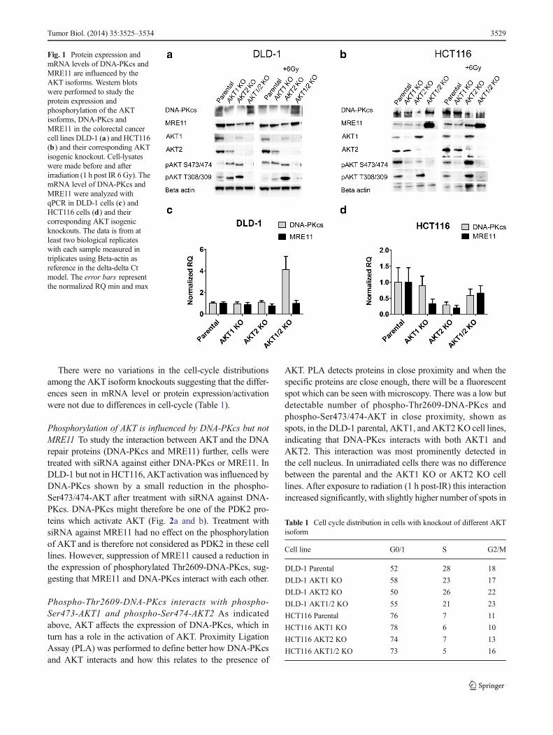
There were no variations in the cell-cycle distributionsamong the AKT isoform knockouts suggesting that the differ-ences seen in mRNA level or protein expression/activationwere not due to differences in cell-cycle (Table 1).
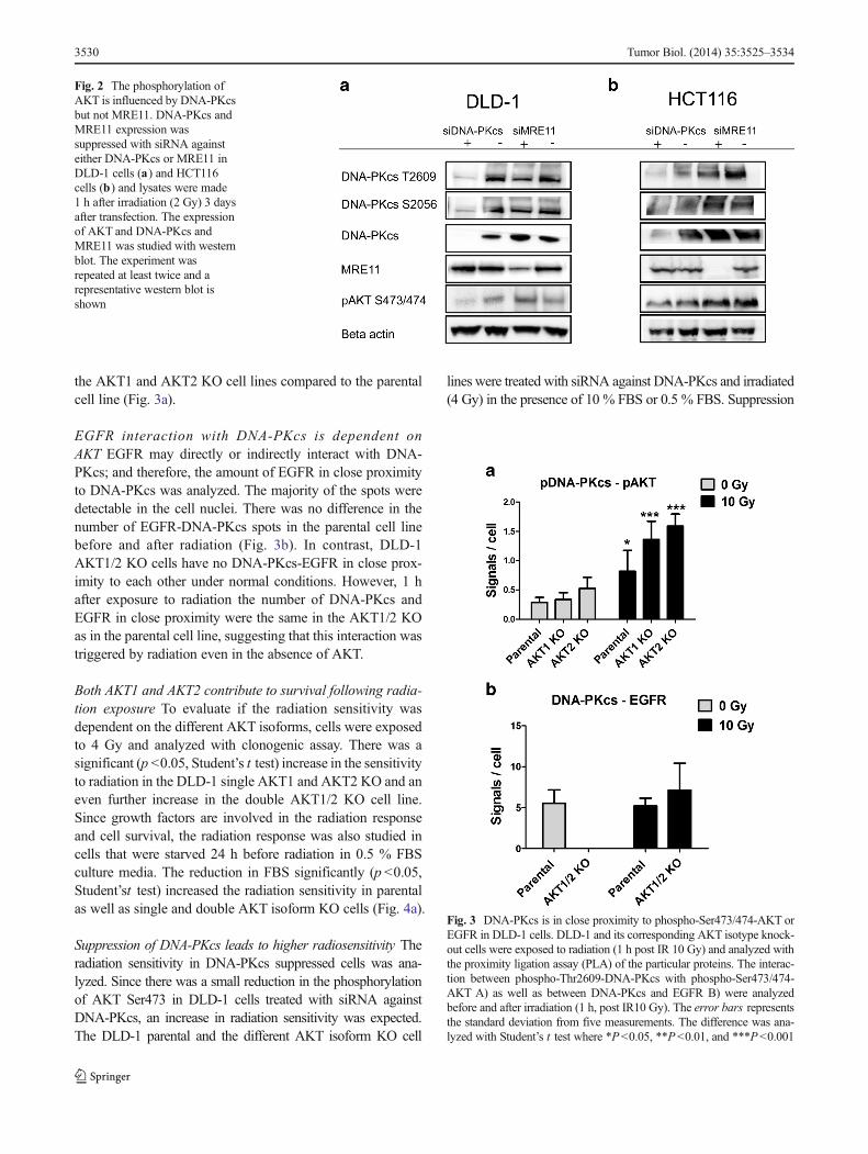
Phosphorylation of AKT is influenced by DNA-PKcs but notMRE11 To study the interaction between AKT and the DNArepair proteins (DNA-PKcs and MRE11) further, cells weretreated with siRNA against either DNA-PKcs or MRE11. InDLD-1 but not in HCT116, AKTactivation was influenced byDNA-PKcs shown by a small reduction in the phospho-Ser473/474-AKT after treatment with siRNA against DNA-PKcs. DNA-PKcs might therefore be one of the PDK2 pro-teins which activate AKT (Fig. 2a and b). Treatment withsiRNA against MRE11 had no effect on the phosphorylationof AKT and is therefore not considered as PDK2 in these celllines. However, suppression of MRE11 caused a reduction inthe expression of phosphorylated Thr2609-DNA-PKcs, sug-gesting that MRE11 and DNA-PKcs interact with each other.
Phospho-Thr2609-DNA-PKcs interacts with phospho-Ser473-AKT1 and phospho-Ser474-AKT2 As indicatedabove, AKT affects the expression of DNA-PKcs, which inturn has a role in the activation of AKT. Proximity LigationAssay (PLA) was performed to define better how DNA-PKcsand AKT interacts and how this relates to the presence of
AKT. PLA detects proteins in close proximity and when thespecific proteins are close enough, there will be a fluorescentspot which can be seen with microscopy. There was a low butdetectable number of phospho-Thr2609-DNA-PKcs andphospho-Ser473/474-AKT in close proximity, shown asspots, in the DLD-1 parental, AKT1, and AKT2KO cell lines,indicating that DNA-PKcs interacts with both AKT1 andAKT2. This interaction was most prominently detected inthe cell nucleus. In unirradiated cells there was no differencebetween the parental and the AKT1 KO or AKT2 KO celllines. After exposure to radiation (1 h post-IR) this interactionincreased significantly, with slightly higher number of spots in
Fig. 1 Protein expression andmRNA levels of DNA-PKcs andMRE11 are influenced by theAKT isoforms. Western blotswere performed to study theprotein expression andphosphorylation of the AKTisoforms, DNA-PKcs andMRE11 in the colorectal cancercell lines DLD-1 (a) and HCT116(b) and their corresponding AKTisogenic knockout. Cell-lysateswere made before and afterirradiation (1 h post IR 6 Gy). ThemRNA level of DNA-PKcs andMRE11 were analyzed withqPCR in DLD-1 cells (c) andHCT116 cells (d) and theircorresponding AKT isogenicknockouts. The data is from atleast two biological replicateswith each sample measured intriplicates using Beta-actin asreference in the delta-delta Ctmodel. The error bars representthe normalized RQ min and max
Table 1 Cell cycle distribution in cells with knockout of different AKTisoform
Cell line G0/1 S G2/M
DLD-1 Parental 52 28 18
DLD-1 AKT1 KO 58 23 17
DLD-1 AKT2 KO 50 26 22
DLD-1 AKT1/2 KO 55 21 23
HCT116 Parental 76 7 11
HCT116 AKT1 KO 78 6 10
HCT116 AKT2 KO 74 7 13
HCT116 AKT1/2 KO 73 5 16
Tumor Biol. (2014) 35:3525–3534 3529
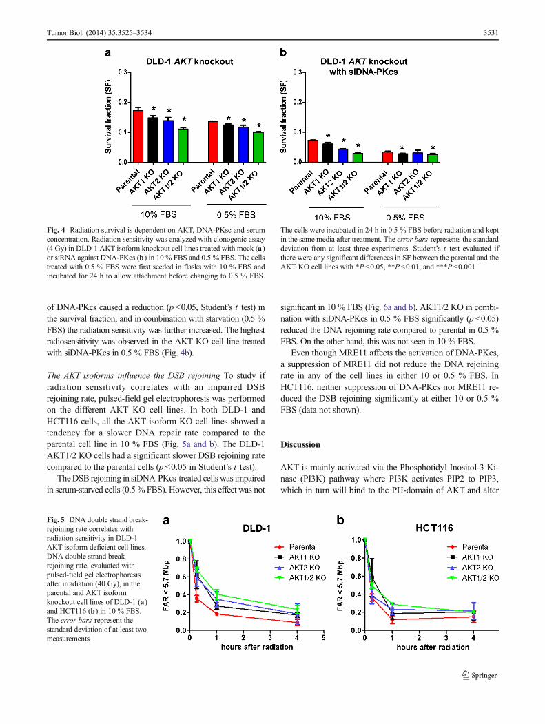
the AKT1 and AKT2 KO cell lines compared to the parentalcell line (Fig. 3a).
EGFR interaction with DNA-PKcs is dependent onAKT EGFR may directly or indirectly interact with DNA-PKcs; and therefore, the amount of EGFR in close proximityto DNA-PKcs was analyzed. The majority of the spots weredetectable in the cell nuclei. There was no difference in thenumber of EGFR-DNA-PKcs spots in the parental cell linebefore and after radiation (Fig. 3b). In contrast, DLD-1AKT1/2 KO cells have no DNA-PKcs-EGFR in close prox-imity to each other under normal conditions. However, 1 hafter exposure to radiation the number of DNA-PKcs andEGFR in close proximity were the same in the AKT1/2 KOas in the parental cell line, suggesting that this interaction wastriggered by radiation even in the absence of AKT.
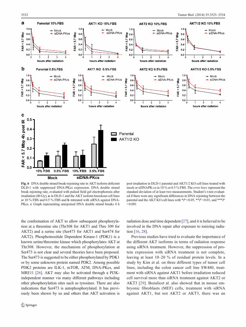
Both AKT1 and AKT2 contribute to survival following radia-tion exposure To evaluate if the radiation sensitivity wasdependent on the different AKT isoforms, cells were exposedto 4 Gy and analyzed with clonogenic assay. There was asignificant (p <0.05, Student’s t test) increase in the sensitivityto radiation in the DLD-1 single AKT1 and AKT2 KO and aneven further increase in the double AKT1/2 KO cell line.Since growth factors are involved in the radiation responseand cell survival, the radiation response was also studied incells that were starved 24 h before radiation in 0.5 % FBSculture media. The reduction in FBS significantly (p <0.05,Student’st test) increased the radiation sensitivity in parentalas well as single and double AKT isoform KO cells (Fig. 4a).
Suppression of DNA-PKcs leads to higher radiosensitivity Theradiation sensitivity in DNA-PKcs suppressed cells was ana-lyzed. Since there was a small reduction in the phosphorylationof AKT Ser473 in DLD-1 cells treated with siRNA againstDNA-PKcs, an increase in radiation sensitivity was expected.The DLD-1 parental and the different AKT isoform KO cell
lines were treated with siRNA against DNA-PKcs and irradiated(4 Gy) in the presence of 10 % FBS or 0.5 % FBS. Suppression
Fig. 2 The phosphorylation ofAKT is influenced by DNA-PKcsbut not MRE11. DNA-PKcs andMRE11 expression wassuppressed with siRNA againsteither DNA-PKcs or MRE11 inDLD-1 cells (a) and HCT116cells (b) and lysates were made1 h after irradiation (2 Gy) 3 daysafter transfection. The expressionof AKT and DNA-PKcs andMRE11 was studied with westernblot. The experiment wasrepeated at least twice and arepresentative western blot isshown
Fig. 3 DNA-PKcs is in close proximity to phospho-Ser473/474-AKT orEGFR in DLD-1 cells. DLD-1 and its corresponding AKT isotype knock-out cells were exposed to radiation (1 h post IR 10 Gy) and analyzed withthe proximity ligation assay (PLA) of the particular proteins. The interac-tion between phospho-Thr2609-DNA-PKcs with phospho-Ser473/474-AKT A) as well as between DNA-PKcs and EGFR B) were analyzedbefore and after irradiation (1 h, post IR10 Gy). The error bars representsthe standard deviation from five measurements. The difference was ana-lyzed with Student’s t test where *P<0.05, **P<0.01, and ***P<0.001
3530 Tumor Biol. (2014) 35:3525–3534
of DNA-PKcs caused a reduction (p<0.05, Student’s t test) inthe survival fraction, and in combination with starvation (0.5 %FBS) the radiation sensitivity was further increased. The highestradiosensitivity was observed in the AKT KO cell line treatedwith siDNA-PKcs in 0.5 % FBS (Fig. 4b).
The AKT isoforms influence the DSB rejoining To study ifradiation sensitivity correlates with an impaired DSBrejoining rate, pulsed-field gel electrophoresis was performedon the different AKT KO cell lines. In both DLD-1 andHCT116 cells, all the AKT isoform KO cell lines showed atendency for a slower DNA repair rate compared to theparental cell line in 10 % FBS (Fig. 5a and b). The DLD-1AKT1/2 KO cells had a significant slower DSB rejoining ratecompared to the parental cells (p <0.05 in Student’s t test).
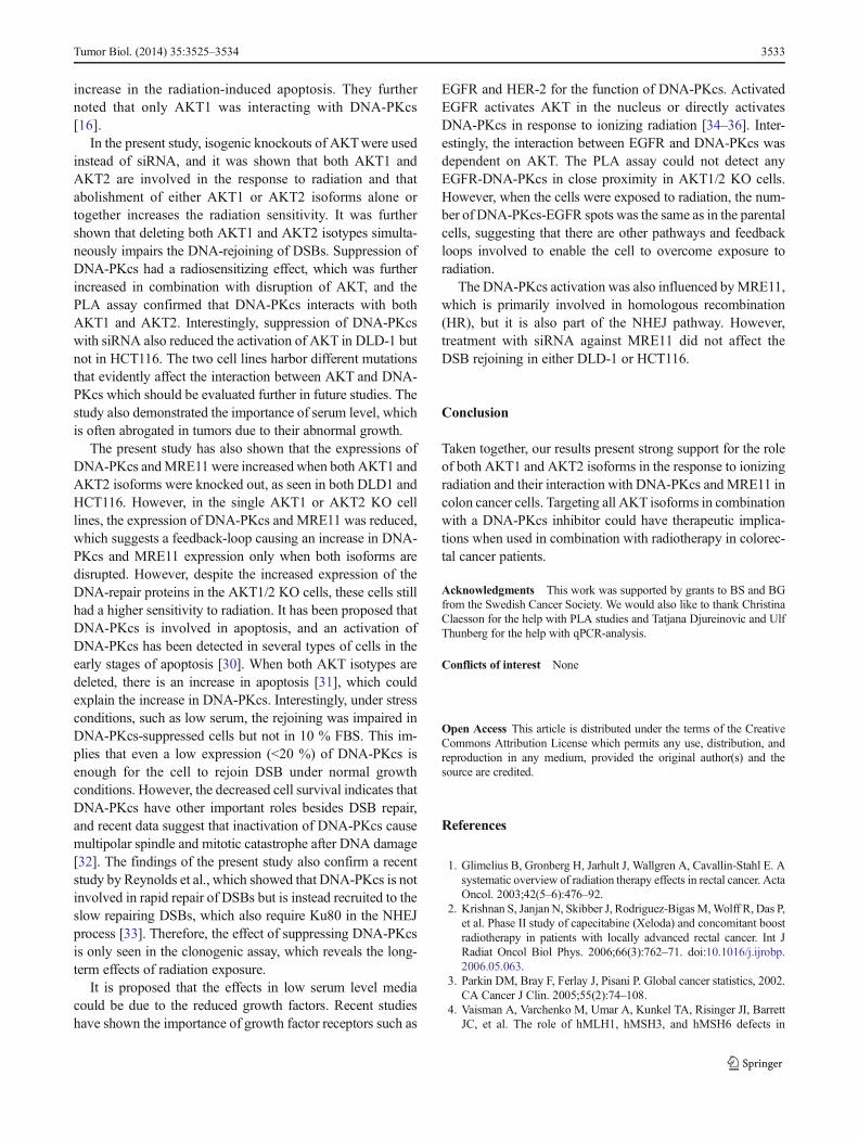
TheDSB rejoining in siDNA-PKcs-treated cells was impairedin serum-starved cells (0.5 % FBS). However, this effect was not
significant in 10 % FBS (Fig. 6a and b). AKT1/2 KO in combi-nation with siDNA-PKcs in 0.5 % FBS significantly (p<0.05)reduced the DNA rejoining rate compared to parental in 0.5 %FBS. On the other hand, this was not seen in 10 % FBS.
Even though MRE11 affects the activation of DNA-PKcs,a suppression of MRE11 did not reduce the DNA rejoiningrate in any of the cell lines in either 10 or 0.5 % FBS. InHCT116, neither suppression of DNA-PKcs nor MRE11 re-duced the DSB rejoining significantly at either 10 or 0.5 %FBS (data not shown).
Discussion
AKT is mainly activated via the Phosphotidyl Inositol-3 Ki-nase (PI3K) pathway where PI3K activates PIP2 to PIP3,which in turn will bind to the PH-domain of AKT and alter
Fig. 4 Radiation survival is dependent on AKT, DNA-PKsc and serumconcentration. Radiation sensitivity was analyzed with clonogenic assay(4 Gy) in DLD-1 AKT isoform knockout cell lines treated with mock (a)or siRNA against DNA-PKcs (b) in 10 % FBS and 0.5 % FBS. The cellstreated with 0.5 % FBS were first seeded in flasks with 10 % FBS andincubated for 24 h to allow attachment before changing to 0.5 % FBS.
The cells were incubated in 24 h in 0.5 % FBS before radiation and keptin the same media after treatment. The error bars represents the standarddeviation from at least three experiments. Student’s t test evaluated ifthere were any significant differences in SF between the parental and theAKT KO cell lines with *P<0.05, **P <0.01, and ***P <0.001
Fig. 5 DNA double strand break-rejoining rate correlates withradiation sensitivity in DLD-1AKT isoform deficient cell lines.DNA double strand breakrejoining rate, evaluated withpulsed-field gel electrophoresisafter irradiation (40 Gy), in theparental and AKT isoformknockout cell lines of DLD-1 (a)and HCT116 (b) in 10 % FBS.The error bars represent thestandard deviation of at least twomeasurements
Tumor Biol. (2014) 35:3525–3534 3531
the conformation of AKT to allow subsequent phosphoryla-tion at a threonine site (Thr308 for AKT1 and Thre 309 forAKT2) and a serine site (Ser473 for AKT1 and Ser474 forAKT2). Phosphoinositide Dependent Kinase-1 (PDK1) is aknown serine/threonine kinase which phosphorylates AKT atThr308. However, the mechanism of phosphorylation atSer473 is not clear and several theories have been proposed.The Ser473 is suggested to be either phosphorylated by PDK1or by some unknown protein named PDK2. Among possiblePDK2 proteins are ILK-1, mTOR, ATM, DNA-PKcs, andMRE11 [26]. AKT may also be activated through a PI3K-independent manner via many different pathways includingother phosphorylation sites such as tyrosines. There are alsoindications that Ser473 is autophosphorylated. It has previ-ously been shown by us and others that AKT activation is
radiation dose and time dependent [27], and it is believed to beinvolved in the DNA repair after exposure to ionizing radia-tion [16, 28].
Previous studies have tried to evaluate the importance ofthe different AKT isoforms in terms of radiation responseusing siRNA treatment. However, the suppression of pro-tein expression with siRNA treatment is not complete,leaving at least 10–20 % of residual protein levels. In astudy by Kim et al. on three different types of tumor celllines, including the colon cancer cell line SW480, treat-ment with siRNA against AKT1 before irradiation reducedcell survival more than siRNA treatment against AKT2 orAKT3 [29]. Bozulicet al. also showed that in mouse em-bryonic fibroblasts (MEF) cells, treatment with siRNAagainst AKT1, but not AKT2 or AKT3, there was an
Fig. 6 DNA double strand break-rejoining rate in AKT isoform deficientDLD-1 with suppressed DNA-PKcs expression. DNA double strandbreak rejoining rate, evaluated with pulsed field gel electrophoresis afterirradiation (40 Gy), a in DLD-1 and the AKT isoform knockout cell linesat 10 % FBS and 0.5 % FBS and b mtreated with siRNA against DNA-PKcs. c Graph representing unrejoined DNA double strand breaks 4 h
post irradiation in DLD-1 parental and AKT1/2 KO cell lines treated withmock or siDNAPK-cs in 10% or 0.5%FBS. The error bars represent thestandard deviation of at least two measurements. Student’s t-test evaluat-ed if there were any significant differences in DNA rejoining between theparental and the AKTKO cell lines with *P <0.05, **P<0.01, and ***P<0.001
3532 Tumor Biol. (2014) 35:3525–3534
increase in the radiation-induced apoptosis. They furthernoted that only AKT1 was interacting with DNA-PKcs[16].
In the present study, isogenic knockouts of AKTwere usedinstead of siRNA, and it was shown that both AKT1 andAKT2 are involved in the response to radiation and thatabolishment of either AKT1 or AKT2 isoforms alone ortogether increases the radiation sensitivity. It was furthershown that deleting both AKT1 and AKT2 isotypes simulta-neously impairs the DNA-rejoining of DSBs. Suppression ofDNA-PKcs had a radiosensitizing effect, which was furtherincreased in combination with disruption of AKT, and thePLA assay confirmed that DNA-PKcs interacts with bothAKT1 and AKT2. Interestingly, suppression of DNA-PKcswith siRNA also reduced the activation of AKT in DLD-1 butnot in HCT116. The two cell lines harbor different mutationsthat evidently affect the interaction between AKT and DNA-PKcs which should be evaluated further in future studies. Thestudy also demonstrated the importance of serum level, whichis often abrogated in tumors due to their abnormal growth.
The present study has also shown that the expressions ofDNA-PKcs andMRE11 were increased when both AKT1 andAKT2 isoforms were knocked out, as seen in both DLD1 andHCT116. However, in the single AKT1 or AKT2 KO celllines, the expression of DNA-PKcs and MRE11 was reduced,which suggests a feedback-loop causing an increase in DNA-PKcs and MRE11 expression only when both isoforms aredisrupted. However, despite the increased expression of theDNA-repair proteins in the AKT1/2 KO cells, these cells stillhad a higher sensitivity to radiation. It has been proposed thatDNA-PKcs is involved in apoptosis, and an activation ofDNA-PKcs has been detected in several types of cells in theearly stages of apoptosis [30]. When both AKT isotypes aredeleted, there is an increase in apoptosis [31], which couldexplain the increase in DNA-PKcs. Interestingly, under stressconditions, such as low serum, the rejoining was impaired inDNA-PKcs-suppressed cells but not in 10 % FBS. This im-plies that even a low expression (<20 %) of DNA-PKcs isenough for the cell to rejoin DSB under normal growthconditions. However, the decreased cell survival indicates thatDNA-PKcs have other important roles besides DSB repair,and recent data suggest that inactivation of DNA-PKcs causemultipolar spindle and mitotic catastrophe after DNA damage[32]. The findings of the present study also confirm a recentstudy by Reynolds et al., which showed that DNA-PKcs is notinvolved in rapid repair of DSBs but is instead recruited to theslow repairing DSBs, which also require Ku80 in the NHEJprocess [33]. Therefore, the effect of suppressing DNA-PKcsis only seen in the clonogenic assay, which reveals the long-term effects of radiation exposure.
It is proposed that the effects in low serum level mediacould be due to the reduced growth factors. Recent studieshave shown the importance of growth factor receptors such as
EGFR and HER-2 for the function of DNA-PKcs. ActivatedEGFR activates AKT in the nucleus or directly activatesDNA-PKcs in response to ionizing radiation [34–36]. Inter-estingly, the interaction between EGFR and DNA-PKcs wasdependent on AKT. The PLA assay could not detect anyEGFR-DNA-PKcs in close proximity in AKT1/2 KO cells.However, when the cells were exposed to radiation, the num-ber of DNA-PKcs-EGFR spots was the same as in the parentalcells, suggesting that there are other pathways and feedbackloops involved to enable the cell to overcome exposure toradiation.
The DNA-PKcs activation was also influenced byMRE11,which is primarily involved in homologous recombination(HR), but it is also part of the NHEJ pathway. However,treatment with siRNA against MRE11 did not affect theDSB rejoining in either DLD-1 or HCT116.
Conclusion
Taken together, our results present strong support for the roleof both AKT1 and AKT2 isoforms in the response to ionizingradiation and their interaction with DNA-PKcs andMRE11 incolon cancer cells. Targeting all AKT isoforms in combinationwith a DNA-PKcs inhibitor could have therapeutic implica-tions when used in combination with radiotherapy in colorec-tal cancer patients.
Acknowledgments This work was supported by grants to BS and BGfrom the Swedish Cancer Society. We would also like to thank ChristinaClaesson for the help with PLA studies and Tatjana Djureinovic and UlfThunberg for the help with qPCR-analysis.
Conflicts of interest None
Open Access This article is distributed under the terms of the CreativeCommons Attribution License which permits any use, distribution, andreproduction in any medium, provided the original author(s) and thesource are credited.
References
1. Glimelius B, Gronberg H, Jarhult J, Wallgren A, Cavallin-Stahl E. Asystematic overview of radiation therapy effects in rectal cancer. ActaOncol. 2003;42(5–6):476–92.
2. Krishnan S, JanjanN, Skibber J, Rodriguez-BigasM,Wolff R, Das P,et al. Phase II study of capecitabine (Xeloda) and concomitant boostradiotherapy in patients with locally advanced rectal cancer. Int JRadiat Oncol Biol Phys. 2006;66(3):762–71. doi:10.1016/j.ijrobp.2006.05.063.
3. Parkin DM, Bray F, Ferlay J, Pisani P. Global cancer statistics, 2002.CA Cancer J Clin. 2005;55(2):74–108.
4. Vaisman A, Varchenko M, Umar A, Kunkel TA, Risinger JI, BarrettJC, et al. The role of hMLH1, hMSH3, and hMSH6 defects in
Tumor Biol. (2014) 35:3525–3534 3533
cisplatin and oxaliplatin resistance: correlation with replicative by-pass of platinum-DNA adducts. Cancer Res. 1998;58(16):3579–85.
5. Manning BD, Cantley LC. AKT/PKB signaling: navigating down-stream. Cell. 2007;129(7):1261–74. doi:10.1016/j.cell.2007.06.009.
6. Santi SA, Lee H. The Akt isoforms are present at distinct subcellularlocations. Am J Physiol Cell Physiol. 2010;298(3):C580–91. doi:10.1152/ajpcell.00375.2009.
7. Lee RS, House CM, Cristiano BE, Hannan RD, Pearson RB, HannanKM. Relative Expression Levels Rather Than Specific Activity Playsthe Major Role in Determining In Vivo AKT Isoform SubstrateSpecificity. Enzyme Res.2011:720985. doi:10.4061/2011/720985.
8. Gonzalez E, McGraw T. The Akt kinases: isoform specificity inmetabolism and cancer. Cell cycle (Georgetown, Tex). 2009;8(16):2502–8.
9. Dummler B, Hemmings B. Physiological roles of PKB/Akt isoformsin development and disease. Biochem Soc Trans. 2007;35(Pt 2):231–5. doi:10.1042/bst0350231.
10. Koseoglu S, Lu Z, Kumar C, Kirschmeier P, Zou J. AKT1, AKT2and AKT3-dependent cell survival is cell line-specific and knock-down of all three isoforms selectively induces apoptosis in 20 humantumor cell lines. Cancer Biol Ther. 2007;6(5):755–62.
11. Hollander MC, Maier CR, Hobbs EA, Ashmore AR, Linnoila RI,Dennis PA. Akt1 deletion prevents lung tumorigenesis by mutant K-ras. Oncogene. 2011;30(15):1812–21. doi:10.1038/onc.2010.556.
12. Carnero A. The PKB/AKT pathway in cancer. Curr Pharm Des.2010;16(1):34–44.
13. Khanna KK, Jackson SP. DNA double-strand breaks: signaling,repair and the cancer connection. Nat Genet. 2001;27(3):247–54.doi:10.1038/85798.
14. Wang C, Lees-Miller SP. Detection and Repair of Ionizing Radiation-Induced DNA Double Strand Breaks: New Developments inNonhomologous End Joining. Int J Radiat Oncol Biol Phys.2013;86(3):440–9. doi:10.1016/j.ijrobp.2013.01.011.
15. Smith G, Jackson S. The DNA-dependent protein kinase. Genes &development. 1999;13(8):916–34. doi:10.1101/gad.13.8.916.
16. Bozulic L, Surucu B, Hynx D, Hemmings BA. PKBalpha/Akt1 actsdownstream of DNA-PK in the DNA double-strand break responseand promotes survival. Mol Cell. 2008;30(2):203–13. doi:10.1016/j.molcel.2008.02.024.
17. ToulanyM, Lee KJ, Fattah KR, Lin YF, Fehrenbacher B, Schaller M,et al. Akt promotes post-irradiation survival of human tumor cellsthrough initiation, progression, and termination of DNA-PKcs-dependent DNA double-strand break repair. Mol Cancer Res.2012;10(7):945–57. doi:10.1158/1541-7786.MCR-11-0592.
18. Fraser M, Harding SM, Zhao H, Coackley C, Durocher D,Bristow RG. MRE11 promotes AKT phosphorylation in directresponse to DNA double-strand breaks. Cell Cycle. 2011;10(13):2218–32.
19. Deng R, Tang J, Ma JG, Chen SP, Xia LP, Zhou WJ, et al. PKB/Aktpromotes DSB repair in cancer cells through upregulating Mre11expression following ionizing radiation. Oncogene. 2011;30(8):944–55. doi:10.1038/onc.2010.467.
20. Day FL, Jorissen RN, Lipton L, Mouradov D, SakthianandeswarenA, Christie M, et al. PIK3CA and PTEN Gene and Exon Mutation-Specific Clinicopathologic and Molecular Associations in ColorectalCancer. Clin Cancer Res. 2013;19(12):3285–96. doi:10.1158/1078-0432.CCR-12-3614.
21. Sartore-Bianchi A, Di Nicolantonio F, Nichelatti M, Molinari F, DeDosso S, Saletti P, et al. Multi-determinants analysis of molecularalterations for predicting clinical benefit to EGFR-targeted monoclo-nal antibodies in colorectal cancer. PLoS One. 2009;4(10):e7287.doi:10.1371/journal.pone.0007287.
22. Soderberg O, Gullberg M, Jarvius M, Ridderstrale K, LeuchowiusKJ, Jarvius J, et al. Direct observation of individual endogenousprotein complexes in situ by proximity ligation. Nature methods.2006;3(12):995–1000. doi:10.1038/nmeth947.
23. Carpenter AE, Jones TR, LamprechtMR, Clarke C, Kang IH, FrimanO, et al. Cell Profiler: image analysis software for identifying andquantifying cell phenotypes. Genome biology. 2006;7(10):R100. doi:10.1186/gb-2006-7-10-r100.
24. Joshi N, Grant SG. DNA double-strand break damage and repairassessed by pulsed-field gel electrophoresis. Methods Mol Biol.2005;291:121–9.
25. Karlsson KH, Radulescu I, Rydberg B, Stenerlow B. Repair ofradiation-induced heat-labile sites is independent of DNA-PKcs,XRCC1 and PARP. Radiat Res. 2008;169(5):506–12. doi:10.1667/RR1076.1.
26. Dong LQ, Liu F. PDK2: the missing piece in the receptor tyrosinekinase signaling pathway puzzle. Am J Physiol Endocrinol Metab.2005;289(2):E187–96. doi:10.1152/ajpendo.00011.2005.
27. Häggblad Sahlberg S, Spiegelberg D, Lennartsson J, Nygren P,Glimelius B. Stenerlöw B. The effect of a dimeric Affibody mole-cule (ZEGFR:1907)2 targeting EGFR in combination with radiationin colon cancer cell lines. Int J Oncol. 2011;40(1):176–84.
28. Toulany M, Kehlbach R, Florczak U, Sak A, Wang S, Chen J, et al.Targeting of AKT1 enhances radiation toxicity of human tumor cellsby inhibiting DNA-PKcs-dependent DNA double-strand break re-pair. Mol Cancer Ther. 2008;7(7):1772–81. doi:10.1158/1535-7163.MCT-07-2200.
29. Kim IA, Bae SS, Fernandes A,Wu J, Muschel RJ, McKennaWG, et al.Selective inhibition of Ras, phosphoinositide 3 kinase, and Akt isoformsincreases the radiosensitivity of human carcinoma cell lines. Cancer Res.2005;65(17):7902–10. doi:10.1158/0008-5472.CAN-05-0513.
30. Mukherjee B, Kessinger C, Kobayashi J, Chen BP, Chen DJ,Chatterjee A, et al. DNA-PK phosphorylates histone H2AX duringapoptotic DNA fragmentation in mammalian cells. DNA Repair(Amst). 2006;5(5):575–90. doi:10.1016/j.dnarep.2006.01.011.
31. Ericson K, Gan C, Cheong I, Rago C, Samuels Y, Velculescu VE,et al. Genetic inactivation of AKT1, AKT2, and PDPK1 in humancolorectal cancer cells clarifies their roles in tumor growth regulation.Proc Natl Acad Sci U S A. 2010;107(6):2598–603. doi:10.1073/pnas.0914018107.
32. Shang ZF, Huang B, Xu QZ, Zhang SM, Fan R, Liu XD, et al.Inactivation of DNA-dependent protein kinase leads to spindle dis-ruption and mitotic catastrophe with attenuated checkpoint protein 2Phosphorylation in response to DNA damage. Cancer Res.2010;70(9):3657–66. doi:10.1158/0008-5472.CAN-09-3362.
33. Reynolds P, Anderson JA, Harper JV, Hill MA, Botchway SW, ParkerAW, et al. The dynamics of Ku70/80 and DNA-PKcs at DSBs inducedby ionizing radiation is dependent on the complexity of damage.Nucleic Acids Res. 2012;40(21):10821–31. doi:10.1093/nar/gks879.
34. Toulany M, Schickfluss TA, Fattah KR, Lee KJ, Chen BP,Fehrenbacher B, et al. Function of erbB receptors and DNA-PKcson phosphorylation of cytoplasmic and nuclear Akt at S473 inducedby erbB1 ligand and ionizing radiation. Radiother Oncol.2011;101(1):140–6. doi:10.1016/j.radonc.2011.06.004.
35. Javvadi P, Makino H, Das AK, Lin YF, Chen DJ, Chen BP, et al.Threonine 2609 phosphorylation of the DNA-dependent proteinkinase is a critical prerequisite for epidermal growth factor receptor-mediated radiation resistance. Mol Cancer Res. 2012;10(10):1359–68. doi:10.1158/1541-7786.MCR-12-0482-T.
36. Rodemann HP, Dittmann K, Toulany M. Radiation-induced EGFR-signaling and control of DNA-damage repair. Int J Radiat Biol.2007;83(11–12):781–91. doi:10.1080/09553000701769970.
3534 Tumor Biol. (2014) 35:3525–3534