Embed Size (px)
Citation preview
1521-0111/91/3/167–177$25.00 http://dx.doi.org/10.1124/mol.116.106161MOLECULAR PHARMACOLOGY Mol Pharmacol 91:167–177, March 2017Copyright ª 2017 by The American Society for Pharmacology and Experimental Therapeutics
Cross-Talk between Alternatively Spliced UGT1A Isoforms andColon Cancer Cell Metabolism s
Yannick Audet-Delage, Michèle Rouleau, Mélanie Rouleau, Joannie Roberge,Stéphanie Miard, Frédéric Picard, Bernard Têtu, and Chantal GuillemettePharmacogenomics Laboratory, Centre Hospitalier Universitaire de Québec Research Center and Faculty of Pharmacy (Y.A.-D.,Mi.R., Me.R., J.R., C.G.), Centre de recherche de l’Institut universitaire de cardiologie et de pneumologie de Québec (S.M., F.P.),and Centre Hospitalier Universitaire de Québec Research Center and Faculty of Medicine (B.T.), Laval University, Québec,Canada
Received July 18, 2016; accepted December 29, 2016
ABSTRACTAlternative splicing at the human glucuronosyltransferase 1 genelocus (UGT1) produces alternate isoforms UGT1A_i2s thatcontrol glucuronidation activity through protein-protein interac-tions. Here, we hypothesized that UGT1A_i2s function as acomplex protein network connecting other metabolic pathwayswith an influence on cancer cell metabolism. This is based on apathway enrichment analysis of proteomic data that identifiedseveral high-confidence candidate interaction proteins ofUGT1A_i2 proteins in human tissues—namely, the rate-limitingenzyme of glycolysis pyruvate kinase (PKM), which plays acritical role in cancer cell metabolism and tumor growth. Thepartnership of UGT1A_i2 and PKM2 was confirmed by coimmu-noprecipitation in the HT115 colon cancer cells and wassupported by a partial colocalization of these two proteins. Insupport of a functional role for this partnership, depletion of
UGT1A_i2 proteins in HT115 cells enforced the Warburg effect,with a higher glycolytic rate at the expense of mitochondrialrespiration, and led to lactate accumulation. Untargetedmetabo-lomics further revealed a significantly altered cellular content of58 metabolites, including many intermediates derived from theglycolysis and tricarboxylic acid cycle pathways. These meta-bolic changes were associated with a greater migration poten-tial. The potential relevance of our observations is supportedby the down-regulation of UGT1A_i2 mRNA in colon tumorscompared with normal tissues. Alternate UGT1A variants maythus be part of the expanding compendium of metabolicpathways involved in cancer biology directly contributing to theoncogenic phenotype of colon cancer cells. Findings uncovernew aspects of UGT functions diverging from their transferaseactivity.
IntroductionThe uridine 59-diphosphate-glucuronosyltransferases (UGTs)
are critical enzymes involved in drug and carcinogen metabo-lism as well as the homeostasis of a wide range of endogenousmolecules, such as bilirubin, steroids, and lipids (Rowland et al.,2013; Rouleau et al., 2014). UGT enzymes catalyze the transferof the sugar moiety of uridine 59-diphosphate-glucuronic acid
(UDPGlcA) to numerous lipophilic substrates, and this reactionleads to inactivationand favors elimination ofmetabolites throughbile and urine. Together with other metabolizing enzymes andtransporters, UGTs are increasingly recognized as key determi-nants of the interindividual variability in pharmacokinetic andpharmacodynamic outcomes of clinically important drugs (Stinglet al., 2014). Findings also reveal that metabolic alterations in theglucuronidation pathwaymaydetermine the degree of exposure ofan individual to toxic or carcinogenic substances over a longperiod, determining individual susceptibility to diseases such ascancer, as well as its progression (Rouleau et al., 2014).Alternative splicing is a powerful means of controlling gene
expression but also creates novel proteins with distinct struc-tural and functional features (Biamonti et al., 2014). Extensiveanalysis of human UGT genomic sequences by our groupuncovered a new class of UGT alternate isoforms derived fromtheUGT1 locus. Novel transcripts are created by the inclusion ofan alternative terminal exon (exon 5b) that leads to the pro-duction of nine shorter UGT1A proteins called isoforms 2 (i2s)(Girard et al., 2007; Lévesque et al., 2007). Expression analyses
This work was supported by the Canadian Institutes of Health [CIHR MOP-142318] and the Natural Sciences and Engineering Research Council ofCanada [NSERC 342176-2012]. YAD received studentship awards from Centrede Recherche en Endocrinologie Moléculaire et Oncologique et GénomiqueHumaine (CREMOGH), Laval University and Fonds de Recherche du Québec –Santé (FRQS). MeR and JR received studentship awards from CIHR. CG holdsa Canada Research Chair in Pharmacogenomics (Canadian Research ChairProgram).
Part of this work was presented as a poster presentation at the followingmeeting: Audet-Delage Y, Rouleau M, Rouleau M, Roberge J and GuillemetteC. (2015) Glucuronosyltransferase splice variant proteins and their roles incancer cell metabolism and progression. Integrating Metabolism and TumorBiology Keystone meeting; 2015 Jan 13-18; Vancouver, BC, Canada.
dx.doi.org/10.1124/mol.116.106161.s This article has supplemental material available at molpharm.
aspetjournals.org.
ABBREVIATIONS: co-IP, coimmunoprecipitation; FBS, fetal bovine serum; FCCP, trifluoromethoxy carbonylcyanide phenylhydrazone; i1, isoform1; i2, isoform 2; KD, knockdown; OXPHOS, mitochondrial oxidative phosphorylation; PBS, phosphate-buffered saline; PCR, polymerase chainreaction; PKM, pyruvate kinase; shRNA, short hairpin RNA; TCA, tricarboxylic acid cycle; UDPGlcA, uridine 59-diphosphate-glucuronic acid; UGT,uridine 59-diphosphate-glucuronosyltransferase.
167
http://molpharm.aspetjournals.org/content/suppl/2017/01/03/mol.116.106161.DC1Supplemental material to this article can be found at:
at ASPE
T Journals on February 10, 2022
molpharm
.aspetjournals.orgD
ownloaded from
revealed that UGT1A enzymes [referred to as isoforms 1 (i1s)]and alternate UGT1A_i2s are coproduced in the same tissuestructures of the liver, kidney, stomach, intestine, and colon(Bellemare et al., 2011). The presence of exon 5b in the mRNAsequence causes a premature end of translation and producesnine alternate UGT1A_i2s with different N-terminal regions(encoded by different exons 1) and lacking the transmembranedomain but comprising 10 novel C-terminal amino acids(R435KKQQSGRQM444). As a consequence, these alternateproteins lack transferase function with UDPGlcA but gainnew biologic activity by negatively modulating cellular glu-curonidation through direct protein-protein interaction withUGT1A enzymes. This represents a new mode of regulation ofthe UGTmetabolic pathway (Bellemare et al., 2010a; Rouleauet al., 2014).Alternative splicing was shown to produce isoforms with
vastly different interaction profiles (Yang et al., 2016).Literature supports oligomerization of UGT enzymes andwith other proteins, such as cytochrome P450 (Rouleauet al., 2014;Miyauchi et al., 2015). This raises the possibility ofother cellular functions for alternate UGT1A proteins and,perhaps, in disease processes such as cancer. Indeed, thehuman protein interactome is of pivotal importance in theregulation of biologic systems and was shown to contribute todifferent pathologies, particularly cancer (Mitsopoulos et al.,2015; Yang et al., 2016). These interactions are consideredpromising therapeutic targets with select protein-proteininteraction inhibitors that have reached clinical development(Scott et al., 2016). Previous untargeted proteomic experi-ments with human liver and kidney normal tissues revealed apartnership between UGT1A_i2s and enzymes of the scav-enging pathway, such as catalase and peroxiredoxin. By theseinteractions, UGT1A_i2 proteins likely interfere with oligo-meric complex formation necessary for scavenging activity ofcatalase and peroxiredoxin (Rouleau et al., 2014). Otherproteins were identified as partners of UGT1A_i2s in humantissues, using an antibody that recognized all nine alternativeUGT1A_i2 proteins but not the nine UGT1A enzymes, includingthemultifunctional protein pyruvate kinase (PKM). PKMplays acritical role in cancer cell metabolism and tumor growth (Guptaand Bamezai, 2010). However, the potential influence of thiscross-talk remains unknown. In addition, it also remains un-definedwhich of the nineUGT1A_i2 proteins interact with theseother proteins. We thus postulated that UGT1A_i2s mightinteract with PKM and influence its activity with potentialconsequences on cancer cell phenotypes given the role the PKMenzyme in the metabolic remodeling of cancer cells.Cancer cells present a distinct metabolic phenotype also
known as the Warburg effect (Warburg et al., 1927; Li andZhang, 2016). In this context, the splice variant PKM2 hasreceived great attention as the major PKM isoform expressedby dividing cancer cells (Anastasiou et al., 2012; Israelsen andVander Heiden, 2015; Taniguchi et al., 2015). In contrast,PKM1 is more abundant in healthy quiescent cells (Bluemleinet al., 2011; Zhan et al., 2015). PKM2 is a rate-limitingglycolytic enzyme and a key metabolic regulator, whereas aclear picture of its function in cancer is still unresolved(Israelsen and Vander Heiden, 2015). PKM2 favors aerobicglycolysis, in which glucose is preferentially catabolizedto lactate, rather than fully metabolized to carbon dioxidevia mitochondrial oxidative phosphorylation (OXPHOS)(Christofk et al., 2008a). This process provides cancer cells
with energy and precursors for the synthesis of nucleic acids,amino acids, and lipids to support cell division. Recent dataindicate that overexpression of PKM2 in colon tumors con-notes poor outcome, as it is associated with advanced tumorstage and lymph node metastasis, and is essential for theaerobic glycolysis of colon cancer cells in vitro (Zhou et al.,2012; Yang et al., 2014, 2015; Cui and Shi, 2015).In this work, we sought to expose potential metabolic and
phenotypic changes associated with UGT1A_i2 proteins in acolon cancer cell model HT115, in which PKM2 is expressedin addition to several different UGT1A proteins, includingUGT1A enzymes and alternative i2 isoforms. We initiallyconfirmed the direct interaction of UGT1A_i2s with pyru-vate kinase by coimmunoprecipitation (co-IP) in HT115 celllysates. Compared with normal colon tissues, downregula-tion of UGT1A_i2 mRNA expression was established incolon tumors by quantitative polymerase chain reaction(PCR) and RNA sequencing. Metabolic alterations associ-ated with depletion of UGT1A_i2 protein levels stablyinduced by short hairpin RNA (shRNA) (targeting the exon5b region common to all UGT1A_i2s) were then investi-gated by complementary approaches, including untargetedmetabolomics and extracellular flux assays, to monitor gly-colysis and oxidative phosphorylation. These findings are thefirst to reveal a significant remodeling of energy metabolismand cellular content of several metabolic intermediates inUGT1A_i2–depleted cancer cells, likely partially explained byan interaction with PKM2. These metabolic changes arefurther associated with a greater migration potential,suggesting that alternate UGT1A proteins might contributeto the oncogenic phenotype of colon cancer cells.
Materials and MethodsImmunofluorescence. HT115 cells were obtained from the Eu-
ropean Collection of Cell Cultures (Salisbury, UK) and grown asdescribed (Bellemare et al., 2010b). Cells grown on coverslips to 60%confluence were fixed with 4% formaldehyde (Sigma-Aldrich, Oak-ville, ON, Canada) for 15 minutes at room temperature, and permea-bilized with 0.5% TX-100 (Sigma-Aldrich) for 5 minutes. All solutionswere prepared in phosphate-buffered saline (PBS). Cells were in-cubated for 1 hour and 30 minutes at room temperature with primaryantibodies diluted in 1% fetal bovine serum (FBS). Primary antibodieswere anti-PKM2 (1:500, #3198S; Cell Signaling, Danvers,MA) and thecustommouse monoclonal anti-i2s (1:100, #4C5E7; GenScript, Piscat-away, NJ) described by Rouleau et al. (2016). Detectionwasmadewithdonkey anti-mouse Alexa Fluor 488 (Life Technologies Inc., Burlington,ON, Canada) and goat anti-rabbit Alexa Fluor 594 (Cell Signaling)antibodies on an LSM510 META NLO laser scanning confocal micro-scope (Zeiss, Toronto, ON, Canada). Images were acquired with the Zen2009 software (version 5.5 SP1; Zeiss) and were analyzed with ImageJ(version 2.0.0; U.S. National Institutes of Health, Bethesda, MD).
Immunoprecipitation and Pathway-Enrichment Analysis.Proteomics experiments in commercial pools of human liver andkidney tissues were conducted according to a previous study (Rouleauet al., 2014). Data were deposited in the PRIDE database under theaccession number PXD000295. In brief, immunoprecipitation wascarried out using 1 mg of total protein lysate of human liver or kidneyS9 fractions (cytosol andmicrosomes; Xenotech, Lenexa, KS) with 4mgof anti-UGT1A_i2 #4863 antibody and protein G–coated magneticbeads (Dynabeads; Life Technologies) for 15 hours at 4°C. Proteinswere identified by mass spectrometry using a TripleTOF 5600 (ABSciex, Concord, ON, Canada) after on-bead trypsin digestion. Pathwayenrichment analysis was performed with Cytoscape version 3.3.0,
168 Audet-Delage et al.
at ASPE
T Journals on February 10, 2022
molpharm
.aspetjournals.orgD
ownloaded from
using ClueGo version 2.2.3 and CluePedia version 1.2.3 apps (Shannonet al., 2003;Bindea et al., 2009, 2013). Pathwayswere retrieved from theKEGG database (updated on January 15, 2016), and an enrichment/depletion two-sided hypergeometric test with the Bonferroni step-downcorrectionmethodwas applied. All presented protein partnerswere partof a significantly enriched pathway (q value,0.05).
Detection of Protein Expression and Complexes. co-IPs toconfirm partnership with PKM2were conducted onHT115 cell lysatesusing the anti-UGT1A_i2s (#4863) and the anti-UGT1A_i1s (#9348)as described (Rouleau et al., 2014). Protein complexes were subjectedto SDS-PAGE and detected by Western blotting with UGT1A_i2s(#4863), biotinylated UGT1A_i1 (#9348), and PKM2 antibodies(#3198S; Cell Signaling). Protein expression was also detected byWestern blot. To monitor protein integrity and phosphorylationstatus, cells were lysed in Tris-buffered solution [175 mM Tris (pH
7.4), 150 mM NaCl, 1% Igepal (Sigma-Aldrich), 1 mM dithiothreitol]supplemented with cOmplete protease inhibitor (Roche, Laval, QC,Canada) and PhosSTOP (Sigma-Aldrich). Proteins were revealedusing anti-PKM1 (#7067; Cell Signaling), anti-PKM2 (Cell Signaling),anti-phospho-PKM2 Y105 (#3827; Cell Signaling), and anti-phospho-PKM2 S37 (#11456; Signalway Antibody, College Park, MD). PKM2dimers and tetramerswere cross-linked according toWang et al. (2014).In brief, cells were lysed in PBS (pH 7.4) containing 0.5% Triton X-100for 30minutes, then centrifuged at 16,000� g for 20minutes to removedebris. Lysates were then diluted to 4 mg/ml, and glutaraldehyde (finalconcentration 0.0035%) was added to permanently cross-link proteins.After 2 minutes of incubation at room temperature, the cross-linkingreaction was stopped using Tris pH 8.0 (final concentration 50 mM).Finally, protein complexes were subjected to SDS-PAGE and revealedusing anti-PKM2 (Cell Signaling).
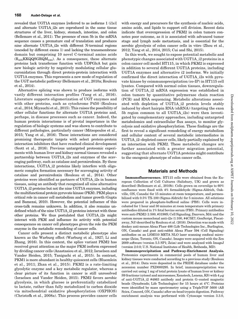
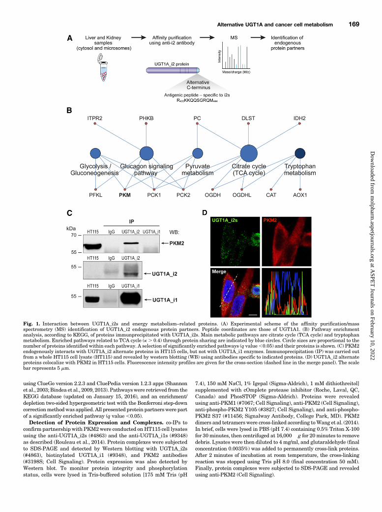
Fig. 1. Interaction between UGT1A_i2s and energy metabolism–related proteins. (A) Experimental scheme of the affinity purification/massspectrometry (MS) identification of UGT1A_i2 endogenous protein partners. Peptide coordinates are those of UGT1A1. (B) Pathway enrichmentanalysis, according to KEGG, of proteins immunprecipitated with UGT1A_i2s. Main metabolic pathways are citrate cycle (TCA cycle) and tryptophanmetabolism. Enriched pathways related to TCA cycle (k . 0.4) through protein sharing are indicated by blue circles. Circle sizes are proportional to thenumber of proteins identified within each pathway. A selection of significantly enriched pathways (q value,0.05) and their proteins is shown. (C) PKM2endogenously interacts with UGT1A_i2 alternate proteins in HT115 cells, but not with UGT1A_i1 enzymes. Immunoprecipitation (IP) was carried outfrom a whole HT115 cell lysate (HT115) and revealed by western blotting (WB) using antibodies specific to indicated proteins. (D) UGT1A_i2 alternateproteins colocalize with PKM2 in HT115 cells. Fluorescence intensity profiles are given for the cross-section (dashed line in the merge panel). The scalebar represents 5 mm.
Alternative UGT1A and cancer cell metabolism 169
at ASPE
T Journals on February 10, 2022
molpharm
.aspetjournals.orgD
ownloaded from
Metabolic Profiling of HT115 Colon Cancer Cells. Metabolicprofiles were established for two HT115 cell lines, a reference HT115cell line (control) and an HT115 cell line in which the expression ofUGT1A_i2 proteins was stably repressed (.90%) using an shRNAspecific to exon 5b ofUGT1A [knockdown (KD)] (Rouleau et al., 2014).Cell lines were grown for a maximum of 15 passages and periodicallytested for mycoplasmas. Cells were seeded at 2 � 106 cells in a 10-cmpetri dish and allowed to adhere and proliferate for 4 days, with onegrowth medium change after 2 days. Cells were trypsinized, centri-fuged, and counted before being washed twice with ice-cold PBS andfrozen on dry ice. Mass spectrometry analyses were performed by theWest CoastMetabolomics Center (University of California, Davis, CA)as reported (Fiehn et al., 2008, 2010). Data were normalized for cellcount and log transformed prior to testing for statistical differencesusing two-sided Student’s t tests.
The analysis of extracellular fluxwas achieved on a Seahorse XFe24using glycolysis and mitochondria stress kits according to themanufacturer’s instructions (Seahorse Bioscience, North Billerica,MA). For the glycolysis stress kit, 100 ml of cells at 2.0 � 106 cells/mlwas seeded in the dedicated plates, and allowed to adhere for 2 hoursin the incubator before the addition of 150 ml of prewarmed culturemedium. Cells were grown overnight, rinsed twice with 1 ml ofSeahorse XF Base Medium (Seahorse Bioscience) supplemented with4 mM glutamine, and a final volume of 450 ml of the medium wasadded. Cells were incubated for 1 hour at 37°C and 100%humidity in anon-CO2 incubator. Test compounds were reconstituted in glutamine-
supplemented Seahorse XF Base Medium, according to instruc-tions. A total of 75 ml of this medium was loaded in the injectionplate port “A,” whereas glucose was added in “B,” oligomycin in“C,” and 2-deoxyglucose in “D.” Three measure phases were donefor each condition, constituting 3 minutes of mixing, 2 minutes ofwaiting, and 3 minutes of measuring. For the mitochondria stresskit, a similar protocol was used, except that the Seahorse XF BaseMedium was supplemented with both 4 mM glutamine and 4 g/lglucose, and injection ports were loaded as follows: assay mediumin “A,” oligomycin in “B,” trifluoromethoxy carbonylcyanide phe-nylhydrazone (FCCP) in “C,” and rotenone/antimycin A in “D.”Data were normalized for protein content. The number of repli-cates and independent experiments is specified in the legend offigure 2.
Lactate was quantified with a Lactate Colorimetric/FluorometricAssay Kit (BioVision, Milpitas, CA). Cells were seeded at 0.6 � 106
cells per well in a six-well plate for 48 hours, rinsed twice with freshmedium, and 3 ml of medium was added per well. Medium wassampled at 0, 60, 120, and 180minutes and immediately frozen on dryice. Cells were then trypsinized and counted. A perchloric acid de-proteinization kit (BioVision) was used prior to sample dilution andlactate dosage. Fluorescence (Excitation/Emission5 535/590 nm) wasmeasured on a TECAN M-1000 (Tecan US Inc., Morrisville, NC).Samples were measured in duplicate for each time point. Resultsrepresent the mean of two independent experiments, and differenceswere assessed by Student’s two-sided t tests.
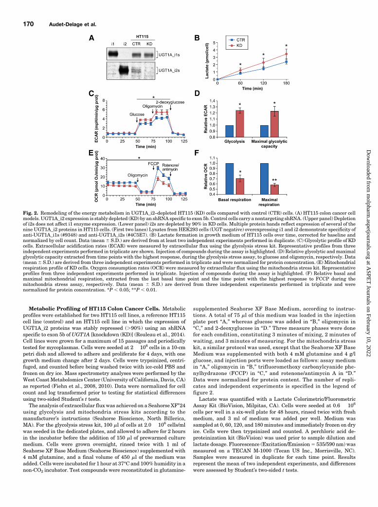
Fig. 2. Remodeling of the energy metabolism in UGT1A_i2–depleted HT115 (KD) cells compared with control (CTR) cells. (A) HT115 colon cancer cellmodels. UGT1A_i2 expression is stably depleted (KD) by an shRNA specific to exon 5b. Control cells carry a nontargeting shRNA. (Upper panel) Depletionof i2s does not affect i1 enzyme expression. (Lower panel) i2s are depleted by 90% in KD cells. Multiple protein bands reflect expression of several of thenine UGT1A_i2 proteins in HT115 cells. (First two lanes) Lysates fromHEK293 cells (UGT negative) overexpressing i1 and i2 demonstrate specificity ofanti-UGT1A_i1s (#9348) and anti-UGT1A_i2s (#4C5E7). (B) Lactate formation in growth medium of HT115 cells over time, corrected for baseline andnormalized by cell count. Data (mean6 S.D.) are derived from at least two independent experiments performed in duplicate. (C) Glycolytic profile of KDcells. Extracellular acidification rates (ECAR) were measured by extracellular flux using the glycolysis stress kit. Representative profiles from threeindependent experiments performed in triplicate are shown. Injection of compounds during the assay is highlighted. (D) Relative glycolytic andmaximalglycolytic capacity extracted from time points with the highest response, during the glycolysis stress assay, to glucose and oligomycin, respectively. Data(mean6 S.D.) are derived from three independent experiments performed in triplicate and were normalized for protein concentration. (E) Mitochondrialrespiration profile of KD cells. Oxygen consumption rates (OCR) were measured by extracellular flux using the mitochondria stress kit. Representativeprofiles from three independent experiments performed in triplicate. Injection of compounds during the assay is highlighted. (F) Relative basal andmaximal mitochondrial respiration, extracted from the last basal time point and the time point with the highest response to FCCP during themitochondria stress assay, respectively. Data (mean 6 S.D.) are derived from three independent experiments performed in triplicate and werenormalized for protein concentration. *P , 0.05; **P , 0.01.
170 Audet-Delage et al.
at ASPE
T Journals on February 10, 2022
molpharm
.aspetjournals.orgD
ownloaded from
Cellular Phenotypes. For proliferation assays, HT115 cells weremonitored for 8 days and counted using a TC10 automated cell counter(Bio-Rad, Hercules, CA) after addition of Trypan Blue (Sigma-Aldrich). Live-cell proliferation assays were conducted on the xCEL-Ligence DP system according to the manufacturer’s instructions(ACEA Biosciences Inc., San Diego, CA). Cells were seeded inE-plates (6.0 � 104 cells/well) and monitored every 5 minutes for thefirst 4 hours, and every 15minutes for 5 days, with the culturemediumbeing changed every 48 hours. In deprivation assays, cells werewashed three times with the experimental culture medium beforeseeding. For glucose deprivation, cells grew in reconstituted glucose-free Dulbecco’s modified Eagle’s medium (Sigma-Aldrich) supple-mented with 15% FBS (Wisent Bioproducts, St-Bruno, QC, Canada)and 5.6 mM glucose (Sigma-Aldrich). For glutamine deprivation, cellsgrew in Dulbecco’s modified Eagle’s medium supplemented with 15%dialyzed FBS and glutamine (Wisent Bioproducts) at a concentrationof 0.75 or 4 mM. Conditions for serine/glycine deprivation wereadapted from Maddocks et al. (2013). In brief, minimum Eagle’smedium (Life Technologies) was supplemented with 15% dialyzedFBS, 25mM glucose (Sigma-Aldrich), 4 mM glutamine, andminimumEagle’s medium vitamins (Life Technologies). Cells grew either in0.4 mM serine (Sigma-Aldrich) and glycine (MP Biomedicals, Solon,OH) or in the absence of those. Doubling time was calculated with theRTCA Software 2.0 (ACEA Biosciences Inc.) using two points withinthe exponential proliferation phase. Data were derived from twoindependent experiments performed at least in triplicate. Differenceswere assessed with Student’s two-sided t tests.
Adhesion assays were conducted with the ECM 545 adhesion arraykit (Millipore Inc., Billerica, MA), according to the manufacturer’sinstructions. Each well was seeded with 5� 104 cells in 100 ml of assaybuffer. Cells were allowed to adhere for 2 hours and then rinsed threetimes with the assay buffer. Along with the provided lysis buffer,CyQuant GR DNA dye (Millipore) was added to each well, and lysateswere transferred in a black plate for fluorescence reading at a specifiedwavelength, i.e., 485 nm (excitation) and 530 nm (emission), on aTECAN M-1000. Data were derived from three independent experi-ments performed in triplicate, and differences were assessed with thetwo-sided Student’s t test.
Wound-healing assays were conducted using ibidi culture inserts(Minitube Canada, Ingersoll, ON, Canada). Cells were seeded at 9.8�104 cells/well in a six-well plate with complete medium, and wereallowed to adhere for 6–8 hours. Cells were then serum-starvedovernight, and complete medium was given 24 hours prior to re-moval of inserts. Migration was monitored every 24 hours on aninverted Diaphot microscope (Nikon, Tokyo, Japan) equipped with a
10� objective and a QICAM video camera (QImaging, Burnaby, BC,Canada). Five microscopic fields per wound were recorded, and thewound area was determined by tracing its contours using ImageJversion 1.48. Data were derived from three independent experimentsperformed in duplicate, and each was analyzed independently by twoinvestigators. Differences were assessed with the two-sided Student’st tests.
mRNA Quantification. Human colon tissues (n 5 6) fromprimary tumors and paired peritumoral normal tissues were analyzedby reverse-transcription and quantitative PCR and are expressed inrelative quantities (Margaillan et al., 2015). Reverse-transcriptionquantitative PCR was performed using UGT1A-specific primers forvariants 2/3 (encoding UGT1A_i2s) as described (Bellemare et al.,2010b). Each participant provided written consent, and the localethics committee approved the study. RNA sequencing data(GSE82292) are from a previous study (Tourancheau et al., 2017)and were collected from three pools of colon tissues, each contain-ing total RNA from five individuals (normal tissues) or threeindividuals (tumor tissues).
ResultsInteraction of UGT1A_i2 Proteins with PKM2 in
Colon Cancer HT115 Cells. Proteomics experiments inhuman liver and kidney tissues were conducted according toa previous study (Fig. 1A). The complete list of identifiedpartners in both tissues is provided (Supplemental Tables1 and 2) based on a previous report (Rouleau et al., 2014). Apathway-enrichment analysis of alternate UGT1A_i2 proteinpartners identified several enzymes linked to pyruvate me-tabolism, citrate metabolism, and the glycolysis pathway,including pyruvate kinase involved in glycolysis (Fig. 1B).We validated the interaction with pyruvate kinase by co-IP inthe HT115 cell model that expresses endogenous PKM2,UGT1A_i2s, as well as UGT1A_i1 enzymes. These exper-iments confirmed that PKM2 interacts specifically with theUGT1A_i2 proteins, but not with UGT1A_i1 enzymes, inHT115 cells (Fig. 1C). Since PKM2 primarily localized in thecytoplasm (Wang et al., 2014), we then sought to establish thecellular distribution of alternate UGT1A_i2 proteins alongwith PKM2 (Fig. 1D). A subset of UGT1A_i2s colocalized withPKM2 and support the potential of UGT1A_i2s to interactwith PKM2 in this cellular context.
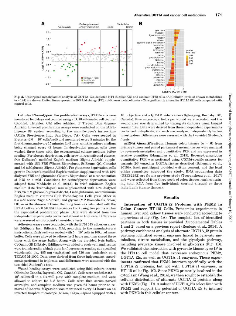
Fig. 3. Untargeted metabolomics analysis of UGT1A_i2s–depleted HT115 cells (KD) and control (CTR) cells. (A) Cellular levels of known metabolites(n = 144) are shown. Dotted lines represent a 20% fold change (FC). (B) Knownmetabolites (n = 24) significantly altered inHT115 KD cells compared withcontrol cells.
Alternative UGT1A and cancer cell metabolism 171
at ASPE
T Journals on February 10, 2022
molpharm
.aspetjournals.orgD
ownloaded from
Depletion of UGT1A_i2s Enhances Glycolytic Activityand Lactate Production but Reduces MitochondrialRespiration in Living Colon Cancer HT115 Cells. Wenext assessed the potential influence of UGT1A_i2s on gly-colysis and compared HT115 colon cancer cells expressing low(KD) and high (reference or control) UGT1A_i2 levels (Fig. 2A)(Rouleau et al., 2014). Given that PKM2 and PKM1 expressionis similar in both cell models (Supplemental Fig. 1), wepostulated that the cellular content in alternate UGT1A_i2scould affect PKM2 activity, thereby modifying glucose metab-olism to produce lactate, the end product of glycolysis. Theaccumulation of lactate by 57% (P5 0.029; Fig. 2B) in the cellmedium of KD cells compared with control cells supports a
functional impact of UGT1A_i2s on PKM2 activity. We thenused an extracellular flux analyzer to measure the extracel-lular acidification rate and oxygen consumption rate in livingHT115 cells, which reflects glycolytic activity and mitochon-drial respiration, respectively. A higher glycolytic activityin the UGT1A_i2–depleted cells was observed as evidenced bya 27% extracellular acidification rate elevation after additionof glucose to the medium. The maximal glycolytic capacity(measured after addition of oligomycin) was also elevated by30%, upholding greater utilization of the glycolysis pathwaycompared with control cells (Fig. 2, C and D). In an assaychallenging themitochondrial capacity andmeasuring oxygenconsumption rate, we detected a lower activity in KD cells
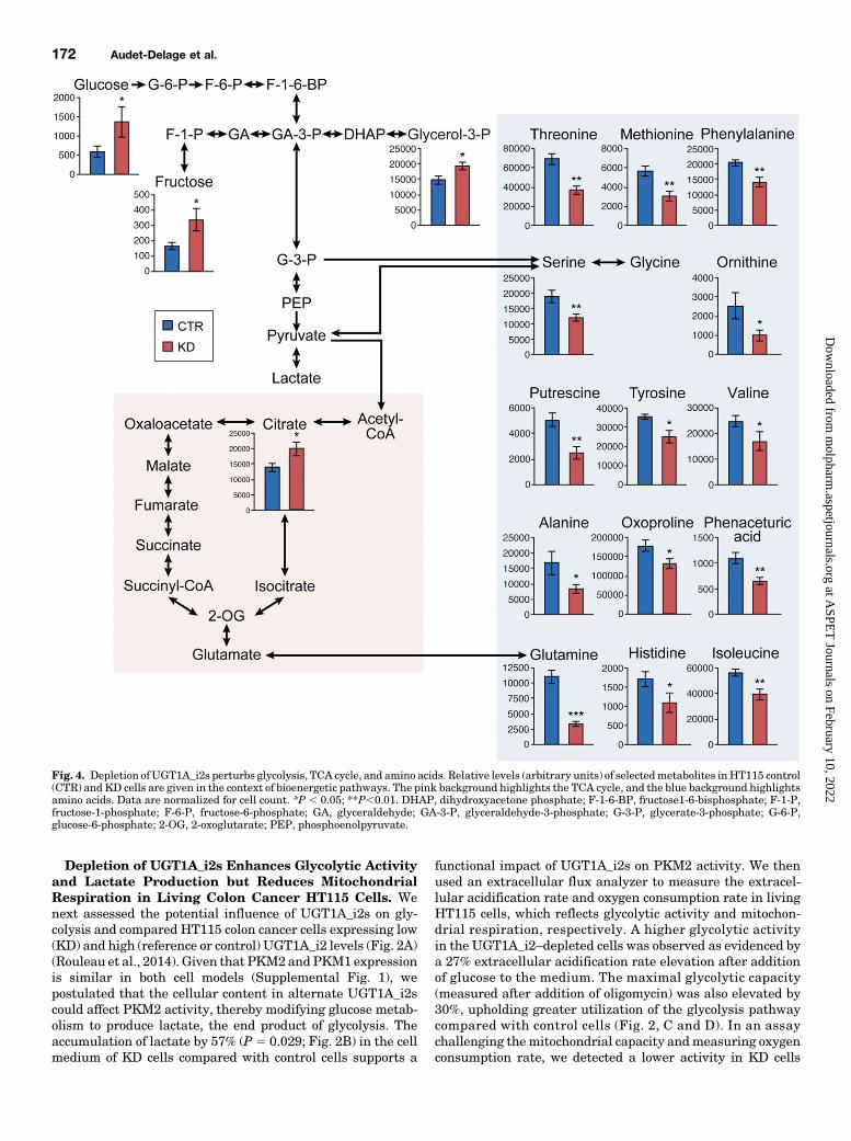
Fig. 4. Depletion ofUGT1A_i2s perturbs glycolysis, TCA cycle, and amino acids. Relative levels (arbitrary units) of selectedmetabolites inHT115 control(CTR) and KD cells are given in the context of bioenergetic pathways. The pink background highlights the TCA cycle, and the blue background highlightsamino acids. Data are normalized for cell count. *P , 0.05; **P,0.01. DHAP, dihydroxyacetone phosphate; F-1-6-BP, fructose1-6-bisphosphate; F-1-P,fructose-1-phosphate; F-6-P, fructose-6-phosphate; GA, glyceraldehyde; GA-3-P, glyceraldehyde-3-phosphate; G-3-P, glycerate-3-phosphate; G-6-P,glucose-6-phosphate; 2-OG, 2-oxoglutarate; PEP, phosphoenolpyruvate.
172 Audet-Delage et al.
at ASPE
T Journals on February 10, 2022
molpharm
.aspetjournals.orgD
ownloaded from
supported by lower baseline (227%) and lower maximalmitochondrial respiration rates (239%; after addition ofFCCP) in these cells compared with control cells (Fig. 2, Eand F). Thus, repression of endogenous UGT1A_i2 levels ledto a higher glycolytic rate at the expense of mitochondrialrespiration.Depletion of UGT1A_i2s Induces Broad Metabolic
Changes in Colon Cancer HT115 Cells and a GreaterMigration Potential. Given the changes in the energeticpathway induced by depletion of endogenous UGT1A_i2levels, we extended these studies to global cell metabolismusing untargeted metabolomics. A significant effect in thecellular levels of 58 metabolites was observed, includingmany intermediates derived from the glycolysis and tri-carboxylic acid cycle (TCA) pathways. These metabolites
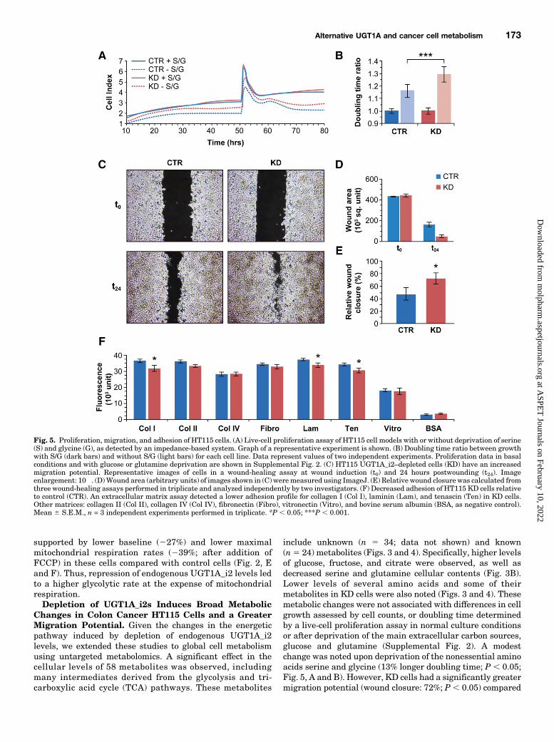
include unknown (n 5 34; data not shown) and known(n5 24) metabolites (Figs. 3 and 4). Specifically, higher levelsof glucose, fructose, and citrate were observed, as well asdecreased serine and glutamine cellular contents (Fig. 3B).Lower levels of several amino acids and some of theirmetabolites in KD cells were also noted (Figs. 3 and 4). Thesemetabolic changes were not associated with differences in cellgrowth assessed by cell counts, or doubling time determinedby a live-cell proliferation assay in normal culture conditionsor after deprivation of the main extracellular carbon sources,glucose and glutamine (Supplemental Fig. 2). A modestchange was noted upon deprivation of the nonessential aminoacids serine and glycine (13% longer doubling time; P , 0.05;Fig. 5, A and B). However, KD cells had a significantly greatermigration potential (wound closure: 72%; P , 0.05) compared
Fig. 5. Proliferation, migration, and adhesion of HT115 cells. (A) Live-cell proliferation assay of HT115 cell models with or without deprivation of serine(S) and glycine (G), as detected by an impedance-based system. Graph of a representative experiment is shown. (B) Doubling time ratio between growthwith S/G (dark bars) and without S/G (light bars) for each cell line. Data represent values of two independent experiments. Proliferation data in basalconditions and with glucose or glutamine deprivation are shown in Supplemental Fig. 2. (C) HT115 UGT1A_i2–depleted cells (KD) have an increasedmigration potential. Representative images of cells in a wound-healing assay at wound induction (t0) and 24 hours postwounding (t24). Imageenlargement: 10�. (D)Wound area (arbitrary units) of images shown in (C) weremeasured using ImageJ. (E) Relative wound closure was calculated fromthree wound-healing assays performed in triplicate and analyzed independently by two investigators. (F) Decreased adhesion of HT115 KD cells relativeto control (CTR). An extracellular matrix assay detected a lower adhesion profile for collagen I (Col I), laminin (Lam), and tenascin (Ten) in KD cells.Other matrices: collagen II (Col II), collagen IV (Col IV), fibronectin (Fibro), vitronectin (Vitro), and bovine serum albumin (BSA, as negative control).Mean 6 S.E.M., n = 3 independent experiments performed in triplicate. *P , 0.05; ***P , 0.001.
Alternative UGT1A and cancer cell metabolism 173
at ASPE
T Journals on February 10, 2022
molpharm
.aspetjournals.orgD
ownloaded from
with control cells (50%) (Fig. 5, C–E). Using an array ofextracellular matrix proteins, data further showed a modestbut significantly reduced adhesion capacity for KD cellstoward collagen I (13%), laminin (10%), and tenascin (11%)compared with control cells (Fig. 5F).Downregulation of UGT1A_i2 mRNA Expression in
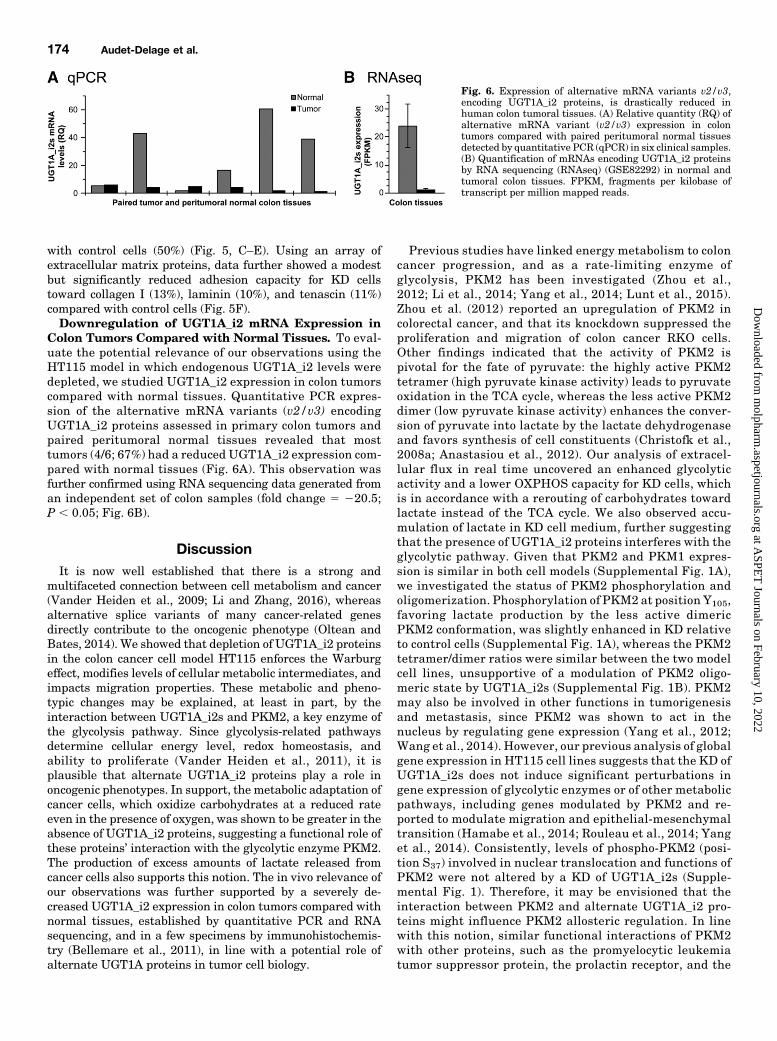
Colon Tumors Compared with Normal Tissues. To eval-uate the potential relevance of our observations using theHT115 model in which endogenous UGT1A_i2 levels weredepleted, we studied UGT1A_i2 expression in colon tumorscompared with normal tissues. Quantitative PCR expres-sion of the alternative mRNA variants (v2/v3) encodingUGT1A_i2 proteins assessed in primary colon tumors andpaired peritumoral normal tissues revealed that mosttumors (4/6; 67%) had a reduced UGT1A_i2 expression com-pared with normal tissues (Fig. 6A). This observation wasfurther confirmed using RNA sequencing data generated froman independent set of colon samples (fold change 5 220.5;P , 0.05; Fig. 6B).
DiscussionIt is now well established that there is a strong and
multifaceted connection between cell metabolism and cancer(Vander Heiden et al., 2009; Li and Zhang, 2016), whereasalternative splice variants of many cancer-related genesdirectly contribute to the oncogenic phenotype (Oltean andBates, 2014). We showed that depletion of UGT1A_i2 proteinsin the colon cancer cell model HT115 enforces the Warburgeffect, modifies levels of cellular metabolic intermediates, andimpacts migration properties. These metabolic and pheno-typic changes may be explained, at least in part, by theinteraction between UGT1A_i2s and PKM2, a key enzyme ofthe glycolysis pathway. Since glycolysis-related pathwaysdetermine cellular energy level, redox homeostasis, andability to proliferate (Vander Heiden et al., 2011), it isplausible that alternate UGT1A_i2 proteins play a role inoncogenic phenotypes. In support, the metabolic adaptation ofcancer cells, which oxidize carbohydrates at a reduced rateeven in the presence of oxygen, was shown to be greater in theabsence of UGT1A_i2 proteins, suggesting a functional role ofthese proteins’ interaction with the glycolytic enzyme PKM2.The production of excess amounts of lactate released fromcancer cells also supports this notion. The in vivo relevance ofour observations was further supported by a severely de-creased UGT1A_i2 expression in colon tumors compared withnormal tissues, established by quantitative PCR and RNAsequencing, and in a few specimens by immunohistochemis-try (Bellemare et al., 2011), in line with a potential role ofalternate UGT1A proteins in tumor cell biology.
Previous studies have linked energy metabolism to coloncancer progression, and as a rate-limiting enzyme ofglycolysis, PKM2 has been investigated (Zhou et al.,2012; Li et al., 2014; Yang et al., 2014; Lunt et al., 2015).Zhou et al. (2012) reported an upregulation of PKM2 incolorectal cancer, and that its knockdown suppressed theproliferation and migration of colon cancer RKO cells.Other findings indicated that the activity of PKM2 ispivotal for the fate of pyruvate: the highly active PKM2tetramer (high pyruvate kinase activity) leads to pyruvateoxidation in the TCA cycle, whereas the less active PKM2dimer (low pyruvate kinase activity) enhances the conver-sion of pyruvate into lactate by the lactate dehydrogenaseand favors synthesis of cell constituents (Christofk et al.,2008a; Anastasiou et al., 2012). Our analysis of extracel-lular flux in real time uncovered an enhanced glycolyticactivity and a lower OXPHOS capacity for KD cells, whichis in accordance with a rerouting of carbohydrates towardlactate instead of the TCA cycle. We also observed accu-mulation of lactate in KD cell medium, further suggestingthat the presence of UGT1A_i2 proteins interferes with theglycolytic pathway. Given that PKM2 and PKM1 expres-sion is similar in both cell models (Supplemental Fig. 1A),we investigated the status of PKM2 phosphorylation andoligomerization. Phosphorylation of PKM2 at position Y105,favoring lactate production by the less active dimericPKM2 conformation, was slightly enhanced in KD relativeto control cells (Supplemental Fig. 1A), whereas the PKM2tetramer/dimer ratios were similar between the two modelcell lines, unsupportive of a modulation of PKM2 oligo-meric state by UGT1A_i2s (Supplemental Fig. 1B). PKM2may also be involved in other functions in tumorigenesisand metastasis, since PKM2 was shown to act in thenucleus by regulating gene expression (Yang et al., 2012;Wang et al., 2014). However, our previous analysis of globalgene expression in HT115 cell lines suggests that the KD ofUGT1A_i2s does not induce significant perturbations ingene expression of glycolytic enzymes or of other metabolicpathways, including genes modulated by PKM2 and re-ported to modulate migration and epithelial-mesenchymaltransition (Hamabe et al., 2014; Rouleau et al., 2014; Yanget al., 2014). Consistently, levels of phospho-PKM2 (posi-tion S37) involved in nuclear translocation and functions ofPKM2 were not altered by a KD of UGT1A_i2s (Supple-mental Fig. 1). Therefore, it may be envisioned that theinteraction between PKM2 and alternate UGT1A_i2 pro-teins might influence PKM2 allosteric regulation. In linewith this notion, similar functional interactions of PKM2with other proteins, such as the promyelocytic leukemiatumor suppressor protein, the prolactin receptor, and the
Fig. 6. Expression of alternative mRNA variants v2/v3,encoding UGT1A_i2 proteins, is drastically reduced inhuman colon tumoral tissues. (A) Relative quantity (RQ) ofalternative mRNA variant (v2/v3) expression in colontumors compared with paired peritumoral normal tissuesdetected by quantitative PCR (qPCR) in six clinical samples.(B) Quantification of mRNAs encoding UGT1A_i2 proteinsby RNA sequencing (RNAseq) (GSE82292) in normal andtumoral colon tissues. FPKM, fragments per kilobase oftranscript per million mapped reads.
174 Audet-Delage et al.
at ASPE
T Journals on February 10, 2022
molpharm
.aspetjournals.orgD
ownloaded from
phosphoprotein (pp60src)-associated protein kinase ofRous sarcoma virus, led to reduced pyruvate kinaseactivity through allosteric regulation (Presek et al., 1980;Glossmann et al., 1981; Mazurek et al., 2001; Christofket al., 2008b; Shimada et al., 2008; Varghese et al., 2010;Gao et al., 2013). Whether this could be mediated by directinteractions with phosphorylated UGT1A_i2s (Basu et al.,2003, 2005; Volak and Court, 2010) or by a UGT1A_i2–dependent modulation of interactions between PKM2and other allosteric modulators will necessitate additionalinvestigations. From a therapeutic perspective, PKM2 expres-sion and activity can be regulated by inhibitors and activa-tors that have been tested in vitro and in vivo (Dong et al.,2016).The greater migration potential provoked by UGT1A_i2 KD
inHT115 cells is in line with a shift from oxidativemetabolismto aerobic glycolysis reported in more aggressive colon cancercells (Hussain et al., 2016). Consistent with our observationthat lactate accumulates in the medium of UGT1A_i2 KDcompared with control cells, this oncometabolite not only wasshown to act as a potent fuel (oxidative) but also functions as asignaling molecule to stimulate tumor angiogenesis (Dohertyand Cleveland, 2013). Moreover, the mitochondrial oxidativemetabolism is viewed as a critical suppressor of metastasis,and thus, lower OXPHOS activity may help promote metas-tasis (Lu et al., 2015). Cell migration is an early requirementfor tumor metastasis (Findlay et al., 2014), but is a complexphenomenon that involves cycling of adhesion and cell de-tachment (Panková et al., 2010; Tozluo�glu et al., 2013;Kurniawan et al., 2016). Strongly adherent cells may increasetheir migration capacity through a decrease of their adhesionto the extracellular matrix. Although subtle, we observed adifferential adhesion capacity of KD cells—namely, for lam-inin, a noncollagenous extracellular matrix critical in colon can-cer and linked to tumor angiogenesis, epithelial-mesenchymaltransition, and metastasis (Kitayama et al., 1999; Guesset al., 2009; Simon-Assmann et al., 2011). Accordingly, thisobservation may partially explain the differential migrationphenotype between cell lines expressing low and high levelsof UGT1A_i2s.A potential limitation is the fact that metabolomics data
were derived from a single time point, and thus represent thesum of numerous metabolic reactions that occurred in a48-hour period. The cellular content of several metabolicintermediates from the glycolysis and TCA cycle pathwayswas changed in UGT1A_i2–depleted HT115 cells comparedwith control cells and is unlikely explained solely by theprotein-protein interaction between UGT1A_i2s and PKM2.Additional partnerships observed by proteomics, such as thosewith enzymes of the gluconeogenesis and TCA cycle thatrequire additional validation by co-IP, may also underlie theseobservations. Of note, data revealed a globally reduced pool ofglucogenic amino acids in KD cells. This is consistent with theenhanced glycolytic potential induced by the KD and with theidentification of additional i2 protein partners associated withgluconeogenesis, such as pyruvate carboxylase and phospho-enolpyruvate carboxykinase 1 and 2. Furthermore, the lowerserine levels in KD cells compared with control suggested thatthe de novo serine synthesis pathway, critical for cancer cellproliferation and metabolism (Mehrmohamadi and Locasale,2015; Yoon et al., 2015), might be perturbed.We thus expectedthat serine andglycine deprivationwould increase themetabolic
pressure on glycolysis through a deviation of its intermedi-ates. Although modest, a significantly decreased cellularproliferation of KD cells was observed in these conditionscompared with control cells. It remains unknown whether aspecific UGT1A_i2 protein or several of those expressed inthe HT115 cell model trigger the observed effects given thatall UGT1A_i2s were simultaneously repressed by shRNA.Additional cell models need to be established to furtherinvestigate our initial findings.To the best of our knowledge, this is the first identification
of a functional link between the UGT pathway and energymetabolism, implying that alternate UGT1A proteins mightbe regulators of PKM2 with consequences for cancer cellmetabolism and phenotype. It may be unexpected that adepletion of alternate UGT1A_i2s is sufficient to alter themetabolic program of colon cancer cells given the numerous,redundant, and efficacious mechanisms in place to limitpyruvate oxidation in cancer cells. In the face of thesecounteractingmechanisms, themetabolic effects of UGT1A_i2depletion are rather impressive and may be explained in partby an interaction with the key glycolytic and multifunctionalenzyme PKM2, andmost likely with other metabolic enzymes.Although there was no change in UDPGlcA cell contentbetween the two cell models, another possibility could in-volve the allosteric modulation by UDPGlcA and/or otherUDP sugars, as reported for other glycolytic enzymes(Wu et al., 2006). It also remains to be demonstrated whetherUGT1A_i2s possess enzyme activity by utilizing other UDPsugars, for instance. Alternatively, the observed effects onmetabolism could be mediated through an impact on endog-enous levels of key metabolic molecules that are unknownsubstrates of UGT enzymes repressed by UGT1A_i2s. Thefindings thus support that alternate UGT1A proteins arepotential metabolic regulators of cancer cell metabolism, andthat they may contribute to the oncogenic phenotype of coloncancer cells. We conclude that alternate UGT proteins may bepart of the expanding compendium of metabolic pathwaysinvolved in cancer biology. However, further investigations ofUGT1A alternate proteins in cancer metabolism are required,as the cross-talk between global cell metabolism and the UGTpathway may likely constitute a key vulnerability in cancercells that could be exploited.
Acknowledgments
The authors acknowledge Lyne Villeneuve, Andréa Fournier,Patrick Caron, and Véronique Turcotte for technical support. Theauthors acknowledge the support from Michèle Orain for help withhandling of human tissues.
Authorship Contributions
Participated in research design: Mi. Rouleau, Guillemette.Conducted experiments: Audet-Delage, Me. Rouleau, Roberge.Contributed new reagents or analytic tools: Miard, Picard, Têtu.Performed data analysis: Audet-Delage, Mi. Rouleau, Me. Rouleau,
Roberge, Miard, Guillemette.Wrote or contributed to the writing of the manuscript: Audet-
Delage, Mi. Rouleau, Me. Rouleau, Guillemette.
References
Anastasiou D, Yu Y, Israelsen WJ, Jiang JK, Boxer MB, Hong BS, Tempel W,Dimov S, Shen M, Jha A, et al. (2012) Pyruvate kinase M2 activators pro-mote tetramer formation and suppress tumorigenesis. Nat Chem Biol 8:839–847.
Basu NK, Kole L, and Owens IS (2003) Evidence for phosphorylation requirement forhuman bilirubin UDP-glucuronosyltransferase (UGT1A1) activity. Biochem Bio-phys Res Commun 303:98–104.
Alternative UGT1A and cancer cell metabolism 175
at ASPE
T Journals on February 10, 2022
molpharm
.aspetjournals.orgD
ownloaded from
Basu NK, Kovarova M, Garza A, Kubota S, Saha T, Mitra PS, Banerjee R, Rivera J,and Owens IS (2005) Phosphorylation of a UDP-glucuronosyltransferase regulatessubstrate specificity. Proc Natl Acad Sci USA 102:6285–6290.
Bellemare J, Rouleau M, Girard H, Harvey M, and Guillemette C (2010a) Alterna-tively spliced products of the UGT1A gene interact with the enzymatically activeproteins to inhibit glucuronosyltransferase activity in vitro. Drug Metab Dispos 38:1785–1789.
Bellemare J, Rouleau M, Harvey M, Popa I, Pelletier G, Têtu B, and Guillemette C(2011) Immunohistochemical expression of conjugating UGT1A-derived isoformsin normal and tumoral drug-metabolizing tissues in humans. J Pathol 223:425–435.
Bellemare J, Rouleau M, Harvey M, Têtu B, and Guillemette C (2010b)Alternative-splicing forms of the major phase II conjugating UGT1A gene nega-tively regulate glucuronidation in human carcinoma cell lines. PharmacogenomicsJ 10:431–441.
Biamonti G, Catillo M, Pignataro D, Montecucco A, and Ghigna C (2014) The alter-native splicing side of cancer. Semin Cell Dev Biol 32:30–36.
Bindea G, Galon J, and Mlecnik B (2013) CluePedia Cytoscape plugin: pathwayinsights using integrated experimental and in silico data. Bioinformatics 29:661–663.
Bindea G, Mlecnik B, Hackl H, Charoentong P, Tosolini M, Kirilovsky A, FridmanWH, Pagès F, Trajanoski Z, and Galon J (2009) ClueGO: a Cytoscape plug-in todecipher functionally grouped gene ontology and pathway annotation networks.Bioinformatics 25:1091–1093.
Bluemlein K, Grüning NM, Feichtinger RG, Lehrach H, Kofler B, and Ralser M(2011) No evidence for a shift in pyruvate kinase PKM1 to PKM2 expression duringtumorigenesis. Oncotarget 2:393–400.
Christofk HR, Vander Heiden MG, Harris MH, Ramanathan A, Gerszten RE, Wei R,Fleming MD, Schreiber SL, and Cantley LC (2008a) The M2 splice isoform ofpyruvate kinase is important for cancer metabolism and tumour growth. Nature452:230–233.
Christofk HR, Vander Heiden MG, Wu N, Asara JM, and Cantley LC (2008b)Pyruvate kinase M2 is a phosphotyrosine-binding protein. Nature 452:181–186.
Cui R and Shi XY (2015) Expression of pyruvate kinase M2 in human colorectalcancer and its prognostic value. Int J Clin Exp Pathol 8:11393–11399.
Doherty JR and Cleveland JL (2013) Targeting lactate metabolism for cancer ther-apeutics. J Clin Invest 123:3685–3692.
Dong G, Mao Q, Xia W, Xu Y, Wang J, Xu L, and Jiang F (2016) PKM2 and cancer:The function of PKM2 beyond glycolysis. Oncol Lett 11:1980–1986.
Fiehn O, Garvey WT, Newman JW, Lok KH, Hoppel CL, and Adams SH (2010)Plasma metabolomic profiles reflective of glucose homeostasis in non-diabetic andtype 2 diabetic obese African-American women. PLoS One 5:e15234.
Fiehn O, Wohlgemuth G, Scholz M, Kind T, Lee DY, Lu Y, Moon S, and Nikolau B(2008) Quality control for plant metabolomics: reporting MSI-compliant studies.Plant J 53:691–704.
Findlay VJ, Wang C, Watson DK, and Camp ER (2014) Epithelial-to-mesenchymal transition and the cancer stem cell phenotype: insights from can-cer biology with therapeutic implications for colorectal cancer. Cancer Gene Ther21:181–187.
Gao X, Wang H, Yang JJ, Chen J, Jie J, Li L, Zhang Y, and Liu ZR (2013) Reciprocalregulation of protein kinase and pyruvate kinase activities of pyruvate kinase M2by growth signals. J Biol Chem 288:15971–15979.
Girard H, Lévesque E, Bellemare J, Journault K, Caillier B, and Guillemette C(2007) Genetic diversity at the UGT1 locus is amplified by a novel 39 alter-native splicing mechanism leading to nine additional UGT1A proteins that actas regulators of glucuronidation activity. Pharmacogenet Genomics 17:1077–1089.
Glossmann H, Presek P, and Eigenbrodt E (1981) Association of the src-gene productof Rous sarcoma virus with a pyruvate-kinase inactivation factor. Mol Cell Endo-crinol 23:49–63.
Guess CM, Lafleur BJ, Weidow BL, and Quaranta V (2009) A decreased ratio oflaminin-332 beta3 to gamma2 subunit mRNA is associated with poor prognosis incolon cancer. Cancer Epidemiol Biomarkers Prev 18:1584–1590.
Gupta V and Bamezai RN (2010) Human pyruvate kinase M2: a multifunctionalprotein. Protein Sci 19:2031–2044.
Hamabe A, Konno M, Tanuma N, Shima H, Tsunekuni K, Kawamoto K, Nishida N,Koseki J, Mimori K, Gotoh N, et al. (2014) Role of pyruvate kinase M2 in tran-scriptional regulation leading to epithelial-mesenchymal transition. Proc NatlAcad Sci USA 111:15526–15531.
Hussain A, Qazi AK, Mupparapu N, Guru SK, Kumar A, Sharma PR, Singh SK,Singh P, Dar MJ, Bharate SB, et al. (2016) Modulation of glycolysis and lipogenesisby novel PI3K selective molecule represses tumor angiogenesis and decreases co-lorectal cancer growth. Cancer Lett 374:250–260.
Israelsen WJ and Vander Heiden MG (2015) Pyruvate kinase: Function, regulationand role in cancer. Semin Cell Dev Biol 43:43–51.
Kitayama J, Nagawa H, Tsuno N, Osada T, Hatano K, Sunami E, Saito H, and MutoT (1999) Laminin mediates tethering and spreading of colon cancer cells in phys-iological shear flow. Br J Cancer 80:1927–1934.
Kurniawan NA, Chaudhuri PK, and Lim CT (2016) Mechanobiology of cell migrationin the context of dynamic two-way cell-matrix interactions. J Biomech 49:1355–1368.
Lévesque E, Girard H, Journault K, Lépine J, and Guillemette C (2007) Regulation ofthe UGT1A1 bilirubin-conjugating pathway: role of a new splicing event at theUGT1A locus. Hepatology 45:128–138.
Li L, Zhang Y, Qiao J, Yang JJ, and Liu ZR (2014) Pyruvate kinase M2 in bloodcirculation facilitates tumor growth by promoting angiogenesis. J Biol Chem 289:25812–25821.
Li Z and Zhang H (2016) Reprogramming of glucose, fatty acid and amino acid me-tabolism for cancer progression. Cell Mol Life Sci 73:377–392.
Lu J, Tan M, and Cai Q (2015) The Warburg effect in tumor progression: mito-chondrial oxidative metabolism as an anti-metastasis mechanism. Cancer Lett 356:156–164.
Lunt SY, Muralidhar V, Hosios AM, Israelsen WJ, Gui DY, Newhouse L, Ogrod-zinski M, Hecht V, Xu K, Acevedo PN, et al. (2015) Pyruvate kinase isoformexpression alters nucleotide synthesis to impact cell proliferation. Mol Cell 57:95–107.
Maddocks OD, Berkers CR, Mason SM, Zheng L, Blyth K, Gottlieb E, and VousdenKH (2013) Serine starvation induces stress and p53-dependent metabolic remod-elling in cancer cells. Nature 493:542–546.
Margaillan G, Rouleau M, Fallon JK, Caron P, Villeneuve L, Turcotte V, Smith PC,Joy MS, and Guillemette C (2015) Quantitative profiling of human renal UDP-glucuronosyltransferases and glucuronidation activity: a comparison of normal andtumoral kidney tissues. Drug Metab Dispos 43:611–619.
Mazurek S, Zwerschke W, Jansen-Dürr P, and Eigenbrodt E (2001) Metabolic co-operation between different oncogenes during cell transformation: interaction be-tween activated ras and HPV-16 E7. Oncogene 20:6891–6898.
Mehrmohamadi M and Locasale JW (2015) Context dependent utilization of serine incancer. Mol Cell Oncol 2:e996418.
Mitsopoulos C, Schierz AC, Workman P, and Al-Lazikani B (2015) Distinctive Be-haviors of Druggable Proteins in Cellular Networks. PLOS Comput Biol 11:e1004597.
Miyauchi Y, Nagata K, Yamazoe Y, Mackenzie PI, Yamada H, and Ishii Y (2015)Suppression of Cytochrome P450 3A4 Function by UDP-Glucuronosyltransferase2B7 through a Protein-Protein Interaction: Cooperative Roles of the CytosolicCarboxyl-Terminal Domain and the Luminal Anchoring Region. Mol Pharmacol88:800–812.
Oltean S and Bates DO (2014) Hallmarks of alternative splicing in cancer. Oncogene33:5311–5318.
Panková K, Rösel D, Novotný M, and Brábek J (2010) The molecular mechanisms oftransition between mesenchymal and amoeboid invasiveness in tumor cells. CellMol Life Sci 67:63–71.
Presek P, Glossmann H, Eigenbrodt E, Schoner W, Rübsamen H, Friis RR, and BauerH (1980) Similarities between a phosphoprotein (pp60src)-associated proteinkinase of Rous sarcoma virus and a cyclic adenosine 39:59-monophosphate-independent protein kinase that phosphorylates pyruvate kinase type M2. Can-cer Res 40:1733–1741.
Rouleau M, Roberge J, Bellemare J, and Guillemette C (2014) Dual roles for splicevariants of the glucuronidation pathway as regulators of cellular metabolism. MolPharmacol 85:29–36.
Rouleau M, Tourancheau A, Girard-Bock C, Villeneuve L, Vaucher J, Duperré AM,Audet-Delage Y, Gilbert I, Popa I, Droit A, et al. (2016) Divergent Expression andMetabolic Functions of Human Glucuronosyltransferases through AlternativeSplicing. Cell Reports 17:114–124.
Rowland A, Miners JO, and Mackenzie PI (2013) The UDP-glucuronosyltransferases:their role in drug metabolism and detoxification. Int J Biochem Cell Biol 45:1121–1132.
Scott DE, Bayly AR, Abell C, and Skidmore J (2016) Small molecules, big targets:drug discovery faces the protein-protein interaction challenge. Nat Rev Drug Dis-cov 15:533–550.
Shannon P, Markiel A, Ozier O, Baliga NS, Wang JT, Ramage D, Amin N,Schwikowski B, and Ideker T (2003) Cytoscape: a software environment for in-tegrated models of biomolecular interaction networks. Genome Res 13:2498–2504.
Shimada N, Shinagawa T, and Ishii S (2008) Modulation of M2-type pyruvate kinaseactivity by the cytoplasmic PML tumor suppressor protein. Genes Cells 13:245–254.
Simon-Assmann P, Orend G, Mammadova-Bach E, Spenlé C, and Lefebvre O (2011)Role of laminins in physiological and pathological angiogenesis. Int J Dev Biol 55:455–465.
Stingl JC, Bartels H, Viviani R, Lehmann ML, and Brockmöller J (2014) Relevance ofUDP-glucuronosyltransferase polymorphisms for drug dosing: A quantitative sys-tematic review. Pharmacol Ther 141:92–116.
Taniguchi K, Ito Y, Sugito N, Kumazaki M, Shinohara H, Yamada N, Nakagawa Y,Sugiyama T, Futamura M, Otsuki Y, et al. (2015) Organ-specific PTB1-associatedmicroRNAs determine expression of pyruvate kinase isoforms. Sci Rep 5:8647.
Tourancheau A, Rouleau M, Guauque-Olarte S, Villeneuve L, Gilbert I, Droit A,and Guillemette C (2017) Quantitative profiling of the UGT transcriptome in hu-man drug metabolizing tissues. Pharmacogenomics J, in press.
Tozluo�glu M, Tournier AL, Jenkins RP, Hooper S, Bates PA, and Sahai E (2013)Matrix geometry determines optimal cancer cell migration strategy and modulatesresponse to interventions. Nat Cell Biol 15:751–762.
Vander Heiden MG, Cantley LC, and Thompson CB (2009) Understanding theWarburg effect: the metabolic requirements of cell proliferation. Science 324:1029–1033.
Vander Heiden MG, Lunt SY, Dayton TL, Fiske BP, Israelsen WJ, Mattaini KR,Vokes NI, Stephanopoulos G, Cantley LC, Metallo CM, et al. (2011) Metabolicpathway alterations that support cell proliferation. Cold Spring Harb Symp QuantBiol 76:325–334.
Varghese B, Swaminathan G, Plotnikov A, Tzimas C, Yang N, Rui H, and Fuchs SY(2010) Prolactin inhibits activity of pyruvate kinase M2 to stimulate cell pro-liferation. Mol Endocrinol 24:2356–2365.
Volak LP and Court MH (2010) Role for protein kinase C delta in the functionalactivity of human UGT1A6: implications for drug-drug interactions between PKCinhibitors and UGT1A6. Xenobiotica 40:306–318.
Wang HJ, Hsieh YJ, Cheng WC, Lin CP, Lin YS, Yang SF, Chen CC, Izumiya Y, YuJS, Kung HJ, et al. (2014) JMJD5 regulates PKM2 nuclear translocation and re-programs HIF-1a-mediated glucose metabolism. Proc Natl Acad Sci USA 111:279–284.
Warburg O, Wind F, and Negelein E (1927) The Metabolism of Tumors in the Body.J Gen Physiol 8:519–530.
176 Audet-Delage et al.
at ASPE
T Journals on February 10, 2022
molpharm
.aspetjournals.orgD
ownloaded from
Wu C, Khan SA, Peng LJ, and Lange AJ (2006) Roles for fructose-2,6-bisphosphate inthe control of fuel metabolism: beyond its allosteric effects on glycolytic and glu-coneogenic enzymes. Adv Enzyme Regul 46:72–88.
Yang P, Li Z, Fu R, Wu H, and Li Z (2014) Pyruvate kinase M2 facilitates coloncancer cell migration via the modulation of STAT3 signalling. Cell Signal 26:1853–1862.
Yang P, Li Z, Wang Y, Zhang L, Wu H, and Li Z (2015) Secreted pyruvate kinase M2facilitates cell migration via PI3K/Akt and Wnt/b-catenin pathway in colon cancercells. Biochem Biophys Res Commun 459:327–332.
Yang W, Zheng Y, Xia Y, Ji H, Chen X, Guo F, Lyssiotis CA, Aldape K, Cantley LC,and Lu Z (2012) ERK1/2-dependent phosphorylation and nuclear translocation ofPKM2 promotes the Warburg effect. Nat Cell Biol 14:1295–1304.
Yang X, Coulombe-Huntington J, Kang S, Sheynkman GM, Hao T, RichardsonA, Sun S, Yang F, Shen YA, Murray RR, et al. (2016) Widespread Expansionof Protein Interaction Capabilities by Alternative Splicing. Cell 164:805–817.
Yoon S, Kim JG, Seo AN, Park SY, Kim HJ, Park JS, Choi GS, Jeong JY, Jun Y, YoonGS, et al. (2015) Clinical Implication of Serine Metabolism-Associated Enzymes inColon Cancer. Oncology 89:351–359.
Zhan C, Yan L, Wang L, Ma J, Jiang W, Zhang Y, Shi Y, and Wang Q (2015) Isoformswitch of pyruvate kinase M1 indeed occurs but not to pyruvate kinase M2 inhuman tumorigenesis. PLoS One 10:e0118663.
Zhou CF, Li XB, Sun H, Zhang B, Han YS, Jiang Y, Zhuang QL, Fang J, and Wu GH(2012) Pyruvate kinase type M2 is upregulated in colorectal cancer and promotesproliferation and migration of colon cancer cells. IUBMB Life 64:775–782.
Address correspondence to: Dr. Chantal Guillemette, Canada ResearchChair in Pharmacogenomics, Centre Hospitalier Universitaire de QuébecResearch Center, R4720, 2705 Boulevard Laurier, Québec, Canada G1V 4G2.E-mail: [email protected]
Alternative UGT1A and cancer cell metabolism 177
at ASPE
T Journals on February 10, 2022
molpharm
.aspetjournals.orgD
ownloaded from