Embed Size (px)
Citation preview

Brief Communications
The extended pectoralis major flap for reconstruction of the upperposterior chest wall and axillaNiklas Iblher, MD, Vincenzo Penna, MD, Arash Momeni, MD, Nestor Torio Padron, MD, and G. Bjoern Stark, MD, Prof,Freiburg, Germany
Supplemental material is available online.
Although the pectoralis major muscle as a basis for a va-
riety of flaps is still a workhorse for head and neck re-
construction1 and a flap of choice for anterior chest
wall reconstruction,2 coverage of dorsal upper chest
wall defects including the axilla are hardly described. In certain
cases when infiltration of a tumor or trauma or previous damage
to the vascular supply precludes the use of adjacent muscle flaps,
a pectoralis major musculocutaneous island flap can offer a valuable
alternative.
AnatomyThe pectoralis major muscle originates from the medial part of the
clavicle, the sternocostal border of the first 6 ribs, and the external
oblique muscle aponeurosis.
The main functions of the muscle are adduction and medial rota-
tion of the arm. Sacrifice of this muscle leads to only minimal func-
tional deficit because adjunct muscles of the shoulder belt can
almost completely compensate for the loss.3
The main vascular supply to the pectoralis muscle and its over-
lying skin derives from the pectoral branch of the thoracoacromial
artery originating from beneath the midportion of the clavicle and
coursing toward the xiphoid. Further vascular supply originates
from perforating branches of the internal thoracic artery, from per-
forating vessels that derive from the 5th to 7th intercostal arteries,
and from the lateral thoracic artery.
The skin island should be centered over the pectoralis major
muscle but can be considerably extended caudally (up to a size of
45 3 18 cm and more) by including the rectus fascia owing to
a rich vascular network anastomosing with the superior epigastric
system,4 as has been shown in injection studies of the thoracodorsal
system.5
From the Department of Plastic and Hand Surgery, University of Freiburg
Medical Center, Freiburg, Germany.
Received for publication Oct 9, 2007; accepted for publication Nov 25,
2007.
Address for reprints: Niklas Iblher, MD, Department of Plastic and Hand Sur-
gery, University of Freiburg Medical Center, Hugstetter Strasse 55, 79106
Freiburg, Germany (E-mail: [email protected]).
J Thorac Cardiovasc Surg 2008;136:790-1
0022-5223/$34.00
Copyright � 2008 by The American Association for Thoracic Surgery
doi:10.1016/j.jtcvs.2007.11.066
790 The Journal of Thoracic and Cardiovascular Surgery c Sep
TechniqueThe estimated defect is marked and an equivalent skin island is de-
signed along the pectoralis major muscle axis (Figure 1). The island
can extend well beyond the lower border of the pectoralis muscle so
long as a sufficient part of the skin island is localized above the pec-
toralis major. If a larger transverse dimension of the skin island is
required, the nipple may be included in the flap in men.
The patient is positioned in a lateral decubitus position. The flap
is raised in a retrograde manner by incising the skin along the pre-
operative markings down to the anterior rectus sheath caudally.
The anterior rectus sheath has to be included in extended flaps. Stay-
ing below the abdominal fascia allows a straightforward dissection.
The origins of the pectoralis major at the caudal ribs and the sternum
in continuation with the fascia are separated, and the subpectoral
plane is entered. If possible, the lateral part of the muscle should
be spared to prevent loss of the anterior axillary fold. At the cranial
edge of the island, the dissection is performed down to the pectoralis
major muscle and from there upward toward the clavicle. The vas-
cular pedicle is visualized on the undersurface of the muscle and iso-
lated, leaving a protective cuff of muscle around it. The insertion of
the muscle to the humerus lateral to the pedicle can then be separated
and the flap transferred to the recipient area (Figure 2). If a transpo-
sition into a dorsal defect is planned, a subcutaneous tunnel is made.
Skin islands of a dimension up to 45 3 18 cm can be safely elevated,
still allowing primary wound closure at the donor site (Figure 3).
In men, large skin islands may incorporate the nipple areola com-
plex, which can later be retransplanted as a free skin transplant in
a secondary procedure with local anesthesia.
Three representative clinical summaries are presented in an
online appendix (E-Appendix).
DiscussionNumerous articles report algorithms for chest wall reconstruction.
Chest wall defects are more common anteriorly because the primary
underlying causes are located here (eg, breast cancer, radiation
ulcer, and sternotomy wounds) and their management is well docu-
mented. Posterior chest wall defects are less common, their
approach far less well documented, and thus are left out of sug-
gested algorithms. Among the regional options for coverage are
the trapezius flap, the latissimus dorsi flap, flaps of the (para)scap-
ular system, gluteus flaps, or paraspinous muscle flaps. If these lo-
cal flaps are precluded by the extent of resection or compromised
vascular pedicles, free tissue transfer can offer a flexible solution,
although the availability of recipient vessels may be difficult in
some cases. For defects of the upper posterior chest wall and shoul-
der, the pectoralis major flap offers a valuable alternative with
minimal donor site defect and functional impairment.3 This is
supported by reports on patients with congenital deficiency of
this muscle.
Flap dissection is quick and straightforward. A wide arc of rota-
tion and a large skin island are among the advantages of the
tember 2008

Brief Communications
Figure 1. Defect and flap design.
Figure 2. Flap transposition.
Figure 3. Wound closure.
pectoralis major flap. Owing to the anastomosis of vascular network
with the epigastric system, large skin islands that extend far below
the pectoralis muscle down to the periumbilical region can be har-
vested, with primary donor site closure being possible in almost
all patients (Figure 3).
The modern armamentarium of plastic surgery offers a wide
range of safe reconstructive flap transfers with minor morbidity for
chest wall reconstruction. Resectability of solid tumors hardly
should have to be excluded on the basis of the impossibility to cover
the resulting defect. In patients with an upper dorsal or axillary defect
in which use of dorsal muscle flaps is precluded owing to resection,
vascular compromise, or other reasons, the extended pectoralis major
musculocutaneous flap offers a safe, fast, and reliable alternative.
The Journal of Tho
References1. Vartanian JG, Carvalho AL, Carvalho SM, Mizobe L, Magrin J,
Kowalski LP. Pectoralis major and other myofascial/myocutaneous flapsin head and neck cancer reconstruction: experience with 437 cases at asingle institution. Head Neck. 2004;26:1018-23.
2. Tobin GR. Pectoralis major muscle-musculocutaneous flap for chest-wallreconstruction. Surg Clin North Am. 1989;69:991-1006.
3. Har-El G, Krespi YP, Har-El R. Physical rehabilitation after myocutane-ous flaps. Head Neck. 1990;12:218-24.
4. Russell RC, Feller AM, Elliott LF, Kucan JO, Zook EG. The extendedpectoralis major myocutaneous flap: uses and indications. Plast ReconstrSurg. 1991;88:814-23.
5. Reid CD, Taylor GI. The vascular territory of the acromiothoracic axis. BrJ Plast Surg. 1984;37:194-212.
racic and Cardiovascular Surgery c Volume 136, Number 3 791
Brief Communications
E-AppendixClinical SummariesPATIENT 1. A 24-year-old man with a history of a mole removal 7
months before presentation and a failed reoperation with axilla
dissection owing to an increasing mass at the incision site several
weeks later was transferred to our department. The histologic
workup of the second operation revealed a malignant melanoma
with lymphatic metastasis to the axilla. Staging showed no signs of
further metastasis. The patient received preoperative chemotherapy.
Figure E1A shows the preoperative situation. Wide excision
included the margo lateralis, spina, and pars membranacea of the
scapula, as well as the periscapular muscles including the teres
major and minor, infraspinatus, supraspinatus, and latissimus dorsi,
which precluded use of dorsally based muscle flaps. Axillary dis-
section was performed and R0 status was achieved (Figure E1B).
Defect coverage was achieved with a musculocutaneous pector-
alis flap with an abdominally extended skin island (Figure E1C). Pri-
mary closure of the donor site was possible. Healing was uneventful
and the patient was discharged on postopeerative day 7. Six months
after the operation, the patient achieved nearly symmetric active
shoulder movement (Figure E1D). Retransplantation of the nipple
to its original location was offered to the patient but he declined.
Seven months after the operation, a local lymph node metastasis
was removed and 20 months after the operation another two suspi-
cious lymph nodes were resected, but did not show any malignant
cells. Up to now, 3.5 years after the operation, the patient is free
of relapse. The patient reports almost normal shoulder function.
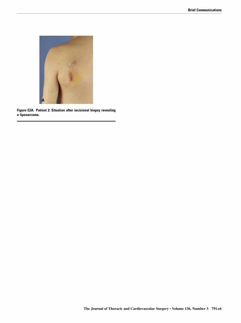
PATIENT 2. A 76-year-old retired oncology professor had a scapu-
lar swelling that he attributed to a recent mosquito bite. Magnetic
resonance imaging showed a suspicious fatty soft tissue tumor. Inci-
sional biopsy was performed and revealed a low-grade liposarcoma
(Figure E2A). The tumor infiltrated the latissimus and infraspinatus
791.e1 The Journal of Thoracic and Cardiovascular Surgery c S
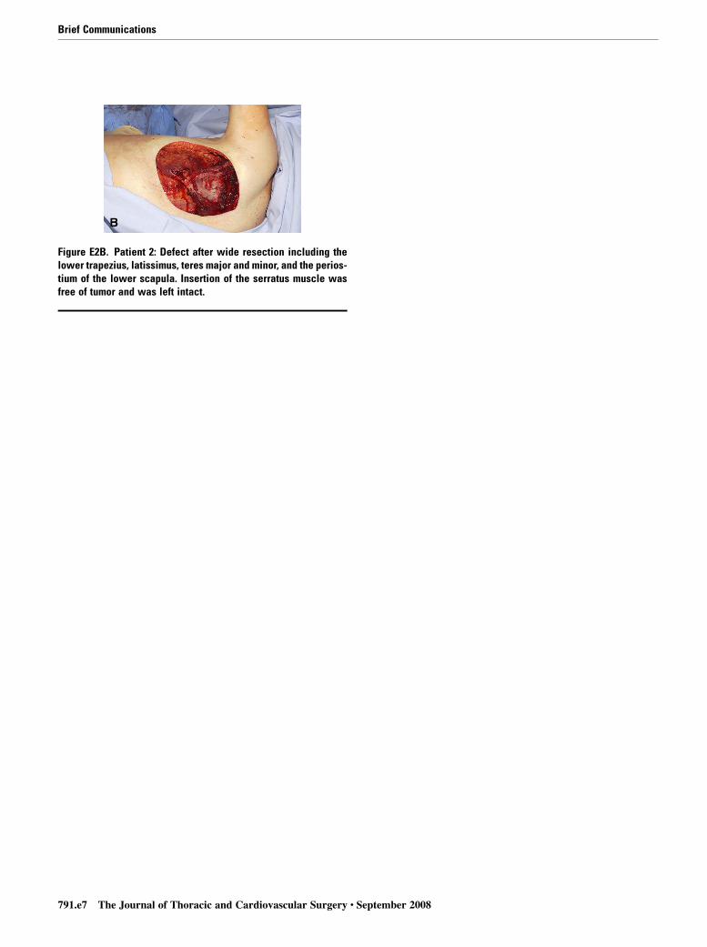
muscles and reached the posteroinferior surface of the scapula. Wide
resection included the caudal part of the trapezius, the latissimus
dorsi, teres minor and major, infraspinatus, and the periostium of
the scapula, which precluded the use of muscle flaps from the dorsal
trunk (Figure E2B). An extended pectoralis major musculocutane-
ous flap was raised, tunneled subcutaneously through the axilla,
and placed into the defect. Primary closure of the donor site was pos-
sible. Postoperative recovery was uneventful. Figure E2C displays
the postoperative result after 3 months. One year after the operation,
the patient is free of relapse.
PATIENT 3. A 66-year-old patient had a growing tumor above
the left scapula. Incisional biopsy demonstrated a pleomorphic sar-
coma. Wide resection was performed by the senior author (GBS).
The latissimus muscle was infiltrated and had to be resected,
thereby being excluded for local coverage. Large parts of the infra-
spinatus fossa were resected, preserving the inferior tip and spine
(Figure E3A). Axillary lymph nodes of levels one and two were
resected en bloc, including a suspicious pectoralis minor muscle.
Because the skin was infiltrated up to the anterior axillary line,
a large island flap (35 3 15 cm) extending far into the hypogastric
region had to be raised (Figure E3B). To assure perfusion of the
skin island, the anterior sheath of the rectus muscle was raised
with it. The flap was rotated 120� and completely covered the de-
fect (Figure E3C ). The donor site defect could be closed primarily
(Figure E3D). Resection margins were free of tumor and wound
healing was uneventful. Again, nipple transplantation was offered
to the patient but has not yet been performed. Postoperative radio-
therapy was performed after complete wound healing and was well
tolerated. Figure E3E shows the result 18 months postoperatively.
A mild bulging of the flap is noticeable but does not disturb the
patient. Three years postoperatively, there is no recurrence of the
sarcoma.
eptember 2008

Brief Communications
Figure E1A. Patient 1: Preoperative situation with R2 resection ofa malignant melanoma.
The Journal of Thoracic and Cardiovascular Surgery c Volume 136, Number 3 791.e2

Brief Communications
Figure E1B. Patient 1: Design of extended musculocutaneouspectoralis major flap.
791.e3 The Journal of Thoracic and Cardiovascular Surgery c September 2008

Brief Communications
Figure E1C. Patient 1: Intraoperative situation. Above wide resec-tion including the margo lateralis, spina, and pars membranaceaof the scapula and the periscapular and parascapular muscles.
The Journal of Thoracic and Cardiovascular Surgery c Volume 136, Number 3 791.e4

Brief Communications
Figure E1D. Patient 1: Six months post-operatively showing almost normalshoulder function.
791.e5 The Journal of Thoracic and Cardiovascular Surgery c September 2008
Brief Communications
Figure E2A. Patient 2: Situation after incisional biopsy revealinga liposarcoma.
The Journal of Thoracic and Cardiovascular Surgery c Volume 136, Number 3 791.e6
Brief Communications
Figure E2B. Patient 2: Defect after wide resection including thelower trapezius, latissimus, teres major and minor, and the perios-tium of the lower scapula. Insertion of the serratus muscle wasfree of tumor and was left intact.
791.e7 The Journal of Thoracic and Cardiovascular Surgery c September 2008

Brief Communications
Figure E2C. Patient 2: Three months postopera-tively.
The Journal of Thoracic and Cardiovascular Surgery c Volume 136, Number 3 791.e8

Brief Communications
Figure E3A. Patient 3: Defect after wide resection of a pleomor-phic sarcoma.
791.e9 The Journal of Thoracic and Cardiovascular Surgery c September 2008

Brief Communications
Figure E3B. Patient 3: Flap design of extended pectoralis majormyocutaneous flap.
The Journal of Thoracic and Cardiovascular Surgery c Volume 136, Number 3 791.e10

Brief Communications
Figure E3C. Patient 3: Flap transposi-tion.
791.e11 The Journal of Thoracic and Cardiovascular Surgery c September 2008

Brief Communications
Figure E3D. Patient 3: Wound closure.
The Journal of Thoracic and Cardiovascular Surgery c Volume 136, Number 3 791.e12

Brief Communications
Figure E3E. Patient 3: Result after 18months.
791.e13 The Journal of Thoracic and Cardiovascular Surgery c September 2008


![Deep Circumflex Iliac Artery Flap for Reconstruction of ... · mandibular reconstruction, the fibula flap [6] has been equally successful. Although fibula and scapula free flaps remains](https://img.pdfslide.us/doc/110x75/5ed54f1f1dbb8245b96a7213/deep-circumflex-iliac-artery-flap-for-reconstruction-of-mandibular-reconstruction.jpg)


![Fascial Flap Reconstruction of the Hand a Single.25[1]](https://img.pdfslide.us/doc/110x75/55147566497959ee1d8b4746/fascial-flap-reconstruction-of-the-hand-a-single251.jpg)
