Embed Size (px)
Citation preview
PECTORALIS MAJOR MYOCUTAJNEUUS FLAP FOR
RECONSTRUCTION OF DEFECTS FOLLOWING
RESECTIONS IN HEAD AND NECK AREA
Pages with reference to book, From 72 To 76 Mohammad Arshad Cheema ( Department of Surgery, King Saud University, College of Medicine, Abha, Saudi Arabia. )
ABSTRACT
Pectoralis major myocutaneous flap (PMMF) has become the standard for reconstruction of major
defects in head and neck area, Eleven cases, operated over a three year period, in which PMMF was
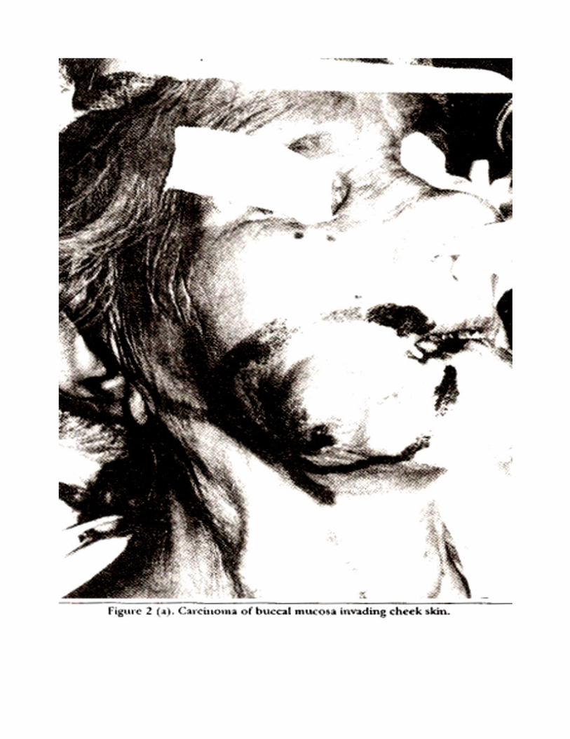
used for reconstruction have been reviewed retrospectively. Nine patients had oral squamous cell
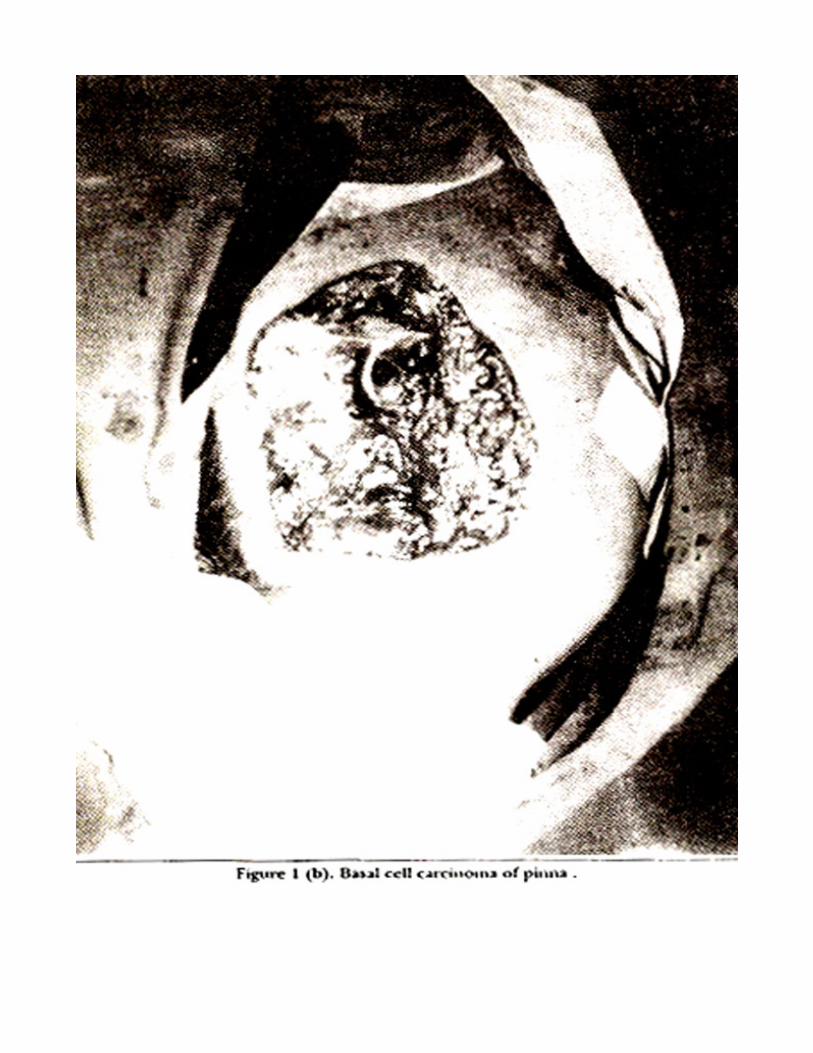
carcinoma, one had a basal cell carcinoma of the external ear and one had lost skin and soft tissue of
neck following synergistic gangrene. Ten of the eleven flaps survived (success rate 91%). One of the
three rib grafts used to reconstruct mandible got infected and had to be removed. Three patients
developed wound infections and one had a temporary orocutaneous fistula which closed spontaneously.
This brief experience confirms the reliability and efficiency of PMMF for head and neck reconstruction
(JMPA 43: 73, 1993).
INTRODUCTION
Resection of advanced cancer in head and neck area is likely to lead to complex defects which usually
require skin for covering and/or lining and may require bone for mandibular reconstruction. Previously
main options available for this purpose have been axial pattern forehead or delto pectoral skin flaps.
Recognition of the fact, that a piece of skin can survive from the blood supply via musculocutaneous
arteries from the underlying muscle, has revolutionized the practice of reconstruction in head and neck
area. PMMF has become the standard for reconstruction since 1979 reports by Ariyan1,2. This paper
reports an initial experience with this flap from a hospital in Southwestern Saudi Arabia.
PATIENTS AND METHODS
All cases of head and neck disease in which PMMF was used for reconstruction, operated over a three
year period from September 1989 to October 1992, were reviewed retrospectively. Data was collected
regarding the site, stage and type of tumour, type of surgical excision and reconstruction. In addition
note was made of flap related postoperative complications.
Technique of PMMF
The flap is elevated, based on thoracoacromial artery (TAA), to pectoralis major muscle. The upper
chest is prepared and draped with the head and neck area. The flap is marked on the chest skin near the
origin of pectoralis major from the 6th rib, according to the size of the defect created after excision of
the primary tumour. TAA exits from the clavipectoral fascia to travel on the undersurface of the
pectoralis major muscle from the midpoint of clavicle downwards and laterally initially to meet a line
drawn from shoulder tip to the xiphoid process (Figure 1c).
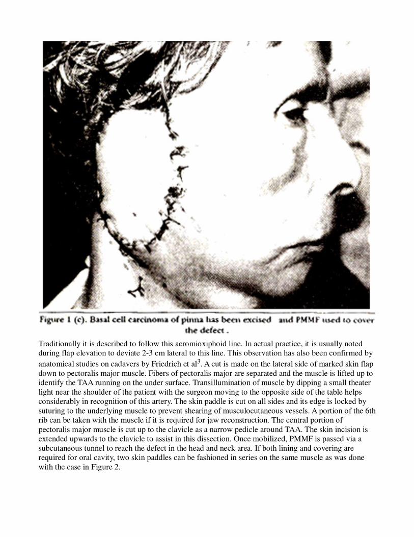
Traditionally it is described to follow this acromioxiphoid line. In actual practice, it is usually noted
during flap elevation to deviate 2-3 cm lateral to this line. This observation has also been confirmed by
anatomical studies on cadavers by Friedrich et al3. A cut is made on the lateral side of marked skin flap
down to pectoralis major muscle. Fibers of pectoralis major are separated and the muscle is lifted up to
identify the TAA running on the under surface. Transillumination of muscle by dipping a small theater
light near the shoulder of the patient with the surgeon moving to the opposite side of the table helps
considerably in recognition of this artery. The skin paddle is cut on all sides and its edge is locked by
suturing to the underlying muscle to prevent shearing of musculocutaneous vessels. A portion of the 6th
rib can be taken with the muscle if it is required for jaw reconstruction. The central portion of
pectoralis major muscle is cut up to the clavicle as a narrow pedicle around TAA. The skin incision is
extended upwards to the clavicle to assist in this dissection. Once mobilized, PMMF is passed via a
subcutaneous tunnel to reach the defect in the head and neck area. If both lining and covering are
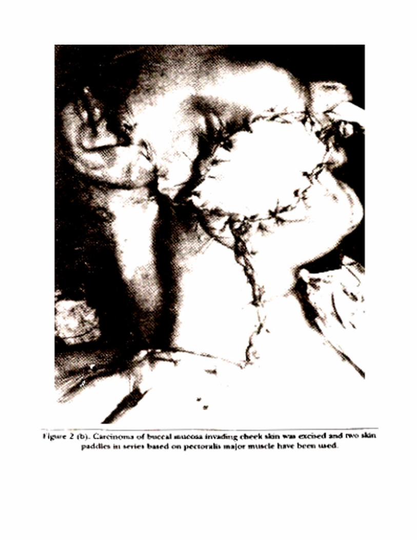
required for oral cavity, two skin paddles can be fashioned in series on the same muscle as was done
with the case in Figure 2.
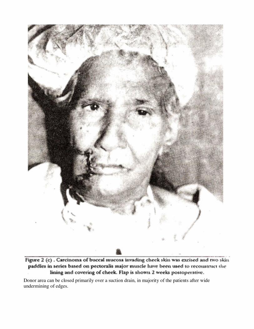
Donor area can be closed primarily over a suction drain, in majority of the patients after wide
undermining of edges.
RESULTS
PMMF was used in 11 patients during the study period. There were 7 males and 4 females with median
age of 60 years (range 54-90). The disease site, stage and resection type are shown in Table I.
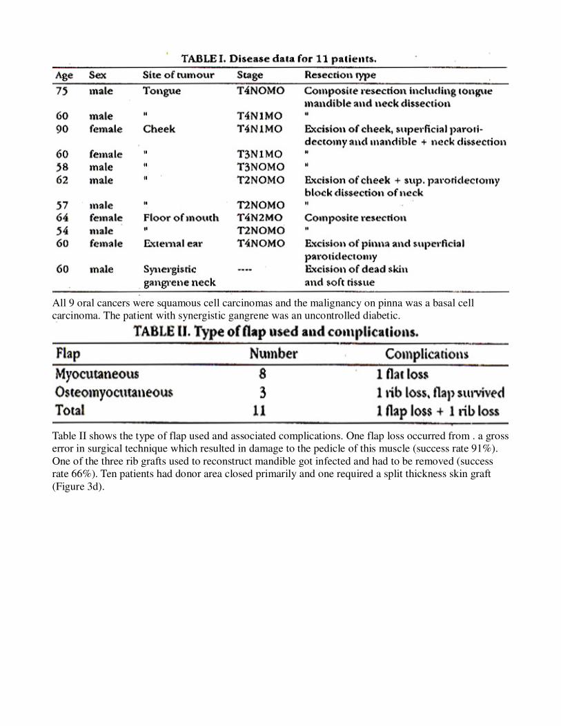
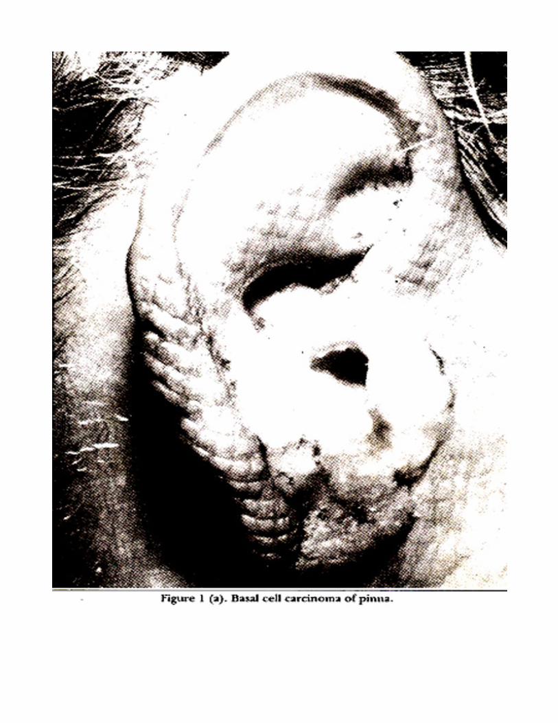
All 9 oral cancers were squamous cell carcinomas and the malignancy on pinna was a basal cell
carcinoma. The patient with synergistic gangrene was an uncontrolled diabetic.
Table II shows the type of flap used and associated complications. One flap loss occurred from . a gross
error in surgical technique which resulted in damage to the pedicle of this muscle (success rate 91%).
One of the three rib grafts used to reconstruct mandible got infected and had to be removed (success
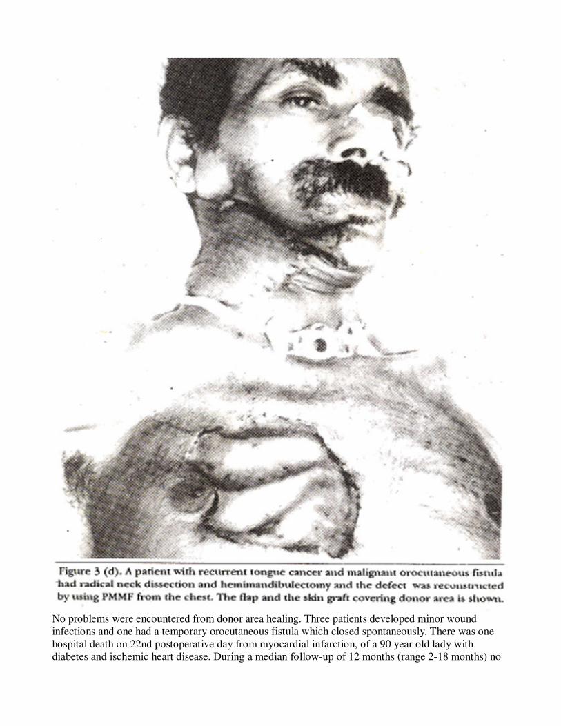
rate 66%). Ten patients had donor area closed primarily and one required a split thickness skin graft
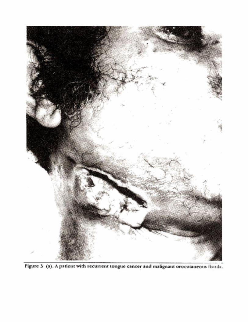
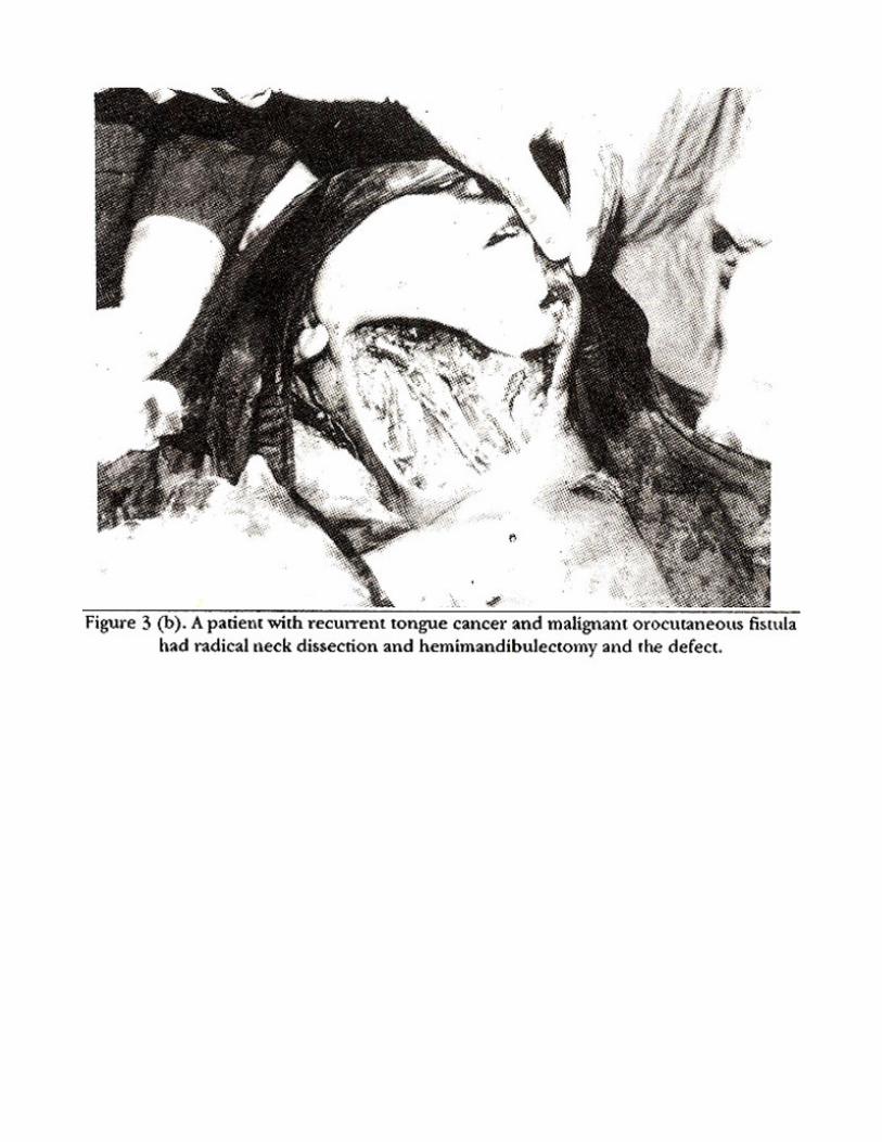
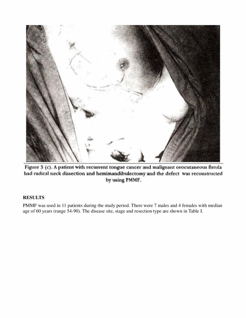
(Figure 3d).
No problems were encountered from donor area healing. Three patients developed minor wound
infections and one had a temporary orocutaneous fistula which closed spontaneously. There was one
hospital death on 22nd postoperative day from myocardial infarction, of a 90 year old lady with
diabetes and ischemic heart disease. During a median follow-up of 12 months (range 2-18 months) no
problems have been encountered with any of flaps.
DISCUSSION
This brief experience confirms the reliability of PMMF for head and neck reconstruction as reported in
literature4-11. The only flap loss occurred from a surgical misadventure. Experience with use of rib
graft has not been that favourable. This is in conformity with other reports. Baek et al4 reported 2 losses
in 5 patients which had ostemyocutaneous flaps and Ariyan11 also had 2 losses from his 5 cases when
rib was used. An additional advantage of PMMF is the muscle cover provided by the pedicle to the bare
carotid vessels following neck dissection, thus greatly diminishing incidence of carotid blow out.
Whole procedure can be accomplished in a single stage. The donor area can be closed primarily in
majority of cases with very insignificant cosmetic or functional deficit. Previously major ablative
procedures in head and neck requiring lengthy and multi stage procedures which also suffered from
cosmetic problems with donor areas. PMMP represents a transformation of that unhappy situation. It
has sound anatomical basis and the technique is not difficult to learn. The use of this flap is strongly
recommended for moderate sized defects of oral cavity, lower face and neck.
REFERENCES
1. Ariyan, 5.The pectoralia majormyocutaneousflap.Theversatileflap forreconstruction in head and
neck.. Plast. Reconstr. Surg., 1979;63:73-81.
2. Ariyan, 5. Further experiences with the pectoralia major myocutaneoua flap for the immediate repair
of defects from excisions of bead and neck cancers. Plaat. Reconstr. Surg., 1979;64:605-12.
3. Friedrich, W., Lierse, W. and Herberhold, C. Myocutaneous vascular territoty of the thoracoacromial
artery. A topographical and morphometric study of the arterial vascularization of the pectoralis major
myocutaneoua flap. Ada. AnaL (Basel), 1988; 131:284-91.
4, Back, SM., Lawson, W. and Biller, H.P. An analysis of 133 pectoralis major myocutaneous flaps.
Plast. Reconstr. Surg., 1982;69:460-9.
5. Wilson, J.S.P., Yiscoumettis, AM. and O\'Neil., T. Some observations on 112 pectorals major
myocutaneous flaps. Am.J. Surg., 1984;147:273-9.
6. Brusati, R., Collini, M., Bozzetti, A, Chiapasco, M. and Galioto, 5. The pectoralis major
myocutaneous flap. Experience in 100 consecutive cases. 3. Crsniomaxillofac. Surg., 1988:16:35-9.
7. Robertson, M.S. and Allison, R.S. The pectoralis major muscle in head and neck reconstruction,
Aust.N.Z.J. Surg.. 1986:56:753-7.
8. Josepls, C.A., Gregor, R.T., Davidge-Pitts. N.J. and Waner, M. The versatility of the pectoralia major
myocutaneoua flap. Head Neck Surg., 1985;7:365-8.
9. Ossoff, R.H., Wurster, CF., Berktold, RE., Krespi, Y.P. and Siason, G.A. Complications after
pectoralis majormyocutaneous flap reconstruction of head and neck defects. Arch. Otolatyngol.,
1983;109:812-4.
10. Mehrhof, A.l. Jr., Rosenstock, A., Neifeld, J.P., Merritt, W.H., Theogaraj, S.D. and Cohen, L.K. The
pectoralis major myocutaneous flap in bead and neck reconstruction. Analysia of complications.
Am.J.Surg., 1983;146:478-82.
11. Ariyan, S. Reconstruction of the oropharyngeal area, in cancer of the head and neck. Edited byS.
Ariyan, St. Louis and c.v. Mosby. 1987, pp. 285-98.


![An accessory muscle of the thoracic wall - Pulsus Group · Key words [pectoralis major muscle] [pectoralis quartus] [pectoral variation] [accessory muscle] [thoracic wall] eISSN 1308-4038](https://img.pdfslide.us/doc/110x75/5e9c4e2e397e311e6b4da4c8/an-accessory-muscle-of-the-thoracic-wall-pulsus-group-key-words-pectoralis-major.jpg)