Embed Size (px)
Citation preview
REVIEW SHEET
e x e r c i s e
10The Axial Skeleton
Review Sheet 10 155
The Sku l l
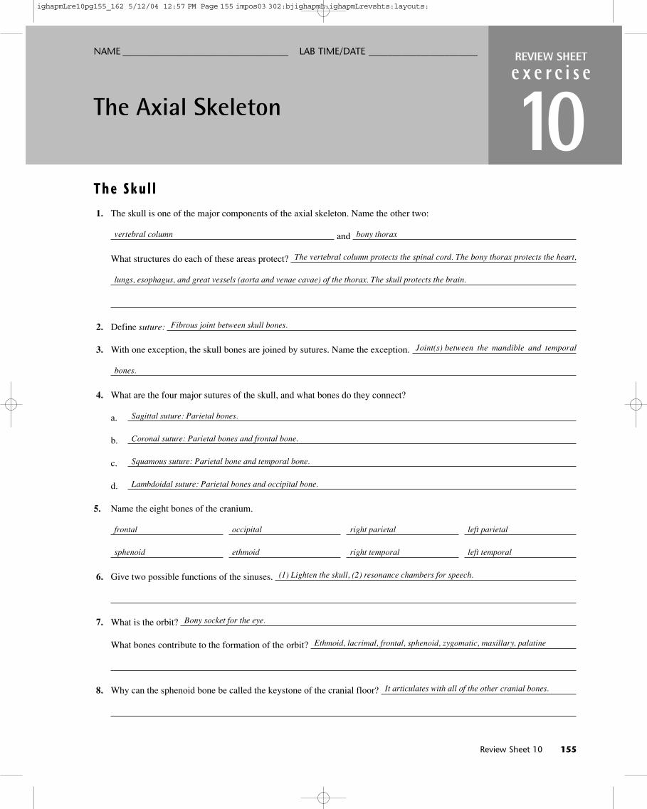
1. The skull is one of the major components of the axial skeleton. Name the other two:
and
What structures do each of these areas protect?
2. Define suture:
3. With one exception, the skull bones are joined by sutures. Name the exception.
4. What are the four major sutures of the skull, and what bones do they connect?
a.
b.
c.
d.
5. Name the eight bones of the cranium.
6. Give two possible functions of the sinuses.
7. What is the orbit?
What bones contribute to the formation of the orbit?
8. Why can the sphenoid bone be called the keystone of the cranial floor?
NAME ___________________________________ LAB TIME/DATE _______________________
vertebral column bony thorax
The vertebral column protects the spinal cord. The bony thorax protects the heart,
lungs, esophagus, and great vessels (aorta and venae cavae) of the thorax. The skull protects the brain.
Fibrous joint between skull bones.
Joint(s) between the mandible and temporal
bones.
Sagittal suture: Parietal bones.
Coronal suture: Parietal bones and frontal bone.
Squamous suture: Parietal bone and temporal bone.
Lambdoidal suture: Parietal bones and occipital bone.
frontal occipital right parietal left parietal
sphenoid ethmoid right temporal left temporal
(1) Lighten the skull, (2) resonance chambers for speech.
Bony socket for the eye.
Ethmoid, lacrimal, frontal, sphenoid, zygomatic, maxillary, palatine
It articulates with all of the other cranial bones.
ighapmLre10pg155_162 5/12/04 12:57 PM Page 155 impos03 302:bjighapmL:ighapmLrevshts:layouts:
9. What is a cleft palate?
10. Match the bone names in column B with the descriptions in column A.
Column A Column B
1. forehead bone
2. cheekbone
3. lower jaw
4. bridge of nose
5. posterior bones of the hard palate
6. much of the lateral and superior cranium
7. most posterior part of cranium
8. single, irregular, bat-shaped bone forming part of the cranial floor
9. tiny bones bearing tear ducts
10. anterior part of hard palate
11. superior and medial nasal conchae formed from its projections
12. site of mastoid process
13. site of sella turcica
14. site of cribriform plate
15. site of mental foramen
16. site of styloid processes
, , , and
17. four bones containing paranasal sinuses
18. condyles here articulate with the atlas
19. foramen magnum contained here
20. small U-shaped bone in neck, where many tongue muscles attach
21. middle ear found here
22. nasal septum
23. bears an upward protrusion, the “cock’s comb,” or crista galli
, 24. contain alveoli bearing teeth
156 Review Sheet 10
a. ethmoid
b. frontal
c. hyoid
d. lacrimal
e. mandible
f. maxilla
g. nasal
h. occipital
i. palatine
j. parietal
k. sphenoid
l. temporal
m. vomer
n. zygomatic
An opening in the palate resulting in a continuity between the oral and nasal cavities due to the failure of the
palatine bones or palatine processes of the maxillary bones to fuse properly.
b; frontal
n; zygomatic
e; mandible
g; nasal
i; palatine
j; parietal
h; occipital
k; sphenoid
d; lacrimal
f; maxilla
a; ethmoid
l; temporal
k; sphenoid
a; ethmoid
e; mandible
l; temporal
a; ethmoid b; frontal f; maxilla
k; sphenoid
h; occipital
h; occipital
c; hyoid
l; temporal
m; vomer (a; ethmoid)
a; ethmoid
e; mandible f; maxilla
ighapmLre10pg155_162 5/12/04 12:57 PM Page 156 impos03 302:bjighapmL:ighapmLrevshts:layouts:
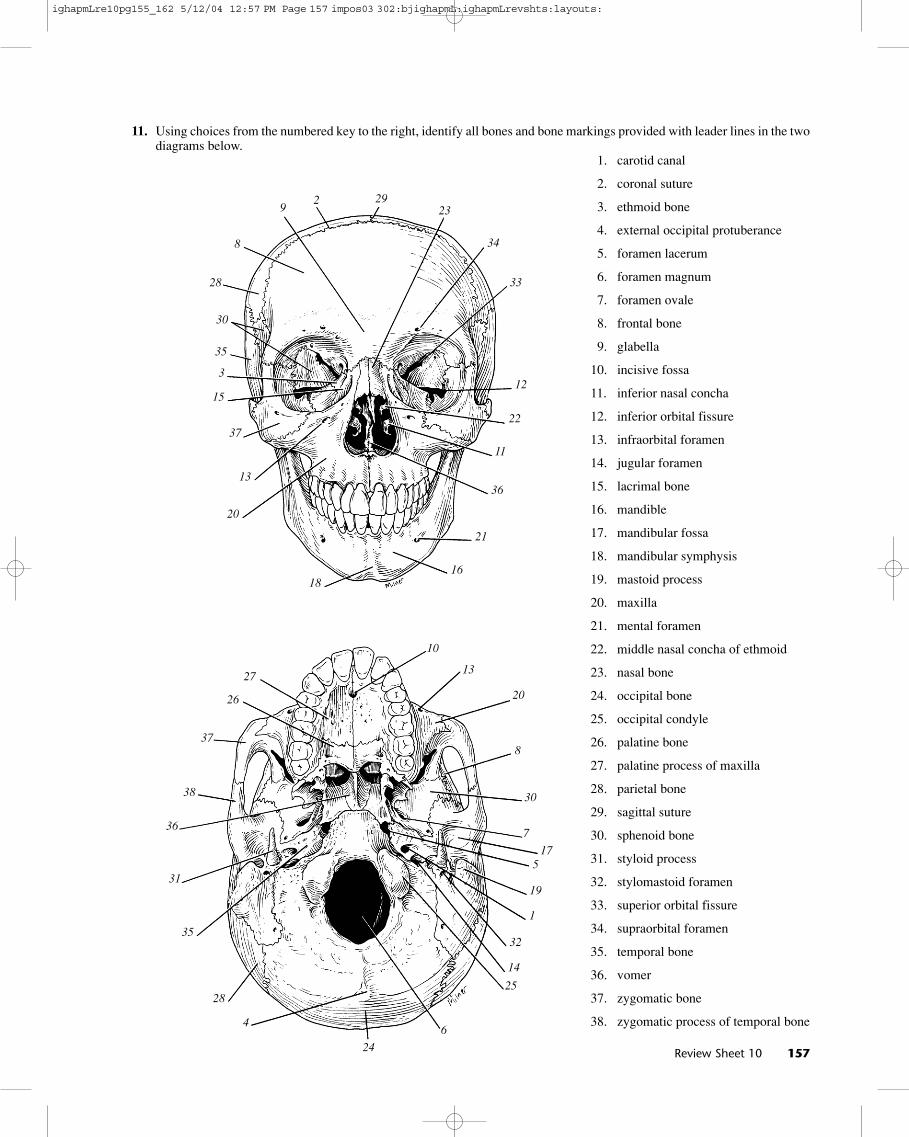
11. Using choices from the numbered key to the right, identify all bones and bone markings provided with leader lines in the twodiagrams below.
1. carotid canal
2. coronal suture
3. ethmoid bone
4. external occipital protuberance
5. foramen lacerum
6. foramen magnum
7. foramen ovale
8. frontal bone
9. glabella
10. incisive fossa
11. inferior nasal concha
12. inferior orbital fissure
13. infraorbital foramen
14. jugular foramen
15. lacrimal bone
16. mandible
17. mandibular fossa
18. mandibular symphysis
19. mastoid process
20. maxilla
21. mental foramen
22. middle nasal concha of ethmoid
23. nasal bone
24. occipital bone
25. occipital condyle
26. palatine bone
27. palatine process of maxilla
28. parietal bone
29. sagittal suture
30. sphenoid bone
31. styloid process
32. stylomastoid foramen
33. superior orbital fissure
34. supraorbital foramen
35. temporal bone
36. vomer
37. zygomatic bone
38. zygomatic process of temporal bone
Review Sheet 10 157
1
3
8
8
35
28
15
16
20
20
30
23
24
26
37
36
28
30
35
37
36
2
14
4
175
18
6
19
7
21
9
22
10
25
11
27
12
29
31
13
32
33
34
38
13
ighapmLre10pg155_162 5/12/04 12:57 PM Page 157 impos03 302:bjighapmL:ighapmLrevshts:layouts:
The Ver tebra l Co lumn
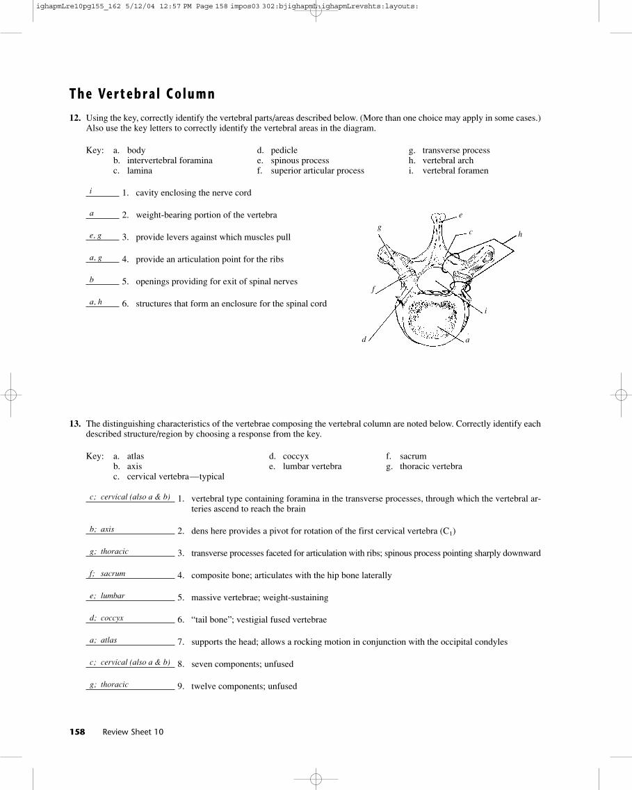
12. Using the key, correctly identify the vertebral parts/areas described below. (More than one choice may apply in some cases.)Also use the key letters to correctly identify the vertebral areas in the diagram.
Key: a. body d. pedicle g. transverse processb. intervertebral foramina e. spinous process h. vertebral archc. lamina f. superior articular process i. vertebral foramen
1. cavity enclosing the nerve cord
2. weight-bearing portion of the vertebra
3. provide levers against which muscles pull
4. provide an articulation point for the ribs
5. openings providing for exit of spinal nerves
6. structures that form an enclosure for the spinal cord
13. The distinguishing characteristics of the vertebrae composing the vertebral column are noted below. Correctly identify eachdescribed structure/region by choosing a response from the key.
Key: a. atlas d. coccyx f. sacrumb. axis e. lumbar vertebra g. thoracic vertebrac. cervical vertebra—typical
1. vertebral type containing foramina in the transverse processes, through which the vertebral ar-teries ascend to reach the brain
2. dens here provides a pivot for rotation of the first cervical vertebra (C1)
3. transverse processes faceted for articulation with ribs; spinous process pointing sharply downward
4. composite bone; articulates with the hip bone laterally
5. massive vertebrae; weight-sustaining
6. “tail bone”; vestigial fused vertebrae
7. supports the head; allows a rocking motion in conjunction with the occipital condyles
8. seven components; unfused
9. twelve components; unfused
158 Review Sheet 10
i
a
e, g
a, g
b
a, h
c; cervical (also a & b)
b; axis
g; thoracic
f; sacrum
e; lumbar
d; coccyx
a; atlas
c; cervical (also a & b)
g; thoracic
hc
e
g
f
d a
i
ighapmLre10pg155_162 5/12/04 12:57 PM Page 158 impos03 302:bjighapmL:ighapmLrevshts:layouts:
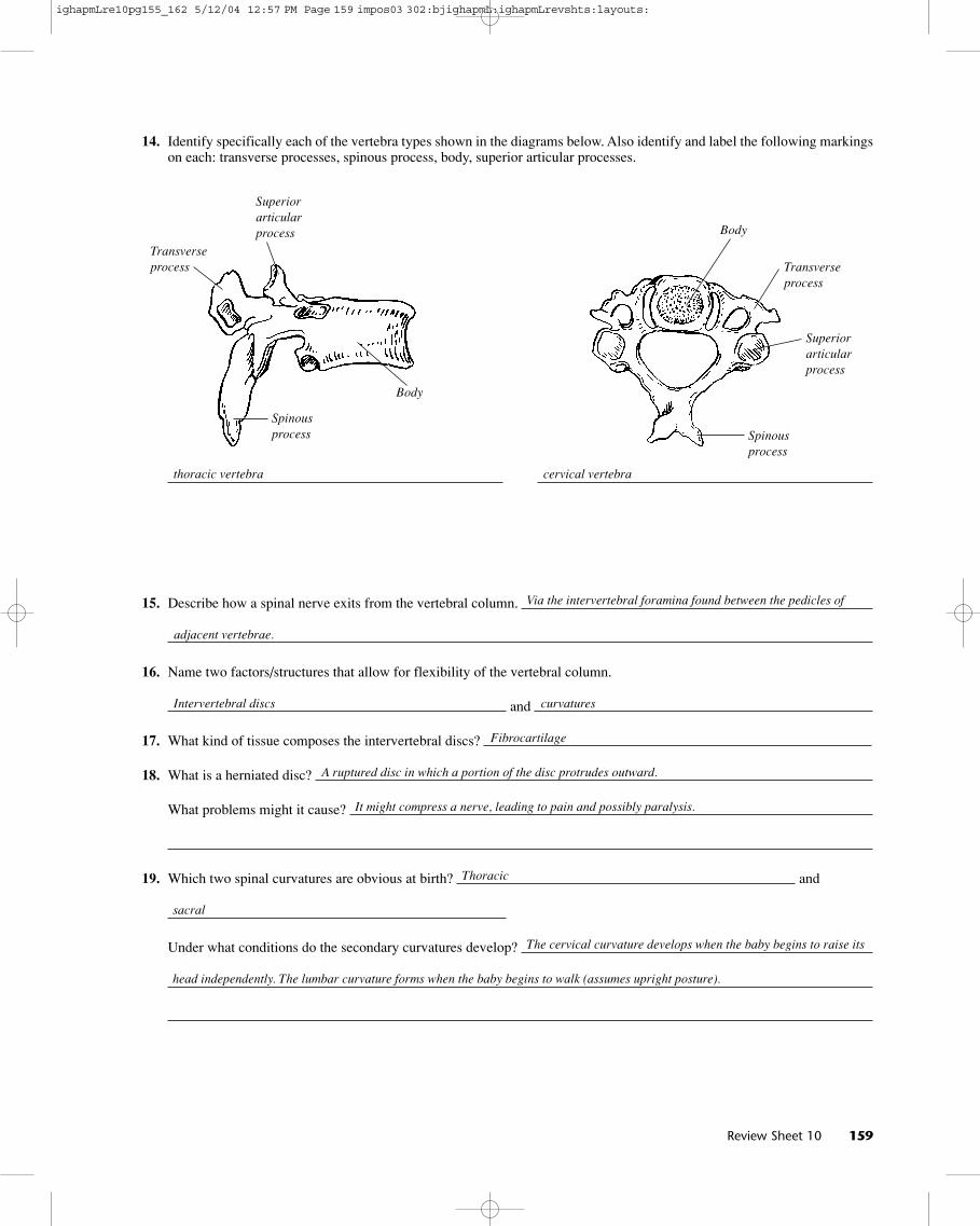
14. Identify specifically each of the vertebra types shown in the diagrams below. Also identify and label the following markingson each: transverse processes, spinous process, body, superior articular processes.
15. Describe how a spinal nerve exits from the vertebral column.
16. Name two factors/structures that allow for flexibility of the vertebral column.
and
17. What kind of tissue composes the intervertebral discs?
18. What is a herniated disc?
What problems might it cause?
19. Which two spinal curvatures are obvious at birth? and
Under what conditions do the secondary curvatures develop?
Review Sheet 10 159
Via the intervertebral foramina found between the pedicles of
adjacent vertebrae.
Intervertebral discs curvatures
Fibrocartilage
A ruptured disc in which a portion of the disc protrudes outward.
It might compress a nerve, leading to pain and possibly paralysis.
The cervical curvature develops when the baby begins to raise its
head independently. The lumbar curvature forms when the baby begins to walk (assumes upright posture).
thoracic vertebra cervical vertebra
Thoracic
sacral
Superiorarticularprocess
Superiorarticularprocess
Transverseprocess Transverse
process
Body
Body
Spinousprocess Spinous
process
ighapmLre10pg155_162 5/12/04 12:57 PM Page 159 impos03 302:bjighapmL:ighapmLrevshts:layouts:
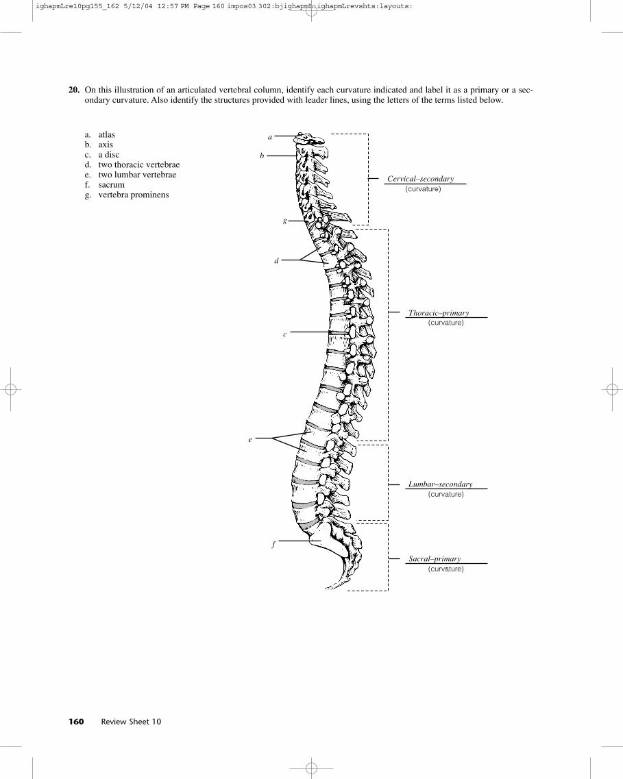
20. On this illustration of an articulated vertebral column, identify each curvature indicated and label it as a primary or a sec-ondary curvature. Also identify the structures provided with leader lines, using the letters of the terms listed below.
a. atlasb. axisc. a discd. two thoracic vertebraee. two lumbar vertebraef. sacrumg. vertebra prominens
160 Review Sheet 10
(curvature)
(curvature)
(curvature)
(curvature)
Cervical–secondary
Thoracic–primary
Lumbar–secondary
Sacral–primary
a
b
c
d
e
f
g
ighapmLre10pg155_162 5/12/04 12:57 PM Page 160 impos03 302:bjighapmL:ighapmLrevshts:layouts:
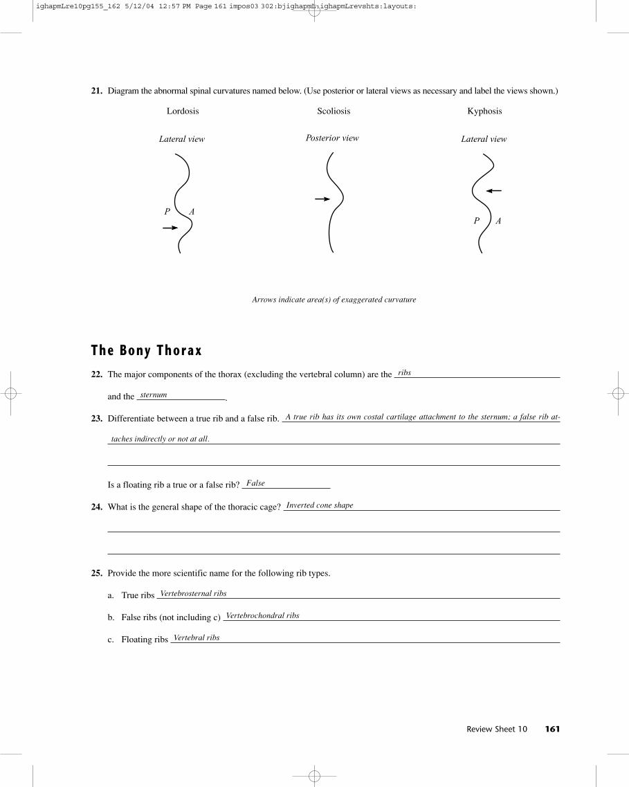
21. Diagram the abnormal spinal curvatures named below. (Use posterior or lateral views as necessary and label the views shown.)
Lordosis Scoliosis Kyphosis
The Bony Thorax
22. The major components of the thorax (excluding the vertebral column) are the
and the .
23. Differentiate between a true rib and a false rib.
Is a floating rib a true or a false rib?
24. What is the general shape of the thoracic cage?
25. Provide the more scientific name for the following rib types.
a. True ribs
b. False ribs (not including c)
c. Floating ribs
Review Sheet 10 161
ribs
sternum
A true rib has its own costal cartilage attachment to the sternum; a false rib at-
taches indirectly or not at all.
False
Inverted cone shape
Vertebrosternal ribs
Vertebrochondral ribs
Vertebral ribs
Arrows indicate area(s) of exaggerated curvature
Lateral view
P A
Posterior view Lateral view
P A
ighapmLre10pg155_162 5/12/04 12:57 PM Page 161 impos03 302:bjighapmL:ighapmLrevshts:layouts:
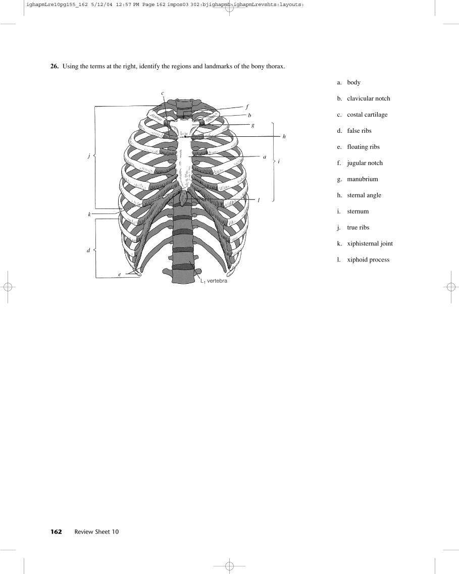
26. Using the terms at the right, identify the regions and landmarks of the bony thorax.
a. body
b. clavicular notch
c. costal cartilage
d. false ribs
e. floating ribs
f. jugular notch
g. manubrium
h. sternal angle
i. sternum
j. true ribs
k. xiphisternal joint
l. xiphoid process
L1 vertebra
162 Review Sheet 10
j
c
f
b
g
h
ia
l
k
d
e
ighapmLre10pg155_162 5/12/04 12:57 PM Page 162 impos03 302:bjighapmL:ighapmLrevshts:layouts: