Embed Size (px)
Citation preview

RESEARCH ARTICLE Open Access
The antibacterial activity and mechanism ofginkgolic acid C15:1Zhebin Hua1,3, Caie Wu1,2*, Gongjian Fan1,2, Zhenxing Tang2 and Fuliang Cao1,3
Abstract
Background: The present study investigated the antibacterial activity and underlying mechanisms of ginkgolic acid(GA) C15:1 monomer using green fluorescent protein (GFP)-labeled bacteria strains.
Results: GA presented significant antibacterial activity against Gram-positive bacteria but generally did not affectthe growth of Gram-negative bacteria. The studies of the antibacterial mechanism indicated that large amounts ofGA (C15:1) could penetrate GFP-labeled Bacillus amyloliquefaciens in a short period of time, and as a result, led tothe quenching of GFP in bacteria. In vitro results demonstrated that GA (C15:1) could inhibit the activity of multipleproteins including DNA polymerase. In vivo results showed that GA (C15:1) could significantly inhibit the biosynthesisof DNA, RNA and B. amyloliquefaciens proteins.
Conclusion: We speculated that GA (C15:1) achieved its antibacterial effect through inhibiting the protein activity of B.amyloliquefaciens. GA (C15:1) could not penetrate Gram-negative bacteria in large amounts, and the lipid solublecomponents in the bacterial cell wall could intercept GA (C15:1), which was one of the primary reasons that GA(C15:1) did not have a significant antibacterial effect on Gram-negative bacteria.
Keywords: GA, Green fluorescent protein, Antibacterial activity
BackgroundPlants can synthesize over 200,000 compounds throughvarious metabolic pathways [1]. Secondary metabolites inplants are derived from primary metabolites, and theircategories and chemical structures are complex and di-verse, including nitrogen-containing organic compounds,terpenoids, phenols and polyacetylenes, of which alkaloids,terpenoids and phenols are the most common. Secondarymetabolites are widely involved in plant growth, develop-ment and defense as well as other physiological and bio-logical processes [2]. Plant secondary metabolites providemany useful natural organic compounds for human use.Because traditional chemical pesticides contaminate soiland water, the development of environmentally friendlybio-pesticides has become a popular research focus. How-ever, the development of synthetic pesticides has manyproblems, such as a low successful rate, long cycle and
huge cost etc. Therefore, discovering lead compounds(plant-derived antibacterial reagents) from natural plantproducts with improved biological activity has become aneffective method to develop new biological pesticides.Self-defense mechanisms have been evolved in plants, andmany secondary plant metabolites are natural antibacterialagents [3–5]. Wilkins et al. [6] reported that 1389 plantscould be used as sources of plant antibacterial agents in-cluding ingredients that could kill or inhibit bacteria, suchas antibiotics, flavonoids, organic acids, polyphenols andspecific proteins. Wilson et al. [7] studied the inhibition ofBotrytis cinerea by 345 crude plant extracts and 49 essen-tial oils, found that 13 crude extracts and 4 essential oilsprovided antibacterial activities.Resorcinolic lipids are widely distributed plant secondary
metabolites produced in large numbers. Recent studieshave shown that they have extraordinarily high antibacterialactivity. Resorcinolic lipids produced by Pseudomonas car-boxydoflava can inhibit the growth of many bacteria spe-cies, such as Micrococcus lysodeictius and Bacillus subtilis[8, 9]. Resorcinolic lipids isolated from cashew apple havestrong antibacterial effects on Gram-positive bacteria,including methicillin-resistant S. aureus strains [10, 11].
* Correspondence: [email protected] Centre for Sustainable Forestry in Southern China, NanjingForestry University, Nanjing 210037, China2College of Light Industry Science and Engineering, Nanjing ForestryUniversity, Nanjing 210037, ChinaFull list of author information is available at the end of the article
© The Author(s). 2017 Open Access This article is distributed under the terms of the Creative Commons Attribution 4.0International License (http://creativecommons.org/licenses/by/4.0/), which permits unrestricted use, distribution, andreproduction in any medium, provided you give appropriate credit to the original author(s) and the source, provide a link tothe Creative Commons license, and indicate if changes were made. The Creative Commons Public Domain Dedication waiver(http://creativecommons.org/publicdomain/zero/1.0/) applies to the data made available in this article, unless otherwise stated.
Hua et al. BMC Biotechnology (2017) 17:5 DOI 10.1186/s12896-016-0324-3
brought to you by COREView metadata, citation and similar papers at core.ac.uk
provided by Springer - Publisher Connector
Sixteen phenolic compounds have been isolated from thecashew Anacardium occidentale (Anacardiaceae) nut shelloil, including various C15 phenolic compounds. Their anti-microbial activity has been tested against four typical mi-croorganisms, Bacillus subtilis, a Gram-positive bacterium;Escherichia coli, a Gram-negative bacterium; Saccharomy-ces cereuisiae, a yeast; and Penicillium chrysogenum, a mold.Most of them exhibited potent antibacterial activity againstonly Gram-positive bacteria [12].Ginkgo is a Chinese-specific rare relict species that is
well known as a “living fossil of gymnosperms” [13]. Thefruit and leaves of ginkgo have relatively high economicand medicinal values. However, its sarcotestas is usuallydiscarded, causing secondary pollution of the environ-ment [14]. GA, which is in high level in sarcotestas, is anatural plant-derived active substance contained in ginkgo,and it belongs to long-chain phenolic compounds that arederivatives of sumac acid [15]. Current studies have shownthat the biological activities of GA include anti-tumoractivity, neuroprotective activity, anxiolytic and anti-bacterial activity [16–20]. These biological activitiesmay make a possibility that increases the utilization ofginkgo sarcotestas and reduces environmental pollu-tion. The potential uses of Ginkgo have been attractedmany concern. Studies of GA antibacterial activityhave found that although GA could inhibit the activityof bacteria and plant pathogens, it just showed selectiveantibacterial activity, with strong inhibition towards toGram-positive bacteria and almost no inhibition to Gram-negative bacteria [21–24].The present study employed GFP-labeled strains and
analyzed the antibacterial activity and mechanisms ofGA C15:1 monomer, high amounts of which was inginkgo sarcotestas and had relatively high antibacterialactivity. Investigations of the selective antibacterial ac-tivity of GA could provide a scientific and theoreticalbasis for the development of new plant-derived pesti-cides using ginkgo sarcotestas as the raw material.
ResultsAntibacterial activity of GA (C15: 1)The antibacterial activity of GA (C15:1) is shown inTable 1. GA (C15:1) had strong antibacterial activityagainst Gram-positive bacteria, the MIC values of all ofthe tested Gram-positive bacteria were not greater than10 μg mL−1..In this study, all of the tested Gram-negativebacteria could grow well after the addition of large dosesof GA (C15:1) (final concentration 500 μg mL−1), and nodifferences were observed compared with the controlssupplemented with salicylic acid, indicating that GA(C15:1) did not have significant antibacterial action againstGram-negative bacteria.
The effect of GA (C15:1) on GFP in bacteriaUsing a GFP-labeled strain as the target, we studied effectof GA (C15:1) on GFP fluorescence in bacteria, and theresults are show in Fig. 1-a). GA (C15:1) could signifi-cantly affect GFP fluorescence in the Gram-positivebacteria B. amyloliquefaciens SQR9-gfp within 1 min.Compared with the results for the CK (bacteria only con-taining DMSO), GA (C15:1) at the concentration of5 μg mL−1 could reduce GFP fluorescence intensity inSQR9 bacteria by more than 50% within 1 min, and GA(C15:1) at higher concentrations could almost completelyquench GFP fluorescence in SQR9 bacteria within 1 min.Although GA (C15:1) could significantly affect GFP
fluorescence in B. amyloliquefaciens SQR9-gfp bacteriawithin 1 min, it did not have a significant effect on GFPfluorescence in Gram-negative bacteria E. coli DH5α-gfpand P. putida KT2440-gfp. Within 1 min, a significantdecrease of fluorescence intensity was not detected inthe studied Gram-negative bacteria, and fluorescenceintensity values in the CK were close to the fluorescenceintensity value in bacteria supplemented with GA.We extended the contact time of Gram-negative bac-
teria E. coli DH5α-gfp and P. putida KT2440-gfp withGA (C15:1) to 4 h. The results (Fig. 1-b) showed that
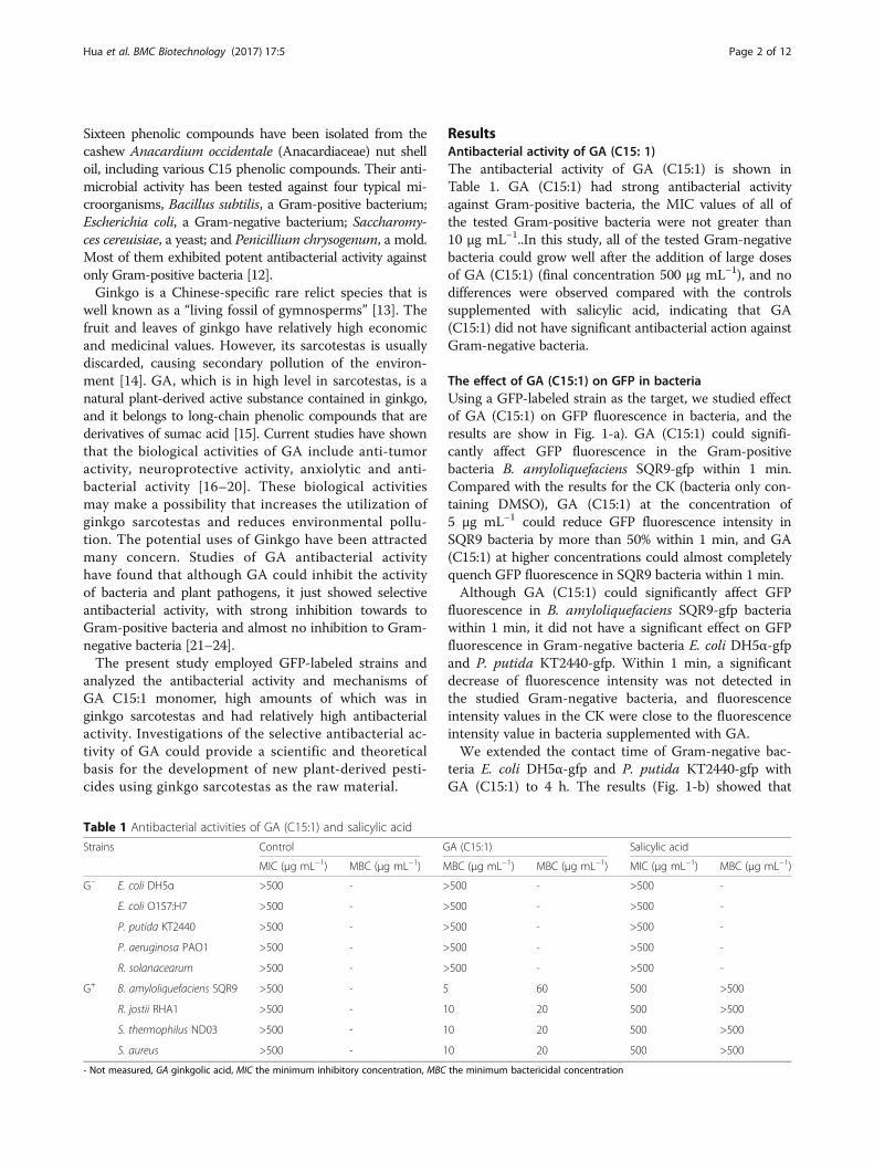
Table 1 Antibacterial activities of GA (C15:1) and salicylic acid
Strains Control GA (C15:1) Salicylic acid
MIC (μg mL−1) MBC (μg mL−1) MBC (μg mL−1) MBC (μg mL−1) MIC (μg mL−1) MBC (μg mL−1)
G− E. coli DH5α >500 - >500 - >500 -
E. coli O157:H7 >500 - >500 - >500 -
P. putida KT2440 >500 - >500 - >500 -
P. aeruginosa PAO1 >500 - >500 - >500 -
R. solanacearum >500 - >500 - >500 -
G+ B. amyloliquefaciens SQR9 >500 - 5 60 500 >500
R. jostii RHA1 >500 - 10 20 500 >500
S. thermophilus ND03 >500 - 10 20 500 >500
S. aureus >500 - 10 20 500 >500
- Not measured, GA ginkgolic acid, MIC the minimum inhibitory concentration, MBC the minimum bactericidal concentration
Hua et al. BMC Biotechnology (2017) 17:5 Page 2 of 12

even with longer incubation times, GA could only reduceGFP fluorescence in Gram-negative bacteria by a smallamount. GA (C15:1) at the concentration of 500 μg mL−1
had the most significant effect on fluorescence in E. coliDH5α-gfp, causing approximately 30% fluorescence re-duction. The fluorescence reduction values at other con-centrations were all less than 25%.The scanning electron microscopy examination
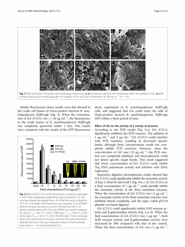
showed that after the addition of GA (C15:1), the cellsof the three bacteria still remained intact withoutapparent cell lysis (Fig. 2). Because GFP protein wasonly present in the bacteria, we speculated that GFPfluorescence decay in Gram-positive bacteria B. amy-loliquefaciens SQR9-gfp was caused by a large amountof GA that entered the bacteria within a short time,whereas the reason that GFP fluorescence in bothGram-negative bacteria did not show decay was thatGA (C15:1) did not enter these bacteria in a largeamount. The lack of a significant reduction in GFP
fluorescence in the two Gram-negative bacteria wascaused by a limited amount of GA entering the cells.
Effect of GA (C15:1) on GFP in bacteria crude extractsTo verify the hypothesis that “GFP fluorescence decay wasrelated to GA entering the bacteria cells”, the bacteria cellsof Gram-negative bacteria E. coli DH5α-gfp and P. putidaKT2440-gfp were lysed and centrifuged, and the crudelysate supernatants which contained GFP, were collected.GA (C15:1) was directly added to the supernatant, and theGFP fluorescence intensity was examined. The same pro-cedure was performed on Gram-positive bacteria B. amylo-liquefaciens SQR9-gfp. The results showed that GA (C15:1)could significantly affect GFP fluorescence in the crudelysates of E. coli DH5α-gfp and P. putida KT2440-gfpwithin 1 min (Fig. 3). Compared with the results for theCK, GA (C15:1) at a final concentration > 25 μg mL−1 couldcompletely quench GFP fluorescence in the crude lysates ofE. coli DH5α-gfp and P. putida KT2440-gfp within 1 min.
Fig. 1 Effect of GA (C15:1) on GFP fluorescence in bacteria. Three independent experiments were conducted (n = 3); the error bars indicate onestandard error. Three individual tubes were collected from LB plates, and each tube was performed in triplicate. Each bar in the gram representsmeans of three individual tubes (mean + − non-log transfoemed SE.). a: Bacteria were incubated at 30 °C for 1 min before GFP fluorescence wasmeasured.10 μL of DMSO without the drug was used as a control. The blank was the E. coli bacteria solution without GFP. One-way ANOVA wasused for analyzing the data (F7,16 = 656.9 P < 0.001(SQR9-gfp); F7,16 = 0.208 P > 0.05(DH5α-gfp); F7,16 = 0.357 P > 0.05 (KT2440-gfp)); (b) Bacteria wereincubated at 30 °C for 4 h before GFP fluorescence was measured. 10 μL of DMSO without the drug was used as a control. The blank was the E. colibacteria solution without GFP. One-way ANOVA was used for analyzing the data (F7,16 = 0.178 P > 0.05(DH5α-gfp); F7,16 = 1.412 P > 0.05 (KT2440-gfp))
Hua et al. BMC Biotechnology (2017) 17:5 Page 3 of 12
Similar fluorescence decay results were also showed inthe crude cell lysates of Gram-positive bacteria B. amy-loliquefaciens SQR9-gfp (Fig. 3). When the concentra-tion of GA (C15:1) was > > 10 μg mL−1, the fluorescencein the crude lysates of B. amyloliquefaciens SQR9-gfpwas completely quenched within 1 min. This resultswere consistent with the results of the GFP fluorescence
decay experiment in B. amyloliquefaciens SQR9-gfpcells, and suggested that GA could enter the cells ofGram-positive bacteria B. amyloliquefaciens SQR9-gfpGFP within a short period of time.
Effect of GA on the activity of a variety of proteinsAccording to the PCR results (Fig. 4-a), GA (C15:1)significantly inhibited the PCR reaction. The addition of1 μg mL−1 and 5 μg mL−1 GA (C15:1) could interferewith PCR reactions, resulting in decreased specificbands, although these concentrations would not com-pletely inhibit PCR reactions. However, when theconcentration of GA was ≥ 10 μg mL−1, the PCR reac-tion was completely inhibited, and electrophoresis couldnot detect specific target bands. This result suggestedthat lower concentration of GA (C15:1) could inhibitTaq DNA polymerase activity and interfere with DNAreplication.Restriction digestion electrophoresis results showed that
GA (C15:1) could significantly inhibit the enzymatic activityof Kpn I, Hind III and EcoR I (Fig. 4-b, c, d). GA (C15:1) ata final concentration of 1 μg mL−1 could partially inhibitthe enzymatic activity of the three restriction enzymes.When the concentration of GA (C15:1) was > 5 μg mL−1,the enzymatic activity of the three restriction enzymes wereinhibited almost completely, and the super coiled pUC19plasmid was barely digested.GA (C15:1) could significantly inhibit SOD enzyme ac-
tivity and β-galactosidase activity (Fig. 5-a, b). When thefinal concentration of GA (C15:1) was 1 μg mL−1, bothSOD enzyme activity and β-galactosidase activity weredecreased by 50% compared with that of the control.When the final concentration of GA was > 5 μg mL−1,
Fig. 2 SEM observation of bacteria cell morphology. respectively, (a, b, c) represent Bacteria cell morphology before the addition of GA; (d, e, f)represent Bacteria cell morphology after the addition of GA at a final concentration of 100 μg mL−1 for 1 min
Fig. 3 Effect of GA (C15:1) on GFP fluorescence in bacteria crude lysates(1 min). Three independent experiments were conducted (n= 3). Theerror bars indicate one standard error. All of the tests were incubated at30 °C for 1 min before GFP fluorescence was measured. 10 μL of DMSOwithout the drug was used as a control. The blank was the E. colibacteria solution without GFP. One-way ANOVA was used for analyzingthe data (F7,16 = 281.4 P < 0.001 (SQR9-gfp); F7,16 = 246.3 P < 0.001(DH5α-gfp); F7,16 = 304.0 P < 0.001 (KT2440-gfp)). Three individualtubes were collected from LB plates, and each tube was performed intriplicate. Each bar in the gram represents means of three individualtubes (mean + − non-log transfoemed SE.)
Hua et al. BMC Biotechnology (2017) 17:5 Page 4 of 12

SOD enzyme activity and β-galactosidase enzyme ac-tivity were almost undetectable.These proteins had different sources and were selected
randomly. Thus, the results of this study suggested thatthe inhibition of GA (C15:1) on protein activities wasnon-selective.
Inhibition of isotope incorporation experimentsThe aforementioned experiments showed that GA couldinhibit DNA polymerase function in vitro. However, itwas unclear whether GA had a similar function in bac-teria, including whether GA (C15:1) could inhibit DNApolymerase activity in vivo, which would inhibit DNAreplication. In addition, it is unclear whether GA couldinhibit RNA polymerase and ribosome activities, whichwould inhibit transcription and translation. To furtherclarify the mechanism of GA (C15:1), we used themethod of the inhibition of isotope incorporation to verifythe effects of GA (C15:1) in vivo. (Methy-3H) thymine([3H] TdR), 3H-uridine ([3H] UR) and 3H-tyrosine ([3H]
Tyr) were used as precursors to determine effect of GA(C15:1) on the biosynthesis of DNA, RNA and B. amyloli-quefaciens SQR9 proteins. The results are showed in Fig. 6.Compared with that of the control, GA (C15:1) couldinhibit DNA replication, RNA synthesis and protein syn-thesis to different extents under all three concentrations(25 μg mL−1, 10 μg mL−1 and 5 μg mL−1). When the con-centration of GA (C15:1) reached 25 μg mL−1, the inhib-ition of [3H] TdR incorporation was approximately 99%,of [3H] UR incorporation was approximately 90%, and ofprotein precursor [3H] tyrosine was approximately 85%.These data indicated that GA (C15:1) could inhibit DNAreplication in vivo as well as RNA transcription andprotein synthesis.
The interception of GA by Gram-negative bacteria cellwallsUsing E. coli as the target, lysozyme was used to destroythe peptidoglycan structure in the cell wall and thus E. coliDH5α-gfp protoplasts were obtained. Effect of GA (C15:1)
Fig. 4 Effect of GA (C15:1) on a variety of proteins. respectively :(a) represents the effect of GA (C15:1) on the activity of Taq DNA Polymerase. TheDNA contration at the bottom of fig represents the PCR reaction was inhibited by GA (C15:1). b represents the effect of GA (C15:1) on the activityof Kpn I. The DNA contration at the bottom of fig represents the enzymatic activity Kpn I was inhibited by GA (C15:1) (c) represents the effect ofGA (C15:1) on the activity of Hind III. The DNA contration at the bottom of fig represents the enzymatic activity Hind III was inhibited by GA(C15:1) (d) represents the effect of GA (C15:1) on the activity of EcoR I. The DNA contration at the bottom of fig represents the enzymatic activityEcoR I was inhibited by GA (C15:1) lane 1, CK; lane 2, addition of DMSO into PCR or digestion system; lane 3, addition of 1 μg mL−1 GA (C15:1)into PCR or digestion system; lane 4, addition of 5 μg mL−1 GA (C15:1) into PCR or digestion system; lane 5, addition of 10 μg mL−1 GA (C15:1)into PCR or digestion system; lane 6, addition of 25 μg mL−1 GA (C15:1) into PCR or digestion system; lane 7, addition of 50 μg mL−1 GA (C15:1)into PCR or digestion system
Hua et al. BMC Biotechnology (2017) 17:5 Page 5 of 12

on GFP fluorescence in the protoplast within 1 minutewas measured. The results (Fig. 7) showed that when thefinal concentration of C15:1 was lower than 25 μg mL−1, itdid not have significant effect on GFP fluorescence in pro-toplasts. When the final concentration of GA was ≥25 μg mL−1, the decreased GFP fluorescence intensity inthe protoplasts was found. It was positively correlatedwith increasing concentration of GA. When the finalconcentration of GA reached 500 μg mL−1, the GFP fluor-escence intensity in the protoplasts decreased to approxi-mately 80% of the control. This result suggested that afterthe peptidoglycan structure in the Gram-negative bacteriacell wall was destroyed, a small amount of high concentra-tion GA could enter the Gram-negative bacteria cell andproduce a low level of GFP fluorescence decay.By soaking E. coli cells in ethanol solution for a short
period of time, the lipid-soluble components (mainlyincluded lipopolysaccharide and phospholipids) in thecell wall were removed/partially removed. Effect of GA
(C15:1) on GFP fluorescence in the E. coli cells that didnot have lipid-soluble components in their cell walls,was measured. The results (Fig. 8) showed that when thefinal concentration of GA reaches was 5 μg mL−1, a largedegree decrease of GFP fluorescence in E. coli occurred.When the final concentration of GA was above
Fig. 5 Effect of GA (C15:1) on the activity of SOD and β-galactosidase.The error bars indicate one standard error. a: Effect of GA (C15:1) on theactivity of SOD.The optical density (OD)560 values were measured. 5 μL ofDMSO (without the drug) was used as a control. One-way ANOVA wasused for analyzing the data (F5,12 = 161.6 P < 0.001);(b) : Effect of GA(C15:1) on the activity of β-galactosidase. The OD value at 420 nm wasread. 5 μL of DMSO (without the drug) was used as a control. One-wayANOVA was used for analyzing the data (F5,12 = 25.15 P < 0.001). Threeindividual tubes were collected from LB plates, and each tube wasperformed in triplicate. Each bar in the gram represents means of threeindividual tubes (mean + − non-log transfoemed SE.)
Fig. 6 Effect of GA (C15:1) on the incorporation of precursors for thesynthesis of macromolecules in B. amyloliquefaciens SQR9. The errorbars indicate one standard error. The final concentrations of GA inthe reaction system were 25 μg mL−1, 10 μg mL−1 and 5 μg mL−1,respectively. 5 μL of DMSO (without the drug) was used as a control.All of the treatments were conducted at 37 °C in a shaker. One-wayANOVA was used for analyzing the data (F2,6 = 8.72 P = 0.017 (TdR);F2,6 = 19.54 P = 0.002 (UR); F2,6 = 28.59 P < 0.001 (Tyr)). Three individualtubes were collected from LB plates, and each tube was performed intriplicate. Each bar in the gram represents means of three individualtubes (mean + − non-log transfoemed SE.)
Fig. 7 Effect of GA (C15: 1) on GFP fluorescence in the protoplastsof E. coli DH5α-gfp (1 min). Three independent experiments wereconducted (n = 3); The error bars indicate one standard error. 5 μL ofDMSO (without the drug) was used as a control. The blank was E.coli protoplast solution without GFP. All the tests were incubated at30 °C for 1 min. One-way ANOVA was used for analyzing the data(F7,16 = 0.106 P > 0.05 (DH5α-gfp); F7,16 = 1.412 P > 0.05 (KT2440-gfp)).Three individual tubes were collected from LB plates, and each tubewas performed in triplicate. Each bar in the gram represents meansof three individual tubes (mean + − non-log transfoemed SE.)
Hua et al. BMC Biotechnology (2017) 17:5 Page 6 of 12

10 μg mL−1, it could completely quench the GFP fluor-escence in E. coli. However, if the lipid-soluble compo-nents were not removed from the E. coli bacteria cellwall, even the final concentration of GA at 500 μg mL−1
could not quench the GFP fluorescence (Fig. 1-a). Theseresults showed that the lipid-soluble components inGram-negative bacteria cell walls could intercept GA.
DiscussionIn this study, E. coli DH5α, E. coli O157: H7, P. putidaKT2440, P. aeruginosa PAO1, R. solanacearum, Rho-dococcus RHA1, S. thermophilus ND03, S. aureus andother common strains were used to study the antibacterialactivity of GA (C15:1), and GA was found to have signifi-cant antibacterial activity against Gram-positive bacteriabut little effect on the growth of Gram-negative bacteria.A relatively strong selective antibacterial mechanism ofGA was observed. The MIC value of B. amyloliquefaciensSQR9 was the smallest among all of the tested Gram-positive bacteria. However, its MBC value (60 μg mL−1)was the largest among all of the tested Gram-positivebacteria. These results might be caused by small amountof endospores that were generated when B. amyloliquefa-ciens SQR9 was cultured. Endospores had relatively strongresistance, and could withstand higher concentrations ofGA without being killed. Therefore, B. amyloliquefaciensSQR9 had significantly higher MBC values than otherGram-positive bacteria. The antibacterial activity of GAhas been reported these years. Himejima and Kubo [12]
found that 2-hydroxy-6-(8-pentadecenyl) salicylic (anothername of ginkgolic acid C15:1) showed lower MICs (about10 μg mL−1) against Gram-positive bacteria and higherMICs (>100 μg mL−1) against Gram-negative bacteria.Choi et al. [23] also showed that GA (C15:1) had significantantibacterial activity against 18 g-positive vancomycin-resistant. The results of the present study are consistentwith above studies.Additional studies on antibacterial mechanisms using
GFP fluorescence-labeled Gram-positive bacteria B.amyloliquefaciens SQR9 and GFP-labeled Gram-negativebacteria E. coli DH5α and P. putida KT2440 showed thatGA (C15:1) could significantly affect GFP fluorescencein the cells of Gram-positive B. amyloliquefaciens SQR9-gfp, whereas it had no significant effect on GFP fluores-cence in the cells of Gram-negative bacteria E. coli DH5α-gfp and P. putida KT2440-gfp. The green fluorescent pro-tein (GFP) has been widely used as a highly useful tool inthe fluorescence studies of living cells, which is found incell cytoplasm of jellyfish and is an extremely stableprotein with 238 amino acids [25, 26]. The fluorescenceproduced by GFP was caused by its protein conformation.In general, as long as the protein conformation of GFP didnot change, the fluorescence would not decay or disappear.Previous reports showed that GA and sumac acids, whichhad a similar structure, could affect the activity ofnumerous enzymes, including protein phosphatase,lipoxygenase and histone acetyltransferase [27–29]. Inaddition, GA affected in vivo regulation mechanism ofsmall ubiquitin-related modifier (SUMO) and alteredprotein conformation, thereby affecting protein expres-sion [30].According to the above test, we suggested that the
mechanism by which GA (C15:1) decayed GFP fluores-cence was through conformation changes in the GFPprotein. In addition, the mechanism by which GA pro-moted antibacterial activity against Gram-positive bacteriawas through conformational changes of the proteins inthe bacteria that inactivated the proteins and inhibited thegrowth of Gram-positive bacteria.The results of crude cell lysate experiments showed that
GFP fluorescence decay might be related to the interactionbetween GFP and GA. The GFP fluorescence in bothGram-negative and Gram-positive bacteria crude lysateswas quenched by GA in a short period of time, which indi-cated that GFP fluorescence would be quenched as long asit had contact with GA and was not related to the micro-organism tagged with GFP. Because the structure betweenGram-negative and Gram-positive bacteria was similar andresults showed that peptidoglycan in Gram-positive bac-teria could not prevent GA (C15:1) from entering the cell,we suggested that the peptidoglycan structure of Gram-negative bacteria also could not block GA (C15:1) fromentering the cell. In the protoplast experiment, a small
Fig. 8 Effect of GA (C15: 1) on GFP fluorescence in E. coli DH5α-gfp(1 min), in which the lipids have been removed from the strains’ cellwall. Three independent experiments were conducted (n = 3). Theerror bars indicate one standard error. 5 μL of DMSO (without thedrug) was used as a control. The blank was E. coli bacteria solutionwithout GFP (after ethanol solubilization). All the tests were incubated at30 °C for 1 min. One-way ANOVA was used for analyzing the data (F7,16= 2.116 P> 0.05 (Black); F7,16 = 121.6 P< 0.001 (White)). Three individualtubes were collected from LB plates, and each tube was performed intriplicate. Each bar in the gram represents means of three individualtubes (mean +− non-log transfoemed SE.)
Hua et al. BMC Biotechnology (2017) 17:5 Page 7 of 12

amount of GA molecules could enter the cells after thepeptidoglycan structure in E. coli cell wall was destroyed bylysozyme, which might be the result of the action of lyso-zyme. After the peptidoglycan structure was destroyed bylysozyme, pores might be present on the surface of thepeptidoglycan layer that allowed GA molecules to passthrough. However, only a small amount of GA moleculescould enter the cells because the number of poresgenerated on the surface of the peptidoglycan layer waslow, and the surface of Gram-negative bacteria was stillcovered by a large amount of lipids (including lipopoly-saccharides and phospholipids), which could intercept alarge amount of GA molecules. In order to furtherconfirm lipid-soluble components in the cell wall ofGram-negative bacteria intercept the majority of GAmolecules, the studies use high resolution electronmicroscopy to observe membrane change or othermethods to study transport of GA through membranewill be carried out.Some studies demonstrated that GA markedly inhibited
the biofilm formation of S. mutans and Escherichia coliO157:H7, and disrupted biofilm integrity [24, 31]. There-fore, we speculate that the GA may affect the secondarymetabolism of Gram-positive and Gram-negative bacteria.Due to the secondary metabolism of bacteria, such as theformation of biofilm, fluorescence formation and synthesisof antibiotics are regulated by quorum-sensing, furtherstudies on this section will be investigated.
ConclusionsGA (C15:1) has a relatively strong selective antibacterialmechanism, which significant antibacterial activity againstGram-positive bacteria but little effect on the growth ofGram-negative bacteria. Additional studies on antibacterialmechanisms showed that GA (C15:1) could inhibit theactivities of the selected proteins to a certain degree, andnon-selectively induce protein conformational changes. GA(C15:1) also inhibit DNA replication in vivo as well as RNAtranscription and protein synthesis. Thus, we suggested thatthe mechanism by which GA (C15:1) promoted antibacter-ial activity against Gram-positive bacteria was throughconformational changes of the proteins in the bacteria thatinactivated the proteins and inhibited the growth of Gram-positive bacteria. The research results indicated that lipid-soluble components (including lipopolysaccharide andphospholipids) in the cell wall of Gram-negative bacteriaintercepted the majority of GA molecules, whereas the pep-tidoglycan layer in the cell wall showed a reduced capacityto intercept GA molecules.
MethodsMedia and reagentsLysogeny broth (LB) medium (g L−1) was composed asfollows: peptone 10.0 g L−1, yeast extract 5.0 g L−1, NaCl
10.0 g L−1, pH 7.2, (solid, addition of 1.5% agar), and de-ionized water 1000 mL, which was sterilized at 121 °Cfor 20 min.Proteinase K, lysozyme, ampicillin (Amp), kanamycin
(Km), gentamicin (Gm), isopropyl-β-D-thiogalactopyrano-side (IPTG), o-nitrophenyl β-D-galactopyranoside (ONPG),and o-nitrophenol (ONP) were purchased from ShanghaiSangon Biotech (Sangon Biotech, Shanghai, China). GAC15:1 standard was purchased from Shanghai TautoBiotechnology (Shanghai, China). Other chemical reagentswere analytical grade.
StrainsEscherichia coli DH5α (E. coli DH5α) (ATCC53338), E. coliO157: H7 (E. coli O157: H7) (ATCC43895), Pseudomonasputida KT2440 (ATTC47054), Pseudomonas aeruginosaPAO1 (ATCC15692), Ralstoniasolanacearum (ATCC11696), Rhodococcusjostii RHA1 [32], Streptococcus thermo-philus ND03 [33], and S. aureus (ATCC25923) were fromour laboratory.Bacillus amyloliquefaciens SQR9 (CGMCC 5808;
China General Microbiology Culture Collection Center)[34], E. coli-gfp (E. coli DH5α-gfp), P. putida KT2440-gfp, and B. amyloliquefaciens SQR9-gfp were providedby the Environmental Microbiology Lab at the Collegeof Resources and Environment, Nanjing AgriculturalUniversity.
Determination of antibacterial activity of GAA conventional broth-dilution method was adopted[35]. GA and salicylic acid were dissolved intodimethylsulfoxide (DMSO) to prepare stock solutionswith different concentrations, respectively. The finalconcentrations of the drug (GA or salicylic acid) inthe medium (500 μg mL−1, 250 μg mL−1, 100 μg mL−1, 80 μg mL−1, 60 μg mL−1, 40 μg mL−1, 20 μg mL−1, 10 μg mL−1, 5 μg mL−1, 2 μg mL−1, 1 μg mL−1,0.5 μg mL−1 and 0.1 μg mL−1) were obtained. 10 μLstock solutions were added to 3 mL liquid LBmedium that was inoculated with bacteria. All of thetests were incubated at 200 rpm. E. coli was culturedat 37 °C for 2 d, whereas other bacteria were cul-tured at 30 °C for 2 d. The lowest concentrationwithout turbidity was defined as the minimum in-hibitory concentration (MIC) of that substance. Themedium without turbidity (50 μL) was inoculatedwith 3 mL of fresh LB liquid medium, and culturedin a shaker. E. coli was cultured at 37 °C for 2 d,and all other bacteria were cultured at 30 °C for 2 d.The lowest concentration without turbidity wasdefined as the minimum bactericidal concentration(MBC) of that substance. Same concentration ofDMSO without GA and salicylic acid was added incontrol groups.
Hua et al. BMC Biotechnology (2017) 17:5 Page 8 of 12

Measurement of fluorescence decay of GFP in bacteriaSingle colonies of E. coli DH5α-gfp, P. Putida KT2440-gfp and B. amyloliquefaciens SQR9-gfp were extractedfrom solid LB plates, inoculated in liquid LB medium,and then 50 μg mL−1 Amp, Gm and Km was added tothe medium to maintain the normal replication of plasmidsin each strain. The medium was then centrifuged, and thebacteria were collected. The supernatant was discarded,and phosphate buffer was added to the collected bacteria toobtain the bacteria concentration of 108 CFU mL−1. GAwas then dissolved in DMSO to prepare stocks withdifferent concentrations.
(1) 10 μL of GA stock solution at different concentrationswas added to 1 mL of bacteria solution. The finalconcentrations of GA in the solution were 500 μg mL−1, 250 μg mL−1, 100 μg mL−1, 50 μg mL−1, 25 μg mL−1, 10 μg mL−1 and 5 μg mL−1, respectively. 10 μL ofDMSO without the drug was used as a control. All ofthe tests were incubated at 30 °C for 1 min before GFPfluorescence was measured. The morphology of thebacteria was monitored under a scanning electronmicroscopy. For strains of E. coli DH5α-gfp and P.Putida KT2440-gfp, the incubation was extended to4 h and additional samples were collected to measureGFP fluorescence. The morphology of the bacteria wasmeasured using a scanning electron microscopy. Bac-teria GFP fluorescence was initially observed by thenaked eye using an LB16 Maestrogen UltraSlim no-damage blue LED transilluminator. Measurements ofbacterial GFP fluorescence were performed using aSpectra ax M5 multifunctional microplate reader.The samples’ fluorescence intensity was measuredwhen the excitation wavelength was 488 nm andthe emission wavelength was 509 nm. The blankwas the E. coli bacteria solution without GFP.
(2) 1 mL of bacteria solution was lysed by sonication.The broken bacteria were centrifuged at 12,000 × g,4 °C for 20 min, and then the supernatant wascollected. GA stock solution was added to thesupernatant. The final concentrations of GA in thebacteria solution were 500 μg mL−1, 250 μg mL−1,100 μg mL−1, 50 μg mL−1, 25 μg mL−1, 10 μg mL−1
and 5 μg mL−1, respectively. 10 μL of DMSO(without the drug) was used as a control. All of thetests were incubated at 30 °C for 1 min before GFPfluorescence was measured. The control was thecrude enzyme solution of the corresponding bacteriawithout GFP.
Effect of GA on the activities of a variety of proteinsEffect of GA on the activity of Taq DNA polymeraseStock solutions at different concentrations were preparedby dissolving GA in DMSO. GA stock solutions (0.5 μL)
at different concentrations were then added into eachPCR reaction system. The final concentrations of GA inthe reaction system were 25 μg mL−1, 10 μg mL−1,5 μg mL−1 and 1 μg mL−1, respectively. 0.5 μL of DMSO(without the drug) was as a control. The PCR reactionconditions were adopted the reference of Chester andMarshak [36]. After the reaction was finished, 3 μL ofPCR product was analyzed on an appropriate agarose gel.GelRed nucleic acid dye was used to stain the gel.
Effect of GA on the activity of restriction enzymesThree common restriction enzymes (Kpn I, Hind IIIand EcoR I) were selected as the targets. GA wasdissolved in DMSO to prepare stock solutions withdifferent concentrations. A single colony of E. coli thatcarried the pUC19 plasmid, was extracted and inocu-lated in 3 mL of LB medium containing Amp, whichwas then cultured at 37 °C overnight with vigorousshaking. 41.5 μL of pUC19 (200 ng μg−1), 5 μL of 10 ×restriction enzyme buffer, 3 μL of corresponding re-striction enzyme, and 0.5 μL of GA stock solution withdifferent concentrations were mixed. The final con-centrations of GA in the reaction system were25 μg mL−1, 10 μg mL−1, 5 μg mL−1 and 1 μg mL−1, re-spectively. 5 μL of DMSO (without the drug) was usedas a control. All of the components in the reactionsystems were digested at 37 °C for 4 h, and then thetemperature was increased to 75 °C for 15 min to stopthe enzyme digestion. Electrophoresis was then carriedout to examine the enzyme digestion.
Effect of GA on the activity of superoxide dismutaseAccording to the method by Beauchamp et al. [37], GAwas dissolved in DMSO to prepare stock solutions withdifferent concentrations. 80 μmol L -1riboflavin,77 μmol L -1 nitro blue tetrazolium (NBT), 13 mmol L−1
methionine, 0.1 mmol L−1 EDTA, 20 μL superoxide dis-mutase (SOD) enzyme solution (1 μg mL−1), and 5 μL ofthe different concentrations of GA stock solution (finalconcentrations of GA in the reaction system of25 μg mL−1, 10 μg mL−1, 5 μg mL−1 and 1 μg mL−1)were mixed. 5 μL of DMSO (without the drug) was usedas a control. After the samples were exposed to light at4500 Lux light intensity for 15 min, the reaction wasstopped by shielding the light. The optical density(OD)560 values were measured. One active unit (U)occurred when NBT was inhibited by 50%, and enzymeactivity = (△ A ×N × 60)/(W × T × V × 50%), where △ Arepresented the difference in OD values between thecontrol and sample, N represented the total volume ofenzyme solution, W represented the protein mass, Trepresented the light reaction time and V representedthe volume of enzyme solution added. Enzyme activitywas represented as OD560 • mg−1 (pro) • min−1.
Hua et al. BMC Biotechnology (2017) 17:5 Page 9 of 12

Effect of GA on the activity of β-galactosidase2 μg mL−1 the enzyme β-galactosidase was prepared in10 mM pH 7.0 phosphate buffer. GA was dissolved inDMSO to prepare the stock solutions with different con-centrations. Enzyme solution (1 mL) was incubated at37 °C for 5 min, then 1 mL of phosphate buffer (pH 7.0)containing 20 mM ONPG preheated to 37 °C was added,finally 5 μL of the different concentrations of GA stocksolution were added. The final concentrations of GA inthe reaction system were 25 μg mL−1, 10 μg mL−1,5 μg mL−1 and 1 μg mL−1, respectively. 5 μL of DMSO(without the drug) was used as a control. All of the sam-ples were incubated in a 37 °C for 10 min, and then3 mL of 0.5 mol L−1 Na2CO3 was added to stop the re-action. The OD value at 420 nm was read. One enzymeunit was defined as the amount of enzyme required torelease 1 μmol of ONP per minute at 37 °C.
Inhibition of isotopic precursor incorporationAccording to the method by Aspedon and Groisman[21], [3H] TdR, [3H] UR and [3H] Tyr were incorpo-rated into precursors to investigate effect of GA(C15:1) on the biosynthesis of DNA, RNA and B.amyloliquefaciens SQR9 protein. The logarithmicgrowth phase SQR9 culture was diluted with sterilewater. In a 96-well plate, 0.9 mL of bacterial suspension(OD600 = 0.1) was added to each well. The isotope-labeledprecursor (final concentration was 0.5 μCi mL−1) and 5 μLof the different concentration of GA (C15: 1) were alsoadded into the well. The final concentrations of GA in thereaction system were 25 μg mL−1, 10 μg mL−1 and5 μg mL−1, respectively. 5 μL of DMSO (without the drug)was used as a control. All of the treatments wereconducted at 37 °C in a shaker. When examiningeffect of GA (C15:1) on the synthesis of DNA andRNA, culture time for bacteria was limited to onegeneration. After growing the culture for 30 min, theculture was centrifuged at 12,000 × g, 4 °C for 5 minto harvest the bacteria pellet. Because protein syn-thesis was relatively slow, the bacteria culture timewas extended to 2 h. OD600 was also adjusted sothat the cultures had the same bacteria concentra-tion. The bacteria pellet was washed three timeswith phosphate buffer and then placed in an ovenovernight to dry it. Scintillation solution was directlyadded to the Eppendorf tubes containing bacteria,and the tubes were then transferred to a scintillationcounter (LS3801, Beckman) to determine the countsper minute (CPM) values. The average CPM valuesof the experimental group and control group werecompared. The incorporation inhibition rate wascalculated. Incorporation inhibition rate = controlgroup CPM - experimental group CPM/control groupCPM× 100%.
Effect of GA (C15:1) on GFP fluorescence in theprotoplasts of E. coliE. coli protoplasts were prepared according to themethod described by Weiss [38]. GA was dissolved inDMSO, and its stock solutions with different concentra-tions were prepared. E. coli protoplasts (1 mL) werediluted with sucrose-magnesium-maleate (SMM) buffer(protoplast number > 107 CFU mL−1), and 5 μL of thedifferent concentration GA stock solutions were added.The final concentrations of GA in the reaction systemwere 500 μg mL−1, 250 μg mL−1, 100 μg mL−1,50 μg mL−1, 25 μg mL−1, 10 μg mL−1 and 5 μg mL−1, re-spectively. 5 μL of DMSO (without the drug) was usedas a control. All the tests were incubated at 30 °C for1 min, and then the samples were analyzed for GFPfluorescence. GFP fluorescence was initially observed bythe naked eye using an LB-16 Maestrogen UltraSlim no-damage blue LED transilluminator. GFP fluorescencewas measured with a Spectra ax M5 multifunctionalmicroplate reader. The samples’ fluorescence intensitywas measured when the excitation wavelength was488 nm and the emission wavelength was 509 nm. Theblank was E. coli protoplast solution without GFP.
The interception of GA by the lipid-soluble component inE. coli cell wallsE. coli DH5α-gfp was cultured in 100 mL of liquid LBmedium at 37 °C, 200 rpm overnight. After centrifuga-tion at 12,000 × g for 5 min, the supernatant wasdiscarded to collect the bacteria. The bacteria pellet wasthen re-suspended in the same volume of phosphatebuffer. The half was retained for the experiments andthe left half was centrifuged at 12,000 × g for 5 min. Thesupernatant was then discarded. A small volume ofphosphate buffer was used to re-suspend the bacteriapellet, and then 10 mL of 70% ethanol solution wasadded, mixed well, and kept at room temperature for30 s before centrifuging at 12,000 × g for 1 min. Thesupernatant ethanol solution was then discarded. Phos-phate buffer (50 mL) was used to re-suspend thebacteria pellet, which was centrifuged at 12,000 × g for5 min. The supernatant was then discarded. Finally,50 mL of phosphate buffer was used to re-suspend thebacteria pellet for the experiments.GA was dissolved in DMSO, and its stock solutions
with different concentrations were prepared. GA stocksolutions (5 μL) at different concentrations were addedto 1 mL of bacteria solution before/after ethanolsolubilization of the lipids. The final concentrations ofGA in the reaction system were 500 μg mL−1,250 μg mL−1, 100 μg mL−1, 50 μg mL−1, 25 μg mL−1,10 μg mL−1 and 5 μg mL−1, respectively. 5 μL of DMSO(without the drug) was used as a control. All the testswere incubated at 30 °C for 1 min, and then the samples
Hua et al. BMC Biotechnology (2017) 17:5 Page 10 of 12

were examined for GFP fluorescence. GFP fluorescencewas initially observed by the naked eye using an LB-16Maestrogen UltraSlim no-damage blue LED transillumi-nator. GFP fluorescence was measured using a Spectraax M5 multifunctional microplate reader. The samples’fluorescence intensity was measured when the excitationwavelength was 488 nm and the emission wavelengthwas 509 nm. The blank was E. coli bacteria solutionwithout GFP (after ethanol solubilization).
Statistical analysesFor all the experiments throughout the study, we collectedthree individual tubes from LB plates, and each tube wasperformed in triplicate. In analysis, comparisons werecarried out using the fluorescent means of each triplicate,then, finally each bar in the gram represents means ofthree individual tubes. The data of each strain wasanalyzed using one-way ANOVA (i.e., B. amyloliquefaciensSQR9-gfp E. coli DH5α-gfp and P. putida KT2440-gfp),with final concentration of GA as the fixed factors (P <0.05). The experimental data were log transformed tomeet the homogeneity of variance or a normal distribu-tion of residuals. All statistical analyses were conductedusing SPSS 13.0 (SPSS, Chicago, IL, USA).
Abbreviations[3H] TdR: (Methy-3H) thymine; [3H] Tyr: 3H-tyrosine; [3H] UR: 3H-uridine;Amp: Ampicillin; CFU: Colony-Forming Units; CK: Control check;CPM: Counts per minute; DMSO: Dimethylsulfoxide; DNA: Deoxyribonucleicacid; EDTA: Ethylenediaminetetraacetic acid; GA: Ginkgo acid; GFP: Greenfluorescent protein; Gm: Gentamicin; IPTG: Isopropyl-β-D-thiogalactopyranoside;Km: Kanamycin; LB: Lysogeny broth; LED: Light emitting diode; MBC: Minimumbactericidal concentration; MIC: Minimum inhibitory concentration; NBT: Nitroblue tetrazolium; OD: Optical density; ONP: O-nitrophenol; ONPG: O-nitrophenyl-β-D-galactopyranoside; PCR: Polymerase chain reaction; RNA: Ribonucleic acid;SMM: Sucrose-magnesium-maleate; SOD: Superoxide dismutase
AcknowledgementsIt is a great pleasure to thank research fund for key projects in the nationalscience & technology pillar program (2012BAD21B04) and the science andtechnology project of jiangsu province (BE2015315). The authors would liketo acknowledge the support of the project funded by the priority academicprogram development (PAPD) of jiangsu higher education institutions andco-innovation entre for sustainable forestry in southern china. The authorsalso would like to thank Dr. Xie lulu of University of Rochester, Dr. Xiedongof Nanjing Forestry University for guidance in the data analysis.
FundingThis work was supported by research fund for key projects in the nationalscience & technology pillar program (2012BAD21B04) and the science andtechnology project of jiangsu province (BE2015315).
Availability of data and materialsAll data generated/analysed during the current study that are not alreadyincluded in this published article, are available from the correspondingauthor on reasonable request.
Authors’ contributionsHZB did the experimental, analyses of experimental data, figures drawing,critical reading of the manuscript and writing of the manuscript; WCE andCFL were responsible for study conception and design, analysis andinterpretation of data work; FGJ contributed to study conception, to analysisof experimental data and to the writing of the manuscript; TZX analyzed thedata, supervised the statistical analyses and contributed to the writing of the
manuscript. All authors discussed and commented the results and gave theirfinal approval for submission.
Competing interestsThe authors declare that they have no competing interests.
Consent for publicationNot applicable.
Ethics approval and consent to participateN/A.
Author details1Co-Innovation Centre for Sustainable Forestry in Southern China, NanjingForestry University, Nanjing 210037, China. 2College of Light Industry Scienceand Engineering, Nanjing Forestry University, Nanjing 210037, China.3College of Forestry, Nanjing Forestry University, Nanjing 210037, China.
Received: 9 December 2015 Accepted: 17 December 2016
References1. Dixon RA, Strack D. Phytochemistry meets genome analysis, and beyond.
Phytochemistry. 2003;62(6):815–6.2. Toni M, Kutchan. Ecological arsenal and developmental dispatcher. The
paradigm of secondary metabolism. Plant Physiol. 2001;125(1):58–60.3. Cowan MM. Plant products as antimicrobial agents. Clin Microbiol Rev.
1999;12(4):564–82.4. Li JWH, Vederas JC. Drug discovery and natural products: end of an era or
an endless frontier? Science. 2009;325(5937):161–5.5. Zhao J, Davis LC, Verpoorte R. Elicitor signal transduction leading to production
of plant secondary metabolites. Biotechnol Adv. 2005;23(4):283–333.6. Wilkins KM, Board RG, Gould GW: Mechanisms of action of food
preservation procedures. London: Elservierapplied Science EA; 1989.7. Wilson CL, Solar JM, EI-Ghaouth A, Wisniewski ME. Rapid evaluation of plant
extracts and essential oils for antifungal activity against Botrytis cinerea. PlantDis. 1997;81(2):204–10.
8. Erin AN, Davitashvili NG, Prilipko LL, Boldyrev AA, Lushchak VI, Batrakov SG,Pridachina NN, Serbinova AE, Kagan VE. Influence of alkylresorcin onbiological membranes during activation of lipid peroxidation. Biokhimiia.1987;52:1180–5.
9. Kaprelyants AS, Suleimenov MK, Sorokina AD, Deborin GA, El-Registan GI,Stoyanovich FM, Lille YE, Ostrovsky DN. Structural functional changes inbacterial and model membranes induced by phenolic lipids. Biol Membr(Moscow). 1987;4:254–61.
10. Kubo I, Komatsu S, Ochi M. Molluscicides from the cashew Anacardiumoccidentale and their large-scale isolation. J Agr Food Chem. 1986;34(6):970–3.
11. Muroi H, Kubo I. Antibacterial activity of anacardic acid and totarol, aloneand in combination with methicillin, against methicillin-resistantStaphylococcus aureus. J Appl Microbiol. 1996;80(4):387–94.
12. Himejima M, Kubo I. Antibacterial Agents from the Cashew Anacardiumoccidentale (Anacardiaceae) Nut Shell Oil. J Agric Food Chem. 1991;39:418–21.
13. Boonkaew T, Camper ND. Biological activities of Ginkgo extracts.Phytomedicine. 2005;12(4):318–23.
14. Chen JJ, Zhang T, Jiang B, Mu WM, Miao M. Characterization andantioxidant activity of Ginkgo biloba exocarp polysaccharides. CarbohydrPolym. 2012;87(1):40–5.
15. van Beek TA, Montoro P. Chemical analysis and quality control of Ginkgobiloba leaves, extracts, and phytopharmaceuticals. J Chromatogr A.2009;1216(11):2002–32.
16. Ahlemeyer B, Krieglstein J. Neuroprotective effects of Ginkgo biloba extract.Cell Mol Life Sci. 2003;60(9):1779–92.
17. Satyan KS, Jaiswal AK, Ghosal S, Bhattacharya SK. Anxiolytic activity ofginkgolic acid conjugates from Indian Ginkgo biloba. Psychopharmacology(Berl). 1998;136(2):148–52.
18. Yang XM, Ye YR, Wang P, Chen J, Guo T. Study on anti-bacterium activitiesof extract of Ginkgo biloba leaves (EGbs) and Ginkgolic Acids (GAs). FoodSci. 2004;25(4):68–71.
19. Itokawa H, Totsuka N, Nakahara K, Takeya K, Lepoittevin JP, Asakawa Y.Antitumor principles from Ginkgo biloba L, vol. 35. Tokyo: JAACC; 1987.
Hua et al. BMC Biotechnology (2017) 17:5 Page 11 of 12

20. Ni XW, Wu MC. Study on isolation identification and the antibacterialactivity of ginkgolic acids. Food Sci. 2004;25(9):59–63.
21. Aspedon A, Groisman EA. The antibacterial action of protamine: evidencefor disruption of cytoplasmic membrane energization in Salmonellatyphimurium. Microbiology. 1996;142(pt12):3389–97.
22. Begum P, Hashidoko Y, Islam MT, Ogawa Y, Tahara S. Zoosporicidal activitiesof anacardic acids against Aphanomyces cochlioides. Z Naturforsch C: Biosci.2002;57(9–10):874–82.
23. Choi JG, et al. Antibacterial activity of hydroxyalkenyl salicylic acids fromsarcotesta of Ginkgo biloba against vancomycin-resistant Enterococcus.Fitoterapia. 2009;80:18–20.
24. He J, Wang S, Wu T, Cao Y, Xu X, Zhou X. Effects of ginkgoneolic acid onthe growth, acidogenicity, adherence, and biofilm of Streptococcus mutansin vitro. Folia Microbiol (Praha). 2013;58:147–53.
25. Jefferson RA, Kavanagh TA, Bevan MW. GUS fusions: beta-glucuronidaseas a sensitive and versatile gene fusion marker in higher plants. EMBOJ. 1987;20:3901–7.
26. Wang S, Hazelrigg T. Implications for bcd mRNA localization from spatialdistribution of exu protein in Drosophila oogenesis. Nature. 1994;369:400–3.
27. Ahlemeyer B, Selke D, Schaper C, Klumpp S, Krieglstein J. Ginkgolic acidsinduce neuronal death and activate protein phosphatase type-2C. Eur JPharmacol. 2001;430(1):1–7.
28. Balasubramanyam K, Swaminathan V, Ranganathan A, Kundu TK. Smallmolecule modulators of histone acetyltransferase p300. J Biol Chem. 2003;278(19):134–40.
29. Grazzini R, Hesk D, Heininger E, Hildenbrandt G, Reddy CC, Cox-Foster D,Medford J, Craig R, Mumma RO. Inhibition of lipoxygenase andprostaglandin endoperoxide synthase by anacardic acids. Biochem BiophRes Co. 1991;176(2):775–80.
30. Fukuda I, Ito A, Hirai G, Nishimura S, Kawasaki H, Saitoh H, Kimura K, SodeokaM, Yoshida M. Ginkgolic acid inhibits protein SUMOylation by blockingformation of the E1-SUMO intermediate. Chem Biol. 2009;16(2):133–40.
31. Lee JH, Kim YG, Ryu SY, Cho MH, Lee J. Ginkgolic acids and Ginkgo bilobaextract inhibit Escherichia coli O157:H7 and Staphylococcus aureus biofilmformation. Int J Food Microbiol. 2014;174:47–55.
32. Seto M, Kimbara K, Shimura M, Hatta T, Fukuda M, Yano K. A noveltransformation of polychlorinated biphenyls by Rhodococcus sp. strainRHA1. Appl Environ Microbiol. 1995;61:3353–8.
33. Sun Z, et al. Identification and characterization of the dominant lactic acidbacteria from kurut: the naturally fermented yak milk in Qinghai, China.J Gen Appl Microbiol. 2010;56:1–10.
34. Rosado A, Duarte GF, Seldin L. Optimization of electroporation procedure totransform B. polymyxa SCE2 and other nitrogen-fixing Bacillus. J MicrobiolMethods. 1994; 19(1–11). doi: 10.1016/0167-7012(94)90020-5.
35. Kubo I, Muroi H, Kubo A. Structural functions of antimicrobial long-chainalcohols and phenols. Bioorgan Med Chem. 1995;3(7):873–80.
36. Chester N, Marshak DR. Dimethyl sulfoxide-mediated primer Tm reduction: amethod for analyzing the role of renaturation temperature in thepolymerase chain reaction. Anal Biochem. 1993;209(2):284–90.
37. Beauchamp C, Fridovich I. Superoxide dismutase: improved assays and anassay applicable to acrylamide gels. Anal Biochem. 1971;44(1):276–87.
38. Weiss RL. Protoplast formation in Escherichia coli. J Bacteriol. 1976;128(2):668–70.
• We accept pre-submission inquiries
• Our selector tool helps you to find the most relevant journal
• We provide round the clock customer support
• Convenient online submission
• Thorough peer review
• Inclusion in PubMed and all major indexing services
• Maximum visibility for your research
Submit your manuscript atwww.biomedcentral.com/submit
Submit your next manuscript to BioMed Central and we will help you at every step:
Hua et al. BMC Biotechnology (2017) 17:5 Page 12 of 12