Embed Size (px)
Citation preview
Inorganic Chemistry Communications 56 (2015) 17–21
Contents lists available at ScienceDirect
Inorganic Chemistry Communications
j ourna l homepage: www.e lsev ie r .com/ locate / inoche
Investigation of antibacterial activity and related mechanism of aruthenium(II) polypyridyl complex
Dongdong Sun ⁎, Weiwei Zhang, Endong Yang, Nuan Li, Haiping Liu, Weiyun Wang ⁎School of Life Sciences, Anhui Agricultural University, Hefei 230036, China
⁎ Corresponding authors.E-mail addresses: [email protected] (D. Sun), we
http://dx.doi.org/10.1016/j.inoche.2015.03.0441387-7003/© 2015 Elsevier B.V. All rights reserved.
a b s t r a c t
a r t i c l e i n f oArticle history:Received 8 February 2015Received in revised form 15 March 2015Accepted 22 March 2015Available online 25 March 2015
Keywords:Ruthenium(II) complexesAntibacterial activityCellular uptakeCell morphology
Metal based drug represents a novel group of antimicrobial agents with potential application for the control ofbacterial and fungal infections. In this study, we fabricate ruthenium(II) complex containing the polypyridyl li-gands, namely [Ru(phen)2(tip)] (ClO4)2 (RuTh) and carefully investigate its antibacterial activities against boththe Gram-negative (G−) bacteria Escherichia coli (E. coli) and the Gram-positive (G+) bacteria Staphylococcusaureus (S. aureus). The RuTh ismore toxic to S. aureus than that to E. coli. The antibacterial effects of RuTh are fur-ther investigated, revealing specific mechanisms. The results demonstrate that RuTh functions as a bactericideagainst the E. coli and S. aureus through disrupting bacterial cell wall integrity and its cellular components.
© 2015 Elsevier B.V. All rights reserved.
Pathogenic bacteria such as Escherichia coli (E. coli) and Staphylococ-cus aureus (S. aureus) have always had a large impact on human health.In addition, they can be the human pathogens that cause the most eco-nomically important food-borne diseases throughout the world [1].E. coli is the most common bacterium from human feces and S. aureusis one of the indigenous microbiota on human skin [2]. These bacteriaare present in foods and can multiply quickly at room temperature.The appearance of bacterial strains with broad antibiotic resistance isbecoming an alarming global health concern. Especially in recentyears, microorganisms resistant to antimicrobial agents are seriousproblems worldwide in the fight against infectious diseases and the de-velopment of materials with the ability to inhibit bacterial growth hasbeen of great interest in recent years due to their potential use in every-day products like paints, kitchenware, school and hospital utensils [3].In the search for drugs with improved clinical effectiveness, reducedtoxicity and a broader spectrum of activity, other metals than platinumhave been considered, such as Ru, Rh, Cu, Cd and Au [4–9]. Rutheniumcomplexes are very promising, especially from the viewpoint of over-coming cisplatin resistance with a low general toxicity [10–13]. Theconsequences of this resulted in the study of many ruthenium com-plexes for biological activities, such as NAMI-A and KP109, which areentered into clinical trials [14,15]. Ruthenium polypyridyl complexeshave received special attention due to their strong metal to ligandcharge transfer (MLCT) absorption and unique emission characteristics,the perturbance of which could be exploited to study their DNA bindingproperties [16–20]. In previous studies, ruthenium complexes ofdiimine ligands such as 2,2′-bipyridine (bpy) and 1,10-phenanthroline
[email protected] (W. Wang).
(phen) are widely used in bioinorganic chemistry particularly as probesfor DNA. Some of these compounds also possess interesting anticancerproperties and may be candidates for drugs [21–26]. This paper de-scribes the synthesis, characterization of one ruthenium(II) complexcontaining the polypyridyl ligands, namely [Ru(phen)2(tip)] (ClO4)2(Scheme S1, Supporting information), in vitro antibacterial activitytests were performed on Gram-negative and Gram-positive bacteria.The aim of the present work is to evaluate antibacterial activity and re-lated mechanism of the ruthenium(II) complex.
The antibacterial activities of the ruthenium complex RuTh againstG− and G+ bacteria were investigated using E. coli and S. aureus asmodel organisms, respectively. Log phase bacterial cells actively divid-ingwere used in all experiments. As shown in Fig. 1A, at higher concen-trations of 30 μM, RuTh significantly decreased viabilities of both E. coliand S. aureus cells; it reduced the viabilities of both E. coli and S. aureuscells by 76% and 91%, respectively. RuTh also showed strong antibacte-rial effect in a dose-dependent manner, at a concentration as low as5 μM, and it reduced the viabilities of both E. coli and S. aureus cells by33% and 42%, respectively. And at a concentration of 10 μM, it decreasedhalf the amount of the viabilities of E. coli and S. aureus cells (Fig. 1B).Wehave also studied the antibacterial activity of the ligand tip and[Ru(phen)2Cl2]·2H2O, the result showed that they cannot inhibit thegrowth of E. coli and S. aureus cells (Fig. S1, Supporting information).The data suggested that RuThhas concentration-dependent antibacteri-al activity.
We explored the effect of time on dictating uptake of RuTh into thetwo cell types. After fixation with 4% paraformaldehyde and threetimes washing with PBS, the RuTh uptake was quantified by flow cy-tometry and 20,000 total events were acquired. As shown in Fig. 2A,E. coli cells were incubated with the 5 μM RuTh under different time
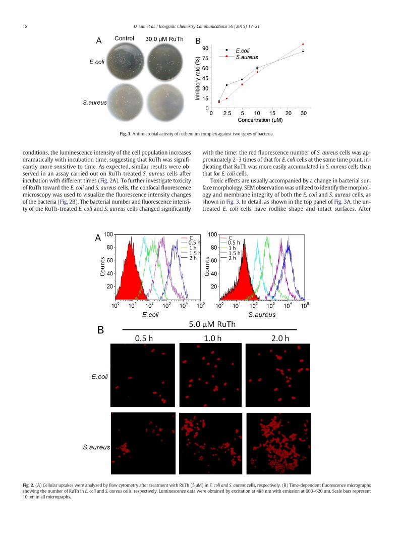
Fig. 1. Antimicrobial activity of ruthenium complex against two types of bacteria.
18 D. Sun et al. / Inorganic Chemistry Communications 56 (2015) 17–21
conditions, the luminescence intensity of the cell population increasesdramatically with incubation time, suggesting that RuTh was signifi-cantly more sensitive to time. As expected, similar results were ob-served in an assay carried out on RuTh-treated S. aureus cells afterincubation with different times (Fig. 2A). To further investigate toxicityof RuTh toward the E. coli and S. aureus cells, the confocal fluorescencemicroscopy was used to visualize the fluorescence intensity changesof the bacteria (Fig. 2B). The bacterial number and fluorescence intensi-ty of the RuTh-treated E. coli and S. aureus cells changed significantly
Fig. 2. (A) Cellular uptakes were analyzed by flow cytometry after treatment with RuTh (5 μMshowing the number of RuTh in E. coli and S. aureus cells, respectively. Luminescence data we10 μm in all micrographs.
with the time; the red fluorescence number of S. aureus cells was ap-proximately 2–3 times of that for E. coli cells at the same time point, in-dicating that RuTh was more easily accumulated in S. aureus cells thanthat for E. coli cells.
Toxic effects are usually accompanied by a change in bacterial sur-facemorphology. SEMobservationwas utilized to identify themorphol-ogy and membrane integrity of both the E. coli and S. aureus cells, asshown in Fig. 3. In detail, as shown in the top panel of Fig. 3A, the un-treated E. coli cells have rodlike shape and intact surfaces. After
) in E. coli and S. aureus cells, respectively. (B) Time-dependent fluorescence micrographsre obtained by excitation at 488 nm with emission at 600–620 nm. Scale bars represent
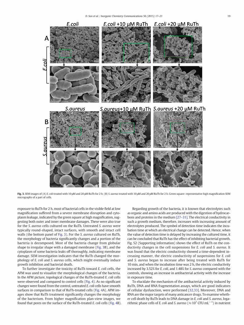
Fig. 3. SEM images of (A) E. coli treatedwith 10 μMand 20 μMRuTh for 2 h; (B) S. aureus treatedwith 10 μMand 20 μMRuTh for 2 h. Green square: representative highmagnification SEMmicrographs of a part of cells.
19D. Sun et al. / Inorganic Chemistry Communications 56 (2015) 17–21
exposure to RuTh for 2 h,most of bacterial cells in the visible field at lowmagnification suffered from a severe membrane disruption and cyto-plasm leakage, indicated by the green square at highmagnification, sug-gesting both outer and inner membrane damages. These were also truefor the S. aureus cells cultured on the RuTh. Untreated S. aureus weretypically round-shaped, intact surfaces, with smooth and intact cellwalls (the bottom panel of Fig. 3). For the S. aureus cultured on RuTh,the morphology of bacteria significantly changes and a portion of thebacteria is decomposed. Most of the bacteria change from globularshape to irregular shape with a damaged membrane (Fig. 3B), and thecytoplasm of some bacteria leaks off thoroughly, indicating membranedamage. SEM investigation indicates that the RuTh changed the mor-phology of E. coli and S. aureus cells, which might eventually inducegrowth inhibition and bacterial death.
To further investigate the toxicity of RuTh toward E. coli cells, theAFM was used to visualize the morphological changes of the bacteria.In the AFM picture, topological changes of the RuTh-treated E. coli cellswere observed and compared to control cells (Fig. 4). As no significantchangeswere found from the control, untreated E. coli cells have smoothsurfaces in comparison to that of RuTh-treated cells (Fig. 4A). AFM im-ages show that RuTh treatment significantly changes the morphologyof the bacterium. From higher magnification plan-view images, wefound that pores on the surface of the RuTh-treated E. coli cells (Fig. 4B).
Regarding growth of the bacteria, it is known that electrolytes suchas organic and amino acids are producedwith thedigestion of hydrocar-bons and proteins in themedium [27–31]. The electrical conductivity insuch a growth medium, therefore, increases with increasing amount ofelectrolytes produced. The symbol of detection time indicates the incu-bation time at which an electrical change can be detected. Hence, whenthe value of detection time is delayed by increasing the cultured time, itcan be concluded that RuTh has the effect of inhibiting bacterial growth.Fig. S2 (Supporting information) shows the effect of RuTh on the con-ductivity changes in the cell suspensions for E. coli and S. aureus. Itwas found that the electric conductivity showed a time-dependent in-creasing manner, the electric conductivity of suspensions for E. coliand S. aureus began to increase after being treated with RuTh for10min, andwhen the incubation timewas 2 h, the electric conductivityincreased by 3.52% for E. coli, and 1.48% for S. aureus compared with thecontrols, showing an increase in antibacterial activity with the increasein exposure time.
To elucidate the mechanism of the antibacterial activity induced byRuTh, DNA and RNA fragmentation assays, which are good indicatorsof cellular dysfunction, were performed [32,33]. Moreover, DNA andRNAare themajor targets ofmany anticancer drugs. To examinewheth-er cell death by RuTh leads to DNA damage in E. coli and S. aureus, loga-rithmic phase cells of E. coli and S. aureus (≈107 CFUmL−1) in nutrient
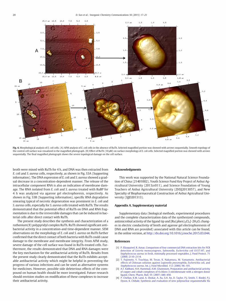
Fig. 4.Morphological analysis of E. coli cells. (A) AFM analysis of E. coli cells in the absence of RuTh. Selectedmagnified portion was showedwith arrows sequentially. Smooth topology ofthe control cell surface was visualized in themagnified photograph. (B) Effect of RuTh (10 μM) on surfacemorphology of E. coli cells. Selectedmagnified portion was showedwith arrowssequentially. The final magnified photograph shows the severe topological damage on the cell surface.
20 D. Sun et al. / Inorganic Chemistry Communications 56 (2015) 17–21
broth were mixed with RuTh for 4 h, and DNA was then extracted fromE. coli and S. aureus cells, respectively, as shown in Fig. S3A (Supportinginformation). The DNA expression of E. coli and S. aureus showed a grad-ual decrease in a concentration-dependent manner. The release of theintracellular component RNA is also an indication of membrane dam-age. The RNA isolated from E. coli and S. aureus treated with RuBP for4 h was analyzed via agarose gel electrophoresis, respectively. Asshown in Fig. S3B (Supporting information), specific RNA degradativesmearing typical of necrotic degeneration was prominent in E. coli andS. aureus cells, especially for S. aureus cells treatedwith RuTh. The resultsdemonstrated that the potential effect of RuTh on DNA and RNA frag-mentation is due to the irreversible damages that can be induced in bac-terial cells after direct contact with RuTh.
The present study describes the synthesis and characterization of aruthenium(II) polypyridyl complex RuTh. RuTh showed significant anti-bacterial activity in a concentration-and time-dependent manner. SEMobservations on the morphology of E. coli and S. aureus on RuTh furtherconfirmed that the direct contact of both bacteriawith RuTh could causedamage to the membrane and membrane integrity. From AFM study,severe damage of the cell surface was found in RuTh-treated cells. Fur-thermore, the results demonstrated that DNA and RNA damages werethe key mechanism for the antibacterial activity of RuTh. Results fromthe present study clearly demonstrated that the RuTh exhibits accept-able antibacterial activity which might be helpful in preventing theprogress of various infections and can be used as alternative systemsfor medicines. However, possible side deleterious effects of the com-pound on human health should be more investigated. Future researchshould envision studies on modification of these complexes to increasetheir antibacterial activity.
Acknowledgments
This work was supported by the National Natural Science Founda-tion of China (21401002), Youth Science Fund Key Project of Anhui Ag-ricultural University (2013zr011), and Science Foundation of YoungTeachers of Anhui Agricultural University (ZHSJ2013057), and NewSpecialty of Biopharmaceutical Construction of Anhui Agricultural Uni-versity (SJJD201313).
Appendix A. Supplementary material
Supplementary data (biological methods, experimental proceduresand the complete characterization data of the synthesized compounds,antimicrobial activity of the ligand tip and [Ru(phen)2Cl2]·2H2O, chang-es in electric conductivity of broth and agarose gel electrophoresesis ofDNA and RNA are provided) associated with this article can be found,in the online version, at http://dx.doi.org/10.1016/j.inoche.2015.03.044.
References
[1] P. Elizaquivel, R. Aznar, Comparison of four commercial DNA extraction kits for PCRdetection of Listeria monocytogenes, Salmonella, Escherichia coli O157:H7, andStaphylococcus aureus in fresh, minimally processed vegetables, J. Food Protect. 71(2008) 2110–2114.
[2] T. Fujimoto, Y. Tsuchiya, M. Terao, K. Nakamura, M. Yamamoto, Antibacterialeffects of Chitosan solution against Legionella pneumophila, Escherichia coli, andStaphylococcus aureus, Int. J. Food Microbiol. 112 (2006) 96–101.
[3] A.T. Kabbani, H.H. Hammud, A.M. Ghannoum, Preparation and antibacterial activityof copper and cobalt complexes of 4-chloro-3-nitrobenzoate with a nitrogen donorligand, Chem. Pharm. Bull. 55 (2007) 446–450.
[4] P. Chellan, K.M. Land, A. Shokar, A. Au, S.H. An, D. Taylor, P.J. Smith, T. Riedel, P.J.Dyson, K. Chibale, Synthesis and evaluation of new polynuclear organometallic Ru
21D. Sun et al. / Inorganic Chemistry Communications 56 (2015) 17–21
(II), Rh (III) and Ir (III) pyridyl ester complexes as in vitro antiparasitic and antitu-mor agents, Dalton Trans. 43 (2014) 513–526.
[5] M.-L. Teyssot, A.-S. Jarrousse, M. Manin, A. Chevry, S. Roche, F. Norre, C. Beaudoin, L.Morel, D. Boyer, R. Mahiou, Metal–NHC complexes: a survey of anti-cancer proper-ties, Dalton Trans. (2009) 6894–6902.
[6] M.J. Hannon, Metal-based anticancer drugs: from a past anchored in platinumchemistry to a post-genomic future of diverse chemistry and biology, Pure Appl.Chem. 79 (2007) 2243–2261.
[7] Z. Zhang, C. Bi, D. Buac, Y. Fan, X. Zhang, J. Zuo, P. Zhang, N. Zhang, L. Dong, Q.P. Dou,Organic cadmium complexes as proteasome inhibitors and apoptosis inducers inhuman breast cancer cells, J. Inorg. Biochem. 123 (2013) 1–10.
[8] G.-Y. Li, K.-J. Du, J.-Q. Wang, J.-W. Liang, J.-F. Kou, X.-J. Hou, L.-N. Ji, H. Chao, Synthe-sis, crystal structure, DNA interaction and anticancer activity of tridentate copper(II) complexes, J. Inorg. Biochem. 119 (2013) 43–53.
[9] V.V. Grushin, Mixed phosphine–phosphine oxide ligands, Chem. Rev. 104 (2004)1629–1662.
[10] D. Sun, Y. Liu, D. Liu, R. Zhang, X. Yang, J. Liu, Stabilization of G-quadruplex DNA, in-hibition of telomerase activity and live cell imaging studies of chiral ruthenium (II)complexes, Chem. Eur. J. 18 (2012) 4285–4295.
[11] D. Sun, R. Zhang, F. Yuan, D. Liu, Y. Zhou, J. Liu, Studies on characterization, telome-rase inhibitory properties and G-quadruplex binding of η 6-arene ruthenium com-plexes with 1, 10-phenanthroline-derived ligands, Dalton Trans. 41 (2012)1734–1741.
[12] T. Joshi, V. Pierroz, C. Mari, L. Gemperle, S. Ferrari, G. Gasser, ABis(dipyridophenazine)(2‐(2‐pyridyl)pyrimidine‐4‐carboxylic acid)ruthenium (II)complex with anticancer action upon photodeprotection, Angew. Chem. Int. Ed.53 (2014) 2960–2963.
[13] R. Trondl, P. Heffeter, C.R. Kowol, M.A. Jakupec, W. Berger, B.K. Keppler, NKP-1339,the first ruthenium-based anticancer drug on the edge to clinical application,Chem. Sci. 5 (2014) 2925–2932.
[14] I. Bratsos, S. Jedner, T. Gianferrara, E. Alessio, Ruthenium anticancer compounds:challenges and expectations, CHIMIA Int. J. Chem. 61 (2007) 692–697.
[15] X. Meng, M.L. Leyva, M. Jenny, I. Gross, S. Benosman, B. Fricker, S. Harlepp, P.Hébraud, A. Boos, P. Wlosik, A ruthenium-containing organometallic compound re-duces tumor growth through induction of the endoplasmic reticulum stress geneCHOP, Cancer Res. 69 (2009) 5458–5466.
[16] D.L. Arockiasamy, S. Radhika, R. Parthasarathi, B.U. Nair, Synthesis and DNA-bindingstudies of two ruthenium (II) complexes of an intercalating ligand, C44 (2009)2044–2051.
[17] L.-F. Tan, H. Chao, H. Li, Y.-J. Liu, B. Sun, W. Wei, L.-N. Ji, Synthesis, characterization,DNA-binding and photocleavage studies of [Ru(bpy)2(PPIP)]2+ and [Ru(phen)2(PPIP)]2+, J. Inorg. Biochem. 99 (2005) 513–520.
[18] L.-S. Ling, Z.-K. He, Y.-E. Zeng, Spectral studies on the interaction of DNA andRu(bipy)2(dppx)2+, Spectrochim. Acta A 55 (1999) 1297–1302.
[19] Y.-J. Liu, H. Chao, L.-F. Tan, Y.-X. Yuan, W. Wei, L.-N. Ji, Interaction of polypyridyl ru-thenium (II) complex containing asymmetric ligand with DNA, J. Inorg. Biochem. 99(2005) 530–537.
[20] Y. Liu, X. Chen, L. Zhang, D. Sun, Y. Zhou, L. Chen, J. Liu, Stabilization of telomere DNA,and mechanism of apoptosis of tumor cells induced by ruthenium complexes, ActaChim. Sin. 72 (2014) 473–480.
[21] K.E. Erkkila, D.T. Odom, J.K. Barton, Recognition and reaction of metallointercalatorswith DNA, Chem. Rev. 99 (1999) 2777–2796.
[22] J. Liu, W. Zheng, S. Shi, C. Tan, J. Chen, K. Zheng, L. Ji, Synthesis, antitumor activityand structure–activity relationships of a series of Ru (II) complexes, J. Inorg.Biochem. 102 (2008) 193–202.
[23] U. Schatzschneider, J. Niesel, I. Ott, R. Gust, H. Alborzinia, S. Wölfl, Cellular uptake,cytotoxicity, and metabolic profiling of human cancer cells treated withruthenium(II) polypyridyl complexes [Ru(bpy)2(N–N)]Cl2 with N–N = bpy,phen, dpq, dppz, and dppn, ChemMedChem 3 (2008) 1104–1109.
[24] Y.-J. Liu, C.-H. Zeng, H.-L. Huang, L.-X. He, F.-H. Wu, Synthesis, DNA-binding,photocleavage, cytotoxicity and antioxidant activity of ruthenium (II) polypyridylcomplexes, Eur. J. Med. Chem. 45 (2010) 564–571.
[25] C. Tan, J. Liu, L. Chen, S. Shi, L. Ji, Synthesis, structural characteristics, DNA bindingproperties and cytotoxicity studies of a series of Ru (III) complexes, J. Inorg.Biochem. 102 (2008) 1644–1653.
[26] D. Liu, Y. Liu, C.Wang, S. Shi, D. Sun, F. Gao, Q. Zhang, J. Liu, Polypyridyl complexes ofruthenium(II): stabilization of G-quadruplex DNA and inhibition of telomerase ac-tivity, ChemPlusChem 77 (2012) 551–562.
[27] O. Yamamoto, M. Komatsu, J. Sawai, Z.-e. Nakagawa, Effect of lattice constant of zincoxide on antibacterial characteristics, J. Mater. Sci. Mater. Med. 15 (2004) 847–851.
[28] D. Miklavcic, M. Snoj, A. Zupanic, B. Kos, M. Cemazar, M. Kropivnik, M. Bracko, T.Pecnik, E. Gadzijev, G. Sersa, Towards treatment planning and treatment of deep-seated solid tumors by electrochemotherapy, Biomed. Eng. Online 9 (2010) 1–12.
[29] K. Mendgen, A. Schneider, M. Sterk, W. Fink, The differentiation of infection struc-tures as a result of recognition events between some biotrophic parasites andtheir hosts, J. Phytopathol. 123 (1988) 259–272.
[30] M. Pavlin, T. Slivnik, D. Miklavcic, Effective conductivity of cell suspensions, IEEETrans. Biomed. Eng. 49 (2002) 77–80.
[31] D. Miklavčič, N. Pavšelj, F.X. Hart, Electric properties of tissues, Wiley Encyclopediaof Biomedical Engineering2006.
[32] R. Modak, S.D. Mitra, M. Vasudevan, P. Krishnamoorthy, M. Kumar, A.V. Bhat, M.Bhuvana, S.K. Ghosh, B.R. Shome, T.K. Kundu, Epigenetic response in mice mastitis:role of histone H3 acetylation and microRNA(s) in the regulation of host inflamma-tory gene expression during Staphylococcus aureus infection, Clin. Epigenetics 6(2014) 12.
[33] S. Gurunathan, J.W. Han, A.A. Dayem, V. Eppakayala, J.-H. Kim, Oxidative stress-mediated antibacterial activity of graphene oxide and reduced graphene oxide inPseudomonas aeruginosa, Int. J. Nanomedicine 7 (2012) 5901–5914.