Embed Size (px)
Citation preview

© 2015 Sudipta Roy and Debdulal Banerjee. This is an open access article distributed under the terms of the Creative Commons Attribution License -NonCommercial-ShareAlikeUnported License (http://creativecommons.org/licenses/by-nc-sa/3.0/).
Journal of Applied Pharmaceutical Science Vol. 5 (07), pp. 006-011, July, 2015 Available online at http://www.japsonline.com DOI: 10.7324/JAPS.2015.50702 ISSN 2231-3354
Broad spectrum antibacterial activity of granaticinic acid, isolated from Streptomyces thermoviolaceus NT1; an endophyte in Catharanthus roseus (L.) G. Don Sudipta Roy1, Debdulal Banerjee2 1Department of Microbiology, Vidyasagar University, Midnapore- 721102, West Bengal, India. 2Department of Botany, Vidyasagar University, Midnapore- 721102, West Bengal, India.
ARTICLE INFO
ABSTRACT
Article history: Received on: 28/03/2015 Revised on: 20/04/2015 Accepted on: 01/07/2015 Available online: 27/07/2015
Searching for endophytic actinomycetes, strain NT1 was isolated from surface sterilized stem of Catharanthus roseus (L.) G. Don collected from Paschim Medinipur, India. The strain was identified as Streptomyces thermoviolaceus NT1 on the basis of morphological, biochemical and 16s rDNA based phylogenetic analysis. It showed potential antagonism against several Gram positive and Gram negative bacterial pathogens along with drug resistant Staphylococcus aureus. Maximum antibacterial production was obtained in ISP2 media at pH 7.2, 35 °C for 10 days. The active antibacterial substance was purified by Silica gel column chromatography and activity guided TLC. IR and NMR analysis identified the active compound as granaticinic acid of m/z 463.26 [M+H]. These results suggest that the antimicrobial produced by the isolated endophytic strain will be useful in near future.
Key words: Endophyte, Streptomyces thermoviolaceus, granaticinic acid, antibacterial, drug resistance.
INTRODUCTION
Screening of antimicrobial compound producing microorganisms is a continual field of research and actinomycetes are playing a promising role as the source of such compounds. Over two third of present day antibiotics were obtained from Streptomyces (Baltz, 1998), a special group within actinomycetes but presently isolation of such compounds are unable to meet the demand for a new class of bioactive compounds. Moreover emergence of drug resistant pathogens, outbreak of opportunistic bacteria and throttle success of combinatorial chemistry creates the situation more severe (Strobel, 2003). Endophytic microorganisms have taken this opportunity as a good source of future antimicrobial compounds. Endophytes reside within the plant’s internal tissues which are still less explored ecosystem harboring diverse microbes. Some of them act synergistically by producing secondary metabolites
* Corresponding Author Debdulal Banerjee, Department of Botany, Vidyasagar University, Midnapore- 721102, West Bengal, India. Email: [email protected]
antagonistic to plant pathogens (Strobel and Daisy, 2003) or by producing plant growth promoting substances (Bhattacharya and Jha, 2012).
Actinomycetes are also found in endophytic association like fungi (Kharwar et al., 2008) and other bacteria and can serve as a prime source of novel class antimicrobial compounds, as chances of getting new compound is relatively higher from new strain and isolation of new strain is advantageous from unexplored or less explored habitat (Stierle et al., 1993). Several studies describe the isolation and identification of endophytic actinomycetes producing metabolites against fungal plant pathogens but few are reported against bacterial pathogens especially drug resistant or opportunistic bacterial pathogens (Castillo et al., 2002; Castillo et al., 2003; Taechowisan et al., 2005, Taechowisan et al., 2014).
Present study illustrates the isolation and identification of endophytic Streptomyces thermoviolaceus NT1 from Catharanthus roseus. Purification of metabolites produced by this strain afforded granaticinic acid, which showed broad spectrum antibacterial property and was also active against drug resistant pathogens.

Roy and Banerjee / Journal of Applied Pharmaceutical Science 5(07); 2015: 006-011 07
MATERIALS AND METHODS
Endophytic actinomycetes isolation Healthy and young plants of Catharanthus roseus (L.) G.
Don. were collected from various places of Psachim Medinipur, India (22.57° N – 87.11° E). Stems (about 1 cm length) of C. roseus were surface sterilized by sequential ethanol and NaOCl treatment and finally washed three times with autoclaved distilled water (Coombs and Franco, 2003).
Authenticity of surface sterilization was verified by plating the final sample washed water on nutrient agar media. The bark was removed of samples and tissues were soaked in 10 % NaHCO3 for 5 min to suppress fungal contamination. Inner tissues were then aseptically transferred to ISP2, ISP5 (International Streptomyces Project) agar and actinomycetes isolation agar media (Himedia, Mumbai) supplemented with cycloheximide and streptomycin (50 µg/ ml). Plates were incubated at 28 ºC for 14 days. Isolated strains were pure cultured and preserved in glycerol based liquid media at -20 ºC. In vitro antimicrobial assay
Isolated strain, NT1 was checked for Antimicrobial potency by agar diffusion methods. This strain was grown in ISP2 broth and 100 µl cell free cultures was applied in wells of Muller Hinton agar media that was previously seeded with bacterial pathogens. Inhibition zones were recorded after 24 h incubation at 35 ºC for each pathogen.
Following pathogenic bacteria were selected in this study: methicillin resistant Staphylococcus aureus, penicillin resistant Staphylococcus aureus (clinical isolate), Bacillus subtilis (ATCC 11774), Bacillus cereus (ATCC 14579), Vibrio parahemolyticus ATCC 1782, Pseudomonas aeruginosa (ATCC 9027), Shigella flexnerii (ATCC 12022), and Escherichia coli (clinical isolate). Morphological, physiological and biochemical characterizations
Characterization of the strain was done based on cultural characteristics, morphological, physiological and biochemical properties. Cultural morphology was established after growing the organism at various media (Shirling and Gottlieb, 1966; Williams et al., 1983) and cellular morphology was determined by the compound and scanning electron microscopy (Vega© TESCAN, Czech Republic). Extracellular enzyme production and sugar utilization test (Gordon et al., 1974) were made by standard methods. The strain was grown in ISP 2 broth at various incubation temperature and pH conditions for determining its optimum growth temperature and pH requirement. Genomic DNA isolation and amplification of 16s rRNA gene
Cells were lysed of 5 days old culture by lysozyme and SDS treatment (5 mg/ ml and 10 % respectively). Genomic DNA was extracted with phenol-chloroform and precipitated with isopropanol. Finally genomic DNA was dissolved in 100 µl TE
(pH 8) buffer. The 16s rRNA gene was amplified using the primers 27F (5’-AGAGTTTGATCCTGGCTCAG-3’) and 1492R (5’-GGTTACCTTGTTACGACTT-3’) (Santhi and Solomon, 2011) in a thermocycler (Eppendorf). PCR products were purified with Hi-PurATM PCR product purification spin kit (Himedia Laboratories, India). Forward and reverse DNA sequencing of amplicon was carried out with the same primers using BDT v3.1 Cycle sequencing kit on ABI 3730xl Genetic Analyzer. Phylogeny construction and sequence submission
Consensus sequences of 1412 bp rRNA gene were edited and assembled from forward and reverse sequence data using aligner software. The 16S rRNA gene sequence was used to carry out BLAST with the ‘nr’ database of NCBI Genbank to determine operational taxonomic units. Based on maximum identity score sequences were selected and aligned using the multiple alignment software program, Clustal W. Distance matrix was generated using RDP (Ribosomal Database Project) database and the phylogenetic tree were constructed using MEGA 6 (Tamura et al., 2007) (Molecular Evolutionary Genetics Analysis). The evolutionary history was inferred using the Neighbor-Joining method (Saitou and Nei, 1987).
The evolutionary distances were computed using the Kimura 2-parameter method (Kimura, 1980). DNA sequences were deposited in GenBank under accession number KJ486841.1 Antimicrobial production at optimum conditions
Optimization of antibacterial production from NT1 was done by culturing it in various physical and chemical conditions. The strain was grown in a wide range of media pH (5- 10) and incubation temperature (20 – 45 ºC). Optimum values were determined by measuring zone of inhibition produced by culture filtrates of each variant against B. cereus. Optimum additional carbon source was determined by adding glucose, galactose, fructose, maltose, lactose and sucrose (1 %, w/v) in ISP2 broth. Tryptone, peptone, soya meal (0.5 %, w/v), KNO3 NH4NO3 and NH4Cl were additionally (0.2 %, w/v) added to the ISP broth to find most suitable nitrogen source for antibacterial production by the strain NT1.
A 1 cm diameter agar plug, full of NT1 spores was inoculated in 30 ml ISP2 broth and cultured at 28 ºC for 3 days. 5 % culture was transferred to 100 ml ISP2 broth in 500 ml conical flask and cultured for 3 days more and used as seed for next step larger production. Finally, 5 % seed was inoculated in 1.5 L ISP2 broth and cultured for 10 days at 28 ºC with 150 rpm. Cell mycelia were separated by filtration with Whatman paper No. 1 and then centrifuged at 12000 rpm for 15 min. to obtain a cell free culture medium. Culture filtrate was extracted with equal volume ethyl acetate and organic fraction was made water free by adding sufficient anhydrous Na2SO4. It was then dried under vacuum in a rotary evaporator at 40 °C (HS – 2005S, HAHNSHIN, Korea) and antibacterial activity was determined by disk diffusion assay.

08 Roy and Banerjee / Journal of Applied Pharmaceutical Science 5(07); 2015: 006-011 Purification of active component
Residual crude was dissolved in 5 ml chloroform, adsorbed by Silica gel and loaded onto a silica gel column (230-400 Å mesh). It was then fractionated with graded hexane – chloroform (60:0, 50:10, 30:30,10:50, 0:60) and then with chloroform-methanol (60:0, 55:5, 50:10, 40:20, 30:30, 20:40, 0:60). About 60 ml of each fraction was collected. Fractions were concentrated and checked for antibacterial property by disk diffusion technique on MHA plates. Active fraction was re-fractioned with more finely graded solvent system (chloroform and methanol; 60:0, 59:1, 58:2, 57:3, 56:4, 55:5, 50:10, 0:60) and active fraction was determined by similar assay. TLC and bioautogram analysis
Purified fraction was concentrated and analyzed by thin layer chromatography with chloroform and methanol (5:1). Separated components were located on UV exposure. Autobiogram was done with B. cereus to find active component on TLC plates. After TLC analysis the plate was overlaid with molten MHA media (45 ºC) containing 50 μl aliquots of B. cereus (106
cfu/ ml). TLC plate was incubated at 37 ºC overnight and inhibition zone was located by methylthiazoletetrazolium (MTT-5 mg/ ml). Spectroscopic analysis of active compound
Structural information of active component was gathered by UV, IR, NMR, and mass spectral data. λmax was determined of active compound after dissolving in chloroform by UV-Vis spectrophotometer (UV-1800, SHIMADZU). FTIR spectra were taken in FT-IR spectrophotometer (Spectrum T, Perkin Elmer). The sample was dissolved in CDCl3 and analyzed for 1H and 13C NMR spectra at 600 MHz (Bruker, Avance 600). ESI Mass was analyzed with positive ion mode. RESULTS AND DISCUSSIONS
The isolated endophytic strain was gram positive, filamentous actinobacteria that grow well aerobically. Substrate mycelia nicely emerge within 3 day after inoculation and aerial formations were grey that turns to white. The strain produces soluble violet pigment in ISP2 media. It produced slow-growing, dry and hard colonies on ISP2 agar. Scanning electron microscopy of the organism revealed the presence of numerous aerial filaments and few smooth, cylindrical spores (0.8 µm in length and 0.2 µm in diameter) that were produced in short curved (Fig. 1).
Other morphological, and biochemical properties of strain NT1 were summarized in table 1. Comparative 16S rRNA gene sequence analysis (Fig. 2) showed that the strain was taxonomically belong to the species of Streptomyces and highest similarities being found with the sequences of Streptomyces thermoviolaceus (AB184685.1), Streptomyces thermocyaneomaculatus (AB184583.1), Streptomyces thermocyaneoviolaceus (AB184582.1), Streptomyces thermophilus
Table 1: Cultural and physiological characteristics of endophytic Streptomyces themoviolaceus NT1.
Colony morphology Elevated, rough surface, dry, brownish to white colony
Growth on ISP2 Substrate mycelia Aerial mycelia Soluble pigment
Good Brownish White Redish violet
Growth on ISP4 Substrate mycelia Aerial mycelia Soluble pigment
Moderate Yellowish White Violet
Growth of ISP5 Substrate mycelia Aerial mycelia Soluble pigment
Very less Gray White No
Growth on ISP7 Substrate mycelia Aerial mycelia Soluble pigment
Very poor Yellowish White No
Spore morphology 3- 5 spores at tip of the filaments Cylindrical, smooth surface About 0.8 to0.2 µm diameter
Cell morphology Highly branched filamentous, Gram positive Extra cellular enzymes
Cellulase, amylase, protease (+)
Carbon source utilization
Dextrose, fructose, galactose, lactose, maltose, sucrose, starch, mannitol (+), xylose, inositol, rhamnose (-)
Growth temperatue 20 ºC 28 ºC 35 ºC 40 ºC
- ++ ++ (optimum*) -
Growth pH 5 6 7 8 9 10
- + ++ (optimum*) + + -
(*Optimum has been determined as mean of triplicate study).
Fig. 1: Endophytic Streptomyces thermoviolaceous NT1; A: SEM view (arrows indicate spores, magnified view), B – colony morphology on ISP2 (arrow indicate redish violet pigmentation). (AB184358.1) about 99 % and with Streptomyces thermodiastaticus (AJ001434.1), Streptomyces sp. E1125 (DQ303453.1), Streptomyces sp. SDCB6 (JN617215.1) 98%. It is evident from the phylogenetic tree based on the neighbor-joining method that the strain NT1 clusters with the nearest neighbors S. thermoviolaceus NBRC 13387 in a separate branch with high bootstrap support.

Roy and Banerjee / Journal of Applied Pharmaceutical Science 5(07); 2015: 006-011 09
The strain fitted, in all respects, the taxonomic position within genus Streptomyces. Cellular and colony morphology, soluble pigments on media and biochemical characterization like enzyme profile and molecular characterization strongly suggest our isolate to be a new strain, Streptomyces thermovilaceous NT1. Few or modest spore production was found from this isolate in different media may be due to some biochemical relationship with its host plant. Previous isolates of this bacterium were of thermophilic range that could be cultivated at 50 ºC with antimicrobial production (James and Edwards, 1989) but NT1 does not grow over 40 ºC. Internal plant tissue is a specialized ecosystem may be responsible of such alteration in gene sequence during co-evolution (Germaine et al., 2004).
Here is the opportunity of getting variations in
metabolites from similar micro-organism isolated from other common habitats. Surprisingly an endophytic fungus, Fusarium oxysporum of C. roseus was interestingly found to produce vinblastine and vincristine the same natural products as host plants which opens a new probability in drug discovery (Kumar et al., 2013).
The isolated endophytic strain may be a good source for the antimicrobial compound due to its broad spectrum antibacterial property and its potency against drug resistant pathogens. Culture filtrate of endophytic S. thermoviolaceus NT1 showed broad spectrum antibacterial activity to all selected pathogens. Table: 2 represent inhibition zones produced by culture filtrate and crude ethyl acetate extract against test pathogenic bacteria. It was found
Fig. 2: Neighbor-joining tree based on 16s ribosomal RNA gene sequences showing relationship between NT1 and members of the genus Streptomyces.
Table. 2: Pathogen growth inhibition by NT1 culture filtrate and column purified active fraction. Sample Zone inhibition (mm) ( Results are represented as means ± standard deviation of three replicates )
PRSA MRSA B. cereus B. subtilis V. parahemolyticus P. aeruginosa S. flexnerii E. coli Culture filtrate† 13.6±0.5 13±0.8 17.3±1 18±0.4 9.6±0.9 8.83±0.8 10.5±1.4 12.5±1 Crude extract# 29.3±1.2 27.6±1.2 30.8±2 33±0.9 13±0.5 16.3±0.8 19.8±0.4 15±0.6 †, At the amount of 100 µl of 14 days old culture supernatant in agar well. #, At the amount of 25 µl on paper discs soaked with tested substance.
010 Roy and Banerjee / Journal of Applied Pharmaceutical Science 5(07); 2015: 006-011 that the antimicrobial compound was more effective for Gram positive bacteria than Gram negative bacteria. S. thermoviolaceus WR-141, a previous isolate of non-endophytic origin (St. Pyrek et al., 1977) was found to produce antimicrobial that solely inhibited Gram positive.
The NT1 is also active against P. aeruginosa, an opportunistic pathogen that has become a major cause of nosocomial infections worldwide and a serious issue of public health. Moreover, only a few drugs are available to treat very serious infection of drug resistant Staphylococci. In this regard anti MRSA and anti PRSA activity of this strain indicates its promising role in future drugs. It was found that this strain produced the antibacterial substance best level at media pH-7, and 35 °C (Fig. 3) with 1 % additional glucose and tryptone (0.5 %). In a separate experiment, S thermoviolaceous NCIB 10076 was found to produce antibacterial substances optimally at pH-7 and at 45 °C incubation temperature (James and Edwards, 1989).
Fig. 3: Most suitable pH and temperature for optimum antibacterial production by NT1.
After repeated column chromatography and activity guided TLC purification (Fig. 4), the active compound was found as amorphous red substance and changes its color from red to blue by pH change. It absorbs UV strongly with UV λmax (CHCl3) nm (log ε) 223 (1.3), 236 (0.32), 523 (1.24), 558 (1.32), 612 (1.43); IR νmax were found with (KBr) cm¯1: 3600 ~ 3400, 3000 ~ 2850, 1712, 1665, 1610, 1458 (Fig. 3); MS m/z (relative intensities): 463.26 [M]+; 1H NMR (CDCl3) δ7.812 (doublet, j = 7.8 Hz), 7.714 (doublet, j = 5.4), 7.541 (triplet, J1 = J2 = 7.2 Hz), 7.455 (triplet, J1 = J2 = 7.8 Hz ), 7.269, 6.427 (doublet, j = 5.4), 3.648, 3.597, 2.374, 1.471 - 0.843; 13C NMR (CDCl3) δ170.11 (-COOH), 154.26,
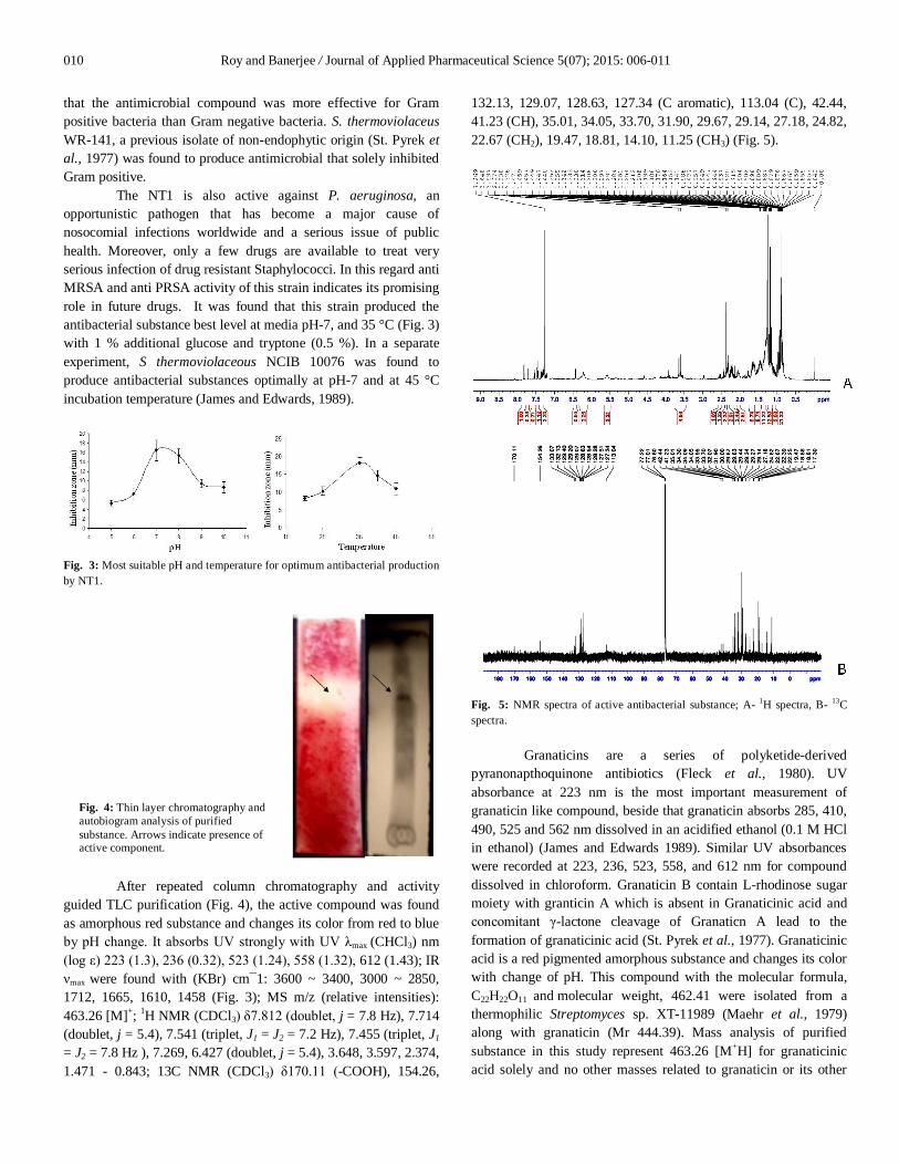
132.13, 129.07, 128.63, 127.34 (C aromatic), 113.04 (C), 42.44, 41.23 (CH), 35.01, 34.05, 33.70, 31.90, 29.67, 29.14, 27.18, 24.82, 22.67 (CH2), 19.47, 18.81, 14.10, 11.25 (CH3) (Fig. 5).
Fig. 5: NMR spectra of active antibacterial substance; A- 1H spectra, B- 13C spectra.
Granaticins are a series of polyketide-derived pyranonapthoquinone antibiotics (Fleck et al., 1980). UV absorbance at 223 nm is the most important measurement of granaticin like compound, beside that granaticin absorbs 285, 410, 490, 525 and 562 nm dissolved in an acidified ethanol (0.1 M HCl in ethanol) (James and Edwards 1989). Similar UV absorbances were recorded at 223, 236, 523, 558, and 612 nm for compound dissolved in chloroform. Granaticin B contain L-rhodinose sugar moiety with granticin A which is absent in Granaticinic acid and concomitant γ-lactone cleavage of Granaticn A lead to the formation of granaticinic acid (St. Pyrek et al., 1977). Granaticinic acid is a red pigmented amorphous substance and changes its color with change of pH. This compound with the molecular formula, C22H22O11 and molecular weight, 462.41 were isolated from a thermophilic Streptomyces sp. XT-11989 (Maehr et al., 1979) along with granaticin (Mr 444.39). Mass analysis of purified substance in this study represent 463.26 [M+H] for granaticinic acid solely and no other masses related to granaticin or its other
Fig. 4: Thin layer chromatography and autobiogram analysis of purified substance. Arrows indicate presence of active component.
Roy and Banerjee / Journal of Applied Pharmaceutical Science 5(07); 2015: 006-011 011
derivative were noticed in the spectrum. The IR spectrum shows typical OH stretching vibrations of the phenolic and carboxylic acid at 3600 - 3400 and 3000 - 2850 cm-1. Peak at 1715 cm-1 is indicative of carbonyl vibrations of a saturated aliphatic carboxylic acid, which is also similar to granaticinic acid reported (US patent). IR absorbance at 1610 cm-1 is indicative of quinine carbonyl with strong intermolecular hydrogen bonds. In 1H NMR, δ 7.269 strongly suggest the presence of p- disubstituted aromatic, δ 2.374 indicates H of adjacent carbon to carboxyl group and δ 1.471 - 0.843 suggest the presence of H in primary or secondary aliphatic carbon. δ 170.11 in 13C NMR clearly represent for a C=O in acid group where as δ 154.26 stands for =C< in aromatic ring structure. So all physical and spectral evidences confirmed that the active antibacterial component is granaticinic acid produced by S. thermoviolaceus NT1. CONCLUSION
This study demonstrates isolation of a new strain Streptomyces thermoviolaceus NT1 that colonized the inner tissues of C. roseus. It also proposes that the strain might be a significant source of antibacterial compound. Granaticinic acid was a major active component in cell free culture filtrate of NT1 which showed effective antagonism to drug resistant pathogens. This is the first report of isolation and identification of an endophytic (stems of C. roseus) Streptomyces thermoviolaceus producing granaticinic acid. ACKNOWLEDGEMENT
Authors are thankful to University Grants Commission (UGC), New Delhi for funding, SR is thankful to Oriental Institute of Science and Technology (OIST) for their help during this work. REFERENCES
Baltz RH. Genetic manipulation of antibiotic-producing Streptomyces. Trends Microbiol. 1998; 6, 76-83.
Bhattacharya PN, Jha DK. Plant growth promoting rhizobacteria (PGPR) emergene in agriculture. World J Microbiol Biotechnol, 2012; 28:1327-1350.
Castillo UF, Strobel GA, Ford EJ, Hess WM, Porter H, Jensen JB, Albert H, Robison R, Condron MA, Teplow DB, Stevens D, Yaver D. Munumbicins, wide-spectrum antibiotics produced by Streptomyces NRRL 30562, endophytic on Kennedia nigriscans. Microbiol, 2002; 148:2675-2685.
Castillo U, Harper JK, Strobel GA, Sears J, Alesi K, Ford E, Lin J, Hunter M, Maranta M, Ge H, Yaver D, Jensen JB, Porter H, Robison R, Milar D, Hess WM, Condron M, Teplow D. Kakadumycins, novel antibiotics from Streptomyces sp NRRL 30566, an endophyte of Grevillea pteridifolia. FEMS Microbiol Lett 2003; 224:183-190.
Coombs JT, Franco CMM. Isolation and identification of actinobacteria from surface-sterilized wheat root. Appl Environ Microbiol, 2003; 69:5603-5608.
Fleck WF, Strauss DG, Prauser H. Naphthoquinone antibiotics from Streptomyces lateritius, fermentation, isolation and characterisation of granatomycins A, C and D. Z. Allg Mikrobiol, 1980; 20:543-551.
Germaine K, Keogh E, Garcia-Cabellos G, Borremans B, van der Lelie D, Barac T, Oeyen L, Vangronsveld J, Moore FP, Moore ER, Campbell CD, Ryan D, Dowling DN. Colonisation of poplar trees by
gfp expressing bacterial endophytes. FEMS Microbiol Ecol, 2004; 48:109-118.
Gordon RE, Barnett DA, Handerhan JE, Pang CHN. Nocardia coeliaca, Nocardia autotrophica and the Nocardin strain. Int J Syst Evol Microbiol, 1974; 24:54-63.
James PDA, Edwards C. The effects of temperature on growth and production of the antibiotic granaticin by a thermotolerant Streptomycete. J Gen Microbiol, 1989; 135:1997-2003.
Kumar A, Patil D, Rajamohana PR, Ahamad A. Isolation, Purification and Characterization of Vinblastine and Vincristine from Endophytic Fungus Fusarium oxysporum Isolated from Catharanthus roseus. 2013; PLoS ONE 8: e71805. doi:10.1371/journal.pone.0071805
Kharwar, R.N., Verma, V.C., Strobel, G., and Ezra, D., 2008. The endophytic fungal complex of Catharanthus roseus (L.) G. Don. Curr. Sci. 95: 228-233
Kimura MA. Simple method for estimating evolutionary rate of base substitutions through comparative studies of nucleotide sequences. J Mol Evol, 1980; 16:111-120.
Maehr H, Cuellar HV, Smallheer J, Williams TH, Sasso GJ, Berger J. Microbial products. II. Granaticinic acid, a new antibiotic from a thermophilic streptomycete. Monatsh Chem, 1979; 110:531 -540.
Saitou N, Nei M. The neighbor-joining method: A new method for reconstructing phylogenetic trees. Mol Biol Evol, 1987; 4:406-425.
Santhi VS, Solomon RDJ. Phylogenetic analysis and antimicrobial activities of Streptomyces isolates from mangrove sediment. J Basic Microbiol, 2011; 51:71-79.
Shirling EB, Gottlieb D. Methods for characterization of Streptomyces species. Int J Syst Bacteriol, 1966; 16:313-340.
Stierle A, Strobel G, Stierle D. Taxol and taxane production by Taxomyces andreanae, an endophytic fungus of Pacific Yew. Science, 1993; 260:214-216.
St. Pyrek JS, Achmatowicz O, Zamojski A. Naphtho- and anthraquinones of Streptomyces thermoviolaceous WR14, Structure and model synthesis. Tetrahedron, 1977; 33:673–680.
Strobel GA. Endophytes as sources of bioactive products. Microbes Infect. 2003; 5, 535-544. Strobel G, Daisy B. Bioprospecting for microbial endophytes and their natural products. Microbiol Mol Biol Rev, 2003; 67:491-502.
Taechowisan T, Lu C, Shen Y, Lumyong S. Secondary metabolites from endophytic Streptomyces aureofaciens CMUAc130 and their antifungal activity. Microbiol, 2005; 151:1691–1695.
Taechowisan T, Chanaphat S, Ruensamran W, Phutdhawong WS. Antibacterial activity of new flavonoids from Streptomyces sp. BT01; an endophyte in Boesenbergia rotunda (L.) Mansf. J Appl Pharmaceutic Sci, 2014; 4:8-13. DOI: 10.7324/JAPS.2014.40402
Tamura K, Dudley J, Nei M, Kumar S. MEGA4: Molecular Evolutionary Genetics Analysis (MEGA) software version 4.0. Mol Biol Evol, 2007; l24:1596-1599.
Williams ST, Goodfellow M, Alderson G, Wellington EMH, Sneath PHA, Sackin MJ. Numerical classification of Streptomyces and related taxa. J Gen Microbiol, 1983; 129:1743-1813.
How to cite this article:
Sudipta Roy, Debdulal Banerjee. Broad spectrum antibacterial activity of granaticinic acid, isolated from Streptomyces thermoviolaceus NT1; an endophyte in Catharanthus roseus (L.) G. Don. J App Pharm Sci, 2015; 5 (07): 006-011.


![Huge Avenues of Opportunities (With Some Potholes ...therapies [30]. On the other hand, silver nanoparticles display potent and broad-spectrum antibacterial [7,31], antifungal [32]](https://img.pdfslide.us/doc/110x75/60da1dddb324712dfa3a7139/huge-avenues-of-opportunities-with-some-potholes-therapies-30-on-the-other.jpg)