Embed Size (px)
Citation preview
British Journal of Ophthalmology 1995; 79: 166-173
Techniques of facial nerve block
Franz Schimek, Manfred Fahle
AbstractThe efficacy of different techniques offacial nerve block for cataract surgery wasinvestigated. Forty four patients under-went either modified O'Brien, Atkinson,van Lint, or lid blocks. Intentional muscleactivity ofthe orbicularis oculi muscle wasrecorded and the area under the EMGcurve calculated for quantitative com-parison of muscle activity between thegroups before and after injection ofligno-caine with the vasoconstrictor naphazo-line nitrate. In addition, the force of lidclosure was measured and lid motilitydetermined on a subjective score scale.Whereas the modified O'Brien and lidblocks nearly abolished the muscleactivity recorded in the EMG (p<0003),the Atkinson and van Lint blocks did notsignificantly affect these variables. TheO'Brien and lid blocks decreased the forceof lid closure and lid movements far moreeffectively than the Atkinson and van Lintblocks (p<O0OOO1). The topographic dis-tribution ofa mixture ofmetrizamide andlignocaine solutions was evaluated radio-graphically in eight additional patients, toassess potential causes for differences inthe efficacy of the block techniques. Theradiological results showed involvementof the region of the facial nerve trunk andits temporal and cervical divisions by themodified O'Brien block. The lid block, onthe other hand, affected terminalbranches of the facial nerve's temporaldivision. In this study, complete lidakinesia was achieved by both themodified O'Brien block and the lid block.However, because the modified O'Brienblock involves the risk of neural injury tothe facial nerve or its main divisions, thelid block is recommended as the mosteffective and safe method to achieveakinesia of the orbicularis oculi muscle.(BrJr Ophthalmol 1995; 79: 166-173)
Several techniques of facial nerve block andtheir combinations are employed in oph-thalmic surgery to achieve lid akinesia - that
Table 1 Age (years (SE)) and sex distribution(male/female) ofpatients with O'Brien (n= 12), Atkinson(n= 10), van Lint (n= 10), and lid blocks (n= 12)
Block Mean ptechnique Age* rank Sext Value
O'Brien 78-3 (9 2) 30 9 7/5 nsAtkinson 54-8 (6 9) 7-1 4/6 nsvan Lint 71-3 (9 3) 22-4 5/5 nsLid 75-3 (2 2) 27-0 6/6 ns
*H=20-377, p=00001 (Kruskal-Wallis test).tDifferences not significant (X2 test).
is, temporary palsy of the orbicularis oculimuscle. In general, the conduction block isperformed at three possible locations topo-graphically related to the facial nerve and itsramifications: (a) proximally, affecting thenerve trunk and/or the temporofacial branch ator beyond the temporal and cervical trunk divi-sion, (b) peripherally, affecting terminalbranches of the temporal division of the facialnerve, and (c) between those two locations.According to injection site, the techniques ofthe facial nerve block are usually referred to as:(a) O'Brien block, (b) van Lint block, and(c) Atkinson block. These blocks differ ininjection site and in efficacy ofthe lid akinesia. 1However, injections close to the facial nervetrunk or its temporofacial branch involve therisks of nerve injury23 and serious neurologicalcomplications.7 Such hazards can be avoidedby blocking the terminal ramifications of thetemporofacial branch at the orbital rim8-11 orby injecting the local anaesthetic into the lids.12The block of facial nerve ramifications or theirterminal branches at the orbital rim seems lesseffective than the block of facial nerve trunk,1however, the risk of complications seemshigher with the latter block technique thanwith the former ones. Therefore, we comparedthe efficacy of the O'Brien, Atkinson, and vanLint blocks with that of the lid block.
Patients and methodsForty four patients underwent conductionblock of the facial nerve before cataract surgerywith implantation of an intraocular lens. Onthe evening before the operation, a detailedexplanation of the scheduled form of anaes-thesia was given to the patients and only thosecapable of fully understanding the procedureparticipated in the study. All participantsgave informed consent in writing which wasapproved by the university's ethics committee.The age of the patients ranged from 50 to89 years; age and sex distributions for eachblock technique are shown in Table 1. Thepatients were assigned to one of the fourgroups according to a preassigned pseudo-randomised scheme based on the order ofadmission to the operation to avoid any pos-sibility of the administrator to bias selection forthe block techniques. Sex distribution did notdiffer among the groups; however, the age ofpatients in the Atkinson group was lower thanthat in the other groups. The patients receivedoral premedication with 0-3 to 0-7 mg fluni-trazepam (Rohypnol), depending on thepatient's age and accompanying diseases. Themean dosage of premedication did not differsignificantly between the groups.
After skin disinfection with a solution of70% alcohol (Kodan), we performed the facial
Eberhard-Karls-University, Hoppe-Seyler-Strasse 3,D-72076 Tubingen,GermanyClinic ofAnaesthesiology andTransfusion Medicine,Department ofAnaesthesiologyF Schimek
Department ofNeuro-OphthalmologyM Fahle
Correspondence to:Dr F Schimek.Accepted for publication15 September 1994
166
on 6 March 2019 by guest. P
rotected by copyright.http://bjo.bm
j.com/
Br J O
phthalmol: first published as 10.1136/bjo.79.2.166 on 1 F
ebruary 1995. Dow
nloaded from
167Techniques offacial nerve block
Fig IA
Figure 1 (A) The modified O'Brien block: injection atthe dorsal rim of the mandible near the tragus of the ear.
5 ml of the anaesthetic solution are injected at a maximaldepth of 1 7 cm. (B) The Atkinson block: subcutaneousinjection at the midpoint ofa line between the lower edge ofthe zygoma and thejawjoint. 2-5 ml of the anaestheticsolution are injected along this line in both directions.(C) The modified van Lint block: injection at the crossingbetween a vertical line 1 cm lateral of the outer orbital rimand a horizontal line 1 cm below the interior orbital rim.2-5 ml of the anaesthetic solution are injected below theorbicularis oculi muscle along either line. (D) The lid block:subcutaneous injection into the lids, 05 cm (lower lid) or
1 0 cm (upper lid) from middle of the canthus. In theclinical setting, the orientation of the needle was parallel tothe eye (horizontal) while the depth of needle insertion wasabout 03 cm.
i~~~~~~~~~~~~~~~~~~~~~~~~~~~~~~~~~~~~~~~~~~~~~~~~~~~~~~........i :
rlg I
Fig IC
nerve block with 5 ml 1O% lignocaine(Xylocaine) plus the vasoconstrictor naphazo-line (Privin, 1:20 000) via a 27 gauge, 17 mmlong, hypodermic needle. One of four tech-niques of facial nerve block was used in eachpatient: (1) proximal block of the facial trunkand/or its temporal and cervical branches asdescribed previously13 and referred to as themodified O'Brien block,14'6 (2) conductionblock of the zygomatic, temporal, and buccalbranches of the facial nerve referred to as theAtkinson block,17-21 (3) peripheral blockade ofthe zygomatic terminal ramifications at the rimof the orbit8-"1 referred to as the van Lintblock, and finally, (4) infiltration anaesthesiaof the lids referred to as the lid block. Twelvepatients received the O'Brien block, twogroups of 10 patients each underwent theAtkinson and van Lint blocks, and 15 patientsreceived the lid block. The patients werepseudorandomly distributed in temporal orderof admission to one of the four block tech-niques. The last (at 10 minutes) recordingscould not be made in three patients under-going lid blocks because of a busy operatingschedule and the unwillingness of the surgeonto wait for the completion of the study, andthese patients were therefore excluded fromthe evaluation. Hence, we present data on 44patients.The study was triple blind as far as the
patient was not informed about the name ofthe administered block and both the investi-gator recording EMG, taking blepharometerreadings, and rating lid motility and the inves-tigator integrating the EMG and evaluating thedata were blind in respect of the block tech-nique used in a particular patient or group ofpatients.
Injection for the O'Brien block (Fig IA) wasat the posterior edge of the lower jaw, near thetragus of the ear; we injected 5 ml of the anaes-thetic solution. The Atkinson block (Fig 1B)was similar to the original description byWright.22 The injection site was in the middleof a line between the lower edge of zygomaand the temporomandibular joint. From there,we injected subcutaneously, towards boththe zygoma and the temporomandibular joint,2*5 ml 1% lignocaine each. Injection for thevan Lint block (Fig 1 C) was in the proximity ofthe orbital rim; the injection site was 1 cmlateral from the lower outer edge of the orbit,at the crossing of lines drawn parallel to thelower and temporal rim of the orbit. Weinjected 2-5 ml lignocaine below the orbicu-laris oculi along each of those lines. The lidblock (Fig 1D) was administered to each lid;we injected 2-5 ml of the anaesthetic solution0 5 cm below (lower lid) and 1-0 cm above(upper lid) the middle of the canthus.An EMG recorder (MS9 la; Medelec Ltd,
England) monitored and stored the electricalactivity of the orbicularis oculi muscle over awide frequency band (2 Hz to 10 kHz). Theinsulated monopolar needle electrode (tip area0-56 mm2, length 25 mm) was located in theorbicularis oculi at the lower rim of the orbit,lateral to the infraorbital foramen. The groundelectrode was located at the lower arm. We
on 6 March 2019 by guest. P
rotected by copyright.http://bjo.bm
j.com/
Br J O
phthalmol: first published as 10.1136/bjo.79.2.166 on 1 F
ebruary 1995. Dow
nloaded from
168
gii?0 , , S . ' ,
N43UX~ ~ ~ ~ ~
6-M~~~~~~~~~~~~~~~-
dN -i .............,. .
MieX ..
r
PIgY 1i1
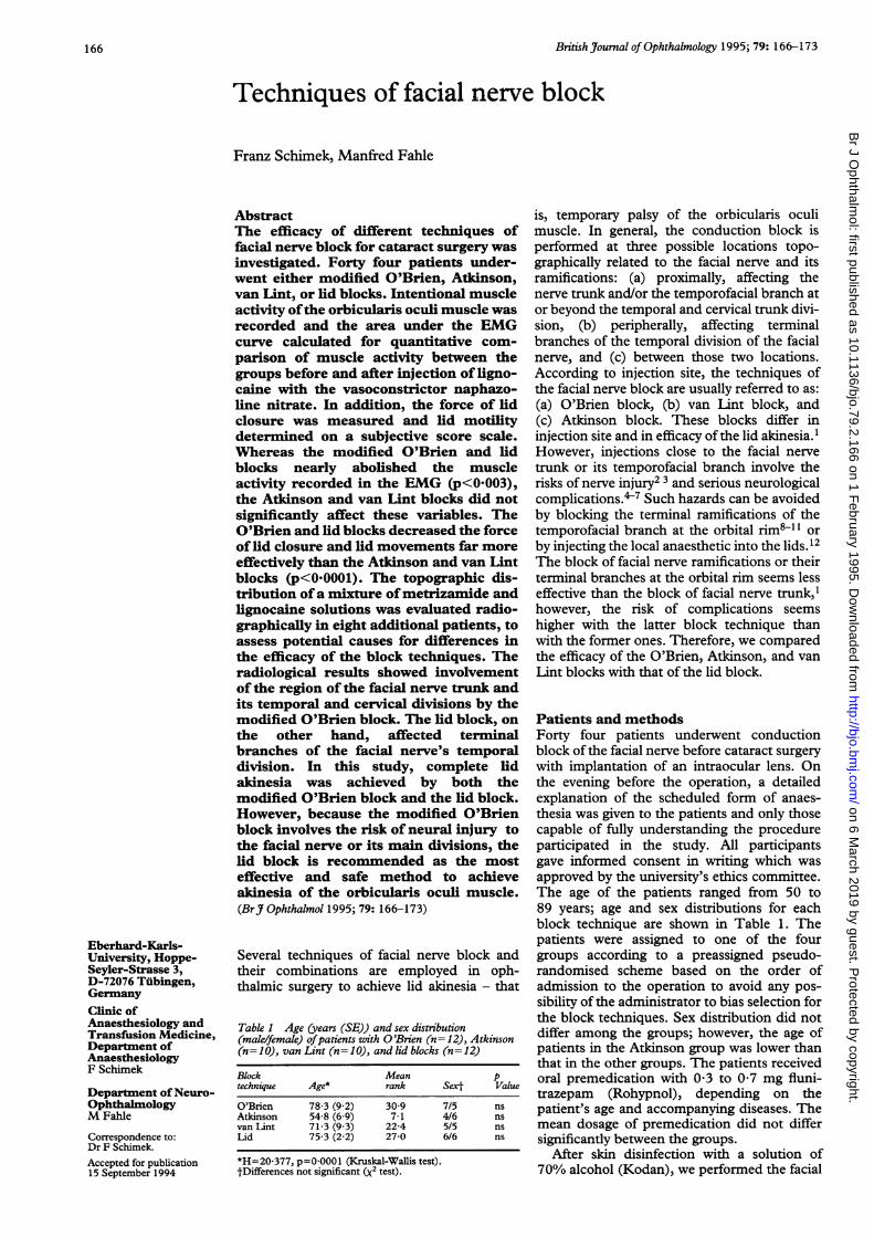
Figure 2 The evaluated area (shaded) under arepresentative EMG curve (replotted). This area representsthe integral of the changes in electrical activity (voltage,,u F7 over time (t, ms) during an intentional contraction ofthe orbicularis oculi muscle.
recorded voluntary activity of the orbicularismuscle in each patient before, 1, 3, 5, and 10minutes after administration of the block. TheEMG curves, plotted on paper, were fed via adigitising tablet into a digital computer (MOP;Kontron, Germany) calculating the area underthe curves (Fig 2). EMG integration wasemployed previously in evaluations of neuro-muscular block23 and studies comparing intra-muscular electric activity and tension of themuscle24; it provides a quantitative measure ofexcitation level in a muscle.25 The needleEMG is an important tool in assessing peri-pheral nerve injuries and the functionalintegrity of a nerve plexus, in determining theextent and severity of a plexus lesion.26A Muller blepharometer27 measured the
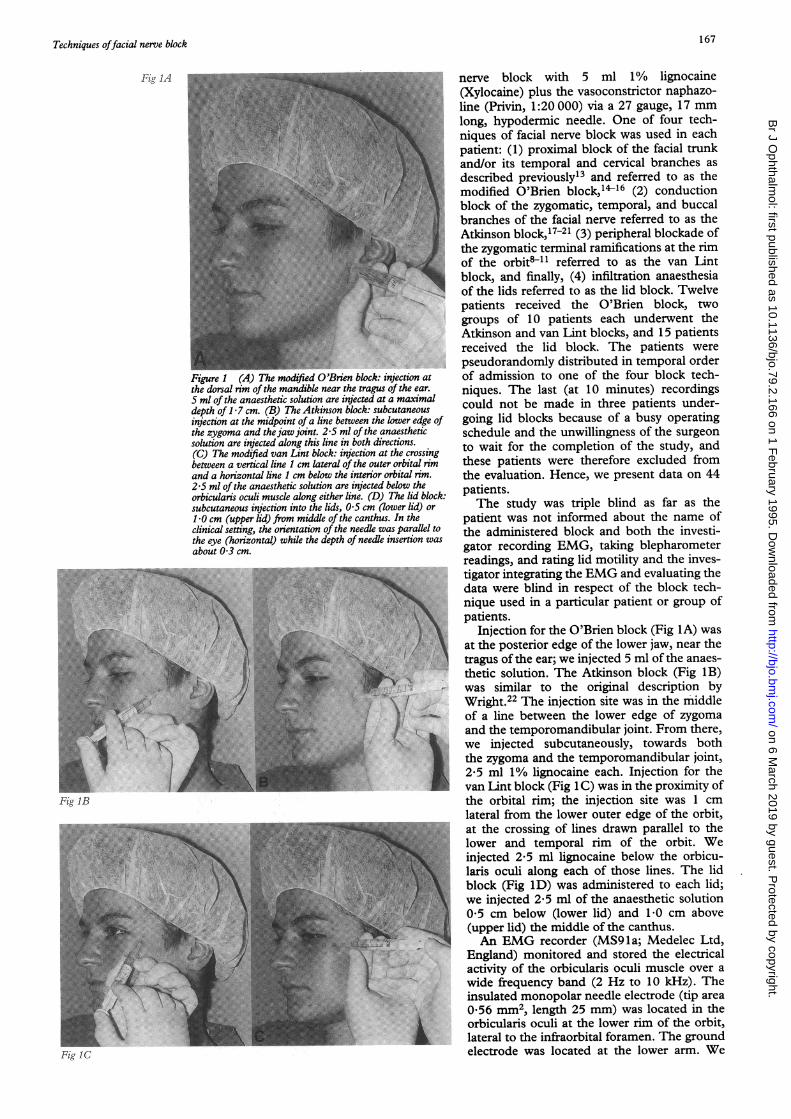
force of lid closure. The instrument has a scalewhich corresponds to a force between 68 and190 pond (Fig 3). The pond, a conventionalunit of force, equals 9-806X 1O-3 newton.28 If
the force of lid closure was below this scale,0 pond was attributed to patients who wereunable to move the lids, since these patientswere obviously unable to close the eyes againstany force. Patients who could move the lids butexerted a force below the measurable rangewere treated as if they had reached a force of68 pond. This was the case in one patientreceiving O'Brien block, who could close theeye 5 minutes after the block but was assignedthe force of 68 pond. We assessed lid move-ment and closure on a subjective scale rangingfrom 1 to 3. Here, '1' corresponds to completeparalysis of the lids (no lid movement), '2'to impaired motility (movement but no lidclosure), and '3' to complete lid closure.The distribution of 2 ml metrizamide
(Solutrast), mixed with 3 ml 1/% lignocainesolution, was investigated in two additionalpatients for each of the O'Brien, Atkinson, vanLint, and lid blocks. Though the diffusion oflignocaine and metrizamide in tissue may differover time, the deposition of such a mixture inseveral locations of the face permits radio-logical correlation of the initial spread of themixture (independent of diffusion) at therespective location with the topography of thefacial nerve and its branches at this location.Therefore, we took x ray pictures within 1minute after the injection of the solution.The Kruskal-Wallis test and one and two
factor analysis of variance (ANOVA) served tocompare the results. The Kruskal-Wallis testcompared the age distribution between groupsof either block technique (Table 1). The sexdistribution was analysed by the x2 test andcontingency table analysis which showed nosex differences among the groups (p=024,Pearson). The force of lid closure was esti-mated on a subjective scale. We treated theresulting values as pseudocontinuous in thestatistical analysis.
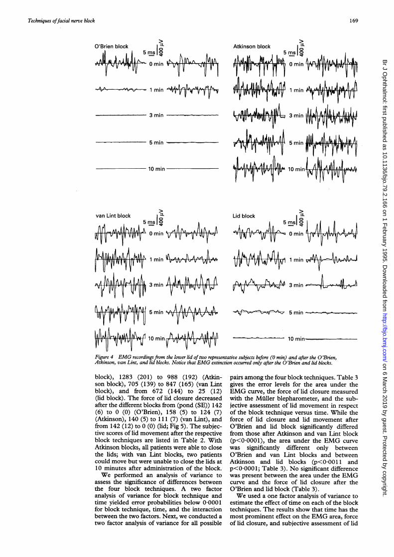
ResultsWe achieved an effective akinesia of lids withthe modified O'Brien and lid blocks. Figure 4shows representative EMG recordings fromthe orbicularis oculi muscle before and aftereach block technique from two differentpatients. The area under the EMG curvesdecreased within 10 minutes after injectionfrom (mm2 (SE)) 725 (132) to 8 (6) (O'Brien
Figure 3 The Miller blepharometer before (left) and during contraction of the orbicularis oculi (right). Before theplacement of the blepharometer specula the conjunctiva was anaesthetised withfive drops of oxybuprocaine 0 45%(Benoxinate SE Thilo). Notice also the EMG electrode inserted into the lower portion of the orbicularis oculi muscle.
Schimek, Fahle
on 6 March 2019 by guest. P
rotected by copyright.http://bjo.bm
j.com/
Br J O
phthalmol: first published as 10.1136/bjo.79.2.166 on 1 F
ebruary 1995. Dow
nloaded from
Techniques offacial nerve block
Atkinson block
.Al I A,_ 1 AK .^AWA ..MIAAV v vv V0IOmin v _g -V-VT
&V 1mmh-A4--- ,v
1 min vy-V t-
3 min
5 min
10 min
0 mnn VVV
1 m*m
3 min1VV W-Wm
5 min V
10 minMm-A
=L Lid block lams0mm V 54s0
1 min
3 minm A A A A.4 -V1A AM A
1 min
hA,AsA.^^ AAA AA k Aw8llrT 115 min VW fV WYVv-s
PIALAAA II.t^ 10 .Ahkm A AA tPv IJVW''v11 ',IJ1 min V vUv
-1 -V L/v NoV'
%e--v - 15 min~~~~~10 min-
Figure 4 EMG recordings from the lower lid of two representative subjects before (O min) and after the O'Brien,Atkinson, van Lint, and lid blocks. Notice that EMG extinction occurred only after the O'Brien and lid blocks.
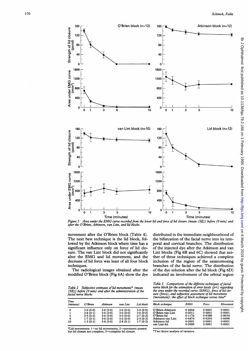
block), 1283 (201) to 988 (192) (Atkin-son block), 705 (139) to 847 (165) (van Lintblock), and from 672 (144) to 25 (12)(lid block). The force of lid closure decreasedafter the different blocks from (pond (SE)) 142(6) to 0 (0) (O'Brien), 158 (5) to 124 (7)(Atkinson), 140 (5) to 111 (7) (van Lint), andfrom 142 (12) to 0 (0) (lid; Fig 5). The subjec-tive scores of lid movement after the respectiveblock techniques are listed in Table 2. WithAtkinson blocks, all patients were able to closethe lids; with van Lint blocks, two patientscould move but were unable to close the lids at10 minutes after administration of the block.We performed an analysis of variance to
assess the significance of differences betweenthe four block techniques. A two factoranalysis of variance for block technique andtime yielded error probabilities below 0 0001for block technique, time, and the interactionbetween the two factors. Next, we conducted atwo factor analysis of variance for all possible
pairs among the four block techniques. Table 3gives the error levels for the area under theEMG curve, the force of lid closure measuredwith the Miller blepharometer, and the sub-jective assessment of lid movement in respectof the block technique versus time. While theforce of lid closure and lid movement afterO'Brien and lid block significantly differedfrom those after Atkinson and van Lint block(p<00001), the area under the EMG curvewas significantly different only betweenO'Brien and van Lint blocks and betweenAtkinson and lid blocks (p<0-0011 andp<0-0001; Table 3). No significant differencewas present between the area under the EMGcurve and the force of lid closure after theO'Brien and lid block (Table 3).We used a one factor analysis of variance to
estimate the effect of time on each of the blocktechniques. The results show that time has themost prominent effect on the EMG area, forceof lid closure, and subjective assessment of lid
3 min
169
A. AN N A &Av -. - -v-w t- VW -
on 6 March 2019 by guest. P
rotected by copyright.http://bjo.bm
j.com/
Br J O
phthalmol: first published as 10.1136/bjo.79.2.166 on 1 F
ebruary 1995. Dow
nloaded from
Schimek, Fahle
O'Brien block (n=12) 160
120
- < Atkinson block (n=12)
80K
40 _
0
1600 r
1200
800
400 _-
0 1 3 -5v
lo o 1 3 5
160 .
120
- van Lint block (n=10)
rI80 _
40
0
1600 r
1200
w E 800
: 400a)
160
120
80
40
0
Lid block (n=12)
_
1600 _
1200 _-
80
I I
0 1 3 5
40noaoIIK
10 0 1 3 5 10
Time (minutes) Time (minutes)Figure 5 Area under the EMG curve recordedfrom the lower lid andforce of lid closure (mean (SE)) before (0 min) andafter the O'Brien, Atkinson, van Lint, and lid blocks.
movement after the O'Brien block (Table 4).The next best technique is the lid block, fol-lowed by the Atkinson block where time has a
significant influence only on force of lid clo-sure. The van Lint block did not significantlyalter the EMG and lid movement, and thedecrease of lid force was least of all four blocktechniques.The radiological images obtained after the
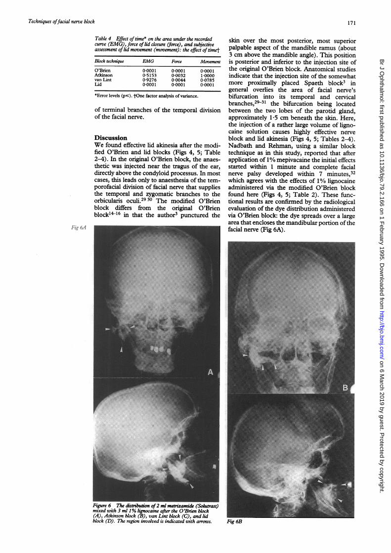
modified O'Brien block (Fig 6A) show the dye
Table 2 Subjective estimates of lid movements* (mean(SE)) before (0 min) and after the administration of thefacial nerve blocks
Time(minutes) O'Brien Atkinson van Lint Lid block
0 30 (00) 30 (00) 30 (00) 3.0 (00)1 2-8 (01) 30 (00) 30 (00) 30 (00)3 2-5 (0 2) 3 0 (0 0) 3 0 (0 0) 2-7 (0 2)5 1-7 (0 1) 3 0 (0 0) 3 0 (0 0) 2-3 (0 2)10 1-3 (0 1) 30 (00) 2-8 (0 1) 1-7 (02)
*Lid movements: 1 =no lid movements, 2=movement presentbut lid closure not complete, 3=complete lid closure.
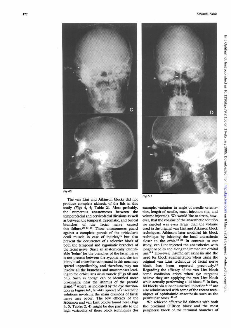
distributed in the immediate neighbourhood ofthe bifurcation of the facial nerve into its tem-poral and cervical branches. The distributionof the injected dye after the Atkinson and vanLint blocks (Fig 6B and 6C) showed that nei-ther of these techniques achieved a completeinclusion of the region of the anastomosingbranches of the facial nerve. The distributionof the dye solution after the lid block (Fig 6D)indicated an involvement of the orbital region
Table 3 Comparisons of the different techniques offacialnerve block for the estimation of error levels (pG) regardingthe area under the recorded curve (EMG), force of lid clo-sure (force), and subjective assessment of lid movement(movement): the effect of block technique versus time*
Block technique EMG Force Movement
O'Brien-Atkinson 0-4664 0 0001 0 0001O'Brien-van Lint 0 0011 0 0001 0 0001O'Brien-lid 0 1176 0-9388 0 0034Atkinson-van Lint 0 4474 0-9294 0-0698Atkinson-lid 0 0001 0 0001 0 0001van Lint-lid 0-0989 0 0001 0 0001
*Two factor analysis of variance.
160 ra)
C0
-0-3c
Q
.Ca)c
n)
120
80
40
0
K1600 r(D
' 1200
CDX Ef 80010 E
CUm 400ax
<
1
0
-
Ca
0,
4- 0a)
(D~
10
170
0
k
on 6 March 2019 by guest. P
rotected by copyright.http://bjo.bm
j.com/
Br J O
phthalmol: first published as 10.1136/bjo.79.2.166 on 1 F
ebruary 1995. Dow
nloaded from
Techniques offacial nerve block
Table 4 Effect of time* on the area under the recordedcurve (EMG), force oflid closure (force), and subjectiveassessment of lid movement (movement): the effect of timet
Block technique EMG Force Movement
O'Brien 0 0001 0 0001 0 0001Atkinson 0-5153 0-0032 10000van Lint 0-9276 010044 0-0785Iid 0 0001 0 0001 0 0001
*Error levels (psV). tOne factor analysis of variance.
of terminal branches of the temporal divisionof the facial nerve.
DiscussionWe found effective lid akinesia after the modi-fied O'Brien and lid blocks (Figs 4, 5; Table2-4). In the original O'Brien block, the anaes-thetic was injected near the tragus of the ear,directly above the condyloid processus. In mostcases, this leads only to anaesthesia of the tem-porofacial division of facial nerve that suppliesthe temporal and zygomatic branches to theorbicularis oculi.29 30 The modified O'Brienblock differs from the original O'Brienblock1416 in that the author3 punctured the
Fig 6A
skin over the most posterior, most superiorpalpable aspect of the mandible ramus (about3 cm above the mandible angle). This positionis posterior and inferior to the injection site ofthe original O'Brien block. Anatomical studiesindicate that the injection site of the somewhatmore proximally placed Spaeth block3 ingeneral overlies the area of facial nerve'sbifurcation into its temporal and cervicalbranches,2931 the bifurcation being locatedbetween the two lobes of the parotid gland,approximately 1-5 cm beneath the skin. Here,the injection of a rather large volume of ligno-caine solution causes highly effective nerveblock and lid akinesia (Figs 4, 5; Tables 2-4).Nadbath and Rehman, using a similar blocktechnique as in this study, reported that afterapplication of 1% mepivacaine the initial effectsstarted within 1 minute and complete facialnerve palsy developed within 7 minutes,32which agrees with the effects of 1% lignocaineadministered via the modified O'Brien blockfound here (Figs 4, 5; Table 2). These func-tional results are confirmed by the radiologicalevaluation of the dye distribution administeredvia O'Brien block: the dye spreads over a largearea that encloses the mandibular portion ofthefacial nerve (Fig 6A).
Figure 6 The disbtion of2 ml metrizamide (Soutrast)mixed with 3 ml 1% lignocaine after the O'Brien block(A), Atkinson block (B), van Lint block (C), and lidblock (D). The region involved is indicated with arrows. Fig 6B
171
on 6 March 2019 by guest. P
rotected by copyright.http://bjo.bm
j.com/
Br J O
phthalmol: first published as 10.1136/bjo.79.2.166 on 1 F
ebruary 1995. Dow
nloaded from
Schimek, Fahle
The van Lint and Atkinson blocks did notproduce complete akinesia of the lids in thisstudy (Figs 4, 5; Table 2). Most probably,the numerous anastomoses between thetemporofacial and cervicofacial divisions as wellas between the temporal, zygomatic, and buccalbranches of the facial nerve causedthis failure.29 33-35 These anastomoses guardagainst a complete paresis of the orbicularisoculi muscle in case of injuries,36 but alsoprevent the occurrence of a selective block ofboth the temporal and zygomatic branches ofthe facial nerve. Since an anatomically identifi-able 'lodge' for the branches of the facial nerveis not present between the zygoma and the jawjoint, local anaesthetics injected in this area mayspread unpredictably, and therefore, may notinvolve all the branches and anastomoses lead-ing to the orbicularis oculi muscle (Figs 6B and6C). Such as 'lodge' can be identified moreproximally, near the isthmus of the parotidgland,37 where, as indicated by the dye distribu-tion in Figure 6A, fan-like spread of anaestheticsolutions involving the main divisions of facialnerve may occur. The low efficacy of theAtkinson and van Lint blocks found here (Figs4, 5; Tables 2, 4) might be due partially to thehigh variability of these block techniques (for
rig OL
example, variation in angle of needle orienta-tion, length of needle, exact injection site, andvolume injected). We would like to stress, how-ever, that the volume of the anaesthetic solutionwe injected was even larger than the volumeused in the original van Lint and Atkinson blocktechniques. Atkinson later modified his blocktechnique by injecting the local anaestheticcloser to the orbit.18-21 In contrast to ourstudy, van Lint injected the anaesthetics withlonger needles and along the immediate orbitalrim.89 However, insufficient akinesia and theneed for block augmentation when using theoriginal van Lint technique of facial nerveblock has been reported previously.38Regarding the efficacy of the van Lint blocksome confusion arises when eye surgeonsbelieve they are applying the van Lint blockwhile actually performing a lid block.39 In fact,lid blocks via subconjunctival injection40 41 arealso administered with some of the recent tech-niques of ophthalmic anaesthesia such as theperibulbar block.42 43We achieved effective lid akinesia with both
the proximal O'Brien block and the mostperipheral block of the terminal branches of
172
on 6 March 2019 by guest. P
rotected by copyright.http://bjo.bm
j.com/
Br J O
phthalmol: first published as 10.1136/bjo.79.2.166 on 1 F
ebruary 1995. Dow
nloaded from
Techniques offacial nerve block
the facial nerve - that is, the lid block (Figs 4,5; Tables 2, 4). Thus, our results agree withprevious reports,12 44 as well as with the recentfindings,45-47 that lid block produces effectiveakinesia of the orbicularis oculi muscle. Theeffects of the lid and O'Brien block on theEMG, the force of lid closure, and lid move-
ments did not differ from each other. Thesevariables were also very similar to each otherafter the Atkinson and van Lint blockswhich were not effective in producing akinesia(Table 3). But overall, the Atkinson blockshowed little or no effect on the EMG and theforce of lid closure or lid movement, respec-tively (Figs 4, 5; Table 4). The age differencebetween the patients in the Atkinson groupand the other groups (Table 1) may be impor-tant since the numbers of fibres in the facialnerve decrease with age,48 and the mimicmustulature rarefies. These facts couldaccount for the larger area under the EMGcurve that we found, and for the larger force oflid closure before block in this group comparedwith the patients with higher mean age(Table 1). However, Jacobs found no differ-ences in the force of lid closure between theyoung and the old, rather a tendency towardshigher force values in male subjects.49 Thestatistics used in the present study - that is, thetwo factor ANOVA for the effects of blocktechnique versus time, eliminate all baselinedifferences between the groups since they are
specific for all the changes over time in eachgroup.The present study shows that the lid block is
as effective as the proximal blocks of the facialnerve, without bearing the risks of direct nerve
trauma2 3 with the destruction of nervefibres50-52 or other neurological complica-tions.4-7 We therefore prefer the administra-tion of the lid block, if the akinesia oforbicularis oculi is required and the safety ofthe patient is of primary concern.
Presented in part at the Annual Meeting of the Association forResearch in Vision and Ophthalmology (ARVO) at Sarasota,Florida, 1992, USA.
1 Schimek F, Steuhl KP, Fahle M, Thiel HJ. DieLindakinesie nach unterschiedlichen Techniken derFacialisblockade. Fortschr Ophthalmol 1990; 87: 696-702.
2 Wright RE. Blocking of the main trunk of the facial nerve incataract operations based on experience of over 150 cases.Arch Ophthalmol 1926; 55: 555-9.
3 Spaeth GL. Total facial nerve palsy following modifiedO'Brien facial nerve block. Ophthalmic Surg 1987; 7:105-9.
4 Wilson ChA, Ruiz RS. Respiratory obstruction followingthe Nadbath facial nerve block. Arch Ophthalmol 1985;103: 1454-6.
5 Shoch D. Complication of the Nadbath facial nerve block.Arch Ophthalmol 1986; 104: 1114-5.
6 Rabinowitz L, Livingston M, Schneider H, Hall A.Respiratory obstruction following the Nadbath facialnerve block. Arch Ophthalmol 1986; 104: 1115.
7 Coper HF. Cord paralysis after Nadbath facial nerve block.Arch Ophthalmol 1986; 104: 337.
8 van Lint H. Paralysie palpebrale temporaire provoquee dansl'operation de la cataracte. Ann Ocul 1914; 151:420-4.
9 van Lint H. II faut toujours paralyser les paupieres dansl'operation de la cataracte. Bull Soc Belge Ophtalmol 1921;43: 23-8.
10 Villard M. Paralysie temporaire des paupieres dansl'operation de la cataracte. Bull Soc Ophtalmol Fr 1919:177-80.
11 Rochat GF. Akinesie bei Augenoperationen. Kin MonatsblAugenheilkd 1920; 65: 177-81.
12 Rubbrecht M. L'operation de la cataracte. Bull Soc BelgeOphtalmol 1927; 53: 44-53.
13 Speath GL. A new method to achieve complete akinesia ofthe facial muscles ofthe eyelids. Ophthalmic Surg 1976; 7:105-9.
14 O'Brien CS. Local anesthesia in ophthalmic surgery. JAMA1928; 90: 8-13.
15 O'Brien CS. Akinesis during cataract extraction.Arch Ophthalmol 1929; 1: 447-9.
16 O'Brien CS. Local anesthesia. Arch Ophthalmol 1934; 12:240-53.
17 Atkinson WS. Akinesia of the orbicularis. Am J Ophthalmol1953; 36: 1255-8.
18 Atkinson WS. Observations on anesthesia for ocularsurgery. Trans Am Acad Ophthalmol Otolaryngol 1956:376-80.
19 Atkinson WS. The development of ophthalmic anesthesia.Am Y Ophthalmol 1961; 51: 1-14.
20 Atkinson WS. Facial nerve block. Am Y Ophthalmol 1964;57: 144-5.
21 Atkinson WS. Akinesia of the orbicularis. In: Atkinson WS,ed. Anesthesia in ophthalmology. 2nd ed. Springfield, IL:Thomas, 1965: 56-63.
22 Wright RE. Blocking of the facial nerve in cataract opera-tions. Am Y Ophthalmol 1921; 4: 445-6.
23 Fink BR. A method for monitoring muscular relaxation bythe integrated abdominal electromyogram. Anesthesiology1960; 21: 178-85.
24 Lippold OCJ. The relation between integrated actionpotentials in a human muscle and its isometric tension.JPhystol (Lond) 1952; 117: 492-9.
25 Lenman JAR. Integration and analysis of the electromyo-gram and related techniques. In: Walton J Sir, ed.Disorders of volumtary muscle. 4th ed. Edinburgh:Livingstone, 1981: 976-1007.
26 Aminoff MJ. Clinical electromyography. In: Aminoff MJ,ed. Electrodiagnosis in clinical neurology. 3rd ed. New York:Livingstone, 1992: 249-81.
27 Muller HK, Droescher J. Ein Lidkraftmesser. Bericht derDeutschen Ophthalmologischen Gesellschaft 1938; 52:438-40.
28 Sykes MK, Vickers MD, Hull CJ. Principles of clinicalmeasurement. 2nd ed. Oxford: Blackwell, 1981:19.
29 Davis RA, Anson BJ, Budinger JM, le Kurth RE. Surgicalanatomy of the facial nerve and parotid gland based upona study of 350 cervicofacial halves. Surg Gynecol Obstet1956; 102: 385-412.
30 Ellis GS. Akinesia of the facial nerve: a laboratory investiga-tion of the surgical anatomy. Trans Am Ophthalmol Soc1968; 66: 746-87.
31 Klein M. Surgical anantomy of the facial nerve with refer-ence to the technique of orbicularis block (palpebralakinesia). Bry Ophthalmol 1946; 30: 668-75.
32 Nadbath RP, Rehman I. Facial nerve block. Am YOphthalmol 1963; 55: 143-6.
33 Tortella EP. Le plexus parotidien du facial. Annalesd'anatomiepathologique 1935; 12: 41-50.
34 McCormack U, Cauldwell EW, Anson BJ. The surgicalanatomy of the facial nerve with special reference to theparotid gland. Surg Gynecol Obstet 1945; 80: 620-30.
35 McKenzie J. The parotid gland in relation to the facialnerve. YAnat 1948; 82: 183-6.
36 Harris WD. Topography of the facial nerve. ArchOtolaryngol 1968; 88: 62-4.
37 Bailey H. The surgical anatomy of the parotid gland.BrMedJY 1948; 2: 245-8.
38 Sanches-Capuchino A, Meadows D, Morgan L. Localanaesthesia for eye surgery without a facial nerve block.Anaesthesia 1993; 48: 428-3 1.
39 Zahl K. Blockade of the orbicularis oculi. OphthalmologyClinics ofNorth America 1990; 3: 93-100.
40 Terson A. Acinesie du droit superieur et de l6rbiculaire parvoie conjunctivale. Ann Oculist 1931; 168: 653-9.
41 Berens C, King JH. An atlas of ophthalmic surgery.Philadelphia: Lippincott, 1961: 22-5.
42 Grizzard WS. Regional ophthalmic anesthesia. In: Gills JP,Hustead RF, Sanders DR, eds. Ophthalmic anesthesia.Thorofare: Slack, 1993: 132-3.
43 Hustead RF. The Wichita technique. In: Gillis JP, HusteadRF, Sanders DR, eds. Ophthalmic anesthesia. Thorofare:Slack, 1993: 141-5.
44 Duverger C, Velter E, Bregeat P. Therapeutique chirurgicaleophthalmologique. 2nd ed. Paris: Masson, 1950: 24-56.
45 Eccarius SC, Gordon ME, Parelman JJ. Bicarbonate-buffered lidocaine-epinephrine-hyaluronidase for eye lidanesthesia. Ophthalmology 1990; 97: 1499-501.
46 Ropo A. Orbicular muscle akinesia: a comparison, usingelectromyography, of three techniques. Ophthalmic Surg1992; 23: 414-7.
47 Sarvela J, Nikki P, Paloheimo M. Orbicular muscle akinesiain regional ophthalmic anaesthesia with pH-adjustedbupivacaine: effects of hyaluronidase and epinephrine.Can J Anaesth 1993; 40: 1028-33.
48 Kullman GL, Dyck PJ, Cody PJ. Anatomy of the mastoidportion of the facial nerve. Arch Otolaryngol 1971; 93:29-33.
49 Jacobs HB. Strength of the orbicularis oculi. Br YOphthalmol 1954; 38: 560-6.
50 Nichols PC, Dyck PJ, Miller DR. Experimental hyper-trophic neuropathy: change in fascicular area and fiberspectrum after acute crush injury. Mayo Clin Proc 1968;43: 297-305.
51 Selander D, Dhuner KG, Lundborg G. Peripheral nerveinjury due to injection needles used for regional anaes-thesia. An experimental study of the acute effects ofneedle point trauma. ActaAnaesth Scand 1977; 21: 182-8.
52 Selander D, Brattsand R, Lundborg G, Nordborg C, OlsonY. Local anesthetics: importance of mode of application,concentration and adrenaline for appearance of nervelesions. Acta Anaesth Scand 1979; 23: 127-36.
173
on 6 March 2019 by guest. P
rotected by copyright.http://bjo.bm
j.com/
Br J O
phthalmol: first published as 10.1136/bjo.79.2.166 on 1 F
ebruary 1995. Dow
nloaded from


![Chapter 173-505 WAClawfilesext.leg.wa.gov/law/WACArchive/2018a/WAC 173... · (8/26/05) [Ch. 173-505 WAC p. 1] Chapter 173-505 Chapter 173-505 WAC INSTREAM RESOURCES PROTECTION AND](https://img.pdfslide.us/doc/110x75/5f3d87ebe97fec5dee3cba18/chapter-173-505-173-82605-ch-173-505-wac-p-1-chapter-173-505-chapter.jpg)