Embed Size (px)
Citation preview
British Journal of Ophthalmology 1996;80:1092-1098
Immunocytochemical study of retinal diode laserphotocoagulation in the rat
P R S Richardson, M E Boulton, J Duvall-Young, D McLeod
AbstractAim-To determine the nature of thecellular infitrate, alterations in cell adhe-sion molecules, and MHC II antigenexpression in the rat retina followingdiode laser retinal photocoagulation.Method-20 normal Lister rats underwentdiode laser photocoagulation ofthe retina.Frozen sections from eyes enucleated at 0,1, 5, 13, and 33 days post laser were exam-ined for T cells (R7.3), CD4 T cells(W3125), activated CD4 T cells (OX-40),CD8 T cells (OX-8), B cells (OX-33), andmacrophages (OX-42), MHC II antigen(OX-6), and E-Selectin-1, VCAM-1, andICAM-1.Results-Retinal diode laser photocoagu-lation stimulated a wound healing re-sponse in the outer retina and choroid.The cellular infiltrate included macro-phages and activated CD4 T cells at 13 and33 days post laser. Glial cells in the innerplexiform and inner nuclear layers ex-pressed MHC II antigen at 24 hours only.ICAM-1 antigen was induced in RPE cellsand in Muller cells in the inner retina at alltime intervals post laser and intensestaining for ICAM-1 was present aroundintraretinal migrated cells at 13 and 33days post laser. VCAM-1 antigen expres-sion was induced in the choroidal vascularendothelium and RPE at 13 and 33 daysafter laser as was E-Selectin-1 antigenexpression which was also evident focallyat the external limiting membrane inassociation with migrated cells adjacent tothe burn.Conclusions-These results suggest thatalterations in cell adhesion molecules mayregulate the migration and activation ofretinal pigment epithelium, macrophagesand CD4 T cells at the outer blood-retinalbarrier and choroid following diode laserphotocoagulation of the normal Lister ratretina.(BrJ Ophthalmol 1996;80:1092-1098)
Retinal laser photocoagulation is generally suc-cessful in the treatment of retinal breaks,preventing retinal detachment by sealing thebreak against the flow of fluid vitreous acrossthe neural retina. It is also effective in the man-agement of vasoproliferative retinopathiesincluding proliferative diabetic retinopathy,ischaemic central or branch retinal vein occlu-sion, and retinopathy of prematurity. However,the mechanisms by which scatter laser photo-
coagulation induces regression of the newblood vessels are not fully understood.
Histological studies have demonstrated thatargon laser photocoagulation of the retina pri-marily destroys the retinal pigment epithelium(RPE) and adjacent outer retina while the useof longer wavelengths (for example, red kryp-ton and some diode lasers) results in additionaldestruction in the choroid. '- The first stage ofthe 'repair' process after laser is the arrival ofsubretinal macrophages and leucocytes in andbelow the wound area.'l0 Within 48 hours postlaser, some RPE cells detach from Bruch'smembrane and enter the wound site while oth-ers flatten, migrate along Bruch's membrane,and re-establish the outer blood-retinal bar-rier.'0 Finally, glial cells proliferate and migrateinto the wound site forming a cellular (non-fibrous) outer retinal scar within 21 days postlaser.58-10
Cell adhesion molecules and integrins areimportant sites of cell to cell and cell to matrixcommunication, mediating cell adhesion andsignal transduction."..." During inflammationthey allow leucocytes to interact with endothe-lial cells to regulate adherence and migrationinto the tissue"" and, together with MHC IIantigens and modified proteins, are necessaryfor antigen presenting cells (APC) to activate Tcells.'3 '9 Dendritic cells, macrophages, and Blymphocytes are most able to achieve T cellactivation and are often called professionalAPCs. The inflammatory cells release growthfactors, lymphokines, and extracellular matrixcomponents which modify the behaviour andfunction of various cells, including retinal cap-illary endothelial cells, RPE, and glial cells invitro.2026
Inflammatory retinal infiltration after laserphotocoagulation has been poorly character-ised and the role of inflammatory cells in therepair processes is unclear. The aim of thisstudy was to determine the nature and tempo-ral profile of the inflammatory cell infiltrate,together with changes in MHC II antigen andcell adhesion molecule expression, in thenormal rat retina following laser photocoagula-tion.
Materials and methodsANIMALTwenty adult Lister rats (200-250 g) weremaintained ad libitum in a 12:12 hourlight:dark cycle. No Home Office regulationregarding animal experimentation was trans-gressed. Lister rats were chosen because (a)they have a pigmented retina and choroid able
UniversityDepartment ofOphthalmology,Manchester University,Manchester M13 9PTP R S RichardsonD McLeod
School of BiologicalSciences, ManchesterUniversity, ManchesterM13 9PTM E Boulton
Department ofOphthalmology,Walton Hospital,Liverpool L9 8XE3J Duvall-Young
Correspondence to:Mr P Richardson, UniversityDepartment ofOphthalmology andOrthoptics, RoyalHallamshire Hospital,Glossop Road,Sheffield S10 2JT.
Accepted for publication16 September 1996
1092
on 28 June 2018 by guest. Protected by copyright.
http://bjo.bmj.com
/B
r J Ophthalm
ol: first published as 10.1136/bjo.80.12.1092 on 1 Decem
ber 1996. Dow
nloaded from
Immunocytochemical study of retinal diode laser photocoagulation in the rat
Table 1 Details of the primary antibodies
Antigen Antibody Dilution Species Source
T cell (rat) R7.3 1/100 Mouse SerotecCD4 T cell (rat) W3/25 1/100 Mouse SerotecActivated CD4 T cell (rat) OX-40 Neat Mouse SerotecCD8 T cell (rat) OX-8 1/100 Mouse SerotecB cell (rat) OX-33 1/100 Mouse SerotecMacrophage (rat) OX-42 1/100 Mouse SerotecMHC II (rat) OX-6 1/100 Mouse SerotecICAM-1 (human) 1/75 Mouse Cambridge ResVCAM-1 (human) 1/75 Mouse Cambridge ResE-Selectin-1 (human) 1/75 Mouse Cambridge ResGFAP (bovine) 1/100 Rabbit Dako
to take up the laser radiation, (b) the availabil-ity of well characterised antibodies to ratimmunocompetent cells, and (c) the retina isvascularised by radial vessels on the surface ofthe internal limiting membrane with capillariesin the inner plexiform layer.
PHOTOCOAGULATIONFollowing halothane anaesthesia via a Boyle'smachine, the rat's head was placed on a modi-fied slit-lamp (Topcon SL 3 E, courtesy ofKeeler, London) and 500-800 scattered appli-cations of laser energy of spot size 50 gm,duration 50 ms, and using power settings tocreate 'moderate' lesions,8 were undertaken inthe retina of the right eye using a Microlasediode laser (Keeler, London). Four animalswere sacrificed at each time interval (0, 1, 5,
Table 2 Number of infiltrating activated CD4 Tlymphocytes (OX-40), macrophages(OX-42), and cells with MHC II antigens (OX-6) per SO,um spot size laser burnimmediately post laser (t=0) and at 1, 5, 13, amd 33 days post laser
Within laser burn Around laser burn
t=O 1 5 13 33 t=O 1 5 13 33
OX-40:Choroid - + + ++ ++ - - - -
RPE - + + ++ + - - - - -
PR - - - + - - - - - -
ELM - + - + - - + - - -
ONL - _ _ _ _ _ _OPL - _ _ _ _ _ _ _INL - -IPL - - - - - -
RGC - - - - - - - - - -
NFL - _ILM - -
OX-42:Choroid - ++ ++ ++ ++ - - + ++PR - ++ ++ + + - - + + +ELM - ++ ++ ++ + - - ++ + +ONL - ++ ++ + + - - + + +OPL - + + - + - - - + +INL - + - - - - - + +IPL - - - - + - - -
RGC - - - - - - - - - -
NFL - --ILM - --
OX-6:Choroid - ++ ++ ++ ++ - + ++ ++ +Sub-RPE - ++ ++ ++ + - + ++ + +RPE - + ++ ++ + - + + + -PR - - - ++ + - - + + -
ELM - - - + + + _ONL - - - + - _ _ _OPL - - - - - - _ _ _ _INL - + - + + - + - - -IPL - + - - - _ _ _RGC - - - - - - - - - -
NFL - _ILM - - - -
Number of cells: - = nil; + = 1 to 3 cells; ++ = 4 to 6 cells; +++ = > 7.PR = photoreceptor layer; ELM = external limiting membrane; ONL = outer nuclear layer; OPL= outer plexiform layer; INL = inner nuclear layer; IPL = inner plexiform layer; RGC = retinalganglion cell layer; NFL = nerve fibre layer; ELM = inner limiting membrane.
13, and 33 days) post laser and the eyes wereimmediately enucleated.
PREPARATION OF TISSUEThe enucleated eyes were embedded in OCT(BDH), immersed in liquid nitrogen cooledisopropane for 6-7 minutes then stored at-80°C. Frozen sections (4 to 7 ptm thickness)were cut on a Reichart-Jung 7400 cryostat andcollected on poly-l-lysine coated slides(Sigma), allowed to dry at room temperature,wrapped in Clingfilm, and stored at -20°C.
ANTIBODIESThe antibodies used in this study wereobtained from a variety of suppliers (Table 1)and, where necessary, diluted in 0.01 M TRISbuffered saline (TBS) pH 7.2 containing 1%bovine serum albumin. The R7.3 antibodyrecognises rat T lymphocytes, the w3/25antibody recognises the CD4 homologue in ratlymphocytes,27 and the OX-40 antibody identi-fies activated CD4 T cells.28 The antibodies forintercellular cell adhesion molecule-i (ICAM-1), vascular cell adhesion molecule-1 (VCAM-1), and E-Selectin-1 were raised against humanantigens but there is cross-reactivity with ratantigens. Rabbit antimouse IgG conjugated tobiotin (Sigma) was used at a dilution of 1/200for the mouse primaries, and mouse antirabbitconjugated to biotin (Sigma) at a dilution of1/150 was used for the rabbit primary anti-body.
IMMUNOSTAININGThe sections were fixed in acetone at -20°Cfor 5 minutes, allowed to air dry then rinsed in0.01 M TBS pH 7.2 for 5 minutes. Normalrabbit (or mouse) serum (1:10 Sigma) wasused for 10 minutes to block non-specificbinding before applying the primary antibodiesfor 1 hour. After three 5 minute washes in TBSthe samples were exposed to the biotinylatedsecondary antibody for 30 minutes. Thesections were washed three times for 5 minutesin TBS and incubated with an avidin-biotinalkaline phosphatase reaction complex (Dako)for 30 minutes. After further washing thetissues were made alkaline by immersing in 0.1M veronal acetate buffer (pH 9.2) for 5minutes and antibody location was determinedusing a naphthol phosphate/fast red substratemixture (Sigma) which, after 40 minutes,resulted in a red product. Endogenous alkalinephosphatase activity was blocked with levami-sole. The washed slides were dried andmounted in UV adhesive (Loctite).
Positive control tissues were rat spleen andliver and negative controls were (i) omission ofthe primary antibody, and (ii) substitution ofthe primary antibody with an affinity purifiedmouse IgG at the same concentration as theprimary antibody.
ASSESSMENT OF IMMUNOSTAININGThe number of cells as identified by RT.7,OX-40, OX-6, and OX-42 antibodies werecounted in three or four 50 gm laser burns inthree or four different animals at each time
1093
on 28 June 2018 by guest. Protected by copyright.
http://bjo.bmj.com
/B
r J Ophthalm
ol: first published as 10.1136/bjo.80.12.1092 on 1 Decem
ber 1996. Dow
nloaded from
Richardson, Boulton, Duvall-Young, McLeod
Table 3 Staining intensity of choroid and retina for VCAM-1, ICAM-l, andE-Selectin-1 adhesion molecules immediately post laser (t=O) and at 1, 5, 13, and 33 dayspost laser
Within laser burn Around laser burn
t=O 1 5 13 33 t0 1 5 13 33
VCAM-1:Choriocapillaris - - ++ +++ +++ - - + ++ +RPE - - ++ ++ ++ - - - - -
PR - - - - - - - - - -
ELM - _ _ _ _ONL - - - - - - - - - -
OPL - - - - - - - - - -
INL - ---- -
IPL - - - - - - - - - -
RGC - - - - - - - - - -
NFLILM - -
ICAM-1:Choriocapillaris - ++ +++ +++ +++ - ++ ++ ++ ++RPE - +++ +++ +++ +++ - ++ ++ ++ ++PR - - - - - - - - - -
ELM - ++ +++ +++ +++ + ++ ++ ++ +ONL - - ++ ++ ++ - - + + -OPL - _ _ _INL - - - - - -IPL - - - - - - - - - -
RGC - - + + + - - - -
NFL - -
ILM - ++ ++ + _ _ _E-Selectin-1:
Choriocapillaris - ++ +++ +++ +++ - - ++ ++ +RPE - ++ +++ +++ +++ - - ++ ++ +PR - - - - - - - - - -
ELM - ++ +++ +++ +++ - ++ ++ ++ ++ONL - ++ ++ ++ +++ - ++ ++ ++ +OPL - - - - - - - - - -
INL - -IPL - - - - - - - - - -
RGC - - - - - - - - - -
NFLILM - ---- -
Intensity score:-= no stain; + = 1/3 contol tissue stain; ++ = 2/3 control tissue stain; +++ =equal to control tissue stain.PR = photoreceptor layer; ELM = external limiting membrane; ONL = outer nuclear layer; OPL= outer plexiform layer; INL = inner nuclear layer; IPL = inner plexiform layer; RGC = retinalganglion cell layer; NFL = nerve fibre layer; ILM = inner limiting membrane.
interval. Average counts of each cell type arepresented in Table 2.The intensity of the staining for ICAM-1,
VCAM-1 and E-Selectin-l in the laseredsections was compared with that of the controltissue and graded as one third, two thirds, or ofequal intensity.
Rat spleen and liver were used as control tis-sues for the adhesion molecule antibodies andthe specimens were viewed under an OlympusVannox microscope and photographed withEktachrome T160.
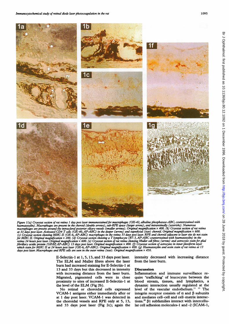
ResultsGENERAL FEATURESWithin 24 hours of diode retinal photocoagula-tion necrosis of the RPE and swelling of thephotoreceptor and outer plexiform layers wasevident with macrophages in the photoreceptorlayer and RPE layer (Fig Ig). At 5 and 13 dayspost laser RPE cells relined Bruch's membranewithin the lesion and pigment bearing cellswere present within the retina. By 33 days theouter retina was no longer swollen and a glialscar had formed. The intensity of the laser wasinsufficient to break Bruch's membrane and nochoroidal new vessels (CNV) were detectedhistologically.
CELLULAR INFILTRATEImmediately following laser (T=O) no T cells,B cells, or macrophages were demonstrated in
either lasered eyes or in non-lasered controleyes (Table 2). At 24 hours post laser OX-42+macrophages were present in the choroid andouter retina within and around the burns (Figla). The macrophages were initially morefrequently identified beneath the laser burns inthe choroid, by 13 days an increasingproportion was found intraretinally.T cell lymphocytes identified by the R7.3
antibody (Fig ld) were found in the choroid,photoreceptor layer, and at the externallimiting membrane (ELM) at all time intervalsother than T=0, with greatest numbers at 24hours. The anti-W3/25 antibody identifiedthese as CD4 T cell lymphocytes and no CD8(OX-8) T lymphocytes were seen. At 24 hoursactivated CD4 T cells (OX-40) were presentbeneath the RPE and in the choroid withincreasing numbers at 13 and 33 days postlaser (Table 2, Fig lb). At no time were B cells(OX-33) detected in the choroid or retina.
MHC II ANTIGEN EXPRESSIONNo retinal or choroidal cells expressed OX-6MHC II antigens before laser. At 24 hours postlaser choroidal endothelial cells around thelaser burn and astrocytes in the inner nuclearlayer around the laser burn stained for OX-6.The astrocytes and Muller cells stained withanti-GFAP antibodies at all intervals post laser(Fig le). Some RPE cells around the laser siteexpressed MHC II at 24 hours post laser andintraretinal pigmented cells were also identifiedby the OX-6 antigen at the inner plexiform andinner nuclear layers.By 5 days post laser MHC II antigen was
present in the choroidal endothelium and insub-RPE macrophages which were in greaternumbers beneath the laser site. The RPE adja-cent to the laser burn also stained for MHC IIantigen, but the intensity rapidly decreasedwith increasing distance away from the burn.The intensity of OX-6 staining in the choroidalendothelium decreased by 33 days post laser.
Increasing numbers of choroidal OX-6 cellswere present beneath the laser burn by 24hours but decreased numbers were found by33 days post laser with occasional OX-6 cells inthe inner and outer plexiform layers at 33 dayspost laser (Fig lc).
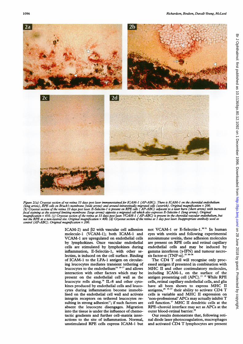
CELL ADHESION ANTIGENSIn the normal rat retina ICAM-1 antigen waspresent at the ELM and was not detected onRPE cells. At 1 and 5 days post laser there wasincreased staining for ICAM-1 in the RPE andchoroidal vessels below the laser burn, in theELM and in the outer nuclear layer (ONL)above the laser burn (Table 3). By 33 daysICAM-1 staining was present in the Mullerfibres which surrounded the migrated intrareti-nal cells in the ONL (Fig 2a). There was alsoincreased staining at 33 days for ICAM-1above the laser burn at the ILM, in the RPEand the choriocapillaris around the laser and atsites away from the lesion.
Before and immediately after laser,E-Selectin-1 antigen could not be identified inthe retina. The RPE and choriocapillarisaround and beneath the laser burn stained for
1094
on 28 June 2018 by guest. Protected by copyright.
http://bjo.bmj.com
/B
r J Ophthalm
ol: first published as 10.1136/bjo.80.12.1092 on 1 Decem
ber 1996. Dow
nloaded from
Immunocytochemical study of retinal diode laserphotocoagulation in the rat
j.iiUfl~I*.@
ElW .. .J.. ...
At'~~~~~~~S& ' si:,.L .,,'M
Figure 1(a) Cryostat section ofrat retina 1 day post laser immunostainedfor macrophages (OX-42, alkaline phosphatase-ABC, counterstained withhaematoxylin). Macrophages are present in the choroid (double arrows), sub-RPE space (larger arrow), and intraretinally (asterisks). Numerousmacrophages are present around the intrascleral posterior ciliary vessels (smaller arrows). Original magnification x 400. (b) Cryostat section of rat retinaat 33 days post laser. Activated CD4 T cells (OX-40,AP-ABC) in the deeper (arrow) and superficial (star) choroid. Original magnification x 600.(c) Cryostat section showingMHC II (OX-6,AP-ABC) macrophages in the retina 33 days post laser. RPE and choroid adjacent to laser site do not stainforMHC II. Original magnification x 200. (d) Cryostat section showing a Tlymphocyte (R7.3,AP-ABC counterstained with haematoxylin) in theretina 24 hours post laser. Original magnification x 600. (e) Cryostat section of rat retina showing Muler cellfibres (arrow) and astrocytes stain for glialfibrillary acidic protein (GFAP1AP-ABC) 13 days post laser. Original magnification x 400. (t) Cryostat section of astrocytes in inner plexiform layerwhich stain forMHC II at 24 hours post laser (OX-6, AP-ABC). Original magnification x 400. (g) Haematoxylin and eosin stain of rat retina at 13days post laser. Macrophages and RPE cells are seen in the outer retina (star). Original magnification x 450.
E-Selectin-1 at 1, 5, 13, and 33 days post laser.The ELM and Muller fibres above the laserburn had increased staining for E-Selectin-1 at13 and 33 days but this decreased in intensitywith increasing distance from the laser burn.Migrated, pigmented cells were in closeproximity to sites of increased E-Selectin-1 atthe level of the ELM (Fig 2b).No retinal or choroidal cells expressed
VCAM-1 antigens either immediately after orat 1 day post laser. VCAM-1 was detected inthe choroidal vessels and RPE only at 5, 13,and 33 days, post laser (Fig 2c); again the
intensity decreased with increasing distancefrom the laser burn.
DiscussionInflammation and immune surveillance re-quire 'trafficking' of leucocytes between theblood stream, tissues, and lymphatics, adynamic interaction usually regulated at thelevel of the vascular endothelium." 13 Theintegrin receptor consists of a and 3 subunitsand mediates cell-cell and cell-matrix interac-tions.29 [1 subfamilies interact with intercellu-lar cell adhesion molecules-i and -2 (ICAM-1,
1095
-s.1! III ".0V
.i.
f^1
*;:;., . I.-d.
VIIf-)ll%.----,_: ....
on 28 June 2018 by guest. Protected by copyright.
http://bjo.bmj.com
/B
r J Ophthalm
ol: first published as 10.1136/bjo.80.12.1092 on 1 Decem
ber 1996. Dow
nloaded from
Richardson, Boulton, Duvall-Young, McLeod
z7 i5- J ''i
.. ...:
.... .k
.z1.s
.4ci; d
^1ob ~~I
. .. .
-w
Figure 2(a) Cryostat section of rat retina 33 days post laser immunostainedfor ICAM-1 (AP-ABC). There is ICAM-1 on the choroidal endothelium(long arrow), RPE cells on Bruch's membrane (wide arrow) and around intraretinally migrated cells (asterisk). Original magnification x 200.(b) Cryostat section of the retina 33 days post laser. E-Selectin-1 is present on RPE cells (AP-ABC) adjacent to a laser burn (short arrow) with increasedfocal staining at the external limiting membrane (large arrow) opposite a migrated cell which also expresses E-Selectin-1 (long arrow). Originalmagnification x 450. (c) Cryostat section of the retina at 33 days post laser. VCAM-1 (AP-ABC) is present in the choroidal vascular endothelium, butnot the RPE at a non-lasered site. Original magnification x 400. (d) Cryostat section of the retina at 1 day post laser. Inappropriate antibody used ascontrol (AP-ABC). Original magnification x 200.
ICAM-2) and 02 with vascular cell adhesionmolecule-i (VCAM-1); both ICAM-1 andVCAM-1 are upregulated on endothelial cellsby lymphokines. Once vascular endothelialcells are stimulated by lymphokines duringinflammation, E-Selectin-1, with other se-lectins, is induced on the cell surface. Bindingof ICAM-1 to the LFA-1 antigen on circulat-ing leucocytes mediates transient tethering ofleucocytes to the endothelium"' -"7 and allowsinteraction with other factors which may bepresent on the endothelial cell wall as theleucocyte rolls along.'6 IL-8 and other cyto-kines produced by endothelial cells and leuco-cytes during inflammation become immobi-lised on the endothelial cell wall and activateintegrin receptors on tethered leucocytes re-sulting in strong adhesion"; if such factors areabsent the leucocyte disengages. Migrationinto the tissue is under the influence of chemo-tactic gradients and further cell-matrix inter-actions to the site of inflammation. Normal,unstimulated RPE cells express ICAM-1 but
not VCAM-1 or E-Selectin-1 3031 In humaneyes with uveitis and following experimentalautoimmune uveitis, these adhesion moleculesare present on RPE cells and retinal capillaryendothelial cells and may be induced bygamma interferon (y-IFN) and tumour necro-sis factor-a (TNF-a) 25 30-34The CD4 T cell will recognise only proc-
essed antigen if presented in combination withMHC II and other costimulatory molecules,including ICAM-1, on the surface of theantigen presenting cell (APC). " '9While RPEcells, retinal capillary endothelial cells, and gliahave all been shown to express MHC IIantigens,25 3 their ability to activate CD4 Tcells is variable and MHC II expression on'non-professional' APCs may actually inhibit Tcell function.37 MHC II dendritic cells at theRPE-choroid interface may act as APCs at theouter blood-retinal barrier."5Our results demonstrate that, following reti-
nal diode laser photocoagulation, macrophagesand activated CD4 T lymphocytes are present
1096
f j
on 28 June 2018 by guest. Protected by copyright.
http://bjo.bmj.com
/B
r J Ophthalm
ol: first published as 10.1136/bjo.80.12.1092 on 1 Decem
ber 1996. Dow
nloaded from
Immunocytochemical study of retinal diode laser photocoagulation in the rat
in the wound during the formation of the cho-rioretinal adhesion. The nature of the inflam-matory infiltrate may be of significance in theproduction of laser scars, since the behaviourof the retinal cells most often involved in thereparative process, the RPE cell and glial cells,can be modified by the released products oftheactivated inflammatory cells.""26 This mayexplain how variations of the power, duration,spot size, and wavelength of the laser, whichdetermine the degree of the laser inducedinjury,8 modify the quality of the wound by dif-ferences in the extent and quality of theinflammatory response. From this immunocy-tochemical study it was not possible todetermine if the activated CD4 T cells in thewound were involved in a specific antigenresponse after activation from an APC.The laser induced inflammation is a focal
wound healing response in which the inflam-matory cells are no longer required once thewound has resolved. It differs in its aetiologyand cellular infiltrate from immunologicallymediated inflammation such as sympatheticophthalmitis, experimental autoimmuneuveitis, and proliferative vitreoretinopathy inwhich CD8 T cells and B cells are alsopresent.""""' In experimental autoimmuneuveitis the CD4 activation is in two stages,42 thefirst by interaction with the CD3/TCR com-plex recognising the peptide/MHC complex.The second signal results from ICAM-1/LFA-1 interaction which completes activationand allows clonal expansion. Monocyte macro-phages are seen at all stages of the inflamma-tion; CD4+ T cells predominate in earlylesions and CD8+ T cells accumulate in laterstages. B cells appear during the repair phasewhen the inflammation is subsiding."6 9The retina is not a passive tissue into which
the inflammatory cells enter during the woundhealing process. Following the laser photo-coagulation, the ability of choroidal vascularendothelial cells, RPE cells, and Muller cells toexpress ICAM-1, VCAM-1, and E-Selectin-Icell surface antigens indicates that they are ableto play an active role in inflammation and mayfunction as regulators of leucocyte adhesion,activation, and migration. The expression ofcell adhesion molecules in the inner retinalvascular endothelium was unchanged com-pared with non-lasered retina, and is consistentwith the laser injury being confined to theouter retina.
Scattered retinal laser photocoagulation resultsin the regression of preretinal new vessel forma-tion by, as yet, incompletely understood mecha-nisms. The presence of macrophages and CD4 Tcells in the laser burn may initially appearparadoxical because of the stimulatory effects ofmany of the lymphokines and growth factorsreleased. However, alterations in retinal andvitreal levels ofgrowth factors, such as transform-ing growth factor 0 have been described follow-ing retinal photocoagulation which may limit theaction ofthe proinflammatory factors.4' 4'This study provides some insight into the
wound healing process in the normal rat retinafollowing diode laser photocoagulation. Fur-ther studies are required to determine the
precise role of the infiltrate in laser inducedregression of neovascularisation.
The authors thank the Lasers for Life Trust, Liverpool, theGuide Dogs for the Blind, and Keeler (London) for their assist-ance in this study.
1 Wallow IH, Sponsel WE, Stevens TS. Clinicopathologicalcorrelation of diode laser burns in monkeys. Arch Ophthal-mol 1991;109:648-53.
2 McHugh DA, Marshall J, fftyche TJ, Hamilton AM, RavenA. Macular photocoagulation ofhuman retina with a diodelaser: a comparative histopathological study. Lasers andLight in Ophthalmol 1990;3:11-28.
3 Marshall J, Bird AC. A comparative histopathological studyofargon and krypton laser irradiations of the human retina.BrJ Ophthalmol 1979;63:657-68.
4 Bresnick GH, Frisch GD, Powell JO, Landers MB, HolstGC, Dallas AG. Ocular effects of argon laser radiation. IRetinal damage threshold studies. Invest Ophthalmol 1970;9:901-10.
5 Marshall J, Hamilton AM, Bird AC. Histopathology ofrubyand argon laser lesions in monkey and human retina. Br JOphthalmol 1975; 59:610-30.
6 Pollack A, Korte GE. Repair of retinal pigment epitheliumand its relationship with capillary endothelium afterkrypton laser photocoagulation. Invest Ophthalmol Vis Sci1990;31:890- 8.
7 Perry DD, Reddick R, Risco JM. Choroidal microvascularrepair after argon laser photocoagulation. Invest OphthalmolVis Sci 1984;25:1019-26.
8 Tso MOM, Wallow IHF, Elgin S. Experimental photo-coagulation of the human retina. fl Correlation of thephysical, clinical and pathological data. Arch Ophthalmol1977;95: 1035-40.
9 Wallow IHF, Tso MOM, Elgin S. Experimental photo-coagulation of the human retina. Arch Ophthalmol 1977;95:1041-50.
10 Wallow IHF. Repair of the pigment epithelial barrier follow-ing photocoagulation. Arch Ophthalmol 1984;102: 126-35.
11 Adams DH, Shaw S. Leucocyte-endothelial interactionsand regulation of leucocyte migration. Lancet 1994;343:831-6.
12 Humphries MJ. The molecular basis and specificity ofintegrin-ligand interactions. J7 Cell Sci 1990;97:585-92.
13 Springer TA. Adhesion receptors of the immune system.Nature 1990;346:425-34.
14 Butcher EC. Cellular and molecular mechanisms that directleukocyte traffic. Am IPathol 1990;136:3-1 1.
15 Belvilacqua MP. Endothelial-leucocyte adhesion molecules.Annu Rev Imunol 1993;11:93-9.
16 Lawrence MB, Springer TA. Leucocytes roll on a selectin atphysiological flow rates: distinction from and prerequesitefor adhesion through integrins. Cell 1991;65:859-73.
17 Lasky LA. Selectins: interpreters of cell-specific carbohy-drates information during inflammation. Science 1992;258:964-9.
18 Shimizu Y, Newman W, Tanka Y, Shaw S. Lymphocyteinteractions with endothelial cells. Immunol Today 1992;13:106-12.
19 Swain SL. T cell subsets and the recognition of MHC class.Immunol Rev 1983;74: 129-42.
20 Boulton ME, Patel B, Khaliq A, Moriarty P, Jarvis-Evans J,McLeod D. Modulators and milieu in preretinal neovascu-larization. Eye 1992;6:560-5.
21 Boulton MB, Lane C, Singh A. Effects of vitreous fromphotocoagulated pig eyes on retinal microvascular cells inculture: a preliminary report. Curr Eye Res 1988;7:465-70.
22 De Vos AF, Hoekzema R, Kijistra A. Cytokines and uveitis,a review. Curr Eye Res 1992;11:581-97.
23 Gilbert C, Hiscott P, Grierson I, McLeod D. Inflammationand the formation of epiretinal membranes. Eye 1988;2:140-56.
24 el-Asar AM, Maimone D, Morse PH, Lascola C, Reder AT.Interferon-gamma and tumour necrosis factor induceexpression of major histocompatability complex antigen onrat retinal astrocytes. BrJ Ophthalmol 1991;75:473-5.
25 Forrester JV, Liversidge J, Dua H S. Regulation of the localimmune response by retinal cells. Curr Eye Res 1990;9(suppl): 183-91.
26 Sunderkotter C, Goebeler M, Schulze-Osthoff K, BhardwajR, Sorg C. Macrophage-derived angiogenesis factors.Pharmacol Ther 1991;51:195-216.
27 Clark SJ, Jeffries WA, Barclay AN, Gagnon J, Williams AF.Peptide and nucleotide sequences of rat CD4 (W3/25)antigen: evidence for derivation from a structure with fourimmunoglobulin-related domains. Proc NadlAcad Sci USA1987;84: 1649-53.
28 Paterson DJ, JefEries WA, Green JR, Brandon MR, CorthesyP, Pulavec M, et al. Antigens of activated rat T lymphocytesincluding a molecule of 50.000 M (r) detected only onCD4 positive T blasts. Mol Immunol 1987;24: 1281-90.
29 Elner SG, Elner VM. The integrin superfamily and the eye.Invest Ophthalmol Vis Sci 1996;37:696-701.
30 Duguid IGM, Boyd AW, Mandel TE. The expresion ofadhesion molecules in the human retina and choroid. AustNZJ' Ophthalmol 1991;19:309-16.
31 Kuppner MC, Liversidge J, McKillop-Smith S, Lumsden L,Forrester JV. Adhesion molecule expression in acute andfibrotic sympathetic ophthalmia. Curr Eye Res 1993;12:923-34.
1097
on 28 June 2018 by guest. Protected by copyright.
http://bjo.bmj.com
/B
r J Ophthalm
ol: first published as 10.1136/bjo.80.12.1092 on 1 Decem
ber 1996. Dow
nloaded from
Richardson, Boulton, Duvall-Young, McLeod
32 Liversidge J, Sewell HF, Forrester JV. Interactions betweenlymphocytes and cells of the blood-retina barrier: mecha-nisms ofT lymphocyte adhesion to human retinal capillaryendothelial cells in vitro. Immunology 1990;71:390-6.
33 Whitcup SM, Chan C-C, Li Q, Nussenblat RB. Expressionof cell adhesion molecules in posterior uveitis. ArchOphthalmol 1992;110:662-6.
34 Forrester JV, Liversidge JM, Dua HS, Dick A, Harper F,McMenamin PG. Experimental autoimmune uveitis: amodel system for immunointervention. Curr Eye Res 1992;11:33-40.
35 Forrester JV, McMenamin PG, Holthouse I, Lumsden L,
Liversidge J. Localization and characterization of majorhistocompatibility complex class II-positive cells in theposterior segment of the eye: implications for induction ofautoimmune uveoretinitis. Invest Ophthalmol Vis Sci1994;35:64-77.
36 Chan C-C, Hooks JJ, Nussenblatt RB, Detrick B. Expres-sion of Ia antigen on retinal pigment epithelium in experi-mental autoimmine uveitis. Curr Eye Res 1986;5:325.
37 Caspi RR, Roberge RG, Nussenblatt RB. Organ-resident,non-lymphoid cells suppress proliferation of autoimmuneT-helper lymphocytes. Science 1987;237:1029-32.
38 Baudouin C, Brignole F, Bayle J, Fredj-Reygrobeller D,Lapalus P, Gastaud P. Class II histocompatability antigenexpression by cellular components of vitreous and sub-retinal fluid in proliferative vitreoretinopathy. Invest Oph-thalmol Vis Sci 1991;32:2065-72.
39 Liversedge J, Forrester JV. Experimental auto-immune uveitis: immunophenotypic analysis of inflamma-tory cells in chorio-retinal lesions. Curr Eye Res 1988;7:1231-41.
40 Peterson JM, Barbul A, Breslin RJ. Significance of Tlymphocytes in wound healing. Surgery 1987;102:300-5.
41 Lightman S, Chan C-C. Immunopathology of ocular inflam-matory disorders. London. Kluwer Academic, 1989.
42 Schwartz RH. A cell culture model for T lymphocyte clonalanergy. Science 1990;248:1349-56.
43 Boulton ME, Xiao M, Khaliq A, Moriarty P, Cranley J,McLeod D. Changes in growth factor expression in pigeyes following scatter laser photocoagulation. Invest Oph-thalmol Vis Sci 1995;36:s95.
44 Matsumoto M, Yoshimura N, Honda Y Increased produc-tion of TGF-[B from cultured human RPE cells byphotocoagulation. Invest Ophthalmol Vis Sci 1994;35:4245-52.
1098
on 28 June 2018 by guest. Protected by copyright.
http://bjo.bmj.com
/B
r J Ophthalm
ol: first published as 10.1136/bjo.80.12.1092 on 1 Decem
ber 1996. Dow
nloaded from