Embed Size (px)
Citation preview

INTRODUCTION
One of the major larval feeding modes of syrphids ispredation. The 1800+ species of Syrphinae make up alarge proportion of the species richness of hoverflies,about 35% of the family. Virtually all of them feed aslarvae on soft-bodied Hemiptera (sensu Sörensen et al.,1995) although some species also attack other arthropods(see Rojo et al., 2003). The species of genera Scaeva Fab-ricius, 1805, Ischiodon Sack, 1913 and Simosyrphus
Bigot, 1882 are predatory syrphids with an important rolein the bio-control of aphid pest in different areas of theworld (e.g. Lal & Haque, 1965; Schmutterer, 1972;Mukhitdinov, 1985; Singh & Mishra, 1988; Sharma &Bhalla, 1991; Xiong & Dong, 1991; Ekukole & Ajayi,1995; Soleyman-Nezhadiyan & Laughlin, 1998). De-scriptions of immature stages of Syrphini are useful forecological studies, e.g. as a method for comparing therole of various natural enemies of aphids (Láska, 1984) orthe study of species relationships, where they provideadditional useful characters for phylogenetic studies,which presently are based mainly on characters of adults.
In 1985 two revisions of the genus Scaeva were pub-lished independently by Dušek & Láska (1985) andKuznetzov (1985). However, these two revisions arequite different in species conceptions and in subgenericclassification. Recent authors usually follow the conceptof Dušek & Láska (e.g. Speight, 2003), but up to now nonomenclatorical acts have been published to clarify thissituation. The defence of Dušek & Láska’s concept(1985) is also part of our contribution with the use ofnewly available data obtained for this study.
According to Kassebeer (1999) and Thompson (2003),19 valid names of the genus Scaeva exist at present.These species of Scaeva are mainly distributed in thePalaearctic (with two species reaching the Oriental regionand one species the Nearctic region) and Neotropical(four species) regions. The genus Ischiodon comprisesthree species only (one endemic of the Cape VerdeIslands, one mainly present in Africa and one in theOriental-Austral region), and of the genus Simosyrphus
(exclusive to the Australian region, including Pacific)only one species is known. These three genera – Scaeva,Ischiodon, Simosyrphus Dušek & Láska (1985) rangedinto Scaeva group.
Eur. J. Entomol. 103: 637–655, 2006ISSN 1210-5759
Taxonomy of the genera Scaeva, Simosyrphus and Ischiodon (Diptera:
Syrphidae): Descriptions of immature stages and status of taxa
PAVEL LÁSKA1, CELESTE PÉREZ-BAÑÓN2, LIBOR MAZÁNEK1, SANTOS ROJO2, GUNILLA STÅHLS3, M. ANGELES
MARCOS-GARCÍA2, VÍT ZSLAV BI ÍK1 and JINDRA DUŠEK4
1Department of Zoology, Natural Sciences Faculty, Palacký University, T . Svobody 26, 771 46 Olomouc, Czech Republic;e-mails: [email protected], [email protected]
2Instituto Universitario de la Biodiversidad (CIBIO), Universidad de Alicante, E-03080 Alicante, Spain; e-mail: celeste.perez@ua. es3Finnish Museum of Natural History, P.O. Box. 17, FIN-00014 University of Helsinki, Finland; e-mail: [email protected]
4Institute of Applied Entomology, University of Agriculture, Zem d lská 1, 613 00 Brno, Czech Republic
Key words. Diptera, Syrphidae, Scaeva, Semiscaeva, Mecoscaeva, Simosyrphus, Ischiodon, taxonomy, immature morphology,chaetotaxy, new combination, new synonymy
Abstract. A review of all known descriptions of immature stages of the species of the genera Scaeva Fabricius, 1805, Ischiodon
Sack, 1913 and Simosyrphus Bigot, 1882 is presented using SEM illustrations. The third instar larval and/or pupal morphology ofScaeva dignota (Rondani, 1857), Scaeva mecogramma (Bigot, 1860) and Simosyrphus grandicornis (Macquart, 1842) are newlydescribed. All species of the genera studied in this paper are very similar for all the studied characters of their immature stages,including the chaetotaxy. Molecular characters of the mitochondrial cox1 gene (1128bp) were used for inferring relationships of thestudied taxa. The nuclear internal transcribed spacer 2 (ITS2) was additionally applied for species delimitation of the closely relatedspecies Scaeva selenitica and S. dignota. The Palaearctic Scaeva species could be split into two groups based on the analysis of mor-phology of posterior respiratory process. These groups were previously diagnosed as S. selenitica-group [i.e., S. selenitica (Meigen,1822), S. dignota (Rondani, 1857), S. mecogramma (Bigot, 1860)] and S. pyrastri-group [i.e., S. pyrastri (Linnaeus, 1758), S.
albomaculata (Macquart, 1842), S. latimaculata (Brunetti, 1923)]. Semiscaeva Kuznetzov, 1985 and Scaeva Fabricius, 1805 are theavailable names for these two natural groups that should be classified as subgenera; the former name is proposed for S. selenitica-group and the latter for S. pyrastri-group. Mecoscaeva Kuznetzov, 1985 syn. n. is transferred as a junior synonym of the subgenusSemiscaeva Kuznetzov, 1985 according to the principle of the first reviser. Based on the analysis of immature stages, the genericname Ischiodon Sack, 1913 syn. n. is proposed as a junior synonym of the genus Simosyrphus Bigot, 1882. The similarity of imma-ture stages between Scaeva s. str. and Simosyrphus grandicornis Macquart, 1842, Simosyrphus aegyptius (Wiedemann, 1830) comb.n. and Simosyrphus scutellaris (Fabricius, 1805) comb. n. is discussed. All the proposed subgeneric and generic taxa based on mor-phological studies received high support employing molecular characters.
637

The single species of Simosyrphus bears a strongresemblance to the species of Ischiodon, with which it hasoften been confused (e.g. Bryan, 1934). In his famousworld revision of the genera of the Syrphini, Vockeroth(1969) was at first inclined to refer all the species of thegenera Ischiodon and Simosyrphus to a single genusdespite the apparent differences in the male terminalia.He finally concluded that both genera are related but notcongeneric. This author also discussed the close relation-ship between Scaeva and Eupeodes Osten Sacken, 1877(including Metasyrphus Matsumura, 1917) but did notsupport a relation between Scaeva and Simosyrphus.Dušek & Láska (1985) first diagnosed these taxaincluding Ischiodon and Simosyrphus as a natural group,using both larval and adult morphology. Monophyly ofthe genera Scaeva, Eupeodes and Ischiodon was recov-ered in the parsimony analysis of Palaearctic Syrphidaelarvae (Rotheray & Gilbert, 1999), but the larvae of thegenus Simosyrphus and some Scaeva species of the Medi-terranean and Oriental regions were undescribed and thusnot included. In the present work we redescribe ordescribe the third instar larvae and/or puparia of the spe-cies of Scaeva, Ischiodon and Simosyrphus and give a keyto known immature stages of the species.
Additionally, in the present study we use DNAsequences of two genes, the mitochondrial cytochrome coxidase subunit I (hereafter cox1), and the nuclear ribo-somal internal transcribed spacer two (ITS2) for under-standing the species limits of very closely related taxa.The mitochondrial cox1 has been used and proven to bevaluable for inferring species-level phylogenies of othersyrphid genera (Ståhls & Nyblom, 2000; Pérez-Bañón etal., 2003), as well as for other insects (e.g. Caterino &Sperling, 1999). Recent findings, however, indicate thatthe cox1 might be too slowly evolving to be informativebetween very closely related species (recent speciation),as interspecific divergences are close to zero (Milankov etal., 2005; Rojo et al., 2006; G. Ståhls, unpubl. data).Hebert et al. (2003), and Megens et al. (2004) showedthat pairwise uncorrected divergence values for cox1
were generally greater than 3% between species of Lepi-doptera in general. Both studies also report geneticallydistinct but very low divergences of the cox1 (0.6–2.4%)for congeneric species pairs, suggesting these low valuesas indicative of very recent origins of species. Thenuclear non-coding ITS2 is a rapidly evolving region andhas proved useful for comparing closely related insectspecies, subspecies or populations in insects (e.g. Álvarez& Hoy, 2002), indicating that the mutation rate couldordinarily be higher for the ITS2 than for any mitochon-drial (protein coding) gene. We explored the utility ofboth gene regions for the specific study of the closelyrelated species pair Scaeva dignota (Rondani, 1857) andS. selenitica (Meigen, 1822), and to use the cox1 forinferring the phylogenetic relationships among the taxa ofthis study using seven representatives.
Proposed taxonomical changes are mainly based on thestudy of immature morphology, but we also find themolecular characters highly informative to address these
questions. Genotypes can be expected to express variationfor recently diverged species even when morphologicalcharacters still show no variation or it is difficult toobserve. The adult morphologies are discussed in relationto results obtained using the two above mentioned char-acter sets. The final conclusions as well as proposed taxo-nomical changes, however, are established in light of allcharacter sets and long term study of Syrphini. The dis-cussion deals also with some paradoxes in relation to evo-lution of larval and adult characters.
MATERIAL AND METHODS
Morphological studies
We have used numerous larvae and puparia of the Scaeva
species and several puparia of the Simosyrphus and Ischiodon
species stored in the entomological collection of the Univer-sidad de Alicante, Alicante, Spain (CEUA) and Department ofZoology and Anthropology, Olomouc, Czech Republic (UP).
Third instar larvae and empty puparia were studied. Larvaewere either reared in laboratory from eggs or were obtained bysearching in the field. Collected gravid females (see UPmaterial) were put in separated boxes with a plantlet of Vicia
faba (Fabaceae) previously infested with the pea aphid Acyrtho-
siphon pisum (Harris, 1776) (Hemiptera: Aphidoidea). Themethodology for laboratory breeding of the aphids was adoptedfrom Department of Entomology at the Academy of Sciences ofthe Czech Republic ( eské Bud jovice). Syrphid field-collectedlarvae were usually fed with aphids from the same colony wherethey were collected. Rearing took place in a growth chamber at16–22°C, 80 ± 5% RH with a constant photo regime of 15L : 9D(CEUA material) or at 22–25°C with 16L : 8D (UP material).Puparia were isolated in individual Petri dishes and inspecteddaily until the adults emerged.
Third instar larvae were selected for preservation after thehindgut was emptied. Obtained larvae were fixed by immersionin boiling water and boiling gently for about four minutes toextend them; they were preserved in 70% alcohol afterwardsand part of the obtained larvae were lyophilised. To study theprothorax, mesothorax and metathorax morphology, weextended these parts by lightly pressing the first abdominal seg-ments.
Descriptions are based on preserved larvae and/or puparia.Larval characters were checked against living specimens, whenpossible. Dimensions were measured on preserved materialusing a binocular microscope (Leica Wild M8) with an eyepiecemicrometer and illustration made with a drawing tube. Thewidth of the posterior respiratory process (below PRP) wasmeasured from anterolateral points of carinae I in posterodorsalview (see Fig. 6).
Terminology used for descriptions of larvae and pupae fol-lows Dušek & Láska (1964). The term orificium (Vimmer,1925) was used instead of spiracular slits, and the term periorifi-cial ornamentation was used instead of interspiracular ornamen-tation that is used in papers by other authors, following Bhatia’s(1939) fundamental work on morphology and anatomy of aphi-dophagous syrphid larvae. The positions of the sensillae werenumbered from the dorsal to the ventral surface of each segment(see Fig. 4), as decribed by Rotheray (1991).
Molecular studies
DNA was extracted usually from legs (of a single individual)of frozen specimens or from specimens preserved in 70–95%alcohol (Table 1). Adults were conserved as DNA voucherspecimens. DNA was extracted using the Nucleospin Tissue Kit
638
(Machery-Nagel, Düren, Germany) according to manufacturer’sprotocols, and resuspended in 50 µl of ultra-pure water.
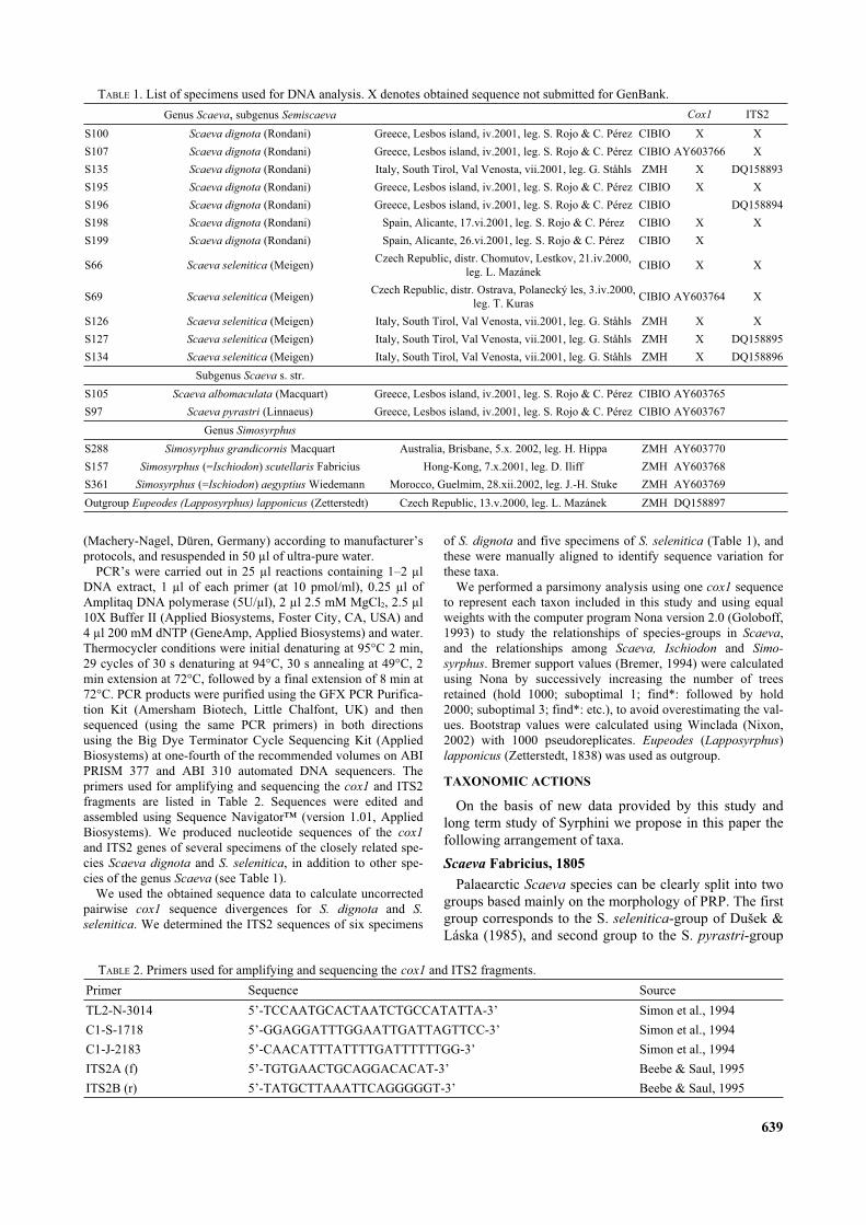
PCR’s were carried out in 25 µl reactions containing 1–2 µlDNA extract, 1 µl of each primer (at 10 pmol/ml), 0.25 µl ofAmplitaq DNA polymerase (5U/µl), 2 µl 2.5 mM MgCl2, 2.5 µl10X Buffer II (Applied Biosystems, Foster City, CA, USA) and4 µl 200 mM dNTP (GeneAmp, Applied Biosystems) and water.Thermocycler conditions were initial denaturing at 95°C 2 min,29 cycles of 30 s denaturing at 94°C, 30 s annealing at 49°C, 2min extension at 72°C, followed by a final extension of 8 min at72°C. PCR products were purified using the GFX PCR Purifica-tion Kit (Amersham Biotech, Little Chalfont, UK) and thensequenced (using the same PCR primers) in both directionsusing the Big Dye Terminator Cycle Sequencing Kit (AppliedBiosystems) at one-fourth of the recommended volumes on ABIPRISM 377 and ABI 310 automated DNA sequencers. Theprimers used for amplifying and sequencing the cox1 and ITS2fragments are listed in Table 2. Sequences were edited andassembled using Sequence Navigator™ (version 1.01, AppliedBiosystems). We produced nucleotide sequences of the cox1
and ITS2 genes of several specimens of the closely related spe-cies Scaeva dignota and S. selenitica, in addition to other spe-cies of the genus Scaeva (see Table 1).
We used the obtained sequence data to calculate uncorrectedpairwise cox1 sequence divergences for S. dignota and S.
selenitica. We determined the ITS2 sequences of six specimens
of S. dignota and five specimens of S. selenitica (Table 1), andthese were manually aligned to identify sequence variation forthese taxa.
We performed a parsimony analysis using one cox1 sequenceto represent each taxon included in this study and using equalweights with the computer program Nona version 2.0 (Goloboff,1993) to study the relationships of species-groups in Scaeva,and the relationships among Scaeva, Ischiodon and Simo-
syrphus. Bremer support values (Bremer, 1994) were calculatedusing Nona by successively increasing the number of treesretained (hold 1000; suboptimal 1; find*: followed by hold2000; suboptimal 3; find*: etc.), to avoid overestimating the val-ues. Bootstrap values were calculated using Winclada (Nixon,2002) with 1000 pseudoreplicates. Eupeodes (Lapposyrphus)lapponicus (Zetterstedt, 1838) was used as outgroup.
TAXONOMIC ACTIONS
On the basis of new data provided by this study andlong term study of Syrphini we propose in this paper thefollowing arrangement of taxa.
Scaeva Fabricius, 1805
Palaearctic Scaeva species can be clearly split into twogroups based mainly on the morphology of PRP. The firstgroup corresponds to the S. selenitica-group of Dušek &Láska (1985), and second group to the S. pyrastri-group
639
DQ158897ZMHCzech Republic, 13.v.2000, leg. L. MazánekEupeodes (Lapposyrphus) lapponicus (Zetterstedt)Outgroup
AY603769ZMHMorocco, Guelmim, 28.xii.2002, leg. J.-H. StukeSimosyrphus (=Ischiodon) aegyptius WiedemannS361
AY603768ZMHHong-Kong, 7.x.2001, leg. D. IliffSimosyrphus (=Ischiodon) scutellaris FabriciusS157
AY603770ZMHAustralia, Brisbane, 5.x. 2002, leg. H. HippaSimosyrphus grandicornis MacquartS288
Genus Simosyrphus
AY603767CIBIOGreece, Lesbos island, iv.2001, leg. S. Rojo & C. PérezScaeva pyrastri (Linnaeus)S97
AY603765CIBIOGreece, Lesbos island, iv.2001, leg. S. Rojo & C. PérezScaeva albomaculata (Macquart)S105
Subgenus Scaeva s. str.
DQ158896XZMHItaly, South Tirol, Val Venosta, vii.2001, leg. G. StåhlsScaeva selenitica (Meigen)S134
DQ158895XZMHItaly, South Tirol, Val Venosta, vii.2001, leg. G. StåhlsScaeva selenitica (Meigen)S127
XXZMHItaly, South Tirol, Val Venosta, vii.2001, leg. G. StåhlsScaeva selenitica (Meigen)S126
XAY603764CIBIOCzech Republic, distr. Ostrava, Polanecký les, 3.iv.2000,
leg. T. KurasScaeva selenitica (Meigen)S69
XXCIBIOCzech Republic, distr. Chomutov, Lestkov, 21.iv.2000,
leg. L. MazánekScaeva selenitica (Meigen)S66
XCIBIOSpain, Alicante, 26.vi.2001, leg. S. Rojo & C. PérezScaeva dignota (Rondani)S199
XXCIBIOSpain, Alicante, 17.vi.2001, leg. S. Rojo & C. PérezScaeva dignota (Rondani)S198
DQ158894CIBIOGreece, Lesbos island, iv.2001, leg. S. Rojo & C. PérezScaeva dignota (Rondani)S196
XXCIBIOGreece, Lesbos island, iv.2001, leg. S. Rojo & C. PérezScaeva dignota (Rondani)S195
DQ158893XZMHItaly, South Tirol, Val Venosta, vii.2001, leg. G. StåhlsScaeva dignota (Rondani)S135
XAY603766CIBIOGreece, Lesbos island, iv.2001, leg. S. Rojo & C. PérezScaeva dignota (Rondani)S107
XXCIBIOGreece, Lesbos island, iv.2001, leg. S. Rojo & C. PérezScaeva dignota (Rondani)S100
ITS2Cox1Genus Scaeva, subgenus Semiscaeva
TABLE 1. List of specimens used for DNA analysis. X denotes obtained sequence not submitted for GenBank.
Beebe & Saul, 19955’-TATGCTTAAATTCAGGGGGT-3’ITS2B (r)
Beebe & Saul, 19955’-TGTGAACTGCAGGACACAT-3’ITS2A (f)
Simon et al., 19945’-CAACATTTATTTTGATTTTTTGG-3’C1-J-2183
Simon et al., 19945’-GGAGGATTTGGAATTGATTAGTTCC-3’C1-S-1718
Simon et al., 1994 5’-TCCAATGCACTAATCTGCCATATTA-3’TL2-N-3014
SourceSequencePrimer
TABLE 2. Primers used for amplifying and sequencing the cox1 and ITS2 fragments.

of Dušek & Láska (1985). These alleged natural groupswere diagnosed by Dušek & Láska (1985: p. 226) usingboth adult and larval characters. We propose to classifythem as subgenera. For genesis of proposed subgenericnames see discussion.
Subgenus Semiscaeva Kuznetzov, 1985
[= S. selenitica group sensu Dušek & Láska (1985: 226)]Semiscaeva Kuznetzov, 1985: 412 (as subgenus of Scaeva Fab-
ricius, 1805). Type species Catabomba odessana Paramonov,1924 (orig. des.) = S. dignota (Rondani, 1857).
Mecoscaeva Kuznetzov, 1985: 418 (as subgenus of Scaeva Fab-ricius, 1805). Type species Lasiophthicus mecogramma
Bigot, 1860 (monotypy). Syn. n. (first reviser).
Subgenus Scaeva s. str.
[= S. pyrastri group sensu Dušek & Láska (1985)]Scaeva Fabricius, 1805: 248. Type species: Musca pyrastri Lin-
naeus, 1758 (des. Curtis, 1834: pl. 509).
Synonymy of Simosyrphus Bigot, 1882 and Ischiodon
Sack, 1913
Vockeroth (1969) provided differential diagnosis anddetailed descriptions of adult characters of both genera.The new data of this paper, larval and pupal morphologyand molecular data confirm the very close relationship ofSimosyrphus and Ischiodon. We propose their synonymyand a sister-group relationship with the genus Scaeva.
Simosyrphus Bigot, 1882
Simosyrphus Bigot, 1882: 68. Type species Syrphus grandi-
cornis Macquart, 1842 (sub. des. Hull, 1949: 291).
Ischiodon Sack, 1913: 6. Type species: Ischiodon trochanterica
Sack, 1913 (monotypy) = I. scutellaris (Fabricius, 1805). Syn.n.
DESCRIPTION OF IMMATURE STAGES
Scaeva Fabricius, 1805
Third instar larva
Length 12–18 mm, maximum width 3.0–4.0 mm.Overall appearance: Oval in cross-section with a littleflattened ventral surface, tapering anteriorly and slightlytruncate posteriorly. Dorsal habitus wrinkled (Fig. 1A),slightly serrate owing to fleshy projections with seg-mental spines (sensillae with setae). Colour pattern vari-able in ground colour even at intraspecific level (green,brown or pink) with a median dorsal white, cream or pinkstripe. Prothorax and mesothorax normally retracted intometathorax. Boundaries between segments obscured bysecondary grooves and folds in integument (Fig. 1A).Abdominal segments usually bearing five secondaryfolds. The pattern of segmental spines is very useful fororientation in primary segmentation, mainly the positionof the segmental spines of each side of abdominal seg-ments. Pairs 1 and 2 of segmental spines both located onsecond fold in metathorax and first abdominal segment; inother abdominal segments, pair 2 of segmental spineslocated just on the next fold (Fig. 1A). Segmental spinesnot pigmented, fully developed (0.15–0.28 mm long),with wide base (about 1/3 length of spine) and a narrowapical part (Fig. 1B). Integumental vestiture distinct inmost species, of cuticle colour or brown pigmented,smaller in groves and on ventral surface. Dorsal body sur-
640
Fig. 1. A, B: Third instar larva of Scaeva selenitica. A – dorsal habitus; B – segmental spine 2A6 with its fleshy projection. C, D:Scaeva albomaculata. C – posterior end of the third instar larva; D – posterior end of puparium.
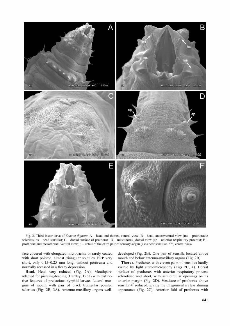
face covered with elongated microtrichia or rarely coatedwith short pointed, almost triangular spicules. PRP veryshort, only 0.15–0.25 mm long, without peritrema andnormally recessed in a fleshy depression.
Head. Head very reduced (Fig. 2A). Mouthpartsadapted for piercing-feeding (Hartley, 1963) with distinc-tive features of predacious syrphid larvae. Lateral mar-gins of mouth with pair of black triangular pointedsclerites (Figs 2B, 3A). Antenno-maxillary organs well-
developed (Fig. 2B). One pair of sensilla located abovemouth and below antenno-maxillary organs (Fig. 2B).
Thorax. Prothorax with eleven pairs of sensillae hardlyvisible by light stereomicroscopy (Figs 2C, 4). Dorsalsurface of prothorax with anterior respiratory processsclerotised and short, with semicircular openings on itsanterior margin (Fig. 2D). Vestiture of prothorax abovesensilla 4P reduced, giving the integument a clear shiningappearance (Fig. 2C). Anterior fold of prothorax with
641
Fig. 2. Third instar larva of Scaeva dignota. A – head and thorax, ventral view; B – head, anteroventral view (ms – prothoracicsclerites, hs – head sensilla); C – dorsal surface of prothorax; D – mesothorax, dorsal view (ap – anterior respiratory process); E –prothorax and mesothorax, ventral view; F – detail of the extra pair of sensory-organ (eso) near sensillae 7 Ms, ventral view.
longitudinal grooves and a ring (extending < 35% ofdorsal surface and < 60% of ventral surface) of small,backwardly directed spicules which become progressivelydensely-aggregated posteriorly on dorsal surface (Fig.2C). Mesothorax with eight pairs of sensillae arranged intwo main transverse rows: dorsal row with short seg-mental spines 1–3 and ventral row located slightly anteri-orly bearing five pairs of sensillae, two pairs ofdorso-lateral segmental spines followed by three pairs ofventral papiliform sensillae (Figs 2E, 3A, 4). Metathoraxwith nine pairs of sensillae arranged in two main trans-versal rows: dorsal row with four pairs of segmentalspines and ventral row located slightly anteriorly withfive pairs of segmental spines of unequal length; setae onsensillae 7Mt and 9Mt shorter than others (Figs 3A, 5A, 4).Contrary to e.g. Syrphus Fabricius, 1775, Megasyrphus
Dušek et Láska, 1967 bearing on metathorax ventrallyonly papilliform sensillae (Fig. 3B). One extra pair ofsensory-organs near sensillae 7Ms and 7Mt (hardly visibleby light stereomicroscope) (Figs 2F, 5A).
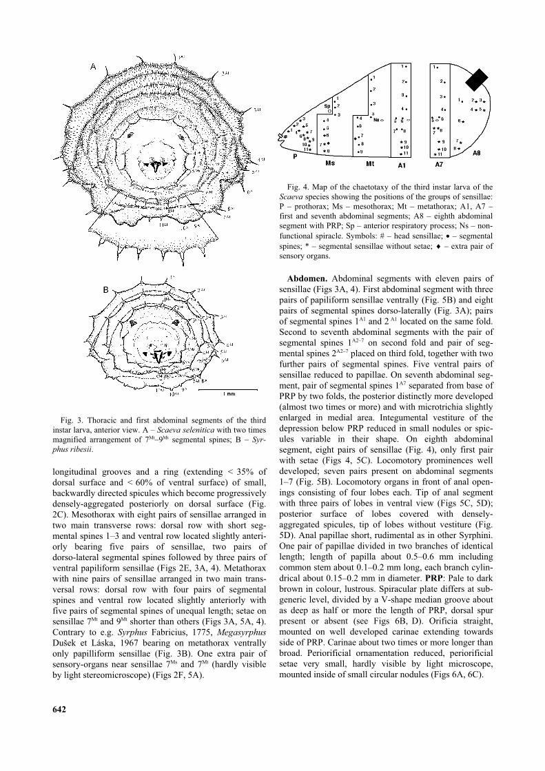
Abdomen. Abdominal segments with eleven pairs ofsensillae (Figs 3A, 4). First abdominal segment with threepairs of papiliform sensillae ventrally (Fig. 5B) and eightpairs of segmental spines dorso-laterally (Fig. 3A); pairsof segmental spines 1A1 and 2 A1 located on the same fold.Second to seventh abdominal segments with the pair ofsegmental spines 1A2–7 on second fold and pair of seg-mental spines 2A2–7 placed on third fold, together with twofurther pairs of segmental spines. Five ventral pairs ofsensillae reduced to papillae. On seventh abdominal seg-ment, pair of segmental spines 1A7 separated from base ofPRP by two folds, the posterior distinctly more developed(almost two times or more) and with microtrichia slightlyenlarged in medial area. Integumental vestiture of thedepression below PRP reduced in small nodules or spic-ules variable in their shape. On eighth abdominalsegment, eight pairs of sensillae (Fig. 4), only first pairwith setae (Figs 4, 5C). Locomotory prominences welldeveloped; seven pairs present on abdominal segments1–7 (Fig. 5B). Locomotory organs in front of anal open-ings consisting of four lobes each. Tip of anal segmentwith three pairs of lobes in ventral view (Figs 5C, 5D);posterior surface of lobes covered with densely-aggregated spicules, tip of lobes without vestiture (Fig.5D). Anal papillae short, rudimental as in other Syrphini.One pair of papillae divided in two branches of identicallength; length of papilla about 0.5–0.6 mm includingcommon stem about 0.1–0.2 mm long, each branch cylin-drical about 0.15–0.2 mm in diameter. PRP: Pale to darkbrown in colour, lustrous. Spiracular plate differs at sub-generic level, divided by a V-shape median groove aboutas deep as half or more the length of PRP, dorsal spurpresent or absent (see Figs 6B, D). Orificia straight,mounted on well developed carinae extending towardsside of PRP. Carinae about two times or more longer thanbroad. Periorificial ornamentation reduced, periorificialsetae very small, hardly visible by light microscope,mounted inside of small circular nodules (Figs 6A, 6C).
642
Fig. 3. Thoracic and first abdominal segments of the thirdinstar larva, anterior view. A – Scaeva selenitica with two timesmagnified arrangement of 7Mt–9Mt segmental spines; B – Syr-
phus ribesii.
Fig. 4. Map of the chaetotaxy of the third instar larva of theScaeva species showing the positions of the groups of sensillae:P – prothorax; Ms – mesothorax; Mt – metathorax; A1, A7 –first and seventh abdominal segments; A8 – eighth abdominalsegment with PRP; Sp – anterior respiratory process; Ns – non-functional spiracle. Symbols: # – head sensillae; – segmentalspines; * – segmental sensillae without setae; – extra pair ofsensory organs.

Puparium
Length 7.5–9 mm, maximum width 3.0–4.0 mm. Rathercask-like than pear-like, sub-cylindrical in cross-section(Fig. 5E). Anterior extreme truncated, slightly taperingposteriorly and flattened ventrally (Fig. 5F). Colour variesfrom cream to dull brown, sometimes with dark seg-mental patterns caused by pigmented cuticle. Dark pat-terns variable even at intraspecific level. Integumentalvestiture and segmental spines persisting. Segmental
spines of cuticle colour, about the same length as in lar-vae, but dried rests of fleshy projections bearing seg-mental spines usually inconspicuous. Sclerotised PRPalmost without changes, only carinae black edging (Fig.6).
Subgenus Semiscaeva Kuznetzov, 1985
Diagnostic characters. Colour pattern of larva variablein ground colour at intraspecific level (green, brown orpink) with a median dorsal white, cream or pink stripe.
643
Fig. 5. Scaeva dignota. A – metathorax of third instar larva, detail of ventral chaetotaxy end extra pair of sensory organs (eso) nearsensilla 7Mt, ventral view; B – locomotory prominences of the first abdominal segment, ventral view; C – anal segment, ventral view;D – detail of locomotory organ on the tip of anal segment, ventral view; E – puparium, dorsal view; F – puparium, lateral view.
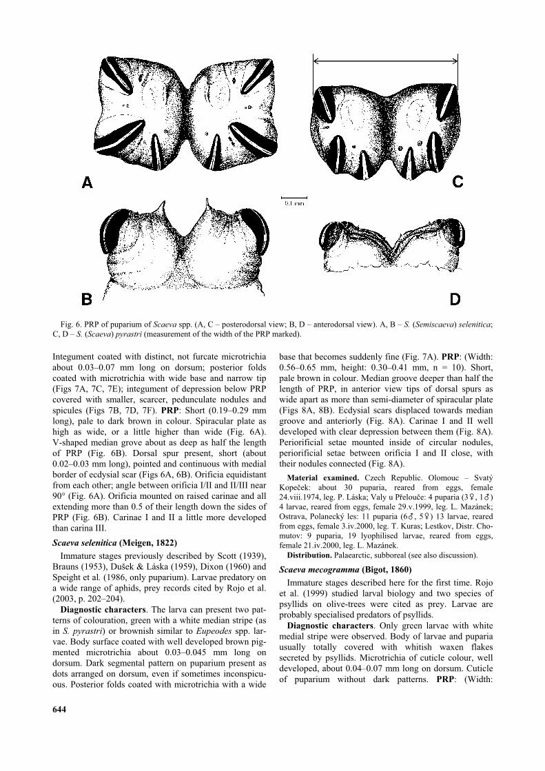
Integument coated with distinct, not furcate microtrichiaabout 0.03–0.07 mm long on dorsum; posterior foldscoated with microtrichia with wide base and narrow tip(Figs 7A, 7C, 7E); integument of depression below PRPcovered with smaller, scarcer, pedunculate nodules andspicules (Figs 7B, 7D, 7F). PRP: Short (0.19–0.29 mmlong), pale to dark brown in colour. Spiracular plate ashigh as wide, or a little higher than wide (Fig. 6A).V-shaped median grove about as deep as half the lengthof PRP (Fig. 6B). Dorsal spur present, short (about0.02–0.03 mm long), pointed and continuous with medialborder of ecdysial scar (Figs 6A, 6B). Orificia equidistantfrom each other; angle between orificia I/II and II/III near90° (Fig. 6A). Orificia mounted on raised carinae and allextending more than 0.5 of their length down the sides ofPRP (Fig. 6B). Carinae I and II a little more developedthan carina III.
Scaeva selenitica (Meigen, 1822)
Immature stages previously described by Scott (1939),Brauns (1953), Dušek & Láska (1959), Dixon (1960) andSpeight et al. (1986, only puparium). Larvae predatory ona wide range of aphids, prey records cited by Rojo et al.(2003, p. 202–204).
Diagnostic characters. The larva can present two pat-terns of colouration, green with a white median stripe (asin S. pyrastri) or brownish similar to Eupeodes spp. lar-vae. Body surface coated with well developed brown pig-mented microtrichia about 0.03–0.045 mm long ondorsum. Dark segmental pattern on puparium present asdots arranged on dorsum, even if sometimes inconspicu-ous. Posterior folds coated with microtrichia with a wide
base that becomes suddenly fine (Fig. 7A). PRP: (Width:0.56–0.65 mm, height: 0.30–0.41 mm, n = 10). Short,pale brown in colour. Median groove deeper than half thelength of PRP, in anterior view tips of dorsal spurs aswide apart as more than semi-diameter of spiracular plate(Figs 8A, 8B). Ecdysial scars displaced towards mediangroove and anteriorly (Fig. 8A). Carinae I and II welldeveloped with clear depression between them (Fig. 8A).Periorificial setae mounted inside of circular nodules,periorificial setae between orificia I and II close, withtheir nodules connected (Fig. 8A).
Material examined. Czech Republic. Olomouc – SvatýKope ek: about 30 puparia, reared from eggs, female24.viii.1974, leg. P. Láska; Valy u P elou e: 4 puparia (3&, 1%)4 larvae, reared from eggs, female 29.v.1999, leg. L. Mazánek;Ostrava, Polanecký les: 11 puparia (6%, 5&) 13 larvae, rearedfrom eggs, female 3.iv.2000, leg. T. Kuras; Lestkov, Distr. Cho-mutov: 9 puparia, 19 lyophilised larvae, reared from eggs,female 21.iv.2000, leg. L. Mazánek.
Distribution. Palaearctic, subboreal (see also discussion).
Scaeva mecogramma (Bigot, 1860)
Immature stages described here for the first time. Rojoet al. (1999) studied larval biology and two species ofpsyllids on olive-trees were cited as prey. Larvae areprobably specialised predators of psyllids.
Diagnostic characters. Only green larvae with whitemedial stripe were observed. Body of larvae and pupariausually totally covered with whitish waxen flakessecreted by psyllids. Microtrichia of cuticle colour, welldeveloped, about 0.04–0.07 mm long on dorsum. Cuticleof puparium without dark patterns. PRP: (Width:
644
Fig. 6. PRP of puparium of Scaeva spp. (A, C – posterodorsal view; B, D – anterodorsal view). A, B – S. (Semiscaeva) selenitica;C, D – S. (Scaeva) pyrastri (measurement of the width of the PRP marked).

0.35–0.4 mm, height: 0.20–0.25 mm, n = 6). PRP smallerand darker in relation to S. selenitica/dignota. Short anddark brown in colour at least in puparium. Median grooveslightly less deep than half the length of PRP, tips of thedorsal spurs as wide apart as less than semi-diameter ofspiracular plate (Figs 8C, 8D). Ecdysial scars displacedtowards median groove and anteriorly (Fig. 8C). Periori-ficial setae mounted inside of circular nodules, periorifi-cial setae between orificia I and II close, but border oftheir nodules separated (Fig. 8C).
Material examined. Spain. Valencia, Moncada: 4 puparia(1%, 3&) leg. as larvae 23.xi.1996, leg. M.J. Verdú; 3 puparia(2%, 1&) leg. as larvae 18.vii.1996, leg. M.A. Marcos-García; 1puparium (%) 8 larvae, leg. as larvae 23.xii.1996, leg. S. Rojo;Alicante, San Vicente del Raspeig: 3 larvae, leg. as larvae13.v.1998, leg. J.V. Falcó; 3 puparia (1%, 2&) leg. as larvae7.vi.1998, leg. J.V. Falcó.
Distribution. South Europe.
645
Fig. 7. Puparium of Scaeva (Semiscaeva) spp. (A, C, E – microtrichia on the central area of the posterior fold, dorsal view; B, D, F– integumental vestiture of the depression below PRP). A, B – S. selenitica; C, D – S. mecogramma; E, F – S. dignota.

Scaeva dignota (Rondani, 1857)
Immature stages described here for the first time.Larvae probably predatory on a wide range of aphids.Prey records cited by Dušek & Láska (1985) and Starý &Havelka (1991). Prey records from southern part ofEurope could be erroneously referred to S. selenitica.
Diagnostic characters. Larva and puparium verysimilar to S. selenitica including coloration variability oflarvae. Dorsal body surface coated with well developedmicrotrichia about 0.03–0.045 mm long, pigmentation of
microtrichia less intense than in S. selenitica, cuticle ofpuparium mainly without dark segmental patterns. Poste-rior folds coated with microtrichia with a wide base thatbecome progressively fine (Fig. 7E). PRP: (Width:0.45–0.55 mm, height: 0.28–0.36 mm, n = 30; PRP sizenot affected by parasitation of Diplazon Ness, 1818(Hymenoptera: Ichneumonidae), contrary to the observa-tion of Dušek et al. (1979), who described the influenceof parasitation on PRP size of puparia in Syrphus).Median groove deeper than half the length of PRP, in
646
Fig. 8. PRP of puparium of Scaeva (Semiscaeva) spp. (A, C, E – posterodorsal view view; B, D, F – anterodorsal view). A, B – S.
selenitica; C, D – S. mecogramma; E–F – S. dignota.
anterior view tips of dorsal spurs as wide apart as morethan semi-diameter of a spiracular plate (Figs 8E, F).Ecdysial scars slightly anterior to centre of each spi-racular plate (Fig. 8E). Periorificial setae mounted insideof circular nodules, periorificial setae between orificia Iand II close, but border of their nodules separated (Fig.8E).
Material examined. Czech Republic. Žlunice, distr. Ji ín: 1puparium (% numbered G 72), leg. as puparium 23.vi.1959 onthe blade of grass in deciduous forest, leg. P. Láska; Olomouc: 1puparium (& numbered 5VZ01/1), leg. as larva 21.vi.2001feeding on Aphis sambuci on Sambucus nigra, leg. M. Štib-narová & M. Hanáková; 1 puparium (&), leg. as larva
10.vi.1994, leg. L. Mazánek. Turkey. zmir, Kar iyaka: 1 pupa-rium, leg. as larva 19.iv.1976 on aphids colony on Prunus
domestica, leg. E. Erkin; zmir, Urla: 1 dried larva, 24.iv.1975on aphis colony on Vicia faba, leg. E. Erkin; zmir, Menemen: 1puparium, leg. as larva 21.iv.1975 on aphis colony on Prunus
communis, leg. E. Erkin. Greece. Lesbos, Mytilene: 3 puparia(1%, 2&) leg. as larvae 10.iii.2001, leg. S. Rojo & C. Pérez-Bañón; Polihnitos: 9 puparia (4%, 5&) leg. as larvae, leg. S.Rojo & C. Pérez-Bañón; Agh. Paraskevi: 10 puparia (6%, 4&)leg. as larvae 27.iii.2001, leg. S. Rojo & C. Pérez-Bañón. Spain.Alicante. Tibi: 7 puparia (4%, 2&) leg. as larvae 21.v.1993; leg.S. Rojo; Villena: 3 puparia (3&) leg. as larvae 16.iv.1993; leg.S. Rojo; Biar: 2 puparia (2&) leg. as larvae 16.iv.1993; leg. S.Rojo.
Distribution. Sub-Mediterranean (see also discussion).
647
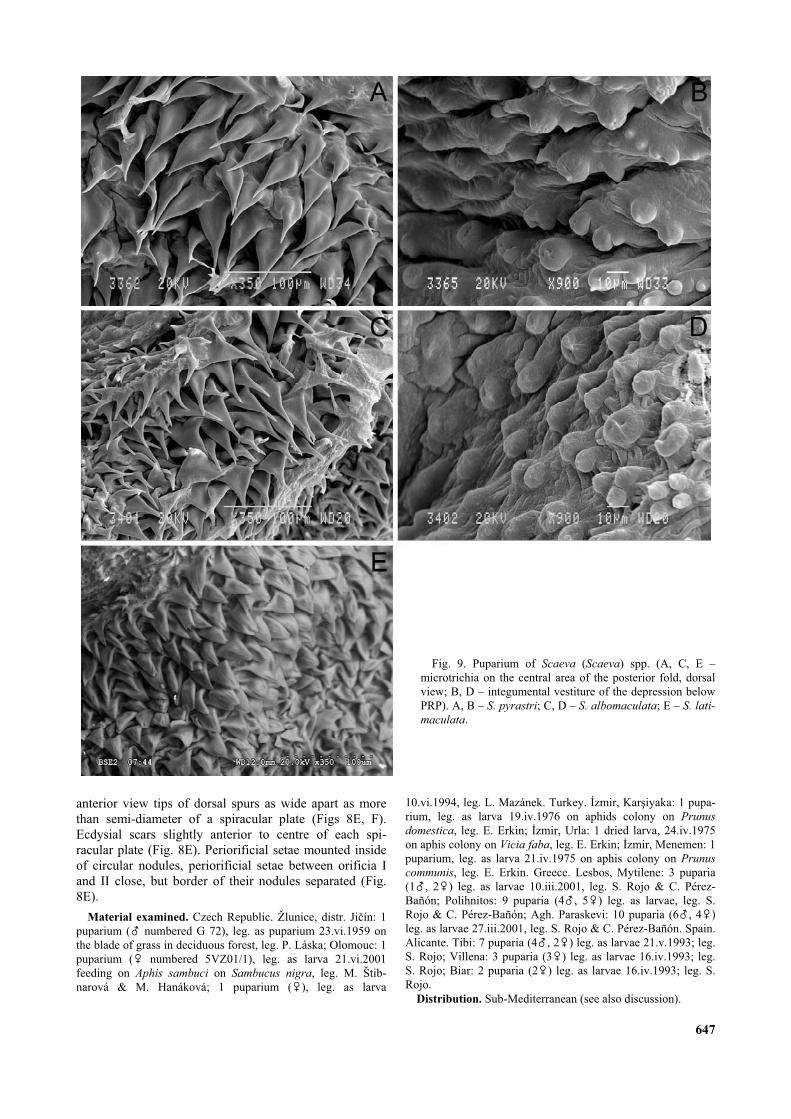
Fig. 9. Puparium of Scaeva (Scaeva) spp. (A, C, E –microtrichia on the central area of the posterior fold, dorsalview; B, D – integumental vestiture of the depression belowPRP). A, B – S. pyrastri; C, D – S. albomaculata; E – S. lati-
maculata.
Subgenus Scaeva s. str.
Diagnostic characters. Colour pattern of larva con-stant, green with white median dorsal stripe. Rarely withpink shade and ochre dorsal stripe. Integument coatedwith microtrichia, about 0.02–0.035 mm long on dorsum,that could be reduced to short, almost triangular spicules;microtrichia of cuticle colour or brown pigmented, oftenfurcate especially on folds around PRP. Integument of thedepression below PRP covered with smaller, scarcer,slightly pedunculate nodules (Figs 9B, 9D) of whitishcolour in puparium. PRP: very short, almost sessile(0.12–0.15 mm long), pale to dark brown in colour. Spi-racular plates divided by a V-shape median groove almostas deep as the length of PRP (Fig. 6D). Dorsal spurabsent; orificia II and III parallel, orificium III inserted onabout 1/3 to nearly 1/2 of length of orificium II posteri-orly than orificium II; carinae I and III extending downthe sides of PRP; carinae I and II distinctly more devel-oped than carina III (Fig. 6C). Periorificial setae mountedinside of circular nodules; periorificial setae between ori-ficia I and II very close, with border of their nodules con-nected (Fig. 6C).
Scaeva pyrastri (Linnaeus, 1758)
Previously described by Martelli (1911), Jones (1922),Krüger (1926), Fluke (1929), Heiss (1938), Bhatia
(1939), Scott (1939), Brauns (1953), Láska (1959),Dušek & Láska (1959), Dixon (1960), Goeldlin deTiefenau (1974). Sharma & Bhalla (1988) dealt withlarval biology on laboratory breeding but did not describeimmature stages. Barkemeyer (1994) provides a compre-hensive literature review of what is known on the biologyof this species. Larvae are predatory on a wide range ofaphids and also Coccidae, Psyllidae and Thysanoptera;prey records listed by Rojo et al. (2003, p. 192–202).
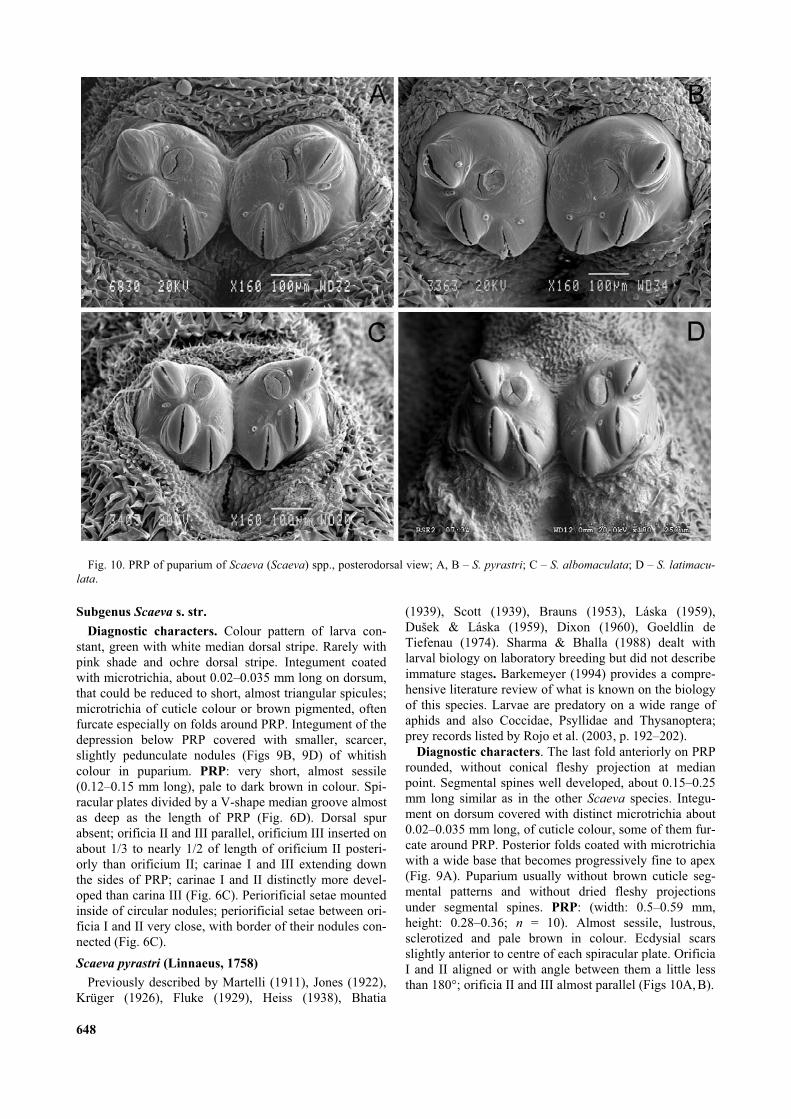
Diagnostic characters. The last fold anteriorly on PRProunded, without conical fleshy projection at medianpoint. Segmental spines well developed, about 0.15–0.25mm long similar as in the other Scaeva species. Integu-ment on dorsum covered with distinct microtrichia about0.02–0.035 mm long, of cuticle colour, some of them fur-cate around PRP. Posterior folds coated with microtrichiawith a wide base that becomes progressively fine to apex(Fig. 9A). Puparium usually without brown cuticle seg-mental patterns and without dried fleshy projectionsunder segmental spines. PRP: (width: 0.5–0.59 mm,height: 0.28–0.36; n = 10). Almost sessile, lustrous,sclerotized and pale brown in colour. Ecdysial scarsslightly anterior to centre of each spiracular plate. OrificiaI and II aligned or with angle between them a little lessthan 180°; orificia II and III almost parallel (Figs 10A, B).
648
Fig. 10. PRP of puparium of Scaeva (Scaeva) spp., posterodorsal view; A, B – S. pyrastri; C – S. albomaculata; D – S. latimacu-
lata.
Material examined. Montenegro. Be i i near Budva: 9puparia, leg. as larvae v.1968 feeding on Uroleucon cichorii onCichorium intybus, leg. P. Láska. Czech Republic. Olomouc: 1puparium (%) leg. as larva on aphid colony on Cirsium arvense,10.vi.1999, leg. L. Mazánek; 53 puparia (24%, 29&) 5 lyophi-lised larvae, 4 larvae in alcohol, reared from eggs, female14.vi.2001, leg. L. Mazánek; 2 puparia (%) 1 larva, leg. aslarvae 4.vii.2003 feeding on Uroleucon cichorii on Cichorium
intybus, leg. L. Mazánek; Mikul in vrch: 1 puparium (&) leg. aslarva feeding on Uroleucon cichorii on Cichorium intybus,
8.ix.2000, leg. L. Mazánek. Greece. Lesbos, Agh. Parakevi: 1puparium (&) leg. as larvae 27.iii.2001, leg. S. Rojo & C. Pérez-Bañón; Mistegna: 1 puparium (&) leg. as larvae 17.iv.2001, leg.S. Rojo & C. Pérez-Bañón; Mandamados: 1 puparium (%) leg.as larvae 18.v.2001, leg. S. Rojo & C. Pérez-Bañón. Spain. Ali-cante, San Vicente del Raspeig: 2 puparia (2&) leg. as larvae13.iii.2000, leg, E.S. Ráez; San Vicente del Raspeig: 1puparium (&) leg. as larvae 21.iii.2000, leg, E.S. Ráez; SanVicente del Raspeig: 1 puparium (%) leg. as larvae 28.iii.2000,
649
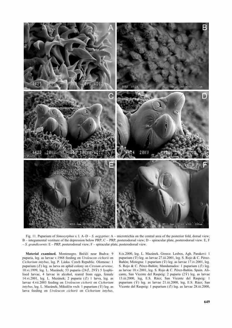
Fig. 11. Puparium of Simosyrphus s. l. A–D – S. aegyptius: A – microtrichia on the central area of the posterior fold, dorsal view;B – integumental vestiture of the depression below PRP; C – PRP, posterodorsal view; D – spiracular plate, posterodorsal view. E, F– S. grandicornis: E – PRP, posterodorsal view; F – spiracular plate, posterodorsal view.
leg, E.S. Ráez; San Vicente del Raspeig: 1 puparium (&) leg. aslarvae 17.xi.2000, leg, E.S. Ráez.
Distribution. Holarctic and marginally Oriental region.
Scaeva albomaculata (Macquart, 1842)
Previously described by Kuznetzov & Daminova(1994). Larvae predatory on a wide range of aphids; preyrecords listed by Rojo et al. (2003, pp. 190–192).
Diagnostic characters. Last fold anterior of PRPenlarged, forming conical fleshy projection at medianpoint (Fig. 1C). Fully developed segmental spines about0.15–0.25 mm long. Integument on dorsum covered withdistinct microtrichia about 0.02–0.035 mm long, brownedtowards top and often furcate on folds around PRP. Pos-terior folds coated with microtrichia with a wide basebecoming progressively fine to apex (Fig. 9C). Pupariumusually with tubercle (= rest of dried fleshy conical pro-jection in larva) before PRP (Fig. 1D) and also driedfleshy projection often persist as microtrichose portionunder base of some segmental spines on posterior part ofpuparium. PRP: (width: 0.39–0.44 mm, height: 0.25–0.3,n = 5). Almost sessile, lustrous, sclerotized and palebrown in colour. Ecdysial scars displaced towards themedian groove and anteriorly; orificia II and III almostparallel (Fig. 10C).
Material examined. Spain. Alicante, San Vicente del Ras-peig: 1 lyophilised larva, reared from eggs, female 27.ii.2000,leg. L. Mazánek; Ibi: 6 puparia (4%, 2&) leg. as larvae6.vii.1992, leg. S. Rojo; Villena: 3 puparia (2%, 1&) leg. aslarvae 26.vi.1992, leg. S. Rojo; Sax: 1 puparium (%) leg. aslarva 28.vi.1992, leg. S. Rojo; Alcoy: 1 puparium (&) leg. aslarva 21.v.1992, leg. S. Rojo. Italy. Roma, park Glyphinia: 1puparium (numbered 63/307), leg. as larva 16.vi.1963, leg. P.Starý.
Distribution. South part of Palaearctic to north part of Ori-ental region.
Scaeva latimaculata (Brunetti, 1923)
Previously described by Kumar et al. (1987). Severaleconomically important aphid species were cited as preyof this species by Agarwala et al. (1984) and Kumar et al.(1987).
Diagnostic characters. The larva is similar to that of S.
pyrastri, from which it differs by less developed integu-mental vestiture, microtichia reduced to short, almost tri-angular spicules not longer than 0.02 mm, spicules ofcuticle colour or browned. Segmental spine shorter thanin the other Scaeva species; most developed spines about0.15 mm long. Posterior folds coated with longer micro-trichia not fine at apex and often furcate (Fig. 9E). Cuticleof puparium with or without darkened segmental patterns,rests of dried fleshy projections absent. PRP: (width:0.33–0.39 mm, height: 0.21–0.26 mm, n = 2). Ecdysialscars displaced towards the median groove and anteriorly;orificium I on most developed carina that overlaps ante-rior margin of spiracular plate in dorsal view; orificia IIand III almost parallel (Fig. 10D).
Material examined. India. Punjab: 1 puparium (& – SEMphotos). leg. as larva, leg. A. Kumar; Himachal Pradesh, 1puparium (& numbered 104), leg. as larva on Hyperomyzus car-
duellinus on Emilia sonchi leg. K. Agarwala. Iran. Mahabad 82
km S., 2600 m: 1 puparium (&) leg. as larva 6.vii.1974 onPapaver sp., leg. C. Buckingham.
Distribution. Southern Palaearctic from Iran eastwards intoOriental region.
Simosyrphus Bigot, 1882
Third instar larva
We could not examine the larvae, but according to pre-vious descriptions of Barbosa (1952), Lal & Gupta(1953), Tawfik et al. (1974), Roy & Basu (1977), Kumaret al. (1987), Soleyman-Nezhadiyan (pers. com.) and ablack and white photo of Schmutterer (1972), the larvaeof this genus are very similar to smaller larvae of Scaeva
s.str. in overall appearance: (Length including PRP7–12.5 mm, maximum width 2–3 mm) oval in cross-section, relatively high; locomotory prominences welldeveloped; body wrinkled and green coloured with mid-dorsal whitish stripe. Some species have on each side ofwhite stripe a line of spots or narrow stripes orange tolight brown coloured and spots or stripes along lateralmargins of larvae. These patterns also visible in fleshyprojection bearing segmental spines. However, Rotheray& Gilbert (1999) give some new characters that show theclose relation between these two genera: presence of setaeon the sensillae 7Mt, 8Mt and 9Mt (that was confirmed byour study of puparia); the locomotory prominences on 6th
and 7th abdominal segments with the tips directed back-wards; and an extra lobe on the locomotory prominencesof abdominal segments 1–7.
We can add some new data to this list of charactersobtained from the puparium: segmental spines relativelylong (about 0.1–0.21 mm long in dorsum, measured inpuparium), with the same pattern and arrangement as inthe Scaeva species, only fleshy projections bearing seg-mental spines are more developed. Integument covered byshort pointed spicules rather than microtrichia, about 0.02mm long, on posterior folds longer and wider and oftenfurcate (Fig. 11A); integument of depression below PRPand also small area close before PRP covered withnodular rounded papillae (Fig. 11B).
PRP: similar to PRP of sg. Scaeva, very short, almostsessile (0.1–0.15 mm long), only width of PRP differsaccording to species. Spiracular plates higher than wide,divided by a V-shape groove as deep as length of PRP;dorsal spur absent; orificia II and III almost parallel; orifi-cium III inserted more posteriorly than in the Scaeva sub-genus, on about half or more of the length of orificium II.Carina I well developed, distinctly overlapping the ante-rior margin of spiracular plate in dorsal view (Figs11C–F). Periorificial ornamentation reduced as in theScaeva species; periorificial setae very small hardlyvisible by light microscope, mounted inside of small cir-cular nodules (Figs 11C–F); periorificial setae betweenorificia I and II very close, with border of their nodulesconnected.
Puparium
Length including PRP 5.5–7.0 mm, maximum width2.5–3.2 mm. Puparia of similar shape as in Scaeva butsmaller. Colour varying from cream to dull brown, also at
650

intraspecific level. Integumental vestiture persisting, ofcuticle colour or a little darkened, rarely forming slightlydarkened segmental patterns with pigmented cuticle. Seg-mental spines elongated by persisting dried fleshy projec-tions that form microtrichose portion under segmentalspines, especially on dorsum of posterior part of pupar-ium. Segmental spines of cuticle colour or browner, vari-able also at intraspecific level. PRP with carinae usuallyblacked, nodular integument of depression below PRPand also small area close before PRP usually whitish.
Simosyrphus grandicornis (Macquart, 1842)
Soleyman-Nezhadiyan (1997) and Soleyman-Nezha-diyan & Laughlin (1998) deal with larval biology of labo-ratory breedings but do not describe immature stages.Larvae predatory on a wide range of aphids and evenlepidopteran larvae; prey records reported by Rojo et al.(2003, pp. 204–205).
Diagnostic characters. Larva green with whitishmedian stripe (E. Soleyman-Nezhadiyan, pers. com.).Segmental spines relatively short, fully developed onlyabout 0.1–0.14 mm long, fleshy projections bearing seg-mental spines less developed, dried persist under severalsegmental spines on posterior part of dorsum of pupariumas microtrichose portion (maximum length 0.05 mm).Microtrichia of cuticle colour or slightly dark brown pig-mented, especially around segmental spines, butpuparium without visible dark coloured pattern. PRP:somewhat smaller than in other species of the genus,about 0.26–0.30 mm wide and 0.14–0.18 mm high (n =2). Length of PRP about 0.1 mm. Carina I welldeveloped, rounded apically (Fig. 11D).
Material examined. Australia. Nethy Bridghe: 1 puparium(&) leg. as larva 11.xi.1974; 1 puparium (& – SEM photos),reared on Hyperomyzus lachicae, leg. as larva 11.xi.1974;Mosman NSU: 1 puparium (%) reared on Schaitedonia lutea,leg. as larva 18.i.1973. All leg. D. Hales.
Distribution. Australia, Oceania.
Simosyrphus aegyptius (Wiedemann, 1830) comb. n.
Previously described by Barbosa (1952), Tawfik et al.(1974). Larvae predatory on a wide range of aphids andalso on Thysanoptera; prey records cited by Rojo et al.(2003, p. 116–118). Even lepidopterous larvae were citedas preys by Randrianandrianina-Razananaivo (1991).
Diagnostic characters. Larva yellowish green withmid-dorsal white stripe flanked by developing narroworange or light brown stripes; yellowish orange fat bodiesdiscerned also in fleshy projections forming lateral orangeline. As the fat bodies extend, larvae become rather yel-lowish orange. Fully developed segmental spines about0.16–0.2 mm long mounted on well developed fleshy pro-jection that dried persist on puparium under segmentalspines as microtrichose portion long almost 0.12 mm,especially on posterior part of dorsum. PRP: about0.38–0.43 mm wide and 0.24–0.30 mm high (n = 5). PRPlength about 0.12–0.14 mm. Carina I rather sharp apically(Fig. 11F).
Material examined. Kenya. Nairobi-Chiromo: 4 puparia(2%, 2&) reared on Rhopalosiphum maidis, leg. as larvae
22.ii.1970, leg. H. Schmutterer; 2 puparia (&) reared on Rhopa-
losiphum maidis, leg. as larvae 20.vi.1970, leg. H. Schmutterer.Senegal. Ziguinchor, Djibélor: 3 puparia (2% – SEM photos,1&) leg. as larvae 18.xi.1979 on Toxoptera aurantii, leg. J. Eti-enne.
Distribution. Afrotropical, including Madagascar, Réunionand South-West Palaearctic (North Africa, Southern Spain,Madeira and Canary Islands).
Simosyrphus scutellaris (Fabricius, 1805) comb. n.
Immature stages were previously described by Lal &Gupta (1953), Okuno (1967), Roy & Basu (1977) andKumar et al. (1987). Ninomyia (1959) described only thepuparium of an aberrant specimen with only two orificiaon one of two spiracular plates. Lal & Haque (1965),Agarwala & Saha (1986), Sharma & Bhalla (1988) andSingh & Mishra (1988) studied biology of immaturestages in detail but did not describe the immature stages.Larvae predatory on a wide range of aphids and evenlepidoptera larvae; prey records listed by Rojo et al.(2003, p. 118–122).
Diagnostic characters. Larvae green to greenish-brown coloured with mid-dorsal whitish stripe and withreddish to brown fat bodies along mid-dorsal stripe, infleshy projections and along lateral margin of larvae.Fully developed segmental spines about 0.13–0.18 mm,mounted on well developed fleshy projection that driedpersist on puparium under base of segmental spines asmicrotrichose portion long almost 0.08 mm, especially onposterior part of dorsum. PRP: about 0.43–0.53 mm wideand 0.26–0.34 mm high, (n = 2); length about 0.13 mm.
Material examined. India. Punjab: 1 puparium (%) leg. aslarva, leg. A. Kumar; Agartala: 1 puparium, leg. as larva 1980on Aphis gossypii on Gossypium sp., leg. K. Agarwala.
Distribution. Southern Palaearctic (from Iran, Turkey toJapan), Oriental region, Australia, Pacific (excl. Hawaiian Is).
KEY FOR THIRD INSTAR LARVAE AND PUPARIA
1 Angles between adjacent orificia about 90°, dorsal spur pre-sent (Figs 6A, B) (sg. Semiscaeva ). . . . . . . . . . . . . . . . . . . 2
– Orificia II and III almost parallel, dorsal spur absent (Figs6C, D). . . . . . . . . . . . . . . . . . . . . . . . . . . . . . . . . . . . . . . . . . 4
2 Width of PRP about 0.5 mm or more; median groove deeperthan half the length of PRP (Figs 8B, F). . . . . . . . . . . . . . . 3
– Width of PRP about 0.4 mm or less; median groove slightlyless deep than half the length of PRP (Fig. 8D). . . . . . . . . . . .. . . . . . . . . . . . . . . . . . . . Scaeva (Semiscaeva) mecogramma
3 Posterior folds coated with microtrichia with a wide basethat become suddenly fine (Fig. 7A); some microtrichia ofintegumental vestiture pigmented apically; puparium usuallywith distinct dark dotted segmental pattern. . . . . . . . . . . . . . .. . . . . . . . . . . . . . . . . . . . . . . Scaeva (Semiscaeva) selenitica
– Posterior folds coated with microtrichia with a wide basethat become progressively fine to the apex (Fig. 7E); bodycoated with microtrichia of cuticle colour or almost so;puparium without dark dotted segmental pattern. . . . . . . . . . .. . . . . . . . . . . . . . . . . . . . . . . . . Scaeva (Semiscaeva) dignota
4 Integumental vestiture distinct, microtrichia about 0.2–0.35mm long on dorsum; microtrichia on posterior folds withthin apex (Figs 9A, C); orificia located on high oval carinae,slightly less developed in orificium III (Figs 10A, B, C) . . 5
651

– Integumental vestiture reduced, microtrichia on dorsumabout 0.02 mm long or shorter; microtrichia on posteriorfolds relatively broad near apex (Figs 9E, 11A); orificialocated on very high oval carinae except orificium IIIlocated on distinctly less developed carina (10D, 11D, F). 6
5 Fold close to anterior part of PRP forming conical fleshyprojection in median point (Fig. 1C), when dried, usuallyalso apparent on puparium as a tubercle overlapping withPRP (Fig. 1D); ecdysial scars displaced towards the mediangroove and anteriorly (Fig. 10C). . . . . . . . . . . . . . . . . . . . . . .. . . . . . . . . . . . . . . . . . . . . . . . Scaeva (Scaeva) albomaculata
– Fold close to anterior part of PRP regularly rounded later-omedially without conical fleshy projection; ecdysial scarsslightly anterior to centre of each spiracular plate (Figs10A, B). . . . . . . . . . . . . . . . . . . . . . Scaeva (Scaeva) pyrastri
6 Orificium III inserted only about 1/3 the length of orificiumII posteriorly to orificium II; carina III distinct and carina Iabout as developed as carina II (Fig. 10D); fleshy projectionunder segmental spines less developed, not persist on pupar-ium, so that segmental spines are sessile on puparium. . . . . .. . . . . . . . . . . . . . . . . . . . . . . . . Scaeva (Scaeva) latimaculata
– Orificium III inserted about half or more the length of orifi-cium II posteriorly to orificium II; carina III almost flat andcarina I enlarged, distinctly overlapping periphery of PRP(Figs 11D, F); fleshy projection under segmental spinesmore prominent, dried persist as microtrichose portion undersegmental spines on puparium, especially on posterior partof dorsum (g. Simosyrphus). . . . . . . . . . . . . . . . . . . . . . . . . 7
7 Width of PRP about 0.3 mm or less; the most developedsegmental spines about 0.1–0.14 mm long, in pupariumelongated by microtrichose rests of dried fleshy projectionsnearly to 0.18 mm, especially on posterior part of dorsum. . .
. . . . . . . . . . . . . . . . . . . . . . . . . . . Simosyrphus grandicornis
– PRP wider than 0.35 mm; segmental spines longer, fullydeveloped about 0.13–0.2 mm long, in puparium elongatedby microtrichose rests of dried fleshy projections to about0.2–0.3 mm, especially on posterior part of dorsum. . . . . . 8
8 Width of PRP about 0.38–0.43 mm; fully developed seg-mental spines about 0.16–0.2 mm long, in puparium elon-gated by microtrichose rests of dried fleshy projection toabout 0.3 mm, especially on posterior part of dorsum. . . . . . .. . . . . . . . . . . . . . . . . . . . . . Simosyrphus aegyptius comb. n.
– Width of PRP about 0.43–0.53 mm or longer; fully devel-oped segmental spines about 0.13–0.18 mm long, inpuparium elongated by microtrichose rests of dried fleshyprojection nearly to 0.24 mm, especially on posterior part ofdorsum. . . . . . . . . . . . . . . . Simosyrphus scutellaris comb. n.
MOLECULAR STUDIES
The parsimony analysis of 1128 nucleotides of the cox1
gene of seven taxa belonging to Scaeva and Simosyrphus,
using Eupeodes (Lapposyrphus) lapponicus as outgroup,produced one most parsimonious tree (Fig. 12A) of 224steps (CI = 0.83; RI = 0.74). All proposed subgeneric andgeneric taxa based on morphological studies receive highbootstrap support (1000 replicates). We did not recover acloser relationship of sg. Scaeva with Simosyrphus thanwith sg. Semiscaeva, even if this node is suggested fromthe immature morphology (see Discussion). Resamplingusing jackknifing spawn from Winclada is shown in Fig.12B.
Cox1 sequences of Scaeva selenitica and S. dignota
were identical except for one nucleotide change in one S.
dignota specimen. The six specimens of S. dignota pre-sented identical ITS2 sequences (genotype 1, Table 3)and the five specimens of S. selenitica were also mono-morphic for ITS2 (genotype 2). These two genotypes aredistinct.
DISCUSSION
Taxonomy of Scaeva
According to our results, the Palaearctic Scaeva speciescan be split into two groups based on the analysis ofimmature stages. One group (S. dignota, S. selenitica, S.
mecogramma) has a distinct angle of about 90° betweenorificia II and III, as is typical for other related genera ofSyrphini. A second group (S. pyrastri, S. albomaculata,S. latimaculata) has the orificia II and III (on spiracularplate) parallel, with insertion of orificium III beneath thelevel of insertion of orificium II (probably apomorphicsituation). These two groups that should be classified assubgenera were designated as natural groups withinScaeva by Dušek & Láska (1985) (S. selenitica-group vsS. pyrastri-group). Independently, Kuznetzov (1985)divided the Palaearctic Scaeva species in three subgenerabased exclusively on adult morphology (Semiscaeva
652
....AGAGGATTATAGTATATAAAATTAAA----------------TTAAAAATATAATTATGTTTAAAAAGTAAAACATAAATTAAAAAAAAAATATACTT....S. selenitica
....AGAGGATTATAGTATATAAAATTAAATCAAAGATATTAATATATAAAAATAAAAATATGTTTAAA--GAAGAAAAACCTAAGAAAAAAAAATATACTT....S. dignota
Sequence varibility in the variable region of the ca 450 nt ITS2 Genotype
TABLE 3. Sequence varibility in the variable regions of the ca 450 nt ITS2 (arbitrary alignment) that were obtained for all of thesequenced S. dignota specimens (genotype 1) and the S. selenitica specimens (genotype 2).
Fig. 12. A – Most parsimonious tree obtained using Nona for1128 bp of the cox1 gene, length = 224 steps, CI = 0.83, RI =0.74. Bootstrap support values and Bremer support (branch sup-port) values are reported above and below nodes, respectively.B – Parsimony jackknifing tree (1000 replicates).

Kuznetzov, 1985: 412, Mecoscaeva Kuznetzov, 1985:418 and Scaeva s. str.). However, his arrangement of thespecies does not agree with the results of the presentpaper. The main differences are that following this authorthe species S. mecogramma (type species of subgen.Mecoscaeva), S. dignota (junior syn. of which is the typespecies of his subgen. Semiscaeva) and S. selenitica (fromhis subgen. Scaeva) are closely related on the basis ofpre-imaginal and molecular data. According to our resultsthey form a natural group that is identical to S. selenitica-group (sensu Dušek & Láska, 1985). The name Semis-
caeva Kuznetzov, 1985 should be the valid name for theS. selenitica-group (sensu Dušek & Láska, 1985) as asubgenus of the genus Scaeva. The subgenus Scaeva s.str. can only be referred to the S. pyrastri-group (sensuDušek & Láska, 1985) and as the first revisers we deter-mine subgen. Mecoscaeva Kuznetzov, 1985 syn. n. as ajunior synonym of subgen. Semiscaeva Kuznetzov, 1985.
Status of Scaeva dignota and S. selenitica
Scaeva dignota was recognized (from variability of S.
selenitica) as a valid species by Dušek & Láska (1985).These authors observed that the anterior angle of approxi-mation of eyes in males of S. dignota is narrow (90°–106°), face is narrower and ocular hairs are shorter incomparison with S. selenitica. The pattern of yellow spotson tergites 3 and 4 differs a little between the species.However, except for size differences of microtrichia ofthe posterior fold, we have been unable to found informa-tive characters to distinguish larvae or pupae of these spe-cies. Moreover, these species present dichroism of larvae(green vs brown colour). We have observed this peculiarphenomenon only in these two species of the genusScaeva. Male terminalia in both taxa are very similar. S.
dignota is mainly a Mediterranean species and S.
selenitica is a sub-boreal species (Dušek & Láska, 1985).The molecular analysis also indicated a very close rela-tionship between these species, as both taxa presentedidentical cox1 sequences in all the studied material(Czech, Spanish, Finnish and Greek specimens). Thisclearly supports that a separation of these two closelyrelated species into different subgenera as done byKuznetzov (1985) is unfounded. However, the nuclearITS2 region sequenced for several S. dignota and S.
selenitica from central Europe presented two differentgenotypes (Table 3). The utility of the mitochondrial cox1
gene is limited when addressing questions about speciesdelimitation between very closely related species, but theITS2 proved to be informative even for these cases. Theidentical mitochondrial cox1 sequences and divergentnuclear ITS2 could demonstrate a case of mitochondrialintrogression between S. selenitica and S. dignota. Thegeographical distributions of both taxa overlap in theCentral European Alps and warmer parts of Palaearctic(particularly Mediterranean).
Taxonomy of Simosyrphus
Vockeroth (1969) stated the close relation of Ischiodon
scutellaris, I. aegyptius and Simosyrphus grandicornis.He mentioned several adult synapomorphies for the three
species: similar habitus, colour pattern, a similar shape ofhead and a reduced antennal pedicel but elongated baso-flagellomere. According to our new data, larval and pupalmorphology also support the close relationship of thesegenera. The comparison of puparia of Ischiodon specieswith the puparium of Simosyrphus grandicornis revealsthat the posterior respiratory process, as well as the wholepuparium, are almost identical. The syrphid male termi-nalia are generally good indicators of relationships, butnot in all cases. For example, Eupeodes volucris OstenSacken, 1877, with enlarged and specialised genitalia, isnow classified with the species from the previous genusMetasyrphus Matsumura, 1917 in a single genus (Vock-eroth, 1986). Similarly, as in the case of E. volucris, weprefer uniting all known species of Ischiodon with Simo-
syrphus grandicornis considering the identical larvalcharacters, even if S. grandicornis differs in the compli-cated and enlarged male terminalia. According to theresults of this study we propose the generic nameIschiodon Sack, 1913 syn. n. as a junior synonym of thegenus Simosyrphus Bigot, 1882.
CONCLUSIONS
The close relationship between Simosyrphus/Ischiodon,
Scaeva and Eupeodes was established by Dušek & Láska(1985: p. 211) based on the special pattern of wing mem-brane and distinct morphology of larval PRP. Rotheray &Gilbert (1999) also showed the relationship of Ischiodon
+ Eupeodes + Scaeva. The character possessed by thesethree taxa is the presence of setae accompanying ventralsensillae of metathorax in larvae. We have found supportfor this feature with the additional taxa. However, there isa paradox: the PRP of Scaeva s. str. is more similar to thatof Simosyrphus s. l. than to PRP of Semiscaeva. The gen-eral similarity in structure of PRP looks like a clear syna-pomorphy. This fact is supported also by the morphologyof Scaeva (Scaeva) latimaculata, with similar thoraciccoloration in adults and similar reduced integumental ves-titure of larvae compared with Simosyrphus species.However, adult morphology of Palaearctic Scaeva s. l.species and Simosyrphus species is very different(including male terminalia). It could indicate that Simo-
syrphus had not long common evolution with most of thespecies of Scaeva s. str. Our molecular results do notresolve the relationship between Palaearctic Scaeva s. l.and Simosyrphus.
Finally, the placement of the Neotropical speciesreferred to genus Scaeva is not resolved here. They forma separate monophyletic group (see Dušek & Láska,1985: p. 226) that is probably sister to all PalaearcticScaeva and Simosyrphus species according to the patternof wing venation and other characters. They should beclassified as a separated taxon (Mazánek & Láska, inprep.). The phylogenetic relationships of Scaeva andrelated genera will be reviewed in a further paper.
ACKNOWLEDGEMENTS. Special thanks to D. Hales (Austra-lia) for rearing specially for us the puparia of Simosyrphus gran-
dicornis, to H. Schmutterer (Giessen, Germany), A. Kumar(Ludhiana, India), B.K. Agarwala (Agartala, India) and E. Erkin
653

(Izmir, Turkey) who generously donated puparia in their care, toM. Hanáková and M. Štibnarová (Olomouc, Czech Republic)for their help with laboratory breedings and to E. Soleyman-Nezhadiyan (Ahwaz, Iran) for valuable information about larvaeof S. grandicornis. We are greatly indebted to T. Petanidou(Greece) for the facilities to study material from Lesbos Island.Financial support was provided by the Spanish Ministry ofEnvironment (040/2002) (C. P-B., M-G.), University of Ali-cante, Spain (GRE04-25) (C. P-B., S.R) and from Finnish Min-istry of Environment (G.S.).
REFERENCES
AGARWALA B.K. & SAHA J.L. 1986: Larval voracity, develop-ment and relative abundance of predators of Aphis gossypiion cotton in India. Series Entomol. (Dordrecht) 35: 339–344.
AGARWALA B.K., LÁSKA P. & RAYCHAUDHURI D.N. 1984: Preyrecords of aphidophagous syrphid flies from India (Diptera,Syrphidae). Acta Entomol. Bohemoslov. 81: 15–21.
ÁLVAREZ J.A. & HOY M.A. 2002: Evaluation of the ribosomalITS2 DNA sequences in separating closely related popula-tions of the parasitoid Ageniaspis (Hymenoptera: Encyrtidae).Ann. Entomol. Soc. Am. 95: 250–256.
BARBOSA A.J. DA S. 1952: Ischiodon aegyptium Wied. (Syrphi-dae, Diptera): Notas breves sobre a sna biologia e o seu valorno control biológio dos Afídeos do algodoeiro. Rev. Fac.
Cienc. Univ. Lisboa (2C) 2: 317–332.BARKEMEYER W. 1994: Untersuchung zum Vorkommen der
Schwebfliegen in Niedersachsen und Bremen (Diptera: Syr-phidae). Naturschutz Landschaft. Niedersachs. 31: 1–514.
BEEBE N.W. & SAUL A. 1995: Discrimination of all members ofthe Anopheles punctulatus complex by polymerase chainreaction-restriction fragment length polymorphism analysis.Am. J. Trop. Med. Hyg. 53: 478–481.
BHATIA M.L. 1939: Biology, morphology and anatomy of aphi-dophagous syrphid larvae. Parasitology 31: 78–129.
BIGOT J.M.F. 1882: Diagnoses de genres et espèces inédits deSyrphides. (4-ème et dernière partie). Annls Soc. Entomol. Fr.
(Ser. 6) 2: 136.BRAUNS A. 1953: Beiträge zur Ökologie und wirtschaftlichen
Bedeutung der aphidivoren Syrphidenarten (Diptera). Ber.
Entomol. 3: 278–303.BREMER K. 1994: Branch support and tree stability. Cladistics
10: 295–304.BRYAN E.H. 1934: A review of the Hawaiian Diptera, with
descriptions of new species. Proc. Hawaii Entomol. Soc. 8:399–468.
CATERINO M.S. & SPERLING F.A.H. 1999. Papilio phylogenybased on mitochondrial cytochrome oxidase I and II genes.Mol. Phylogen. Evol. 11: 122–137.
CURTIS J. 1834: British Entomology; Being Illustrations and
Descriptions of the Genera of Insects Found in Great Britain
and Ireland. Vol. 11. J. Curtis, London, pl. 509.DIXON T.J. 1960: Key to and descriptions of the third instar
larvae of some species of Syrphidae (Diptera) occurring inBritain. Trans. R. Soc. Entomol. Lond. 112: 345–379.
DUŠEK J. & LÁSKA P. 1959: Beitrag zur Kenntnis einigerunbekannter aphidophager Syrphiden-Larven (Diptera, Syr-phidae). Acta Soc. Entomol. echoslov. 56: 279–292.
DUŠEK J. & LÁSKA P. 1964: A contribution to distinguishing theEuropean species of the subgenus Syrphus Fabricius (Diptera,Syrphidae) according to male genitalia and larvae. Acta Soc.
Entomol. echoslov. 61: 58–70.DUŠEK J. & LÁSKA P. 1985: A review of the genus Scaeva Fabri-
cius (Diptera, Syrphidae) with the description of a new spe-cies from Chile. Acta Entomol. Bohemoslov. 82: 206–228.
DUŠEK J., LÁSKA P. & ŠEDIVÝ J. 1979: Parasitization of aphido-phagous Syrphidae (Diptera) by Ichneumonidae(Hymenoptera) in the Palaearctic region. Acta Entomol. Bohe-
moslov. 76: 366–378.EKUKOLE G. & AJAYI O. 1995: Some observations on Scymnus
(Scymnus) floralis (F.) and Ischiodon aegyptius(Wiedemann), predators of the cotton aphid in North Camer-oon. J. Afr. Zool. 109: 93–97.
FABRICIUS J.C. 1805: Systema antliatorum secundum ordines,
genera, species adiectis synonymis, locis, observationibus,
descriptionibus. Brunsvigae (= Brunswick), XIV+15–372+30.FLUKE C.L. 1929: The known predaceous and parasites of the
pea aphid in North America. Res. Bull. Agric. Exp. Sta. Univ.
Wisc. 93: 1–47.GOELDLIN de TIEFENAU P. 1974: Contributión à l’étude systema-
tique et écologique des Syrphidae (Dipt.) de la Suisseoccidentale. Mitt. Schweiz. Entomol. Ges. 47: 151–252.
GOLOBOFF P.A. 1993: NONA, version 2.0 (32 bit version). Pro-gram and documentation. Computer program distributed by J.M. Carpenter, American Museum of Natural History, NewYork.
HARTLEY J.C. 1963: The cephalopharyngeal apparatus of syrphidlarvae and its relationship to other Diptera. Proc. Zool. Soc.
Lond. 33: 81–87.HEBERT D.N.P., CYWINSKA A., BALL S.L. & DE WAARD J.R.
2003: Biological identifications through DNA barcodes. Proc.
R. Soc. Lond. (B) 270: 313–321.HEISS E.H. 1938: A classification of the larvae and puparia of
the Syrphidae of Illinois exclusive of aquatic forms. Univ.
Illinois Bull. 36(1), Illinois Biol. Monograph 16(4), Univer-sity of Illinois, Urbana, 142 pp.
HULL F.M. 1949: The morphology and inter-relationship of thegenera of Syrphid flies, recent and fossil. Trans. Zool. Soc.
Lond. 26: 257–408.JONES C.R. 1922: A contribution to our knowledge of the Syr-
phidae of Colorado. Bull. Colorado Agric. Exp. Sta. 269:1–72.
KASSEBEER C.F. 1999: The neotropical species of the genusScaeva Fabricius, 1805 (Diptera, Syrphidae). Dipteron 2:93–108.
KRÜGER F. 1926: Biologie und Morphologie einiger Syrphiden-larven. Z. Morph. Oekol. Tiere 6: 83–149.
KUMAR A., KAPOOR V.C. & LÁSKA P. 1987: Immature stages ofsome aphidophagous syrphid flies of India (Insecta, Diptera,Syrphidae). Zool. Scripta 16(1): 83–88.
KUZNETZOV S.Y. 1985: Hoverflies of the genus Scaeva Fabricius(Diptera, Syrphidae) of the Palaearctic fauna. Entomol.
Obozr. (Rev. Entomol. URSS) 64(2): 398–418. [In Russian,English translation: Entomol. Rev. Washington 64(4): 67–87.]
KUZNETZOV S.Y. & DAMINOVA D. 1994: A description of thepuparium of Scaeva albomaculata Mcq. (Diptera, Syrphidae).Dipt. Res. (Riga) 5: 101–104.
LAL R. & GUPTA S.B.L. 1953: Morphology of the immaturestages of Sphaerophoria scutellaris (Fabr.) – (Syrphidae) withnotes on its biology. Indian J. Entomol. 15: 207–218.
LAL R. & HAQUE E. 1965: Effect of nutrition under controlledconditions of temperature and humidity on longevity andfecundity of Sphaerophoria scutellaris (Fab.) (Syrphidae: Dip-tera) – Efficacy of its maggots as aphid predators. Indian J.
Entomol. 17: 317–325.LÁSKA P. 1959: P ísp vky k poznání aphidofágních pest enek,
zvlášt k potravní ekologii larev (Syrphidae, Diptera). [Con-tributions to the knowledge of aphidophagous hoverflies,especially to the food ecology of larvae.] Bohem. Central. (A)
1: 321–344 [in Czech, English abstr.].
654
LÁSKA P. 1984: A method of comparing the role of aphid parasi-toids and predators exemplified by the cabbage aphid,Brevicoryne brassicae. Acta Entomol. Bohemoslov. 81:81–89.
MARTELLI G. 1911: Notizie sull Aphis brassicae L. e su alcunisuoi parassiti ed iperparassiti. Portici Boll. Lab. Zool. 5:40–54.
MEGENS H.-J., VAN MOORSEL C.H.M., PIEL W.H., PIERCE N.E. &DE JONG R. 2004: Tempo of speciation in a butterfly genusfrom the Southeast Asian tropics, inferred from mitochondrialand nuclear DNA sequence data. Mol. Phylogen. Evol. 31:1181–1196.
MILANKOV V., STAMENKOVI J., LUDOŠKI J., STÅHLS G. & VUJI A.2005: Diagnostic molecular markers and the genetic relation-ships among three species of the Cheilosia canicularis group(Diptera: Syrphidae). Eur. J. Entomol. 102: 125–131.
MUKHITDINOV M. 1985: [Influence of defoliation on larvaldynamics of syrphid flies in cotton agrobiocoenosis]. Izv.
Akad. Nauk Tadzhikskoi SSR Otd. Biol. Nauk 1985(2): 79–81[in Russian].
NINOMYIA E. 1959: Further notes on the immature stages of aphi-dophagous syrphid flies of Japan. Sci. Bull. Fac. Lib. Arts
Educ. Nagasaki Univ. 10: 23–52.NIXON K.C. 2002: WinClada ver. 1.00.08. Published by the
author, Ithaca, New York.OKUNO T. 1967: On the syrphid larvae attacking the aphids in
Japan (Diptera). Mushi. Fukuoka 41(10): 123–141.PÉREZ-BAÑÓN C., ROJO S., STÅHLS G. & MARCOS-GARCÍA M.A.
2003: Taxonomy of European Eristalinus (Diptera:Syrphidae) based on larval morphology and molecular data.Eur. J. Entomol. 100: 417–428.
RANDRIANANDRIANINA-RAZANANAIVO L. 1991: Parasitoids of Spo-doptera littoralis Boisduval (Lep. Noctuidae) on cotton inMadagascar. Redia 74: 245–248.
ROJO S., PÉREZ-BAÑÓN C. & MARCOS-GARCÍA M.A. 1999: Firstobservations on the biology of Scaeva mecogramma (Bigot,1860) (Diptera, Syrphidae) and notes on some other syrphidspreying on psyllids (Hemiptera, Aphalaridae and Triozidae).Volucella 4: 105–111.
ROJO S., GILBERT F.S., MARCOS-GARCÍA M.A., NIETO J.M. &MIER M.P. 2003: A World Review of Predatory Hoverflies
(Diptera, Syrphidae: Syrphinae) and their Prey. Cibio, Ali-cante, 319 pp.
ROJO S., STÅHLS G., PÉREZ-BAÑÓN C. & MARCOS-GARCÍA M.A.2006: Testing molecular barcodes: Invariant mitochondrialDNA sequences vs the larval and adult morphology of WestPalaearctic Pandasyopthalmus species (Diptera: Syrphidae:Paragini). Eur. J. Entomol. 103: 443–458.
ROTHERAY G.E. 1991: Larval stages of 17 rare poorly knownBritish hoverflies (Diptera: Syrphidae). J. Nat. Hist. 25:945–969.
ROTHERAY G.E. & GILBERT F. 1999: Phylogeny of PalaearticSyrphidae (Diptera): evidence from larval stages. Zool. J.
Linn. Soc. 127: 1–112.ROY P. & BASU S.K. 1977: Bionomics of aphidophagous syrphid
flies. Indian J. Entomol. (New Delhi) 39: 165–174.SACK P. 1913: H. Sauter’s Formosa-Ausbeute. Syrphiden I.
(Dipt.). Entomol. Mitt. 2(1): 1–10.SCHMUTTERER H. 1972: Zur Beutespezifität polyphager, räuberi-
scher Syrphiden Ostafrikas. Z. Angew. Entomol. 71: 278–286.SCOTT E.I. 1939: An account of the development of some aphi-
dophagous Syrphidae (Dipt.) and their parasites (Hymenopt.).Ann. Appl. Biol. 26: 509–532.
SHARMA K.C. & BHALLA O.P. 1988: Biology of six syrphidpredators of cabbage aphid (Brevicoryne brassicae) on seed
crop of cauliflower (Brassica oleracea var. botrytis). Indian J.
Agric. Sci. 58: 652–654.SHARMA K.C. & BHALLA O.P. 1991: Predatory potential of syr-
phid species on different aphids of cruciferous crops in theMid Hill Regions of Himachal Pradesh. Indian J. Plant Prot.
19: 73–75SIMON C., FRATI F., BECKENBACK A., CRESPI B., LIU H. & FLOOK
P. 1994: Evolution, weighting, and phylogenetic utility ofmitochondrial gene sequences and a compilation of conservedPCR primers. Ann. Entomol. Soc. Am. 87: 651–701.
SINGH R. & MISHRA S. 1988: Development of a syrphid fly,Ischiodon scutellaris (Fabricius) on Rhopalosiphum maidis(Fitch). J. Aphidology 2(1–2): 28–34.
SOLEYMAN-NEZHADIYAN E. 1997: The Ecology of Melangyna vir-
idiceps and S. grandicornis (Diptera: Syrphidae) and their
Impact on Populations of the Rose Aphid, Macrosiphum
rosae. PhD thesis, University of Adelaide, Adelaide, 254 pp.SOLEYMAN-NEZHADIYAN E. & LAUGHLIN R. 1998: Voracity of lar-
vae, rate of development in eggs, larvae and pupae, and flightseasons of adults of the hoverflies Melangyna viridiceps Mac-quart and Simosyrphus grandicornis Macquart (Diptera: Syr-phidae). Aust. J. Entomol. 37: 243–248.
SÖRENSEN J.T., CAMPBELL B.C., GILL R.J. & STEFFEN-CAMPBELL
J.D. 1995: Non-monophyly of Auchenorrhyncha (“Homop-tera”), based upon 18S rDNA phylogeny: eco-evolutionaryand cladistic implications within pre-Heteropterodea Hemip-tera (s. l.) and a proposal for new monophyletic suborders.Pan-Pac. Entomol. 71: 31–60.
SPEIGHT M.C.D. 2003: Species accounts of European Syrphidae(Diptera) 2003. In Speight M.C.D., Castella E., Sarthou J.-P.& Ball S. (eds): Syrph the Net, the Database of European Syr-
phidae. Vol. 39. Syrph the Net publications, Dublin, 209 pp.SPEIGHT M.C.D., DE COURCY WILLIAMS M. & LEGRAND J. 1986:
Scaeva dignota and S. mecogramma new to France, with akey for determining species of the genus (Diptera:Syrphidae). Entomologiste 42: 359–364.
STÅHLS G. & NYBLOM K. 2000: Phylogenetic analysis of thegenus Cheilosia (Diptera, Syrphidae) using mitochondrialCOI sequence data. Mol. Phylogen. Evol. 15: 235–241.
STARÝ P. & HAVELKA J. 1991: Macrosiphum albifrons Essig, aninvasive lupin aphid and its natural-enemey complex inCzechoslovakia (Homoptera, Aphididae). Acta Entomol.
Bohemoslov. 88: 111–120.TAWFIK M.F.S., AZAB A.K. & AWADALLAH K.T. 1974: Studies on
the life-history and description of the immature forms of theEgyptian aphidophagous syrphids. III. Xanthogramma aegyp-tium Wied. Bull. Entomol. Soc. Egypt 58: 73–83.
THOMPSON F.C. 2003: Syrphidae. Biosystematic Database ofWorld Diptera (BDWD). http://www.sel.barc.usda.gov/names. July 2003.
VIMMER A. 1925: Larvy a kukly dvojk ídlého hmyzu st edoev-
ropského se zvláštním z etelem na šk dce rostlin kulturních.
[Larvae and Pupae of Central-European Diptera with Special
Reference to Pests of Cultivated Plants]. Prague, 379 pp. [inCzech].
VOCKEROTH J.R. 1969: A revision of the genera of the Syrphini(Diptera: Syrphidae). Mem. Entomol. Soc. Canada 62: 1–176.
VOCKEROTH J.R. 1986: Nomenclatural notes on nearcticEupeodes (including Metasyrphus) and Dasysyrphus (Diptera,Syrphidae). Can. Entomol. 118: 199–204.
XIONG H. & DONG H. 1991: The oviposition behaviour ofScaeva pyrastri (Hym.: Syrphidae) and its control effect onaphids in greenhouses. Chinese J. Biol. Control 7(2): 49–52.
Received February 1, 2005; revised and accepted January 30, 2006
655