Embed Size (px)
Citation preview
674 Copyright © 2020 Korean Neurological Association
Background and Purpose Orbital and cranial form of idiopathic inflammatory pseudotu-mors (IIPs) are rare disorders with heterogeneous clinical presentations. Corticosteroids have been the first-line treatment for IIPs, but they are not always effective. Methods We reviewed the medical records of three patients with orbital or cranial form of IIP who were treated with tacrolimus as an adjuvant treatment. Results The three patients showed favorable outcomes with the addition of tacrolimus, which is a calcineurin inhibitor that inhibits T-cell activation and T-cell-dependent B-cell ac-tivation. Conclusions Tacrolimus may be a safe and effective immunosuppressant for refractory or relapsing form of orbital or cranial IIPs.Key Words tacrolimus, inflammatory pseudotumor, Tolosa-Hunt syndrome,
orbital pseudotumor.
Tacrolimus for Treating Orbital and Cranial Form of Idiopathic Inflammatory Pseudotumors
INTRODUCTION
Idiopathic inflammatory pseudotumor (IIP) is a nonneoplastic inflammatory lesion with histological features of polymorphous inflammatory cell infiltration, fibrosis, necrosis, and a granulomatous reaction.1 Such lesions commonly involve the orbit and so are often called orbital IIP, but the lesion may also spread through the superior orbital fissure to the intra-cranial structures. IIP can also develop solely within the cranium, and Tolosa-Hunt syn-drome (THS)—which is an idiopathic granulomatous inflammation mostly involving the cavernous sinus and the superior orbital fissure—can be considered as cranial IIP because THS shares histological, radiological, and clinical features with orbital IIPs.1 Both orbital and cranial IIPs are known to respond favorably to short-term systemic corticosteroids.1,2 In refractory or relapsing forms of these disorders, treatment with long-term corticosteroids and second-line immunosuppressants may be attempted like they are in other autoimmune disorders.1-4
Tacrolimus forms a complex with the immunophilin-FK-binding protein that inhibits calcineurin, which is essential for activating NF-AT, the T-cell-specific transcription factor.5 Therefore, tacrolimus exerts selective inhibitory effects on T-cell activation and T-cell-de-pendent B-cell activation, which becomes an immunological basis for immunosuppressive treatment. Tacrolimus is currently commonly administered to patients with organ trans-plantation, rheumatoid arthritis, and myasthenia gravis.5 However, the effectiveness of ta-crolimus has not been documented in orbital and cranial IIPs.
Here we report three patients with orbital or cranial IIP who showed favorable outcomes
Hyun Jae Kima,b Seonkyung Leea Yu Jin Kooa Eunjin Kwona Hyo-Jung Kimc Jeong-Yoon Choia Ji-Soo Kima,d
a Dizziness Center, Clinical Neuroscience Center, Department of Neurology, Seoul National University Bundang Hospital, Seongnam, Korea
b Department of Medical Sciences, Neurology, Graduate School of Ajou University, Suwon, Korea
c Research Administration Team, Seoul National University Bundang Hospital, Seongnam, Korea
d Department of Neurology, Seoul National University College of Medicine, Seoul, Korea
pISSN 1738-6586 / eISSN 2005-5013 / J Clin Neurol 2020;16(4):674-680 / https://doi.org/10.3988/jcn.2020.16.4.674
Received June 19, 2020Revised August 8, 2020Accepted August 10, 2020
CorrespondenceJeong-Yoon Choi, MD, PhDDepartment of Neurology, Seoul National University Bundang Hospital, 82 Gumi-ro 173beon-gil, Bundang-gu, Seongnam 13620, KoreaTel +82-31-787-7562Fax +82-31-787-4059E-mail [email protected]
cc This is an Open Access article distributed under the terms of the Creative Commons Attribution Non-Com-mercial License (https://creativecommons.org/licenses/by-nc/4.0) which permits unrestricted non-commercial use, distribution, and reproduction in any medium, provided the original work is properly cited.
JCN Open Access ORIGINAL ARTICLE
www.thejcn.com 675
Kim HJ et al. JCNJCN Open Access
with the administration of tacrolimus as an adjuvant treatment.
METHODS
This study is a case series of three patients who were diag-nosed with orbital or cranial IIP and treated with tacrolimus. The clinical course of each patient is depicted in Figs. 1A, 2A, and 3A. The details of each patient are reported in the Results and summarized in Table 1. The Institutional Review Board of Seoul National University Bundang Hospital approved the protocol applied in this study and did not require con-sent to be obtained from each patient (IRB number: B-2004-604-108).
RESULTS
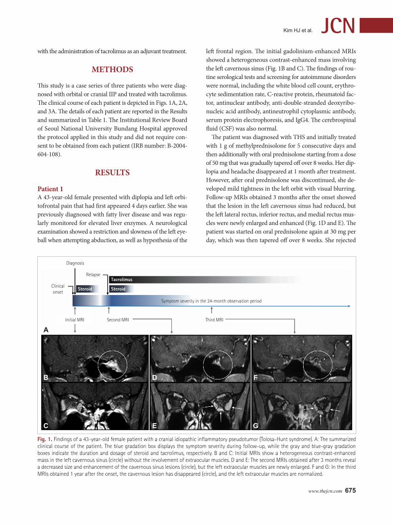
Patient 1A 43-year-old female presented with diplopia and left orbi-tofrontal pain that had first appeared 4 days earlier. She was previously diagnosed with fatty liver disease and was regu-larly monitored for elevated liver enzymes. A neurological examination showed a restriction and slowness of the left eye-ball when attempting abduction, as well as hypesthesia of the
left frontal region. The initial gadolinium-enhanced MRIs showed a heterogeneous contrast-enhanced mass involving the left cavernous sinus (Fig. 1B and C). The findings of rou-tine serological tests and screening for autoimmune disorders were normal, including the white blood cell count, erythro-cyte sedimentation rate, C-reactive protein, rheumatoid fac-tor, antinuclear antibody, anti-double-stranded deoxyribo-nucleic acid antibody, antineutrophil cytoplasmic antibody, serum protein electrophoresis, and IgG4. The cerebrospinal fluid (CSF) was also normal.
The patient was diagnosed with THS and initially treated with 1 g of methylprednisolone for 5 consecutive days and then additionally with oral prednisolone starting from a dose of 50 mg that was gradually tapered off over 8 weeks. Her dip-lopia and headache disappeared at 1 month after treatment. However, after oral prednisolone was discontinued, she de-veloped mild tightness in the left orbit with visual blurring. Follow-up MRIs obtained 3 months after the onset showed that the lesion in the left cavernous sinus had reduced, but the left lateral rectus, inferior rectus, and medial rectus mus-cles were newly enlarged and enhanced (Fig. 1D and E). The patient was started on oral prednisolone again at 30 mg per day, which was then tapered off over 8 weeks. She rejected
Fig. 1. Findings of a 43-year-old female patient with a cranial idiopathic inflammatory pseudotumor (Tolosa-Hunt syndrome). A: The summarized clinical course of the patient. The blue gradation box displays the symptom severity during follow-up, while the gray and blue-gray gradation boxes indicate the duration and dosage of steroid and tacrolimus, respectively. B and C: Initial MRIs show a heterogeneous contrast-enhanced mass in the left cavernous sinus (circle) without the involvement of extraocular muscles. D and E: The second MRIs obtained after 3 months reveal a decreased size and enhancement of the cavernous sinus lesions (circle), but the left extraocular muscles are newly enlarged. F and G: In the third MRIs obtained 1 year after the onset, the cavernous lesion has disappeared (circle), and the left extraocular muscles are normalized.
Diagnosis
Initial MRI
RelapseTacrolimus
SteroidSteroid
Second MRI
Symptom severity in the 24-month observation period
Third MRI
Clinical onset
B
C
D
E
F
G
A
676 J Clin Neurol 2020;16(4):674-680
Tacrolimus for Orbital and Cranial Inflammatory PseudotumorsJCN
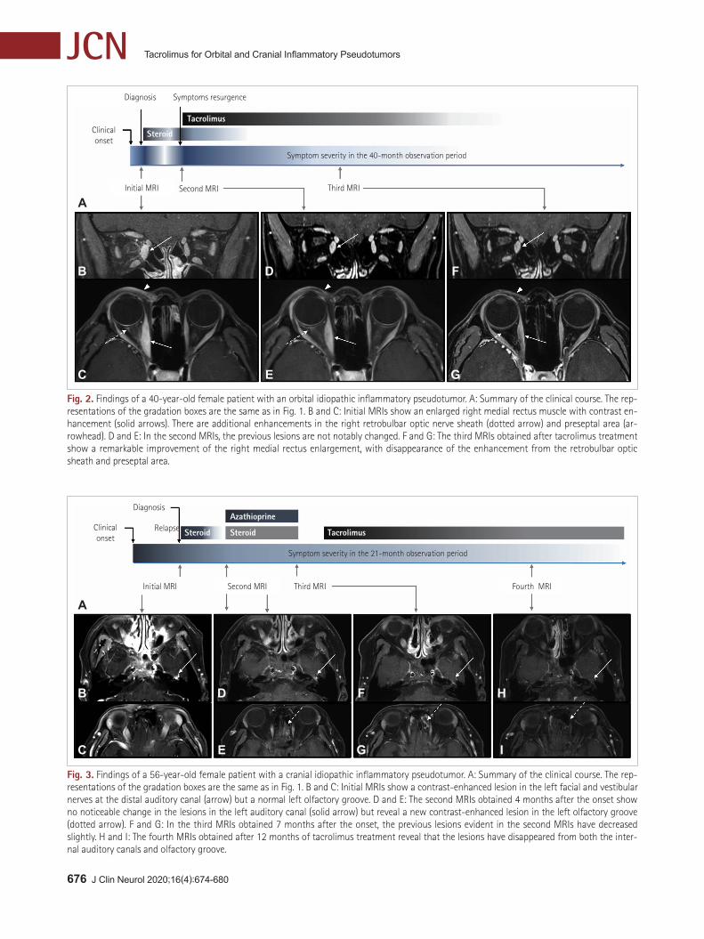
Fig. 3. Findings of a 56-year-old female patient with a cranial idiopathic inflammatory pseudotumor. A: Summary of the clinical course. The rep-resentations of the gradation boxes are the same as in Fig. 1. B and C: Initial MRIs show a contrast-enhanced lesion in the left facial and vestibular nerves at the distal auditory canal (arrow) but a normal left olfactory groove. D and E: The second MRIs obtained 4 months after the onset show no noticeable change in the lesions in the left auditory canal (solid arrow) but reveal a new contrast-enhanced lesion in the left olfactory groove (dotted arrow). F and G: In the third MRIs obtained 7 months after the onset, the previous lesions evident in the second MRIs have decreased slightly. H and I: The fourth MRIs obtained after 12 months of tacrolimus treatment reveal that the lesions have disappeared from both the inter-nal auditory canals and olfactory groove.
Diagnosis
Initial MRI
Relapse TacrolimusSteroid
Azathioprine
Steroid
Second MRI Fourth MRI
Symptom severity in the 21-month observation period
Third MRI
Clinical onset
B
C
D
E
F H
G I
A
Fig. 2. Findings of a 40-year-old female patient with an orbital idiopathic inflammatory pseudotumor. A: Summary of the clinical course. The rep-resentations of the gradation boxes are the same as in Fig. 1. B and C: Initial MRIs show an enlarged right medial rectus muscle with contrast en-hancement (solid arrows). There are additional enhancements in the right retrobulbar optic nerve sheath (dotted arrow) and preseptal area (ar-rowhead). D and E: In the second MRIs, the previous lesions are not notably changed. F and G: The third MRIs obtained after tacrolimus treatment show a remarkable improvement of the right medial rectus enlargement, with disappearance of the enhancement from the retrobulbar optic sheath and preseptal area.
Diagnosis Symptoms resurgence
Initial MRI
Tacrolimus
Steroid
Second MRI
Symptom severity in the 40-month observation period
Third MRI
Clinical onset
B
C
D
E
F
G
A
www.thejcn.com 677
Kim HJ et al. JCN
taking azathioprine or mycophenolate mofetil due to poten-tial liver toxicity, and instead was maintained on 3 mg of ta-crolimus along with the prednisolone. The second follow-up MRIs performed 1 year after the onset revealed that the le-sions had disappeared (Fig. 1F and G). Tacrolimus was grad-ually tapered for 6 months before being discontinued, and no relapse occurred during a subsequent 1-year follow-up.
Patient 2A 40-year-old female with chronic hepatitis C was referred for an evaluation of right periorbital pain, right eye ptosis, and diplopia that had first appeared 20 days earlier. She had re-stricted adduction of the right eyeball, but her eye velocity was normal within the available ocular motor range. There was painful tenderness of the right medial orbital region. The initial MRIs showed enlargement of the right medial rectus muscle with mild contrast enhancement, and additional le-sions in the right retrobulbar optic nerve sheath and presep-tal area (Fig. 2B and C). Routine serological tests, autoimmune screening tests, and CSF studies all produced normal find-ings. However, her hepatitis C virus RNA level was 1,724,673 IU/mL in the quantification test, indicating active hepatitis C.
The patient was diagnosed with orbital IIP and started on 60 mg of prednisone daily for 10 days, which greatly improved her symptoms. The dosage was gradually reduced over the next 8 weeks, but her symptoms relapsed when prednisone was reduced to below 20 mg per day. Follow-up MRIs ob-tained at the resurgence of symptoms showed no noticeable change compared with the previous MRIs (Fig. 2D and E). Be-cause of its harmful effect on chronic hepatitis C, long-term corticosteroid treatment could not be administered. Azathi-oprine and mycophenolate mofetil also could not be admin-istered because of potential direct hepatotoxicity. The patient was started on 3 mg of tacrolimus while discontinuing pred-
nisolone over 8 weeks. After 6 months of tacrolimus admin-istration, the right periorbital pain had completely disap-peared, and the restriction of right eye abduction had resolved. The second follow-up MRIs were obtained at 16 months af-ter the onset, by which time the lesions had nearly resolved (Fig. 2F and G). Tacrolimus was gradually reduced over a pe-riod of 7 months before being discontinued, and the patient had no further relapse during the 1-year follow-up.
Patient 3A 56-year-old previously healthy female presented with spon-taneous vertigo that had first appeared more than 2 weeks earlier. She had experienced left-sided facial palsy 2 months previously. A bedside examination showed spontaneous right-beating nystagmus that became stronger during rightward gaze or in darkness without visual fixation, and positive right-ward head-impulse signs indicative of acute left vestibulop-athy, in addition to preexisting complete left facial palsy. The initial MRIs revealed segmental enhancement in the left fa-cial and vestibular nerves at the distal auditory canal (Fig. 3B and C). The findings of serological and CSF evaluations for infectious and autoimmune disorders were normal.
The patient was diagnosed with an inflammatory disorder of uncertain origin that involved the left internal auditory ca-nal. She initially took oral prednisone at 1 mg/kg for 7 days. The dosage was then rapidly reduced over the following 4 weeks due to excessive weight gain, formation of a moon face, nervousness, and stomach irritation. The spontaneous nys-tagmus disappeared, but she still suffered from fluctuating dizziness. Follow-up MRIs obtained 4 months after the on-set showed stable lesions in the left auditory canal and newly developed contrast-enhanced lesions in the left olfactory groove and right high-cervical lymph node (Fig. 3D and E). The findings of the second evaluations for autoimmune and
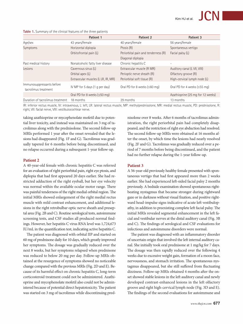
Table 1. Summary of the clinical features of the three patients
Patient 1 Patient 2 Patient 3Age/sex 43 years/female 40 years/female 56 years/female
Symptoms Horizontal diplopia Ptosis (R) Spontaneous vertigo
Orbitofrontal pain (L) Periorbital pain and tenderness (R) Facial palsy (L)
Diagonal diplopia
Past medical history Nonalcoholic fatty liver disease Chronic hepatitis C -
Lesions Cavernous sinus (L) Extraocular muscle (R MR) Auditory canal (L VII, VIII)
Orbital apex (L) Perioptic nerve sheath (R) Olfactory groove (R)
Extraocular muscles (L LR, IR, MR) Periorbital soft tissue (R) High-cervical lymph node (L)
Immunosuppressants before tacrolimus treatment
IV MP for 5 days (1 g per day) Oral PD for 8 weeks (≤60 mg) Oral PD for 4 weeks (≤55 mg)
Oral PD for 8 weeks (≤50 mg) Azathioprine (25 mg for 12 weeks)
Duration of tacrolimus treatment 18 months 29 months 13 months
IR: inferior rectus muscle, IV: intravenous, L: left, LR: lateral rectus muscle, MP: methylprednisolone, MR: medial rectus muscle, PD: prednisolone, R: right, VII: facial nerve, VIII: vestibulocochlear nerve.
678 J Clin Neurol 2020;16(4):674-680
Tacrolimus for Orbital and Cranial Inflammatory PseudotumorsJCN
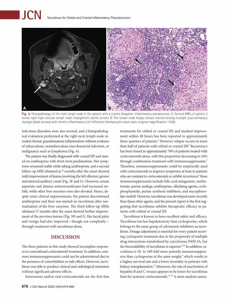
infectious disorders were also normal, and a histopatholog-ical evaluation performed at the right neck lymph node re-vealed chronic granulomatous inflammation without evidence of tuberculosis, nontuberculous mycobacterial infection, or malignancy such as lymphoma (Fig. 4).
The patient was finally diagnosed with cranial IIP and start-ed on azathioprine with short-term prednisolone. Her symp-toms remained stable while taking azathioprine, and a second follow-up MRI obtained at 7 months after the onset showed mild improvement of lesions involving the left olfactory groove and internal auditory canal (Fig. 3F and G). However, serum aspartate and alanine aminotransferases had increased six-fold, while other liver enzymes were also elevated. Hence, de-spite some clinical improvements, the patient discontinued azathioprine and then was started on tacrolimus after nor-malization of the liver enzymes. The third follow-up MRIs obtained 17 months after the onset showed further improve-ment of the previous lesions (Fig. 3H and I). Her facial palsy and vertigo had also improved—though not completely—through treatment with tacrolimus alone.
DISCUSSION
The three patients in this study showed incomplete respons-es to conventional corticosteroid treatment. In addition, com-mon immunosuppressants could not be administered due to the presence of comorbidities or side effects. However, tacro-limus was able to produce clinical and radiological remission without significant adverse effects.
Intravenous and/or oral corticosteroids are the first-line
treatments for orbital or cranial IIP, and marked improve-ment within 48 hours has been reported in approximately three-quarters of patients.6 However, relapse occurs in more than half of patients with orbital or cranial IIP.2 Recurrence has been found in approximately 78% of patients treated with corticosteroids alone, with this proportion decreasing to 16% through combination treatment with immunosuppressants.7 Therefore, immunosuppressants could be empirically used with corticosteroids to improve symptoms, at least in patients who are resistant to corticosteroids or exhibit recurrence;8 these immunosuppressants include folic acid antagonists, metho-trexate, purine analogs, azathioprine, alkylating agents, cyclo-phosphamide, purine synthesis inhibitors, and mycopheno-late mofetil. However, tacrolimus was developed more recently than these other agents, and the present report is the first sug-gesting that tacrolimus exhibits therapeutic efficacy in pa-tients with orbital or cranial IIP.
Tacrolimus is known to have excellent safety and efficacy. Tacrolimus has less hepatotoxicity than cyclosporine, which belongs to the same group of calcineurin inhibitors as tacro-limus. Dosage adjustment is essential for every patient receiv-ing cyclosporin treatment due to the propensity of multiple drug interactions metabolized by cytochrome P450 3A, but the bioavailability of tacrolimus is superior.9,10 In addition, ta-crolimus is 10- to 100-fold more potently immunosuppres-sive than cyclosporine at the same weight,9 which results in a higher survival rate and a lower mortality in patients with kidney transplantation.11 Moreover, the rate of reactivation of hepatitis B and C viruses appears to be lower for tacrolimus than for systemic corticosteroids.12-16 A meta-analysis assess-
A B Fig. 4. Histopathology of the neck lymph node in the patient with a cranial idiopathic inflammatory pseudotumor. A: Second MRIs of patient 3 reveal right high-cervical lymph node enlargement (white arrow). B: The lymph node biopsy shows nonnecrotizing multiple granulomatous changes (black arrows) with chronic inflammatory cell infiltration (hematoxylin-eosin stain, original magnification ×200).

www.thejcn.com 679
Kim HJ et al. JCNing the risk of adverse events in patients with systemic lupus erythematosus found that the rate of serious adverse events was lower for tacrolimus than for other immunosuppressants such as glucocorticoids, cyclosporine, azathioprine, cyclo-phosphamide, mycophenolate mofetil, methotrexate, and rituximab.17 Tacrolimus was also suitable for controlling oc-ular inflammatory diseases including Behçet’s disease and Vogt-Koyanagi-Harada syndrome without inducing increas-es in infection and malignancy.18
The therapeutic mechanism of tacrolimus in orbital or cra-nial IIP remains to be determined, but at least two mecha-nisms can be speculated. First, the immunosuppressive mech-anism of tacrolimus might be similar to that of cyclosporine, which can also be efficacious in orbital IIP. Second, IIP occurs in the CNS and was histologically revealed to be associated with a high density of IgG4-positive plasma cells, which sug-gests that a considerable proportion of IIP cases belong to the subgroup of IgG4-related syndromes.19 Specimens obtained from patients with orbital IIP show high levels of interleukin (IL)-2, IL-8, IL-10, IL-12, interferon (IFN)-γ, and tumor ne-crosis factor (TNF)-α.1 Tacrolimus is known to inhibit IL-2, IL-3, IL-4, TNF-α, CD40L, granulocyte-macrophage colony-stimulating factor, and IFN-γ. It also prevents T-cell activa-tion and inhibits follicular helper T cells, which play a key role in IgG4 production.20 Therefore, unlike steroid and azathio-prine that interrupt the cell cycle of lymphoid cells and in-hibit the production of both B and T cells, tacrolimus might exert its therapeutic effects on IIP mainly by affecting T-cell immune mechanisms.
There are some aspects to consider when interpreting the findings of this study. First, the effect of tacrolimus observed in all three patients could be distinguished from the delayed effects of steroids or azathioprine. There would have been a belated partial effect of prednisolone, but the duration of an-ti-inflammatory effects of prednisolone is known to be rela-tively short, at typically 12–36 hours.21 While tapering ste-roids, all patients showed the aggravation or recurrence of symptoms. The effects of azathioprine are usually expected to occur at 2–3 months after initiating the therapy.22 In pa-tient 3, azathioprine might have been discontinued before the effects fully developed. Thus, the previous immunosup-pressive treatments with azathioprine do not necessarily dis-prove the effect of tacrolimus in our patients.
The second aspect to consider is whether relapsing or re-fractory IIP can be clearly differentiated from other disorders including IgG4-related diseases, sarcoidosis, lymphoma, or chronic infection such as tuberculosis. The lack of a histolog-ical confirmation would not completely exclude these disor-ders. However, performing a biopsy of a brain lesion is often difficult. Thus, extensive serological and imaging evaluations
along with careful monitoring of treatment responses are necessary, and this protocol can lead to a reliable diagnosis of IIP, as shown by the present study.
In summary, tacrolimus can be an alternative option when treating patients with orbital or cranial IIP who exhibit an insufficient response to steroids, need long-term treatments, or have comorbidities restricting the use of other immuno-suppressants. Further investigations are necessary to confirm the efficacy of tacrolimus.
Author Contributions Conceptualization: Jeong-Yoon Choi. Data curation: Jeong-Yoon Choi, Hyun Jae Kim, Eunjin Kwon. Formal analysis: Seonkyung Lee, Yu Jin Koo, Eunjin Kwon, Hyo-Jung Kim, Jeong-Yoon Choi, Ji-Soo Kim. Supervision: Jeong-Yoon Choi. Writing—original draft: Hyun Jae Kim, Jeong-Yoon Choi. Writing—review & editing: Jeong-Yoon Choi, Ji-Soo Kim.
ORCID iDsHyun Jae Kim https://orcid.org/0000-0002-3508-9856Seonkyung Lee https://orcid.org/0000-0002-6880-6217Yu Jin Koo https://orcid.org/0000-0003-4650-8849Eunjin Kwon https://orcid.org/0000-0001-9057-9009Hyo-Jung Kim https://orcid.org/0000-0002-2027-6341Jeong-Yoon Choi https://orcid.org/0000-0003-2159-9967Ji-Soo Kim https://orcid.org/0000-0002-1508-2024
Conflicts of InterestDr. JS Kim serves as an associate editor of Frontiers in Neuro-otology and on the editorial boards of the Journal of Clinical Neurology, Fron-tiers in Neuro-ophthalmology, Journal of Neuro-ophthalmology, Journal of Vestibular Research, Journal of Neurology, and Medicine. The other authors have nothing to disclose.
AcknowledgementsThis study was supported by the Basic Science Research Program through the National Research Foundation of Korea (NRF) funded by the Ministry of Science and ICT (2020R1A2C4002281).
REFERENCES1. Yeşiltaş YS, Gündüz AK. Idiopathic orbital inflammation: review of
literature and new advances. Middle East Afr J Ophthalmol 2018;25: 71-80.
2. Carreón E, Muñiz S, Di Capua D, Porta-Etessam J. Tolosa-Hunt syn-drome with spontaneous remission and recurrence. Neurologia 2018; 33:68-70.
3. Mombaerts I, Koornneef L. Current status in the treatment of orbital myositis. Ophthalmology 1997;104:402-408.
4. Yan J, Wu P. Idiopathic orbital myositis. J Craniofac Surg 2014;25: 884-887.
5. Dheer D, Jyoti, Gupta PN, Shankar R. Tacrolimus: an updated review on delivering strategies for multifarious diseases. Eur J Pharm Sci 2018;114:217-227.
6. Chaudhry IA, Shamsi FA, Arat YO, Riley FC. Orbital pseudotumor: distinct diagnostic features and management. Middle East Afr J Oph-thalmol 2008;15:17-27.
7. Arthur A, Sivadasan A, Mannam P, Prabakhar AT, Aaron S, Mathew V, et al. Tolosa-Hunt syndrome: long-term outcome and role of ste-roid-sparing agents. Ann Indian Acad Neurol 2020;23:201-205.
8. Schoser BG. Ocular myositis: diagnostic assessment, differential di-agnoses, and therapy of a rare muscle disease - five new cases and re-
680 J Clin Neurol 2020;16(4):674-680
Tacrolimus for Orbital and Cranial Inflammatory PseudotumorsJCNview. Clin Ophthalmol 2007;1:37-42.
9. Lake DF, Briggs AD. Immunopharmacology. In: Katzung BG, editor. Basic & Clinical Pharmacology. 14th ed. New York, NY: McGraw-Hill, 2017.
10. Sánchez-Román J, Varela-Aguilar JM, Bravo-Ferrer J, Sequeiros Ma-dueño E, Fernández de Bobadilla M. Idiopathic orbital myositis: treat-ment with cyclosporin. Ann Rheum Dis 1993;52:84-85.
11. Boots JM. Is tacrolimus superior to ciclosporin microemulsion in pre-venting long-term acute renal transplant rejection? Nat Clin Pract Nephrol 2005;1:16-17.
12. Vento S, Cainelli F, Mirandola F, Cosco L, Di Perri G, Solbiati M, et al. Fulminant hepatitis on withdrawal of chemotherapy in carriers of hepatitis C virus. Lancet 1996;347:92-93.
13. Perrillo RP. Hepatitis B reactivation from immunosuppressive drug therapy: a global menace. Clin Liver Dis (Hoboken) 2015;5:39-42.
14. Bessone F, Dirchwolf M. Management of hepatitis B reactivation in immunosuppressed patients: an update on current recommendations. World J Hepatol 2016;8:385-394.
15. Loomba R, Liang TJ. Hepatitis B reactivation associated with im-mune suppressive and biological modifier therapies: current concepts, management strategies, and future directions. Gastroenterology 2017; 152:1297-1309.
16. Tran-Minh ML, Sousa P, Maillet M, Allez M, Gornet JM. Hepatic complications induced by immunosuppressants and biologics in in-flammatory bowel disease. World J Hepatol 2017;9:613-626.
17. Tian J, Luo Y, Wu H, Long H, Zhao M, Lu Q. Risk of adverse events from different drugs for SLE: a systematic review and network meta-analysis. Lupus Sci Med 2018;5:e000253.
18. Hornbeak DM, Thorne JE. Immunosuppressive therapy for eye dis-eases: effectiveness, safety, side effects and their prevention. Taiwan J Ophthalmol 2015;5:156-163.
19. Lui PC, Fan YS, Wong SS, Chan AN, Wong G, Chau TK, et al. In-flammatory pseudotumors of the central nervous system. Hum Pathol 2009;40:1611-1617.
20. Takanashi S, Kaneko Y, Takeuchi T. Effectiveness of tacrolimus on IgG4-related disease. Mod Rheumatol 2019;29:892-894.
21. Liu D, Ahmet A, Ward L, Krishnamoorthy P, Mandelcorn ED, Leigh R, et al. A practical guide to the monitoring and management of the complications of systemic corticosteroid therapy. Allergy Asthma Clin Immunol 2013;9:30.
22. Anstey AV, Wakelin S, Reynolds NJ; British Association of Derma-tologists Therapy, Guidelines and Audit Subcommittee. Guidelines for prescribing azathioprine in dermatology. Br J Dermatol 2004;151: 1123-1132.