Embed Size (px)
Citation preview
RESEARCH Open Access
Impaired mitochondrial calcium uptakecaused by tacrolimus underlies beta-cellfailureAngela Lombardi1, Bruno Trimarco2, Guido Iaccarino3 and Gaetano Santulli1,2*
Abstract
Background: One of the most common side effects of the immunosuppressive drug tacrolimus (FK506) is the increasedrisk of new-onset diabetes mellitus. However, the molecular mechanisms underlying this association have not been fullyclarified.
Methods: We studied the effects of the therapeutic dose of tacrolimus on mitochondrial fitness in beta-cells.
Results: We demonstrate that tacrolimus impairs glucose-stimulated insulin secretion (GSIS) in beta-cells througha previously unidentified mechanism. Indeed, tacrolimus causes a decrease in mitochondrial Ca2+ uptake, accompaniedby altered mitochondrial respiration and reduced ATP production, eventually leading to impaired GSIS.
Conclusion: Our observations individuate a new fundamental mechanism responsible for the augmented incidence ofdiabetes following tacrolimus treatment. Indeed, this drug alters Ca2+ fluxes in mitochondria, thereby compromisingmetabolism-secretion coupling in beta-cells.
Keywords: Mitochondrial calcium, ATP, Diabetes, Insulin release, Immunosuppressive regimen, Ca2+ leak
BackgroundTacrolimus (also known as fujimycin and FK506) is amacrolide lactone isolated from Streptomyces tsukubaensis,currently used as potent immunosuppressant in organtransplantation to reduce rejection rates [1, 2]. One of itsmost common adverse effects is new-onset diabetes melli-tus following transplantation [3–7], a serious complicationthat also increases the risk of infection and cardiovasculardisease [8]. Indeed, a 5-year follow-up study monitoring pa-tients treated with tacrolimus after transplant revealed anincidence of diabetes of 41% [9].The exact mechanisms underlying the diabetogenic
effects of tacrolimus have not been fully elucidated. Vari-ous studies have suggested that tacrolimus side effects areattributable to its peripheral action, engendering a mark-edly reduced insulin sensitivity [10–12]. We hypothesizethat tacrolimus at therapeutic dosage has also a direct
detrimental effect on beta-cells, in particular on mitochon-drial Ca2+ dynamics. To test our hypothesis, we evaluatedthe specific effects of tacrolimus on beta-cells.
MethodsCell culture and drugsINS-1 beta-cells (AddexBio, San Diego, CA) were cul-tured in a humidified atmosphere (37 °C) containing 5%CO2 in RPMI-1640 medium, and insulin levels were de-termined as previously described and validated by ourgroup [13–15]. In some experiments, cells were treatedwith glucose (Bio-Techne, Abingdon, UK), L-leucine andglutamine (both from MyBioSource, San Diego, CA,USA), KCl (Merck KGaA, Darmstadt Germany) ortacrolimus (LC Laboratories, Woburn, MA, dissolved indymethylsulfoxide).
Cell viability assayCell viability was evaluated using the [3-(4,5-dimethyl-thiazol-2-yl)-2,5-diphenyl tetrazolium bromide (MTT)assay, as described [16, 17].
* Correspondence: [email protected] of Medicine, Albert Einstein College of Medicine, New York, NY,USA2Department of Advanced Biomedical Sciences, “Federico II” University ofNaples, Naples, ItalyFull list of author information is available at the end of the article
© The Author(s). 2017 Open Access This article is distributed under the terms of the Creative Commons Attribution 4.0International License (http://creativecommons.org/licenses/by/4.0/), which permits unrestricted use, distribution, andreproduction in any medium, provided you give appropriate credit to the original author(s) and the source, provide a link tothe Creative Commons license, and indicate if changes were made. The Creative Commons Public Domain Dedication waiver(http://creativecommons.org/publicdomain/zero/1.0/) applies to the data made available in this article, unless otherwise stated.
Lombardi et al. Cell Communication and Signaling (2017) 15:47 DOI 10.1186/s12964-017-0203-0
Ca2+ measurementsCa2+ imaging experiments were carried out as previouslydescribed [15, 18–20]. Briefly, to assess mitochondrialCa2+, cells were loaded with Rhod-2 AM (3 μM, ThermoFisher Scientific, Waltham, MA) at 37 °C for 30 min,followed by washout and 1 h rest at room temperaturefor de-esterification. Because of its delocalized positivecharge, Rhod-2 AM accumulates preferentially withinthe mitochondrial matrix, where it is hydrolyzed andtrapped [15, 20, 21]. Fluorescence was detected using apass-band filter of 545–625 nm in response to excitationat 542 nm. Ca2+ mobilization from the ER followingcaffeine (10 mM, Biorbyt, Cambridge, UK) stimulationwas assessed loading the cells with Fura-2 acetoxymethyl(AM) ester (Thermo Fisher Scientific, 5 μM, 15 min,37 °C), as described [15, 18]; images were obtained usinga dual excitation fluorescence imaging system: changesin intracellular Ca2+ were reflected in the ratio of fluor-escence emission acquired above 510 nm in response toexcitation at 340 nm and 380 nm. Intracellular Ca2+ leakwas assessed spectrophotometrically in microsomesobtained from pancreatic beta-cells, as previously de-scribed [15]. Besides the above mentioned indirectevaluation of ER Ca2+ in response to caffeine, ER Ca2+
content was assessed using the FRET-based camaleonD1ER (Addgene, Cambridge, MA) [22].
Mitochondrial respirationMitochondrial respiration was assessed using the SeahorseAnalyzer (Agilent Technologies, Santa Clara, CA, USA),adding to each well glucose (16.7 mM), oligomycin (1 μM,Merck KGaA), carbonyl cyanide 4-(trifluoromethoxy)phenylhydrazone (FCCP, 0.5 μM, Merck KGaA), rotenoneand antimycin A (both 1 μM, Merck KGaA). Aftereach assay, cells were collected to quantify DNAusing QuantiFluor dsDNA System (Promega,Madison, WI, USA).
Cytochrome c oxidase activity assayThe cells were permeabilized by freeze-thaw cycle threetimes and mixed with detergent solution (0.1% bovineserum albumin, 250 mM sucrose, 10 mM KH2PO4
2.5 mM laurylmaltoside, all from Merck KGaA). Theenzymatic activity of cytochrome c oxidase wasspectrophotometrically measured at 550 nm.
ATP/ADP measurementThe genetically encoded sensors PercevalHR and pHRed(Addgene) were co-transfected in the cells in order tomeasure relative intracellular changes in ATP/ADP andpH, respectively. PercevalHR was excited at 405 nm(ADP) and 488 nm (ATP) and emission was collected at530 nm. pHRed was excited using 405 nm and 546 nmand emission was collected at 630 nm band pass filter.
Since PercevalHR has been reported to be sensitive topH [23], a correction for pH was performed.
Statistical analysisExperiments were performed in a blinded fashion atleast three times, unless otherwise noted. All results arepresented as mean ± SEM. Statistical analysis wasperformed via unpaired t test when comparing twogroups (normal distribution was confirmed byAnderson-Darling test) and one-way ANOVA, followedby Tukey-Kramer post hoc correction, when comparingmore than two groups. A P value <0.05 was consideredstatistically significant.
ResultsGlucose-induced insulin secretion (GSIS) is reducedfollowing tacrolimus treatmentTo evaluate the effects of tacrolimus on beta-cellfunction, we measured GSIS in INS-1 beta-cells. In adose-response assay, we found that the treatment with5 nM tacrolimus, which is the average level that hasbeen measured in the blood of patients undergoingorgan transplantation [24], was sufficient to determine asignificant reduction of GSIS compared with vehicle(Fig. 1a). The same dose (5 nM) was then tested in atime-course assay, which revealed that 24-h incubationmarkedly impaired GSIS (Fig. 1b). Importantly, tacrolimushad a significantly detrimental effect on the metabolic re-sponse of beta-cells to leucine and glutamine (Fig. 1c),strongly suggesting an alteration in mitochondrialoxidative metabolism. Supporting this view, insulinsecretion in response to cell depolarization obtained viaKCl, thereby bypassing mitochondria, was comparablebetween groups (Fig. 1c).
Therapeutic doses of tacrolimus do not affect beta-cellviabilityTo test the effect of 5 nM tacrolimus on cell viability, weperformed an MTT assay and we did not observe anysignificant effect on beta-cell viability (Fig. 1d).
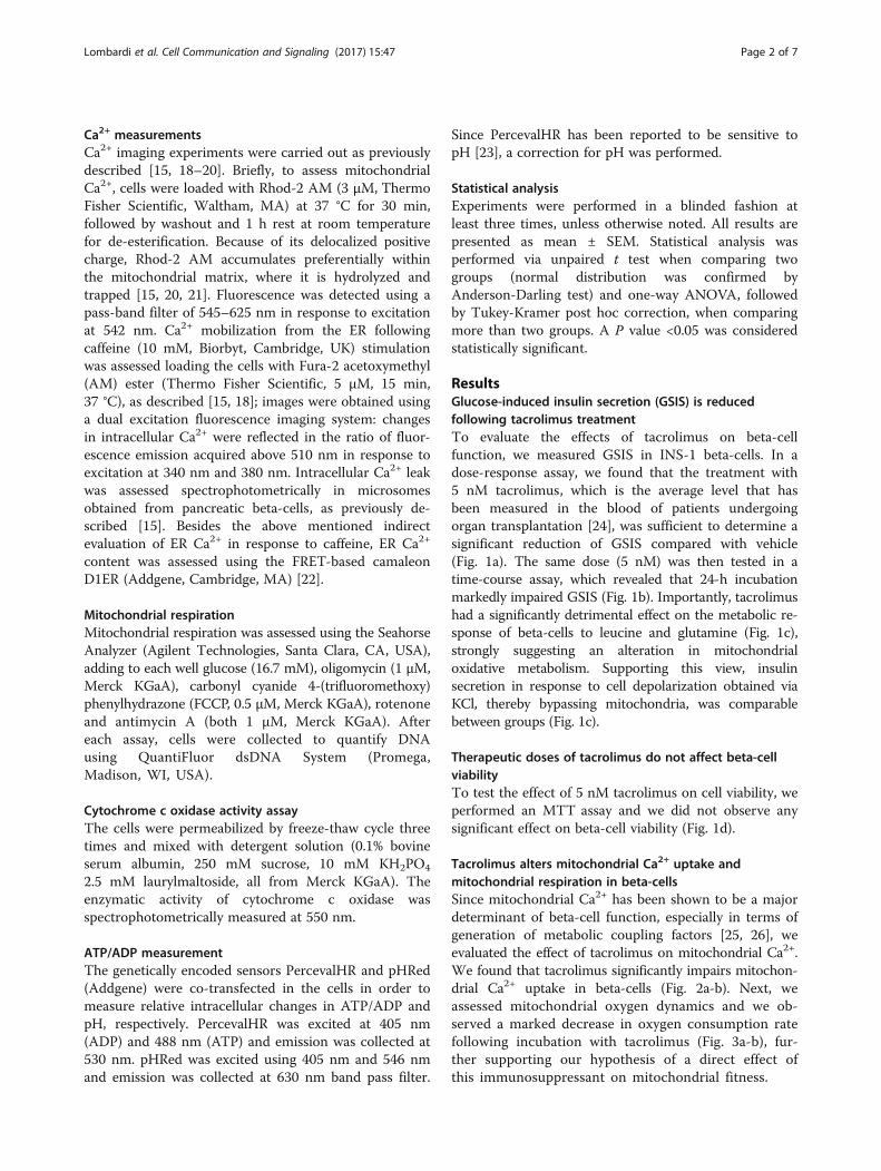
Tacrolimus alters mitochondrial Ca2+ uptake andmitochondrial respiration in beta-cellsSince mitochondrial Ca2+ has been shown to be a majordeterminant of beta-cell function, especially in terms ofgeneration of metabolic coupling factors [25, 26], weevaluated the effect of tacrolimus on mitochondrial Ca2+.We found that tacrolimus significantly impairs mitochon-drial Ca2+ uptake in beta-cells (Fig. 2a-b). Next, weassessed mitochondrial oxygen dynamics and we ob-served a marked decrease in oxygen consumption ratefollowing incubation with tacrolimus (Fig. 3a-b), fur-ther supporting our hypothesis of a direct effect ofthis immunosuppressant on mitochondrial fitness.
Lombardi et al. Cell Communication and Signaling (2017) 15:47 Page 2 of 7

Fig. 1 Therapeutic doses of tacrolimus alter insulin secretion without affecting cell viability. Insulin release measured in beta-cells incubated for 24 hwith vehicle or tacrolimus at the indicated doses (a) and with vehicle or 5 nM tacrolimus at the indicated times (b). Effect of fuel secretagogues leucine(Leu, 10 mM) and glutamine (Gln, 2 mM) on beta-cells treated for 24 h with vehicle or 5 nM tacrolimus (c). Cell viability assessed in beta-cells treatedfor 24 h with vehicle or 5 nM tacrolimus (d). Data are presented as mean ± S.E.M. of experiments performed at least in triplicate; *:p < 0.01 vs vehicle,ANOVA. In panel d, whiskers represent 1% to 99% spread of the data; NS: non-significant; t test
Fig. 2 Tacrolimus impairs mitochondrial Ca2+ uptake. Representative traces (a) of glucose-induced mitochondrial Ca2+ uptake evaluated in beta-cellsincubated for 24 h with vehicle or 5 nM Tacrolimus. b quantification of experiments (at least 35 cells per group were analyzed): whiskers represent 1%to 99% spread of the data; *:p < 0.001 vs vehicle
Lombardi et al. Cell Communication and Signaling (2017) 15:47 Page 3 of 7
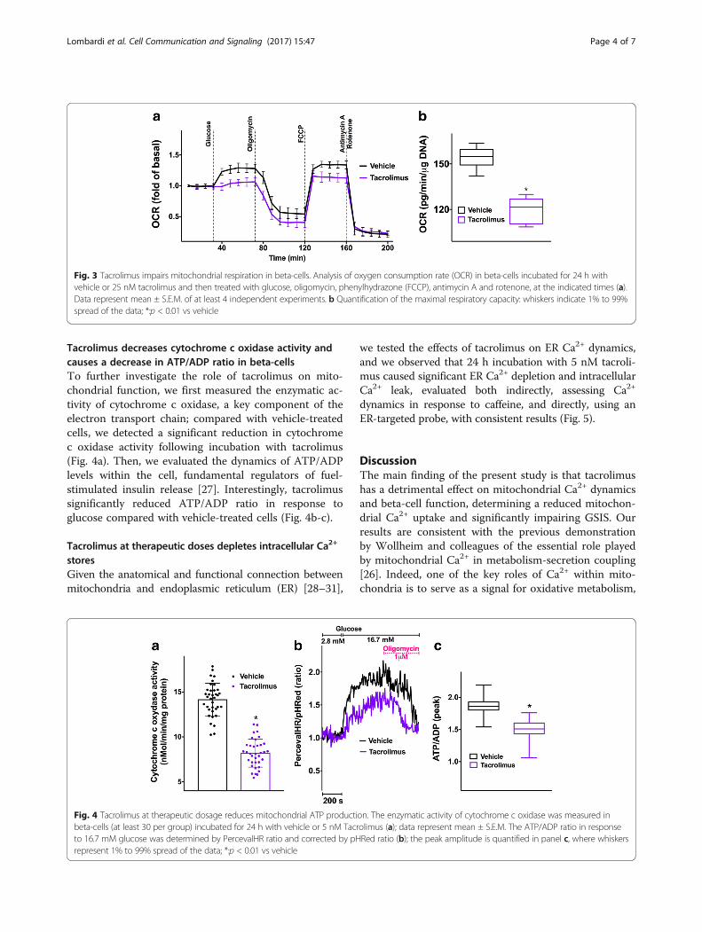
Tacrolimus decreases cytochrome c oxidase activity andcauses a decrease in ATP/ADP ratio in beta-cellsTo further investigate the role of tacrolimus on mito-chondrial function, we first measured the enzymatic ac-tivity of cytochrome c oxidase, a key component of theelectron transport chain; compared with vehicle-treatedcells, we detected a significant reduction in cytochromec oxidase activity following incubation with tacrolimus(Fig. 4a). Then, we evaluated the dynamics of ATP/ADPlevels within the cell, fundamental regulators of fuel-stimulated insulin release [27]. Interestingly, tacrolimussignificantly reduced ATP/ADP ratio in response toglucose compared with vehicle-treated cells (Fig. 4b-c).
Tacrolimus at therapeutic doses depletes intracellular Ca2+
storesGiven the anatomical and functional connection betweenmitochondria and endoplasmic reticulum (ER) [28–31],
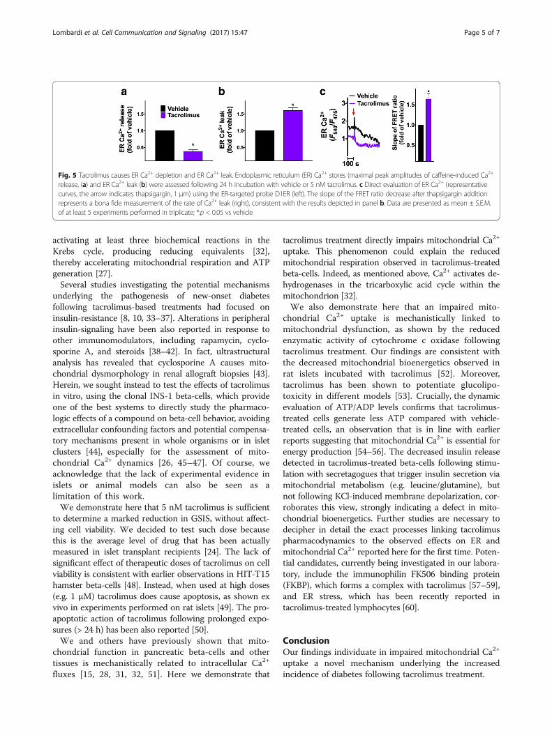
we tested the effects of tacrolimus on ER Ca2+ dynamics,and we observed that 24 h incubation with 5 nM tacroli-mus caused significant ER Ca2+ depletion and intracellularCa2+ leak, evaluated both indirectly, assessing Ca2+
dynamics in response to caffeine, and directly, using anER-targeted probe, with consistent results (Fig. 5).
DiscussionThe main finding of the present study is that tacrolimushas a detrimental effect on mitochondrial Ca2+ dynamicsand beta-cell function, determining a reduced mitochon-drial Ca2+ uptake and significantly impairing GSIS. Ourresults are consistent with the previous demonstrationby Wollheim and colleagues of the essential role playedby mitochondrial Ca2+ in metabolism-secretion coupling[26]. Indeed, one of the key roles of Ca2+ within mito-chondria is to serve as a signal for oxidative metabolism,
Fig. 3 Tacrolimus impairs mitochondrial respiration in beta-cells. Analysis of oxygen consumption rate (OCR) in beta-cells incubated for 24 h withvehicle or 25 nM tacrolimus and then treated with glucose, oligomycin, phenylhydrazone (FCCP), antimycin A and rotenone, at the indicated times (a).Data represent mean ± S.E.M. of at least 4 independent experiments. b Quantification of the maximal respiratory capacity: whiskers indicate 1% to 99%spread of the data; *:p < 0.01 vs vehicle
Fig. 4 Tacrolimus at therapeutic dosage reduces mitochondrial ATP production. The enzymatic activity of cytochrome c oxidase was measured inbeta-cells (at least 30 per group) incubated for 24 h with vehicle or 5 nM Tacrolimus (a); data represent mean ± S.E.M. The ATP/ADP ratio in responseto 16.7 mM glucose was determined by PercevalHR ratio and corrected by pHRed ratio (b); the peak amplitude is quantified in panel c, where whiskersrepresent 1% to 99% spread of the data; *:p < 0.01 vs vehicle
Lombardi et al. Cell Communication and Signaling (2017) 15:47 Page 4 of 7
activating at least three biochemical reactions in theKrebs cycle, producing reducing equivalents [32],thereby accelerating mitochondrial respiration and ATPgeneration [27].Several studies investigating the potential mechanisms
underlying the pathogenesis of new-onset diabetesfollowing tacrolimus-based treatments had focused oninsulin-resistance [8, 10, 33–37]. Alterations in peripheralinsulin-signaling have been also reported in response toother immunomodulators, including rapamycin, cyclo-sporine A, and steroids [38–42]. In fact, ultrastructuralanalysis has revealed that cyclosporine A causes mito-chondrial dysmorphology in renal allograft biopsies [43].Herein, we sought instead to test the effects of tacrolimusin vitro, using the clonal INS-1 beta-cells, which provideone of the best systems to directly study the pharmaco-logic effects of a compound on beta-cell behavior, avoidingextracellular confounding factors and potential compensa-tory mechanisms present in whole organisms or in isletclusters [44], especially for the assessment of mito-chondrial Ca2+ dynamics [26, 45–47]. Of course, weacknowledge that the lack of experimental evidence inislets or animal models can also be seen as alimitation of this work.We demonstrate here that 5 nM tacrolimus is sufficient
to determine a marked reduction in GSIS, without affect-ing cell viability. We decided to test such dose becausethis is the average level of drug that has been actuallymeasured in islet transplant recipients [24]. The lack ofsignificant effect of therapeutic doses of tacrolimus on cellviability is consistent with earlier observations in HIT-T15hamster beta-cells [48]. Instead, when used at high doses(e.g. 1 μM) tacrolimus does cause apoptosis, as shown exvivo in experiments performed on rat islets [49]. The pro-apoptotic action of tacrolimus following prolonged expo-sures (> 24 h) has been also reported [50].We and others have previously shown that mito-
chondrial function in pancreatic beta-cells and othertissues is mechanistically related to intracellular Ca2+
fluxes [15, 28, 31, 32, 51]. Here we demonstrate that
tacrolimus treatment directly impairs mitochondrial Ca2+
uptake. This phenomenon could explain the reducedmitochondrial respiration observed in tacrolimus-treatedbeta-cells. Indeed, as mentioned above, Ca2+ activates de-hydrogenases in the tricarboxylic acid cycle within themitochondrion [32].We also demonstrate here that an impaired mito-
chondrial Ca2+ uptake is mechanistically linked tomitochondrial dysfunction, as shown by the reducedenzymatic activity of cytochrome c oxidase followingtacrolimus treatment. Our findings are consistent withthe decreased mitochondrial bioenergetics observed inrat islets incubated with tacrolimus [52]. Moreover,tacrolimus has been shown to potentiate glucolipo-toxicity in different models [53]. Crucially, the dynamicevaluation of ATP/ADP levels confirms that tacrolimus-treated cells generate less ATP compared with vehicle-treated cells, an observation that is in line with earlierreports suggesting that mitochondrial Ca2+ is essential forenergy production [54–56]. The decreased insulin releasedetected in tacrolimus-treated beta-cells following stimu-lation with secretagogues that trigger insulin secretion viamitochondrial metabolism (e.g. leucine/glutamine), butnot following KCl-induced membrane depolarization, cor-roborates this view, strongly indicating a defect in mito-chondrial bioenergetics. Further studies are necessary todecipher in detail the exact processes linking tacrolimuspharmacodynamics to the observed effects on ER andmitochondrial Ca2+ reported here for the first time. Poten-tial candidates, currently being investigated in our labora-tory, include the immunophilin FK506 binding protein(FKBP), which forms a complex with tacrolimus [57–59],and ER stress, which has been recently reported intacrolimus-treated lymphocytes [60].
ConclusionOur findings individuate in impaired mitochondrial Ca2+
uptake a novel mechanism underlying the increasedincidence of diabetes following tacrolimus treatment.
Fig. 5 Tacrolimus causes ER Ca2+ depletion and ER Ca2+ leak. Endoplasmic reticulum (ER) Ca2+ stores (maximal peak amplitudes of caffeine-induced Ca2+
release, (a) and ER Ca2+ leak (b) were assessed following 24 h incubation with vehicle or 5 nM tacrolimus. c Direct evaluation of ER Ca2+ (representativecurves, the arrow indicates thapsigargin, 1 μm) using the ER-targeted probe D1ER (left). The slope of the FRET ratio decrease after thapsigargin additionrepresents a bona fide measurement of the rate of Ca2+ leak (right), consistent with the results depicted in panel b. Data are presented as mean ± S.E.M.of at least 5 experiments performed in triplicate; *:p < 0.05 vs vehicle
Lombardi et al. Cell Communication and Signaling (2017) 15:47 Page 5 of 7
AbbreviationsCa2+: Calcium; ER: Endoplasmic reticulum; FCCP: Carbonyl cyanide4-(trifluoromethoxy) phenylhydrazone; Fura-2 AM: Fura-2-acetoxymethylester; GSIS: Glucose-stimulated insulin secretion; Rhod2-AM: 1-[2-Amino-5-(3-dimethylamino-6- dimethylammonio- 9-xanthenyl) phenoxy]-2-(2-amino-5-methylphenoxy) ethane-N,N,N′,N′-tetra-acetic acid, tetra-acetoxymethyl ester
AcknowledgementsWe thank Drs. X. Du, TV. McDonald, RN. Kitsis, and Y. Tomer (Albert EinsteinCollege of Medicine) for helpful discussions.
FundingG.S. is supported by the NIH (R00DK107895) and ES-DRC P30DK20541.
Availability of data and materialsThe datasets supporting the conclusions of this article are included withinthis article.
Authors’ contributionsAL designed and performed experiments, analyzed data and wrote thepaper; BT and GI analyzed data and contributed to discussion; GS conceivedof the project and experimental plans, analyzed data, and wrote the paper.All authors read and approved the final manuscript.
Ethics approval and consent to participateNot applicable.
Consent for publicationNot applicable.
Competing interestsThe authors declare that they have no competing interests.
Publisher’s NoteSpringer Nature remains neutral with regard to jurisdictional claims inpublished maps and institutional affiliations.
Author details1Department of Medicine, Albert Einstein College of Medicine, New York, NY,USA. 2Department of Advanced Biomedical Sciences, “Federico II” Universityof Naples, Naples, Italy. 3Department of Medicine, Surgery and Dentistry,“Scuola Medica Salernitana”, University of Salerno, Fisciano, Italy.
Received: 20 September 2017 Accepted: 2 November 2017
References1. Wallemacq PE, Reding R. FK506 (tacrolimus), a novel immunosuppressant in
organ transplantation: clinical, biomedical, and analytical aspects. Clin Chem.1993;39:2219–28.
2. Nankivell BJ, Borrows RJ, Fung CL, O'Connell PJ, Allen RD, Chapman JR. Thenatural history of chronic allograft nephropathy. N Engl J Med. 2003;349:2326–33.
3. Chanchlani R, Joseph Kim S, Kim ED, Banh T, Borges K, Vasilevska-Ristovska J,Li Y, Ng V, Dipchand AI, Solomon M, et al. Incidence of hyperglycemia anddiabetes and association with electrolyte abnormalities in pediatric solidorgan transplant recipients. Nephrol Dial Transplant. 2017;32:1579–86.
4. Regelmann MO, Goldis M, Arnon R. New-onset diabetes mellitus afterpediatric liver transplantation. Pediatr Transplant. 2015;19:452–9.
5. Andrade AR, Bittencourt PL, Codes L, Evangelista MA, Castro AO, Sorte NB,Almeida CG, Bastos JA, Cotrim HP. New onset diabetes and non-alcoholicfatty liver disease after liver transplantation. Ann Hepatol. 2017;16:932–40.
6. Rangel EB. Tacrolimus in pancreas transplant: a focus on toxicity,diabetogenic effect and drug-drug interactions. Expert Opin Drug MetabToxicol. 2014;10:1585–605.
7. Santos AH Jr, Chen C, Casey MJ, Womer KL, Wen X. New-onset diabetesafter kidney transplantation: can the risk be modified by choosingimmunosuppression regimen based on pretransplant viral serology?Nephrol Dial Transplant. 2017; in press. https://www.ncbi.nlm.nih.gov/pubmed/29045704.
8. Sessa A, Esposito A, Giliberti A, Iavicoli G, Costa C, Bergallo M, Lettieri E,Rossano R, Capuano M. Immunosuppressive agents and metabolic factorsof cardiovascular risk in renal transplant recipients. Transplant Proc. 2009;41:1178–82.
9. Weir M. Impact of immunosuppressive regimes on posttransplant diabetesmellitus. Transplant Proc. 2001;33:23S–6S.
10. Montero N, Pascual J. Immunosuppression and post-transplanthyperglycemia. Curr Diabetes Rev. 2015;11:144–54.
11. Bhat M, Pasini E, Copeland J, Angeli M, Husain S, Kumar D, Renner E, TeterinaA, Allard J, Guttman DS, Humar A. Impact of immunosuppression on themetagenomic composition of the intestinal microbiome: a systems biologyapproach to post-transplant diabetes. Sci Rep. 2017;7:10277.
12. Rodriguez-Rodriguez AE, Trinanes J, Velazquez-Garcia S, Porrini E, VegaPrieto MJ, Diez Fuentes ML, Arevalo M, Salido Ruiz E, Torres A. The higherdiabetogenic risk of tacrolimus depends on pre-existing insulin resistance. Astudy in obese and lean Zucker rats. Am J Transplant. 2013;13:1665–75.
13. Santulli G, Lombardi A, Sorriento D, Anastasio A, Del Giudice C, Formisano P,Beguinot F, Trimarco B, Miele C, Iaccarino G. Age-related impairment ininsulin release: the essential role of beta(2)-adrenergic receptor. Diabetes.2012;61:692–701.
14. Lombardi A, Ulianich L, Treglia AS, Nigro C, Parrillo L, Lofrumento DD,Nicolardi G, Garbi C, Beguinot F, Miele C, Di Jeso B. Increased hexosaminebiosynthetic pathway flux dedifferentiates INS-1E cells and murine islets byan extracellular signal-regulated kinase (ERK)1/2-mediated signaltransmission pathway. Diabetologia. 2012;55:141–53.
15. Santulli G, Pagano G, Sardu C, Xie W, Reiken S, D'Ascia SL, Cannone M,Marziliano N, Trimarco B, Guise TA, et al. Calcium release channel RyR2regulates insulin release and glucose homeostasis. J Clin Invest. 2015;125:1968–78.
16. De Vitis S, Sonia Treglia A, Ulianich L, Turco S, Terrazzano G, Lombardi A,Miele C, Garbi C, Beguinot F, Di Jeso B. Tyr phosphatase-mediated P-ERKinhibition suppresses senescence in EIA + v-raf transformed cells, which,paradoxically, are apoptosis-protected in a MEK-dependent manner.Neoplasia. 2011;13:120–30.
17. Fiory F, Lombardi A, Miele C, Giudicelli J, Beguinot F, Van Obberghen E.Methylglyoxal impairs insulin signalling and insulin action on glucose-induced insulin secretion in the pancreatic beta cell line INS-1E.Diabetologia. 2011;54:2941–52.
18. Xie W, Santulli G, Guo X, Gao M, Chen BX, Marks AR. Imaging atrial arrhythmicintracellular calcium in intact heart. J Mol Cell Cardiol. 2013;64:120–3.
19. Umanskaya A, Santulli G, Xie W, Andersson DC, Reiken SR, Marks AR.Genetically enhancing mitochondrial antioxidant activity improves musclefunction in aging. Proc Natl Acad Sci U S A. 2014;111:15250–5.
20. Santulli G, Xie W, Reiken SR, Marks AR. Mitochondrial calcium overload is a keydeterminant in heart failure. Proc Natl Acad Sci U S A. 2015;112:11389–94.
21. Xie W, Santulli G, Reiken SR, Yuan Q, Osborne BW, Chen BX, Marks AR.Mitochondrial oxidative stress promotes atrial fibrillation. Sci Rep. 2015;5:11427.
22. Takeuchi A, Kim B, Matsuoka S. The mitochondrial Na+−Ca2+ exchanger,NCLX, regulates automaticity of HL-1 cardiomyocytes. Sci Rep. 2013;3:2766.
23. Tantama M, Martinez-Francois JR, Mongeon R, Yellen G. Imaging energystatus in live cells with a fluorescent biosensor of the intracellular ATP-to-ADP ratio. Nat Commun. 2013;4:2550.
24. Desai NM, Goss JA, Deng S, Wolf BA, Markmann E, Palanjian M, Shock AP,Feliciano S, Brunicardi FC, Barker CF, et al. Elevated portal vein drug levels ofsirolimus and tacrolimus in islet transplant recipients: localimmunosuppression or islet toxicity? Transplantation. 2003;76:1623–5.
25. Wiederkehr A, Wollheim CB. Impact of mitochondrial calcium on thecoupling of metabolism to insulin secretion in the pancreatic beta-cell. CellCalcium. 2008;44:64–76.
26. Kennedy ED, Rizzuto R, Theler JM, Pralong WF, Bastianutto C, Pozzan T,Wollheim CB. Glucose-stimulated insulin secretion correlates with changesin mitochondrial and cytosolic Ca2+ in aequorin-expressing INS-1 cells. JClin Invest. 1996;98:2524–38.
27. Sweet IR, Cook DL, DeJulio E, Wallen AR, Khalil G, Callis J, Reems J.Regulation of ATP/ADP in pancreatic islets. Diabetes. 2004;53:401–9.
28. Pizzo P, Pozzan T. Mitochondria-endoplasmic reticulum choreography:structure and signaling dynamics. Trends Cell Biol. 2007;17:511–7.
29. Gambardella J, Trimarco B, Iaccarino G, Santulli G. New insights in cardiaccalcium handling and excitation-contraction coupling. Adv Exp Med Biol.2017; in press. https://www.ncbi.nlm.nih.gov/pubmed/28956314
Lombardi et al. Cell Communication and Signaling (2017) 15:47 Page 6 of 7
30. Santulli G. Mitochondrial dynamics in cardiovascular medicine. Berlin:Springer Nature; 2017.
31. Marchi S, Patergnani S, Pinton P. The endoplasmic reticulum-mitochondriaconnection: one touch, multiple functions. Biochim Biophys Acta. 1837;2014:461–9.
32. Santulli G, Marks AR. Essential roles of intracellular calcium release channels inmuscle, brain, metabolism, and aging. Curr Mol Pharmacol. 2015;8:206–22.
33. Li Z, Sun F, Zhang Y, Chen H, He N, Chen H, Song P, Wang Y, Yan S, ZhengS. Tacrolimus induces insulin resistance and increases the glucoseabsorption in the jejunum: a potential mechanism of the diabetogeniceffects. PLoS One. 2015;10:e0143405.
34. Bogan JS. Endocytic cycling of glucose transporters and insulinresistance due to immunosuppressive agents. J Clin Endocrinol Metab.2014;99:3622–4.
35. Chen QJ, Li J, Zuo SR, Zhang YP, Jia SJ, Yuan H, Liu SK, Cheng K, Ming YZ,Zuo XC, et al. Tacrolimus decreases insulin sensitivity without reducingfasting insulin concentration: a 2-year follow-up study in kidney transplantrecipients. Ren Fail. 2015;37:601–6.
36. Sharif A, Ravindran V, Moore RH, Dunseath G, Luzio S, Owens DR, BaboolalK. Insulin resistance indexes in renal transplant recipients maintained ontacrolimus immunosuppression. Transplantation. 2010;89:327–33.
37. Bianchi G, Marchesini G, Marzocchi R, Pinna AD, Zoli M. Metabolic syndromein liver transplantation: relation to etiology and immunosuppression. LiverTranspl. 2008;14:1648–54.
38. Pereira MJ, Palming J, Rizell M, Aureliano M, Carvalho E, Svensson MK,Eriksson JW. The immunosuppressive agents rapamycin, cyclosporin A andtacrolimus increase lipolysis, inhibit lipid storage and alter expression ofgenes involved in lipid metabolism in human adipose tissue. Mol CellEndocrinol. 2013;365:260–9.
39. Garcia-Casarrubios E, de Moura C, Arroba AI, Pescador N, Calderon-Dominguez M, Garcia L, Herrero L, Serra D, Cadenas S, Reis F, et al.Rapamycin negatively impacts insulin signaling, glucose uptake anduncoupling protein-1 in brown adipocytes. Biochim Biophys Acta. 2016;1861:1929–41.
40. Fuhrmann A, Lopes P, Sereno J, Pedro J, Espinoza DO, Pereira MJ, Reis F,Eriksson JW, Carvalho E. Molecular mechanisms underlying the effects ofcyclosporin A and sirolimus on glucose and lipid metabolism in liver,skeletal muscle and adipose tissue in an in vivo rat model. BiochemPharmacol. 2014;88:216–28.
41. Galindo RJ, Wallia A. Hyperglycemia and diabetes mellitus following organtransplantation. Curr Diab Rep. 2016;16:14.
42. Kockx M, Glaros E, Leung B, Ng TW, Berbee JF, Deswaerte V, Nawara D,Quinn C, Rye KA, Jessup W, et al. Low-density lipoprotein receptor-dependent and low-density lipoprotein receptor-independent mechanismsof cyclosporin A-induced dyslipidemia. Arterioscler Thromb Vasc Biol. 2016;36:1338–49.
43. Nacar A, Kiyici H, Ogus E, Zagyapan R, Demirhan B, Ozdemir H, Haberal M.Ultrastructural examination of glomerular and tubular changes in renalallografts with cyclosporine toxicity. Ren Fail. 2006;28:543–7.
44. Poitout V, Olson LK, Robertson RP. Insulin-secreting cell lines: classification,characteristics and potential applications. Diabetes Metab. 1996;22:7–14.
45. Kibbey RG, Pongratz RL, Romanelli AJ, Wollheim CB, Cline GW, Shulman GI.Mitochondrial GTP regulates glucose-stimulated insulin secretion. CellMetab. 2007;5:253–64.
46. Alam MR, Groschner LN, Parichatikanond W, Kuo L, Bondarenko AI, Rost R,Waldeck-Weiermair M, Malli R, Graier WF. Mitochondrial Ca2+ uptake 1(MICU1) and mitochondrial ca2+ uniporter (MCU) contribute tometabolism-secretion coupling in clonal pancreatic beta-cells. J Biol Chem.2012;287:34445–54.
47. Nishi Y, Fujimoto S, Sasaki M, Mukai E, Sato H, Sato Y, Tahara Y, Nakamura Y,Inagaki N. Role of mitochondrial phosphate carrier in metabolism-secretioncoupling in rat insulinoma cell line INS-1. Biochem J. 2011;435:421–30.
48. Redmon JB, Olson LK, Armstrong MB, Greene MJ, Robertson RP. Effects oftacrolimus (FK506) on human insulin gene expression, insulin mRNA levels,and insulin secretion in HIT-T15 cells. J Clin Invest. 1996;98:2786–93.
49. Grunnet LG, Aikin R, Tonnesen MF, Paraskevas S, Blaabjerg L, Storling J,Rosenberg L, Billestrup N, Maysinger D, Mandrup-Poulsen T.Proinflammatory cytokines activate the intrinsic apoptotic pathway in beta-cells. Diabetes. 2009;58:1807–15.
50. Choi SJ, You HS, Chung SY. Tacrolimus-induced apoptotic signaltransduction pathway. Transplant Proc. 2008;40:2734–6.
51. Granatiero V, De Stefani D, Rizzuto R. Mitochondrial calcium handling inphysiology and disease (Editor: G. Santulli). Adv Exp Med Biol. 2017;982:25–47.
52. Rostambeigi N, Lanza IR, Dzeja PP, Deeds MC, Irving BA, Reddi HV, Madde P,Zhang S, Asmann YW, Anderson JM, et al. Unique cellular andmitochondrial defects mediate FK506-induced islet beta-cell dysfunction.Transplantation. 2011;91:615–23.
53. Trinanes J, Rodriguez-Rodriguez AE, Brito-Casillas Y, Wagner A, De Vries APJ,Cuesto G, Acebes A, Salido E, Torres A, Porrini E. Deciphering tacrolimus-induced toxicity in pancreatic beta cells. Am J Transplant. 2017;17:2829.
54. Tarasov AI, Semplici F, Li D, Rizzuto R, Ravier MA, Gilon P, Rutter GA.Frequency-dependent mitochondrial Ca(2+) accumulation regulates ATPsynthesis in pancreatic beta cells. Pflugers Arch. 2013;465:543–54.
55. Vandecasteele G, Szabadkai G, Rizzuto R. Mitochondrial calciumhomeostasis: mechanisms and molecules. IUBMB Life. 2001;52:213–9.
56. Liu JC, Parks RJ, Liu J, Stares J, Rovira II, Murphy E, Finkel T. The in vivobiology of the mitochondrial calcium uniporter. Adv Exp Med Biol.2017;982:49–63.
57. Tong M, Jiang Y. FK506-binding proteins and their diverse functions. CurrMol Pharmacol. 2015;9:48–65.
58. Yuan Q, Chen Z, Santulli G, Gu L, Yang ZG, Yuan ZQ, Zhao YT, Xin HB, DengKY, Wang SQ, Ji G. Functional role of Calstabin2 in age-related cardiacalterations. Sci Rep. 2014;4:7425.
59. Chiasson VL, Pakanati AR, Hernandez M, Young KJ, Bounds KR, Mitchell BM.Regulatory T-cell augmentation or Interleukin-17 inhibition preventscalcineurin inhibitor-induced hypertension in mice. Hypertension. 2017;70:183–91.
60. Lee HK, Chung MW, Chung YW, Choi SK, Choi SJ, Chung SY. Expression ofendoplasmic reticulum-mediated stress proteins in FK506-treated T-lymphocytes.Transplant Proc. 2016;48:1292–6.
• We accept pre-submission inquiries
• Our selector tool helps you to find the most relevant journal
• We provide round the clock customer support
• Convenient online submission
• Thorough peer review
• Inclusion in PubMed and all major indexing services
• Maximum visibility for your research
Submit your manuscript atwww.biomedcentral.com/submit
Submit your next manuscript to BioMed Central and we will help you at every step:
Lombardi et al. Cell Communication and Signaling (2017) 15:47 Page 7 of 7