Embed Size (px)
Citation preview

ORI GIN AL PA PER
Synthesis of Monodisperse Lanthanum HydroxideNanoparticles and Nanorods by Sonochemical Method
Masoud Salavati-Niasari • Ghader Hosseinzadeh •
Omid Amiri
Received: 18 January 2012 / Published online: 3 March 2012
� Springer Science+Business Media, LLC 2012
Abstract In this work, monodisperse nanoparticles and nanorods of lanthanum
hydroxide was synthesized from the reaction of lanthanum(III) nitrate and sodium
hydroxide by sonochemical method. The effect of some of the parameters such as
feeding rate of precursors, different solvents of reaction, time of sonication, and
various surfactants on the particle size and morphology of products was studied. The
as-prepared products were characterized by X-ray diffraction, field emission scan-
ning electron microscopy, transmission electron microscopy.
Keywords La(OH)3 � Sonochemical � Nanoparticles � Nanorods �Monodisperse
Introduction
The synthesis, production and manipulation of nanocrystals (crystals with at least
one dimension (1D) between 1 and 100 nm) is currently one of the favorable areas
of research which also attracts the industrialists for designing and fabricating new
functional materials with novel special properties [1, 2]. As the properties of
nanostructures depend to their shape and size, therefore the ability to prepare
nanostructures with various shapes is central to advances in many areas of modern
science and technology. Among the nanostructures with various shape 1D
nanostructures such as wires, rods, belts and tubes have unique properties and
applications and therefore have become focus of intensive research in the field of
nanotechnology [3, 4]. Because of their unique electronic configuration [4f elec-
trons] lanthanides have been applied in various fields; these lanthanide-based
M. Salavati-Niasari (&) � G. Hosseinzadeh � O. Amiri
Institute of Nano Science and Nano Technology, University of Kashan,
P.O. Box. 87317-51167, Kashan, I. R. Iran
e-mail: [email protected]
123
J Clust Sci (2012) 23:459–468
DOI 10.1007/s10876-012-0454-2
materials have also attractive and interesting magnetic [5], optical [6, 7], electrical
and therapeutic [8] properties. Among the lanthanides, lanthanum has been
extensively examined for its unique properties [9–14]. And lanthanum have been
synthesized in various compositions such as La(OH)3 [15], LaF3 [16], La2(CO3)3
[17], LaPO4 [18–20], LaBO3 [21], LaOF [22], La2Sn2O7 [23], La2O3 [24]
nanoparticles.
Although many methods have been developed for the synthesis of lanthanum
nanostructures including hydrothermal [25], solvothermal [26], micro emulsion or
reverse micelles [27], sol gel [28], laser deposition [29] and other chemical and
physical methods; some of these methods are affected by long reaction time, high
temperature, high pressure, expensive surface materials (surfactant) and so on.
In 1999 a simple, effective and novel route, i.e. sonochemical method, was developed
to prepare nanostructures [30]. Recently, Li et al. have successfully prepared ZnO
nanorod/Ag nanoparticle composites via sonochemical process [31]. Zhu and coworkers
[32] have used a sonochemical method for synthesized MnO2 nanoparticles inside the
pore channels of carbon. Nanosized copper aluminate particles were synthesized using a
precursor method with the aid of ultrasound irradiation [33].
In this work monodisperse, lanthanum hydroxide nanoparticles and nanorods were
successfully prepared from the reaction of lanthanum nitrate and NaOH by sonochem-
ical method. In addition, the effect of some of the parameters such as feeding rate of
precursors, different solvents of reaction, time of sonication, and various surfactants on
the particle size and morphology of products were examined. As-prepared products
were characterized by powder X-ray diffraction (XRD), field emission scanning
electron microscopy (FESEM(transmission electron microscopy (TEM).
Experimental
Materials and Physical Measurements
All the chemicals reagents used in our experiments were of analytical grade and
were used as received without further purification. A multiwave ultrasonic generator
(Sonicator 3000; Bandeline, MS 72, Germany), equipped with a converter/
transducer and titanium oscillator (horn), 12.5 mm in diameter, operating at
20 kHz with a maximum power output of 60 W, was used for the ultrasonic
irradiation. The ultrasonic generator automatically adjusted the power level. The
wave amplitude in each experiment was adjusted as needed. A Rigaku D-max C III,
XRD using Ni-filtered Cu Ka radiation, recorded XRD patterns. FE-SEM images
were obtained on LEO 1455VP equipped with an energy dispersive X-ray
spectroscopy. TEM images were obtained on a Philips EM208 transmission electron
microscope with an accelerating voltage of 100 kV.
Preparation Procedure
To prepare, lanthanum nitrate solution 130 mg La(NO3)3.6H2O (Aldrich) was
dissolved in 25 ml water and sodium hydroxide solution was prepared by dissolving
460 M. Salavati-Niasari et al.
123

of 72 mg NaOH (Merck) in 25 ml water. NaOH solution was added to the under
sonication lanthanum nitrate solution drop wise at a rate of 2 ml/min. To investigate
the effect of sonication time, solution was sonicated at various times according to
Table 1. We also examined the effects of feeding rate, different solvents, and
various surfactants (Table 1). Note that in investigation of feeding rate effect we
added NaOH solution at various rate to the under sonication lanthanum nitrate
solution and in examination of different solvent effect we dissolved NaOH at
various solvents (methanol, ethanol, and acetone) and then added this solution to the
under sonication lanthanum nitrate solution. In consideration of surfactant effect
various surfactants (CTAB = Cetyl trimethylammonium bromide, SDS = Sodium
dodecyl sulfate, and PVP = Poly(vinylpyrrolidone)) was added to lanthanum nitrate
solution before the sonication. In all of the above condition, the resulted products
were collected by centrifuge, were washed several times with distilled water, and
were dried at 50 �C in oven.
Results and Discussion
Shown in Fig. 1a is the wide-angle XRD pattern of sample no. 2. All of the
diffraction peaks can be indexed to the hexagonal structure of La(OH)3 (space group
P-3m1) which is very close to the values in the literature (JCPDS no. 36-1481 with
cell constant a = 6.5286 A, b = 6.5286 A and c = 3.8588 A). The broadening of
Table 1 Experimental condition for the preparation of La(OH)3 nanoparticles and nanorods
Sample no. Time (min) Feeding rate
(ml/min)
NaOH in 25ml Surfactant Surfactant (mg)
1 20 2 Water PVP 100
2 30 2 Water PVP 100
3 40 2 Water PVP 100
4 50 2 Water PVP 100
5 30 2 water PVP 100
6 30 2 Acetone PVP 100
7 30 2 Ethanol PVP 100
8 30 2 Methanol PVP 100
9 30 1 Water PVP 100
10 30 3 Water PVP 100
11 30 4 Water PVP 100
12 30 5 Water PVP 100
13 30 2 Water CTAB 109
14 30 2 Water CTAB 218
15 30 2 Water SDS 434
16 30 2 Water SDS 868
17 30 2 Water PVP 100
18 – 2 Water PVP 100
Synthesis of Monodisperse Lanthanum Hydroxide 461
123

the peaks indicated that the particles were of nanometric scale. The average particle
size of the obtained products was about 15 nm which was estimated from Debeys–
Scherrer equation: Dc = Kk/bcosh; where b is the breadth of the observed diffraction
line at its half-intensity maximum, K is the so-called shape factor, which usually takes
a value of about 0.9, and k is the wavelength of X-ray source used in XRD [34].
Figure 1b shows the XRD pattern of lanthanum hydroxide nanorods, sample no. 12.
From which we concluded that the La(OH)3 with high purity obtained and the pattern
matches with hexagonal structure (space group P-3m1 with cell constant a = 6.5286
A, b = 6.5286 A and c = 3.8588 A, JCPDS no.36-1481), the sharp diffraction peaks
of sample indicated that well crystallized lanthanum hydroxide crystals can be
obtained under current synthetic procedure. The broadening of the peaks indicated that
the particles were of nanometer scale. For lanthanum hydroxide nanorods, the
intensity ratio between the (100) and the (110) diffractions is 0.85, and between the
(100) and the (101) diffractions is 1.19 which is significantly smaller than the
conventional bulk intensity ratios (1.38 and 2.12, respectively) and lanthanum
hydroxide nanoparticles ratios. This trend reveal that there is a direct relation between
XRD patterns and shape of nanostructures because preferential growth of one face
cause formation of specific shape and in the case of La(OH)3 this preferential growth
along the [100] direction forms 1D structure [35–37].
Figure 2 shows TEM image of lanthanum hydroxide nanorods, sample no. 12,
also shows that the average aspect ratio of rods is about 6 nm and diameter of rods
is 1 nm. According to 1D growth mechanism that was proposed in Cheon et al.
review the surface energy of the crystallographic faces of a seed strongly effects the
anisotropic growth of nanocrystals [38] and in the case of La(OH)3 surface energy
of (100) face in mixed solvents is larger than those of other faces. Since the growth
rate of crystals is exponentially depended to the surface energy, such surface energy
differences causes’ fast growth along the [-100] direction resulting nanorods
elongated along [-100] direction.
According to classical nucleation theory (CNT) [39, 40], in obtaining monodis-
perse structures, saturation ratio must be constant and for this propose continues
Fig. 1 XRD patterns of sample no. 2 (a) and sample no. 12 (b)
462 M. Salavati-Niasari et al.
123

Fig. 2 TEM images of sample no. 12
Fig. 3 SEM images of La(OH)3 nanoparticles: a sample no. 1, b sample no. 2, c sample no. 3, andd sample no. 4
Synthesis of Monodisperse Lanthanum Hydroxide 463
123
source of precursors has needed. In this work we added NaOH solution drop wise so
that the saturation ratio of La(OH)3 in solution were kept constant and by this way
we obtained monodisperse nanostructures. And according to nucleation and growth
mechanism of LaMer [41, 42] for obtaining ultrafine structures concentration of
La(OH)3 in solution should be above minimum limit of nucleation. In this way the
added precursors will consumed to generate nuclei and more nuclei will produced,
the more the nuclei the low the growth, with adding drop wise we don’t allow the
concentration of La(OH)3 to rich the minimum of nucleation and we obtained
ultrafine structures. In this work, the role of sonication is preventing from secondary
growth (i.e. agglomeration).
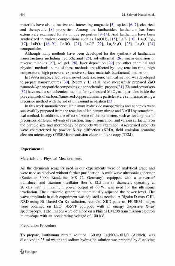
In addition, some other conditions were examined to investigate the morphology
of products, if any, and compare them with each other. One of key parameters,
which can be effective on particle size, is sonication time. Figure 3 shows
sonication time effects on the size and morphology of nanoparticles from the image
we concluded that there is an optimum time for sonication because in low sonication
time (Fig. 3a), we have agglomerated particles. Because power of sonication is not
sufficient for separate them. On the other hand, in high sonication times (Fig. 3c, d),
collision of particles occurs and particles stick with together. Therefore we chose
30 min (Fig. 3b) as an optimum time for preparation of La(OH)3 nanoparticles
because resulted particles are monodisperse, and fine.
Fig. 4 SEM images of La(OH)3 nanoparticles: a sample no. 5, b sample no. 6, c sample no. 7, andd sample no. 8
464 M. Salavati-Niasari et al.
123

For investigating the effects of feeding rate, we carried out the experiment in
different feeding rats of 1, 2, 3, 4, and 5 ml/min. From Fig. 5a–d we see that with
increasing of feeding rate, size of nanoparticles increase. Figure 5a shows the
lowest feeding rate effect in this condition very fine particles can obtained but this
particles are very unstable and agglomerated with together and ultrasonic power
isn’t sufficient to separate them from one another. Figure 3b shows optimum
feeding rate because we have fine, monodisperse, and separated particles.
The effects of various mixed solvents have been shown in Fig. 4. From this
image we can see that in mixed solvents an obvious shape change occurs and rod
like morphology obtain. With increasing vapor pressure of solvents (Fig. 4 a–d) the
aspect ratio and size of this rods increase because in solvents with high vapor
pressure the formed bubbles are filled with molecules of solvents and when these
bubbles collapse more energy is released. The best morphology obtains in sample
no. 12 [43, 44].
Figure 6 shows the surfactant effect on the morphology and size of lanthanum
hydroxide nanoparticles. In Fig. 6a, b (sample no. 13 and 14) we can see that in
presence of CTAB 1D structures can obtain but at low concentration of surfactant
this rod like particles agglomerate therefore in the case of La(OH)3 CTAB has two
roles firstly induce preferential growth (at low concentration) and secondly doesn’t
allow the particles to agglomerate (at high concentration). In Fig. 6c, d the effect of
SDS as surfactant has been shown in this case the addition of surface capping agents
Fig. 5 SEM images of the La(OH)3 nanorods: a sample no. 9, b sample no. 10, c sample no. 11, andd sample no. 12
Synthesis of Monodisperse Lanthanum Hydroxide 465
123

has unfavorable effect because particles has grown and agglomerated. In presence of
PVP, we have very fine particles therefore role of PVP is to stabilizing fine particles
Fig. 6e. In summery we have mastery on size and shape of nanostructures in
sonochemical method via addition of various surfactants. The SEM image of sample
no. 18, which was synthesized without ultrasonic treatment as a reference
experiment, has been shown in Fig. 6f. This image can help us to see the advantage
of using ultrasonic treatment in the synthesis of La(OH)3 nanoparticles. As can be
observed, the successful preparation of nanosized products is indeed due to the
ultrasonic treatments.
Fig. 6 SEM images of the La(OH)3 nanoparticles: a sample no. 13, b sample no. 14, c sample no. 15,d sample no. 16, e sample no. 17, and f sample no. 18
466 M. Salavati-Niasari et al.
123
Summary
In summary, La(OH)3 nanoparticles and nanorods with the hexagonal structure type
were synthesized by a sonochemical method. This method brings forward a broad
idea to synthesize other rare-earth compounds with various morphologies and novel
properties. The XRD, TEM, and FESEM were used to characterize the products.
The effect of some of the parameters such as feeding rate of precursors, different
solvents of reaction, time of sonication, and various surfactants on the size and
morphology of obtained products were also investigated.
Acknowledgments Authors are grateful to the council of Iran National Science Foundation and
University of Kashan for their unending effort to provide financial support to undertake this work.
References:
1. X. S. Fang and L. Zhang (2006). J. Mater. Sci. Technol. 22, 1.
2. X. S. Fang, C. H. Ye, L. D. Zhang, Y. H. Wang, and Y. C. Wu (2005). Adv. Funct. Mater. 15, 63.
3. Y. Xia, Y. Xiong, B. Lim, and S. E. Skrabalak (2009). Chem. Int. Ed. 48, 60.
4. Y. Xia, P. Yang, and Y. Sun (2003). Adv. Mater. 15, 353.
5. N. Wang, Q. Zhang, and W. Chen (2007). J. Cryst. Res. Technol. 42, 138.
6. H. X. Mai, Y. W. Zhang, R. Si, Z. G. Yan, L. D. Sun, L. P. You, and C. H. Yan (2006). J. Am. Chem.Soc. 128, 6426.
7. X. Wang, J. Zhuang, Q. Peng, and Y. D. Li (2006). Inorg. Chem. 45, 6661.
8. S. P. Fricker (2006). Chem. Soc. Rev. 35, 524.
9. B. Lai, A. C. Johnson, H. Xiong, and S. Ramanathan (2009). J. Power Sour. 186, 115.
10. Y. C. Liu and Y. W. Chen (2006). Ind. Eng. Chem. Res. 45, 2973–2980.
11. C. L. Kuo, C. L. Wang, T. Y. Chena, G. J. Chen, I. M. Hung, C. J. Shih, and K. Z. Fung (2007).
J. Alloys Compd. 440, 367.
12. H. Wei, Y. Wu, N. Lun, and F. Zhao (2004). J. Mater. Sci. 39, 1305.
13. K. Rajesh, K. V. Baiju, M. Jayasankar, and K. G. Warrierw (2008). J. Am. Ceram. Soc. 91, 2415.
14. J. Sun, X. P. Qiu, and W. T. Zhu (2005). Int. J. Hydrogen Energy 30, 437.
15. X. Wang, Y. D. Li, and Angew (2002). Chem. Int. Ed. 41, 4790.
16. X. Wang, J. Zhuang, Q. Peng, and Y. D. Li (2006). Inorg. Chem. 45, 6661.
17. P. Jeevanandam, Y. Koltypin, O. Palchik, and A. Gedanken (2001). J. Mater. Chem. 11, 869.
18. Y. P. Fang, A. W. Xu, R. Q. Song, H. X. Zhang, L. P. You, J. C. Yu, and H. Q. Liu (2003). J. Am.Chem. Soc. 125, 16025.
19. K. Rajesh, P. Shajesh, O. Seidel, P. Mukundan, and G. K. Warrier (2007). Adv. Funct. Mater. 17,
1682.
20. W. Bu, L. Zhang, Z. Hua, H. Chen, and J. Shi (2007). Cryst. Growth Des. 7, 2305.
21. J. Lin, Y. Huang, J. Zhang, X. Ding, S. Qi, and C. Tang (2007). Mater. Lett. 61, 1596.
22. J. Lee, Q. Zhang, and F. Saito (2003). J Alloys Compd. 348, 214.
23. S. Wang, G. Zhou, M. Lu, Y. Zhou, S. Wang, and Z. Yang (2006). J. Am. Ceram. Soc. 89, 2956.
24. J. Sheng, S. Zhang, S. Lv, and W. Sun (2007). J. Mater. Sci. 42, 9565.
25. Y. Zhang, K. Han, T. Cheng, and Z. Fang (2007). Inorg. Chem. 46, 4713.
26. B. Tang, J. Ge, C. Wu, L. Zhuo, Z. Chen, Z. Shi, and Y. Dong (2004). Nanotechnology 15, 1273.
27. G. Guo, F. Gu, Z. Wang, and H. Guo (2005). J. Cryst. Growth 277, 631.
28. X. Wang, M. Wang, H. Song, and B. Ding (2006). Mater. Lett. 60, 2261.
29. M. F. Vignolo, S. Duhalde, M. Bormioli, and G. Quintana (2002). Appl. Surf. Sci. 197, 522.
30. K. S. Suslick and G. J. Price (1999). Rev. Mater. Sci. 29, 295.
31. F. Li, X. Liu, Q. Qin, J. Wu, Z. Li, and X. Huang (2009). Cryst. Res. Technol. 44, 1249.
32. S. Zhu, H. Zhou, M. Hibino, I. Honma, and M. Ichihara (2005). Adv. Funct. Mater. 15, 381.
33. W. Lv, B. Liu, Q. Qiu, F. Wang, Z. Luo, P. Zhang, and S. Wei (2009). J. Alloys Compd. 479, 480.
34. R. Jenkins, and R. L Snyder (1996). Introduction to X-ray Powder Diffractometry, Chap. 12. p. 90.
35. F. Kim, S. Connor, H. Song, T. Kuykendall, and P. Yang (2004). Angew. Chem. Int. Ed. 43, 3673.
Synthesis of Monodisperse Lanthanum Hydroxide 467
123

36. Y. Sui, W. Fu, and H. Yang (2010). Cryst. Growth Des. 10, 99.
37. D. Seo, J. H. Park, and H. Song (2009). J. Phys. Chem. C 113, 3449.
38. J. Cheon, Y. W. Jun, and J. S. Choi (2006). Angew. Chem. Int. Ed. 45, 3414.
39. C. Burda, X. Chen, R. Narayanan, and M. A. El-Sayed (2005). Chem. Rev. 105, 1025.
40. M. Volmer and A. Weber (1926). Phys. Chem. 119, 227.
41. V. K. LaMer and R. H. Dinegar (1950). J. Am. Chem. Soc. 72, 484.
42. E. E. Finney and R. G. Finke (2008). J. Colloid Interface Sci. 317, 351.
43. F. Dang, N. Enomoto, J. Hojo, and K. Enpuku (2009). Ultrason. Sonochem. 16, 649.
44. A. Starczewska, R. Wrzalik, and M. Nowak (2009). Ultrason. Sonochem. 16, 537.
468 M. Salavati-Niasari et al.
123