Embed Size (px)
Citation preview

Editor:Daniel Silbergeld, University of Washington Medical Center, Seattle, Washington, USA
OPEN ACCESSFor entire Editorial Board visit : http://www.surgicalneurologyint.com
SNI: Neuro-Oncology, a supplement to Surgical Neurology International
© 2016 Surgical Neurology International | Published by Wolters Kluwer - MedknowS596
AbstractBackground: Epithelioid hemangioendothelioma (EHE) is a rare sarcoma of vascular origin, which is clinically and histologically intermediate between benign hemangioma and angiosarcoma. It is most commonly found in the liver, lung, and bone, however, 46 intracranial cases have been reported in the literature, of which this is the fifth reported suprasellar tumor.Case Description: A 45‑year‑old woman developed progressive lethargy, somnolence, and memory decline over the course of 6 months. On computed tomography (CT), she was found to have a large hypothalamic mass and underwent subtotal resection via a bifrontal craniotomy.Conclusions: While primary intracranial EHE is an uncommon presentation of a rare tumor, the suprasellar region does not seem to be an unusual location when it does occur. Prognosis is generally good, and may be better for primary intracranial disease than that for EHE originating elsewhere. Surgery is the first line of therapy, with variable benefit from adjuvant chemotherapy or radiation when total resection is not possible. Chemotherapeutic approaches in current use are directed at preventing endothelial proliferation.
Key Words: Epithelioid hemangioendothelioma, intracranial, suprasellar, review, vascular tumor
INTRODUCTION
Epithelioid hemangioendothelioma (EHE) is an uncommon neoplasm of vascular origin which may arise in a number of locations; most frequently the liver, lungs, and bones but also intracranially.[3] While less aggressive than angiosarcoma, it may metastasize and in some cases demonstrates quite rapid growth.[17] Management is centered on surgical resection with adjuvant chemotherapy, usually with antiangiogenic agents.[46] We herein present a case of EHE arising in the suprasellar region in a 45-year-old woman, summarize previously published cases of intracranial EHEs, and review the literature on the clinical course and management of EHE.
CASE DESCRIPTION
Over the course of six months, a 45-year-old Vietnamese woman with a history of type 2 diabetes mellitus and hyperlipidemia became progressively lethargic, somnolent,
How to cite this article: Barger J, Tanweer O, Liechty B, Snuderl M, Jafar JJ. Suprasellar epithelioid hemangioendothelioma: Case report and review of the literature. Surg Neurol Int 2016;7:S596-602.http://surgicalneurologyint.com/Suprasellar-epithelioid-hemangioendothelioma:-Case-report-and-review-of-the-literature/
This is an open access article distributed under the terms of the Creative Commons Attribution-NonCommercial-ShareAlike 3.0 License, which allows others to remix, tweak, and build upon the work non-commercially, as long as the author is credited and the new creations are licensed under the identical terms.
For reprints contact: [email protected]
Suprasellar epithelioid hemangioendothelioma: Case report and review of the literatureJames Barger, Omar Tanweer, Benjamin Liechty1, Matija Snuderl1, Jafar J. Jafar
Departments of Neurosurgery and 1Pathology, New York University School of Medicine, New York, USA
E‑mail: *James Barger ‑ [email protected]; Omar Tanweer ‑ [email protected]; Benjamin Liechty ‑ [email protected]; Matija Snuderl ‑ [email protected]; Jafar J. Jafar ‑ [email protected] *Corresponding author
Received: 17 May 16 Accepted: 24 May 16 Published: 01 September 16
Access this article onlineWebsite:www.surgicalneurologyint.comDOI: 10.4103/2152-7806.189729 Quick Response Code:

S597
SNI: Neuro-Oncology 2016, Vol 7, Suppl 23 - A Supplement to Surgical Neurology International
and forgetful. Originally thought by her physicians to have an endocrine issue, she was diagnosed with an intracranial mass on computed tomography (CT) scan when her husband found it difficult to arouse her at home and brought her to a local emergency department. She was referred to our institution for neurosurgical evaluation, at which time she was sleeping 16 hours a day, and was noted to ask the same questions repeatedly, forgetting the answers each time. She had gained 25 pounds during the months prior to the presentation. She did not complain of headaches or visual changes, and had no symptoms of diabetes insipidus. Her last menstrual period had been 3–4 years prior to presentation. Neurological exam was unremarkable [Extraocular movements were intact, visual fields were full, and there was no nystagmus. Muscle strength was 5/5 throughout, reflexes were brisk and symmetric, and gait was normal-based. There was no dysmetria or pronator drift.]
Magnetic resonance imaging (MRI) with contrast was obtained, revealing a 3.6 × 3.7 × 3.2 cm lobulated, heterogeneous, fluid-attenuated inversion recovery hyperintense, and avidly enhancing hypothalamic mass extending into the anterior third ventricle [Figure 1]. The patient underwent subtotal resection via bifrontal craniotomy. Intraoperatively, the tumor was found to be rubbery and vascular, and it appeared continuous with portions of the hypothalamus and optic nerves. Postoperative course was notable for a triphasic water balance response and new-onset adrenal insufficiency treated with hydrocortisone. She also developed new psychiatric symptoms including paranoia and irritability beginning approximately 1 month postoperatively.
PathologyHistologic examination demonstrated a predominantly epithelioid neoplasm with areas of spindled cytology with a dense inflammatory infiltrate [Figure 2a]. The tumor demonstrated several architectural patterns, including retiform [Figure 2b], chordoid [Figure 2c], and strands, often embedded in a myxoid matrix, giving an appearance reminiscent of chordoma at low power. At high power, many cells demonstrated intracytoplasmic lumina [Figure 2d] with occasional erythrocytes. The tumor was
sharply demarcated from the surrounding brain, with reactive changes, including gliosis and accumulation of Rosenthal fibers, suggesting slow growth [Figure 2e]. The tumor cells were strongly and diffusely positive for CD34 [Figure 2f], and more focally positive for CD31 [Figure 2g], FLI-1, and factor VIII, compatible with a tumor of endothelial origin; however, there was only scattered reactivity for Erg [Figure 2h]. An immunostain for SMA [Figure 2i] to rule out a fibroblastic process or leiomyosarcoma were negative, and an immunostain for ALK-1 performed to exclude inflammatory pseudotumor was negative. Immunostains for EMA [Figure 2j], progesterone receptor, and S-100 were negative, which are less compatible with diagnoses of chordoid meningioma, chondrosarcoma, and chordoma. Immunostains for Oct-4 and PLAP performed to exclude a germ cell tumor were negative. Immunostains for cytokeratins CAM 5.2 and AE1/3 were performed to exclude a neoplasm of epithelial origin, and demonstrated only focal immunoreactivity, and an immunostain for TTF-1 was performed to exclude a metastatic carcinoma from a lung or thyroid primary was negative. Immunostains for CD163, CD3, CD20, and CD68 [Figure 2k-m] highlighted a marked lymphohistiocytic infiltrate throughout the tumor, however, immunostains for CD15 and CD30 were negative, arguing against a lymphoproliferative disease such as Hodgkin lymphoma, and an immunostain for CD1a to exclude a histiocytic process such as Langerhan’s cell histiocytosis was negative. Immunostain for Ki-67 shows scattered positivity, demonstrating the moderate proliferative characteristics of this tumor [Figure 2n]. An immunostain for glial fibrillary acidic protein [Figure 2o] is negative in the tumor cells, but highlights the sharp demarcation of the tumor from the adjacent brain.
DISCUSSION
Epithelioid hemangioendothelioma (EHE) is a rare sarcoma of vascular origin which is clinically and histologically intermediate between benign hemangioma and angiosarcoma. It can present at any age but most commonly presents in the fourth and fifth decades.[3] A slight overall predilection for females
Figure 1: (a) Sagittal T1 precontrast, (b) sagittal T1 postcontrast, (c) axial fluid-attenuated inversion recovery
ba c

S598
SNI: Neuro-Oncology 2016, Vol 7, Suppl 23 - A Supplement to Surgical Neurology International
has been reported, however, the majority of intracranial cases have occurred in males.[3] The pathophysiology of tumor development is poorly understood, though the fusion of the WWTR1 gene, part of the hippo signaling pathway, to the CAMTA1 tumor suppressor gene via a t (1;3) (p36;q25) translocation seems to be present in most cases.[3] EHE is most commonly located in the liver, lung, and bone, though 46 previous cases of intracranial EHEs have been reported in the literature [Table 1]. This is the fifth reported case of EHE occurring in the suprasellar region, suggesting that while rare, this is not an unusual location for the tumor to arise. The patient’s symptoms, however, were quite different from previous suprasellar EHEs, which presented with headache and visual loss;[4] loss of libido and asthenia;[15] headaches, ptosis, and diplopia;[32] and headache, diplopia, and visual loss.[1]
Radiologically, EHEs typically demonstrate uniform contrast enhancement on CT, which in at least one case led to misdiagnosis as a meningioma.[50] On
MRI, the lesion may be isointense, hyperintense, and/or heterogeneous on precontrast T1 and there is intense enhancement with contrast.[15] The tumor may appear hyperintense and/or heterogeneous on T2. The differential diagnosis based on MRI may include choroid glioma or an ectopically located craniopharyngioma.
The clinical course of EHE is usually somewhat indolent compared to other sarcomas; overall, 5-year survival is 73% (Lau 2011) vs 35% for angiosarcoma.[17,23] Only five patients with intracranial EHE reported in the literature died from tumor complications, three of whom had multiorgan system disease. 20–30% of EHEs metastasize hematogenously to other organs.[3] While EHEs elsewhere in the body present with multiple tumors in the same organ system, in up to 50% of cases (shown by a recent study to be monoclonal local metastases rather than synchronous primaries), primary intracranial EHE appears to be unifocal.[11] The seven reported cases of multiple intracranial lesions were all associated with EHE of other organs and were likely metastases.[19,20,28,32,38,41,45]
Figure 2: (a) Hematoxylin and Eosin (H and E), low power, (b) H and E, medium power, (c) H and E, medium power, (d) H and E, medium power, (e) H and E, low power, (f) CD34, (g) CD31, (h) Erg, (i) SMA, (j) EMA, (k) CD163, (l) CD3, (m) CD20, (n) Ki-67, (o) GFAP
d
h
c
g
b
f
a e
i j
k l m n o

S599
SNI: Neuro-Oncology 2016, Vol 7, Suppl 23 - A Supplement to Surgical Neurology International
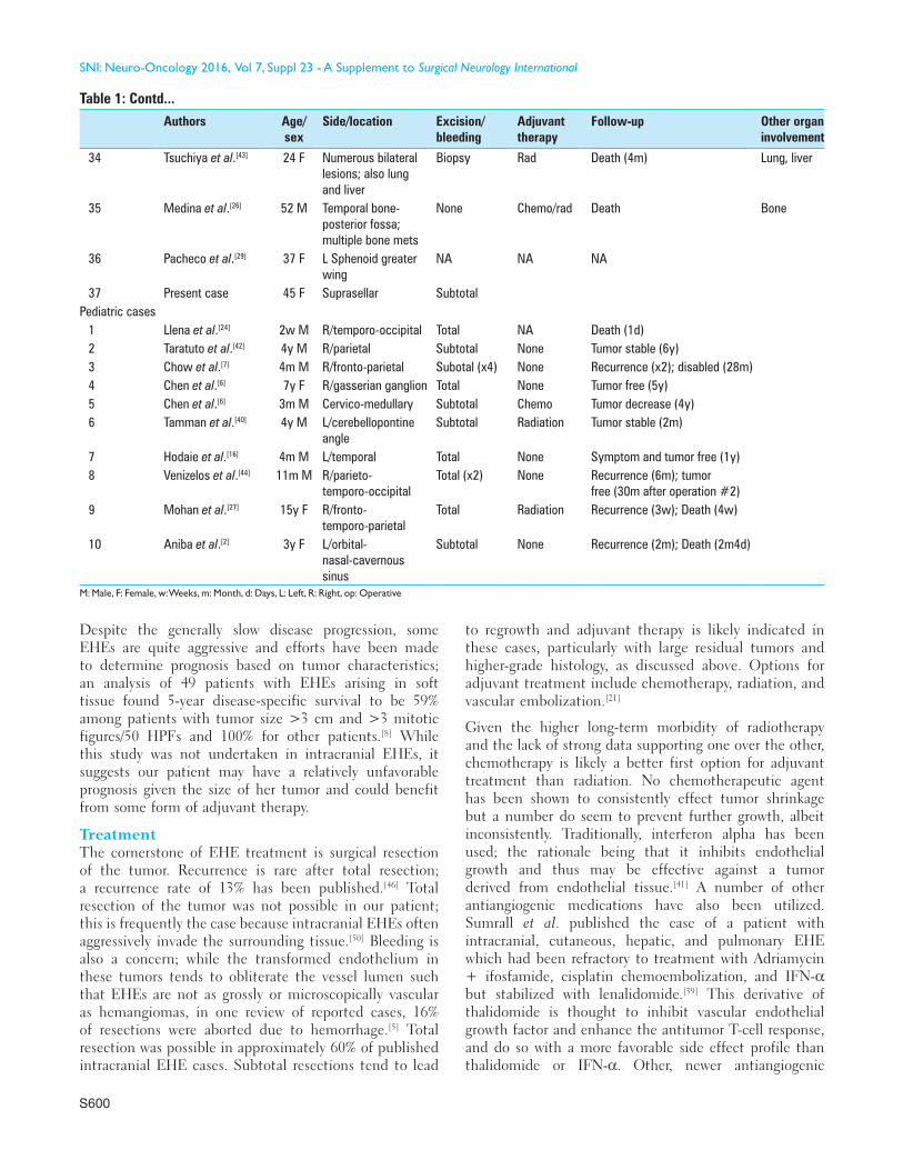
Table 1: Reported intracranial epithelioid hemangioendothelioma
Authors Age/sex
Side/location Excision/bleeding
Adjuvant therapy
Follow‑up Other organ involvement
Adult cases1 Pearl et al.[32] 36 M R/fronto‑parietal Biopsy
(1st op/term)Radiation Improvement; tumor decrease
2 Pearl et al.[32] 73 M Suprasellar Subtotal Radiation Improvement; tumor stable3 Kepes et al.[19] 58 M L/temporal Resection None NA Liver4 Kepes et al.[19] 74 M L/temporal Resection None NA5 Hurley et al.[18] 23 F Multiple Total (x2) None Recurrence (6y); A (10y) Heart6 Nora et al.[28] 28 F R/frontal Total None Symptom free and tumor
stable (30m)7 Nora et al.[28] 62 M L/frontal Total None Symptom and tumor free (1y)8 Puca et al.[35] 27 M R/temporal Total (x2; 1st
op terminated)Radiation and VE
Symptom and tumor free (18m)
9 Phookan et al.[33] 36 F R/cavernous sinus Total None Disabled and tumor free (4m)10 Fryer et al.[13] 61 M R/fronto‑parietal Total Radiation Recurrence (8w); D (6m)11 Golash et al.[14] 33 M L/frontal Total (x2; 1st
op terminated)None Symptom and tumor free (2m)
12 Rushing et al.[37] 38 F Clivus Biopsy Radiation NA13 Tancredi et al.[41] 20 F Bilateral frontal Total Chemo Alive (3y) Skull14 Palmieri et al.[30] 20 F Bilateral parietal Total Chemo Symptom free and tumor
stable (30m)Bone
15 Chan et al.[5] 20 M L/frontal Total None Symptom and tumor free (2y)16 Koh et al.[20] 26 F L/sphenoid bone Total (x2) Failed VE Improvement17 Watanabe et al.[45] 55 F Petroclival Subtotal Radiation Improvement and tumor
stable (1y)18 Kubota et al.[21] 24 F R/parieto‑occipital Total (x2; 1st
op terminated)Radiation and VE
Symptom and tumor free (9y)
19 Baehring et al.[4] 49 F Suprasellar Subtotal None Improvement and tumor stable (6m)
20 Hamlat et al.[15] 53 M Suprasellar Biopsy (1st op/term)
Radiation and Chemo
Improvement and tumor stable (21m)
21 Endo et al.[10] 69 M Multiple Subtotal Chemo Recurrence (1.5m); Death (3m)22 Fernandes et al.[12] 27 M L/temporal Subtotal None Recurrence (3m); Death (8m)23 Yeo et al.[48] 55 M L/multiple Total (frontal
tumor)NA NA
24 Parajon et al.[31] 58 M R/sphenoid bone Total None Symptom and tumor free (1y)25 Wong et al.[47] 50 M L/multiple Total None NA26 Zhang et al.[49] 57 F L/temporal Total Radiation Recurrence (2w);
tumor decrease (2m)27 Sumrall et al.[39] 31 F Multiple Total (largest
tumor)Radiation and Chemo
Tumor stable (11y) Scalp, liver, skull, lung
28 Zheng et al.[50] 25 M R/temporo‑parietal Total None Symptom and tumor free (5m)29 Zheng et al.[50] 44 F Petroclival Subtotal None Symptom free and tumor
stable (1.5y)30 Ma et al.[25] 58 F Clival Subtotal Gamma knife
radiotherapyNo recurrence or metastasis (6m)
31 Ahmed et al.[1] 42 F Sellar/suprasellar NA NA NA32 Rocha Oliveira
et al.[36]37 F L paracentral w/
concurrent lung involvement
Total Sunitinib Slight RLL paresis and tumor free (14m)
Lung
33 Drazin et al.[9] 62 M L Mastoid/posterior fossa
Total Rad following recurrence
Recurrence; symptom improvement and tumor free after 2nd resection (8y)
Contd...
S600
SNI: Neuro-Oncology 2016, Vol 7, Suppl 23 - A Supplement to Surgical Neurology International
Despite the generally slow disease progression, some EHEs are quite aggressive and efforts have been made to determine prognosis based on tumor characteristics; an analysis of 49 patients with EHEs arising in soft tissue found 5-year disease-specific survival to be 59% among patients with tumor size >3 cm and >3 mitotic figures/50 HPFs and 100% for other patients.[8] While this study was not undertaken in intracranial EHEs, it suggests our patient may have a relatively unfavorable prognosis given the size of her tumor and could benefit from some form of adjuvant therapy.
TreatmentThe cornerstone of EHE treatment is surgical resection of the tumor. Recurrence is rare after total resection; a recurrence rate of 13% has been published.[46] Total resection of the tumor was not possible in our patient; this is frequently the case because intracranial EHEs often aggressively invade the surrounding tissue.[50] Bleeding is also a concern; while the transformed endothelium in these tumors tends to obliterate the vessel lumen such that EHEs are not as grossly or microscopically vascular as hemangiomas, in one review of reported cases, 16% of resections were aborted due to hemorrhage.[5] Total resection was possible in approximately 60% of published intracranial EHE cases. Subtotal resections tend to lead
to regrowth and adjuvant therapy is likely indicated in these cases, particularly with large residual tumors and higher-grade histology, as discussed above. Options for adjuvant treatment include chemotherapy, radiation, and vascular embolization.[21]
Given the higher long-term morbidity of radiotherapy and the lack of strong data supporting one over the other, chemotherapy is likely a better first option for adjuvant treatment than radiation. No chemotherapeutic agent has been shown to consistently effect tumor shrinkage but a number do seem to prevent further growth, albeit inconsistently. Traditionally, interferon alpha has been used; the rationale being that it inhibits endothelial growth and thus may be effective against a tumor derived from endothelial tissue.[41] A number of other antiangiogenic medications have also been utilized. Sumrall et al. published the case of a patient with intracranial, cutaneous, hepatic, and pulmonary EHE which had been refractory to treatment with Adriamycin + ifosfamide, cisplatin chemoembolization, and IFN-α but stabilized with lenalidomide.[39] This derivative of thalidomide is thought to inhibit vascular endothelial growth factor and enhance the antitumor T-cell response, and do so with a more favorable side effect profile than thalidomide or IFN-α. Other, newer antiangiogenic
Table 1: Contd...
Authors Age/sex
Side/location Excision/bleeding
Adjuvant therapy
Follow‑up Other organ involvement
34 Tsuchiya et al.[43] 24 F Numerous bilateral lesions; also lung and liver
Biopsy Rad Death (4m) Lung, liver
35 Medina et al.[26] 52 M Temporal bone‑ posterior fossa; multiple bone mets
None Chemo/rad Death Bone
36 Pacheco et al.[29] 37 F L Sphenoid greater wing
NA NA NA
37 Present case 45 F Suprasellar SubtotalPediatric cases
1 Llena et al.[24] 2w M R/temporo‑occipital Total NA Death (1d)2 Taratuto et al.[42] 4y M R/parietal Subtotal None Tumor stable (6y)3 Chow et al.[7] 4m M R/fronto‑parietal Subotal (x4) None Recurrence (x2); disabled (28m)4 Chen et al.[6] 7y F R/gasserian ganglion Total None Tumor free (5y)5 Chen et al.[6] 3m M Cervico‑medullary Subtotal Chemo Tumor decrease (4y)6 Tamman et al.[40] 4y M L/cerebellopontine
angleSubtotal Radiation Tumor stable (2m)
7 Hodaie et al.[16] 4m M L/temporal Total None Symptom and tumor free (1y)8 Venizelos et al.[44] 11m M R/parieto‑
temporo‑occipitalTotal (x2) None Recurrence (6m); tumor
free (30m after operation #2)9 Mohan et al.[27] 15y F R/fronto‑
temporo‑parietalTotal Radiation Recurrence (3w); Death (4w)
10 Aniba et al.[2] 3y F L/orbital‑ nasal‑cavernous sinus
Subtotal None Recurrence (2m); Death (2m4d)
M: Male, F: Female, w: Weeks, m: Month, d: Days, L: Left, R: Right, op: Operative

S601
SNI: Neuro-Oncology 2016, Vol 7, Suppl 23 - A Supplement to Surgical Neurology International
treatments have been reported to be effective against EHE, including capecitabine + bevacizumab,[23] Pazopanib,[38] and sunitinib.[34] In a recent review of 36 patients with EHE treated with antiangiogenic therapy (thalidomide, lenalidomide, sorafenib, or bevacizumab alone or in combination), 6 experienced a partial response, 14 stable disease, and 16 progressive disease.[38] Metronomic cyclophosphamide therapy has also been used.[22]
Radiotherapy seems to have similar rates of success; out of seven patients in the literature with intracranial EHE who received adjuvant radiotherapy, one had tumor shrinkage, three had a stable tumor, and three experienced recurrent tumor growth and symptoms.[50] Vascular embolization has mostly been used in a neoadjuvant manner to reduce tumor size preoperatively.
CONCLUSION
While primary intracranial EHE is an uncommon presentation of a rare tumor, the suprasellar region does not seem to be an unusual location when it does occur. Prognosis is generally good, and may be better for primary intracranial disease than for EHE originating elsewhere. Surgery is the first line of therapy, with variable benefit from adjuvant chemotherapy or radiation when total resection is not possible.
Financial support and sponsorshipNil.
Conflicts of interestThere are no conflicts of interest.
REFERENCES
1. Ahmed S, Epari S, Shah M, Rao KS. Epithelioid hemangioendothelioma of sphenoid bone: A case report of an unusual case. Neurol India 2012;60:344-6.
2. Aniba K, Laghmari M, Lmejjati M, Ghannane H, Ait Benali S. A tragical paediatric case history of intraorbital and intracranial epithelioid hemangioendothelioma. Case Rep Neurol Med 2012;2012:396097.
3. Antonescu C. Malignant vascular tumors--An update. Mod Pathol 2014;27(Suppl 1):S30-8.
4. Baehring JM, Dickey PS, Bannykh SI. Epithelioid hemangioendothelioma of the suprasellar area: A case report and review of the literature. Arch Pathol Lab Med 2004;128:1289-93.
5. Chan YL, Ng HK, Poon WS, Cheung HS. Epithelioid haemangioendothelioma of the brain: A case report. Neuroradiology 2001;43:848-50
6. Chen TC, Gonzalez-Gomez I, Gilles FH, McComb JG. Pediatric intracranial hemangioendotheliomas: Case report. Neurosurgery 1997;40:410-4.
7. Chow LT, Chow WH, Fong DT. Epithelioid hemangioendothelioma of the brain. Am J Surg Pathol 1992;16:619-25.
8. Deyrup AT, Tighiouart M, Montag AG, Weiss SW. Epithelioid hemangioendothelioma of soft tissue: A proposal for risk stratification based on 49 cases. Am J Surg Pathol 2008;32:924-7.
9. Drazin D, Gandhi R, S lodkowska E, Boulos AS. Epithel ioid hemangioendothelioma of the mastoid: Resection for recurrence and adjuvant radiation with 8-year followup. Case Rep Surg 2013;2013:469201.
10. Endo T, Su CC, Numagami Y, Shirane R. Malignant intracranial epithelioid hemangioendothelioma presumably originating from the lung: Case report. J Neurooncol 2004;67:337-43.
11. Errani C, Sung YS, Zhang L, Healey JH, Antonescu CR. Monoclonality of multifocal epithelioid hemangioendothelioma of the liver by analysis of WWTR1-CAMTA1 breakpoints. Cancer Genet 2012;205:12-7.
12. Fernandes AL, Ratilal B, Mafra M, Magalhaes C. Aggressive intracranial and extra-cranial epithelioid hemangioendothelioma: A case report and review of the literature. Neuropathology 2006;26:201-5.
13. Fryer JA, Biggs MT, Katz IA, Brazier DH, Shakespeare TP. Intracranial epithelioid hemangioendothelioma arising at site of previously excised atypical meningioma. Pathology 1998;30:95-9.
14. Golash A, Strang FA, Reid H. Intracranial haemangioendothelioma mimicking a meningioma. Br J Neurosurg 1999;13:594-7.
15. Hamlat A, Casallo-Quilliano C, Saikali S, Lesimple T, Brassier G. Epithelioid hemangioendothelioma of the infundibular-hypothalamic region: Case report and literature review. J Neurooncol 2004;67:361-6.
16. Hodaie M, Becker L, Teshima I, Rutka JT. Total resection of an intracerebral hemangioendothelioma in an infant. Case report and review of the literature. Pediatr Neurosurg 2001;34:104-12.
17. Huntington JT, Jones C, Liebner DA, Chen JL, Pollock RE. Angiosarcoma: A rare malignancy with protean clinical presentations. J Surg Oncol 2015;111:941-50.
18. Hurley TR, Whisler WW, Clasen RA, Smith MC, Bleck TP, Doolas A, Dampier MF. Recurrent intracranial epithelioid hemangioendothelioma associated with multicentric disease of liver and heart: Case report. Neurosurgery 1994;35:148-51.
19. Kepes JJ, Rubinstein LJ, Maw G, Burdick. Epithelioid hemangiomas (hemangioendotheliomas) of the central nervous system and its coverings, a report of three cases. J Neuropathol Exp Neurol 1986;45:319.
20. Koh YC, Yoo H. Epithelioid haemangioendothelioma of the sphenoid bone. J Clin Neurosci 2001;8(Suppl 1):63-6.
21. Kubota T, Sato K, Takeuchi H, Handa Y. Successful removal after radiotherapy and vascular embolization in a huge tentorial epithelioid hemangioendothelioma: A case report. J Neurooncol 2004;68:177-18.
22. Lakkis Z, Kim S, Delabrousse E, Jary M, Nguyen T, Mantion G, et al. Metronomic cyclophosphamide: An alternative treatment for hepatic epithelioid hemangioendothelioma. J Hepatol 2013;58:1254-7.
23. Lau A, Malangone S, Green M, Badari A, Clarke K, Elquza E. Combination capecitabine and bevacizumab in the treatment of metastatic hepatic epithelioid hemangioendothelioma. Ther Adv Med Oncol 2015;7:229-36.
24. Llena JF, Hirano A, Inoue A. Vasoformative tumor of the brain-immunohistology and ultrastructure. Clin Neuropathol 1984;3:155-9.
25. Ma SR, Li KC, Xu YQ, Wang YM, Ma WL, Li Q. Primary epithelioid hemangioendothelioma in the clival region: A case report and literature review. Neuropathology 2011;31:519-22.
26. Medina M, Polo R, Reyes P, Vaca M, Alonso A, Cobeta I. Imaging case of the month. Multifocal epithelioid hemangioendothelioma with massive lateral skull base involvement. Otol Neurotol 2015;36:e67-9.
27. Mohan SM, Symss NP, Pande A, Chakravarthy VM, Ramamurthi R. Intracranial epithelioid hemangioendothelioma. Childs Nerv Syst 2008;24:863-8au.
28. Nora FE, Scheithauer BW. Primary epithelioid hemangioendothelioma of the brain. Am J Surg Pathol 1996;20:707-14.
29. Pacheco JM, Goodman JC, Mandel J. Intracranial epithelioid hemangioendothelioma causing subacute loss of vision. Neurology 2015;85:735-6.
30. Palmieri G, Montella L, Martignetti A, Bianco AR. Interferon alpha-2b at low doses as long-term antiangiogenic treatment of a metastatic intracranial hemangioendothelioma: A case report. Oncol Rep 2000;7:145-9.
31. Parajon A, Vaquero J. Meningeal intracranial epithelioid hemangioendothelioma: Case report and literature review. J Neurooncol 2008;88:169-73.
32. Pearl GS, Takei Y, Tindall GT, O’Brien MS, Payne NS, Hoffman JC. Benign hemangioendothelioma involving the central nervous system: “Strawberry nevus” of the neuraxis. Neurosurgery 1980;7:249-56.
33. Phookan G, Davis AT, Holmes B. Hemangioendothelioma of the cavernous sinus: Case report. Neurosurgery 1998;42:1153-5.
34. Prochilo T, Savelli G, Bertocchi P, Abeni C, Rota L, Rizzi A, Zaniboni A. Targeting VEGF-VEGFR pathway by sunitinib in peripheral primitive neuroectodermal tumor, paraganglioma and epithelioid hemangioendothelioma: Three case reports. Case Rep Oncol 2013;6:90-7.
35. Puca A, Meglio M, Rollo M, Zannoni GF. Intracranial epithelioid hemangioendothelioma: Case report. Neurosurgery 1996;38:399-401.
36. Rocha Oliveira PC, Alcantara FP, Souza-Vianna PE, Brito AP. Cerebral

S602
SNI: Neuro-Oncology 2016, Vol 7, Suppl 23 - A Supplement to Surgical Neurology International
epithelioid hemangioendothelioma with thoracic simultaneous involvement: Advanced MRI features. Arq Neuropsiquiatr 2012;70:637-8.
37. Rushing EJ, White JA, D’ Alise MD, Chason DP, White CL 3rd, Bigio EH. Primary epithelioid hemangioendothelioma of the clivus. Clin Neuropathol 1998;17:110-4.
38. Semenisty V, Naroditsky I, Keidar Z, Bar-Sela G. Pazopanib for metastatic pulmonary epithelioid hemangioendothelioma-A suitable treatment option: Case report and review of anti-angiogenic treatment options. BMC Cancer 2015;15:402.
39. Sumrall A, Fredericks R, Berthold A, Shumaker G. Lenalidomide stops progression of multifocal epithelioid hemangioendothelioma including intracranial disease. J Neurooncol 2010;97:275-7.
40. Tammam AG, Lewis PD, Crockard HA. Cerebellopontine angle epithelioid haemangioendothelioma in a 4-year-old boy. Childs Nerv Syst 1997;13:648-50.
41. Tancredi A, Puca A, Carbone A. Multifocal cerebral hemangioendothelioma. Case report and review of the literature. Acta Neurochir 2000;142:1157-61.
42. Taratuto AL, Zurbriggen G, Sevlever G, Saccoliti M. Epithelioid hemangioendothelioma of the central nervous system. Immunohistochemical and ultrastructural observations of a pediatric case. Pediatr Neurosci 1998;14:11-4.
43. Tsuchiya T, Oya S, Mori H, Matsui T. Multiple hemorrhagic intraparenchymal tumors presenting with fatal intracranial hypertension: A rare manifestation of systemic epithelioid hemangioendothelioma. Surg Neurol Int 2015;6:156.
44. Venizelos ID, Paradinas FJ. Primary paediatric intracranial epithelioid haemangioendothelioma. Histopathology 2002;41:172-4.
45. Watanabe T, Saito N, Shimaguchi H, Fujimaki H, Kamiya M, Nakazato Y, Sasaki T. Primary epithelioid hemangioendothelioma originating in the lower petroclival region: Case report. Surg Neurol 2003;59:429-33.
46. Weiss SW, Enzinger FM. Epithelioid hemangioendothelioma: A vascular tumor often mistaken for a carcinoma. Cancer 1982;50:970-81.
47. Wong DS, Chiu TW, Wong GK, Zhu XL, Kwok MW, Ho CM, et al. Epithelioid haemangioendothelioma of the anterior skull base: What is the optimal treatment? Hong Kong Med J 2009;15:308-10.
48. Yeo SK, Kim JH, Kim CJ, Lee JK. Intracranial epithelioid hemangioendothelioma. J Korean Neurosurg Soc 2007;42:129-31.
49. Zhang J, Wang Y, Geng D. Intracranial epithelioid hemangioendothelioma: An unusual CTA finding in one case. Br J Neurosurg 2010;24:294‑5.
50. Zheng J, Liu L, Wang J, Wang S, Cao Y, Zhao J. Primary intracranial epithelioid hemangioendothelioma: A low-proliferation tumor exhibiting clinically malignant behavior. J Neurooncol 2012;110:119-27.