Embed Size (px)
Citation preview

Suprasellar Rathke Cleft Cysts: ClinicalPresentation and Treatment Outcomes
BACKGROUND: Rathke cleft cysts (RCCs), benign remnants of the Rathke pouch typicallyarising in the sella, sometimes have suprasellar extension. Purely suprasellar RCCs arerarely reported.
OBJECTIVE: To compare the presentations, surgical outcomes, and pathology of purelysuprasellar RCCs and sellar-based RCCs.
METHODS: We retrospectively reviewed records, magnetic resonance images, labora-tory results, and pathology of 151 RCC patients surgically managed at our institutionfrom 1989 to 2009. The RCCs were classified as purely sellar (type I, n = 76), sellar withsuprasellar extension (type II, n = 56), or purely suprasellar (type III, n = 19).
RESULTS: The RCCs with a suprasellar component (types II and III) more commonlypresented with visual dysfunction (P , .001). Complete cyst drainage occurred in89%, 55%, and 38% of type I, II, and III RCCs, respectively (P , .001). Vision improvedin 100%, 55%, and 33% and headache improved in 74%, 64%, and 29% of type I, II, and IIIpatients, respectively (P = .02). Temporary or permanent postoperative diabetesinsipidus occurred in 5%, 16%, and 21% of type I, II, and III patients, respectively.(P, .001). In a multivariate analysis, RCC type was the only factor predicting recurrence.Kaplan-Meier 3-year recurrence/progression rates were 0%, 16%, and 29% for type I, II,and III RCCs, respectively (P , .001, type I vs II, type I vs III; P = .5 type II vs III).
CONCLUSION: The RCCs with a suprasellar component are neurosurgically challengingbecause of their proximity to the optic chiasm and infundibulum. Compared with sellar-based RCCs, RCCs with a suprasellar component more frequently present with visualdysfunction, are more difficult to completely eliminate, recur more frequently, and areassociated with higher postoperative endocrine morbidity, and their preoperative visualdysfunction and headache less frequently improve with surgery. These factors must beconsidered during the treatment of RCCs with a suprasellar component.
KEY WORDS: Pituitary, Rathke cleft cyst, Suprasellar, Transsphenoidal surgery, Visual symptoms
Neurosurgery 69:1058–1069, 2011 DOI: 10.1227/NEU.0b013e318228bcea www.neurosurgery-online.com
Rathke cleft cysts (RCCs) are benign lesionsthat typically arise within the sellabetween the anterior and posterior lobes
of the pituitary.1 Most often asymptomatic,RCCs have been found incidentally in 4% to33% of autopsies.2-5 These lesions, however, cancause mass effect on surrounding structures suchas the pituitary gland and optic chiasm, leadingto headache, pituitary dysfunction, or visualdisturbance.6-8 Although asymptomatic RCCscan safely be followed up with serial imaging,6
the standard treatment for symptomatic RCCs issurgical decompression, typically througha transsphenoidal approach. Several large seriesof symptomatic RCCs have demonstrated goodresolution of headache, hormonal dysfunction,and visual disturbances with surgical manage-ment with acceptable surgical morbidity.6,7,9-16
Rathke cleft cysts are remnants of the Rathkepouch, a structure of ectodermal origin formedduring the fourth week of gestation (Figure 1).1
The Rathke pouch extends caudally to fuse withthe infundibulum around the eighth week ofgestation, forming the craniopharyngeal duct.The Rathke pouch then leads to the formation of
Matthew B. Potts, MD*‡
Arman Jahangiri, BS*§
Kathleen R. Lamborn, PhD‡
Lewis S. Blevins, MD‡
Sandeep Kunwar, MD‡
Manish K. Aghi, MD, PhD‡
‡Department of Neurosurgery and
California Center for Pituitary Disorders
(CCPD), University of California,
San Francisco, California; §University of
Texas Southwestern Medical School,
Dallas, Texas
*These authors have contributed equally
to this article.
Correspondence:
Manish K. Aghi, MD, PhD,
Assistant Professor of Neurological
Surgery,
University of California at San Francisco
(UCSF),
California Center for Pituitary Disorders
(CCPD),
505 Parnassus Ave, Room M779,
San Francisco, CA 94143-0112.
E-mail: [email protected]
Received, September 20, 2010.
Accepted, March 25, 2011.
Published Online, June 11, 2011.
Copyright ª 2011 by the
Congress of Neurological Surgeons
ABBREVIATION: RCC, Rathke cleft cyst
1058 | VOLUME 69 | NUMBER 5 | NOVEMBER 2011 www.neurosurgery-online.com
RESEARCH—HUMAN—CLINICAL STUDIESTOPIC RESEARCH—HUMAN—CLINICAL STUDIES
Copyright © Congress of Neurological Surgeons. Unauthorized reproduction of this article is prohibited.
Copyright © Congress of Neurological Surgeons. Unauthorized reproduction of this article is prohibited.

FIGURE 1. Pituitary gland development. A, the pituitary gland is derived from 2 sources. The anterior lobe originates from anupgrowth of ectoderm from the roof of the stomodeum (pharyngeal epithelium), whereas the posterior lobe (along with the rest ofthe diencephalon) originates from a downgrowth of neuroectoderm. In the middle of the fourth week, a diverticulum, the Rathkepouch, begins as a dorsal evagination from the pharyngeal epithelium and then grows upward from the roof of what will becomethe mouth toward the developing brain. As the upgrowth contacts a ventral evagination or downgrowth from the diencephalon ofthe brain, the infundibular process, it begins to pinch off from its connection with the stomodeum. B, by the sixth week, theconnection between the Rathke pouch and the oral cavity of the pharyngeal epithelium degenerates, after which (C) the cells of theRathke pouch proliferate to form the pars distalis (also called the anterior pituitary or adenohypophysis), and the infundibularprocess forms the neurohypophysis (the posterior lobe of the pituitary gland). D, the cells of the Rathke pouch also extend up theanterior aspect of the infundibulum as the pars tuberalis. The posterior surface of the Rathke pouch does not proliferate but formsthe poorly developed pars intermedia. The infundibulum, having grown down from the floor of the diencephalon, expands as theaxons of cells in the diencephalon grow down into it. Although the Rathke pouch normally closes early in fetal development,a remnant often persists as a Rathke cleft in the pars intermedia between the anterior and posterior lobes. A Rathke cleft (persistentmaterial from Rathke pouch) can sometimes expand to form a Rathke cleft cyst, which can be found in a purely sellar locationcentered in the pars intermedia (type I cysts), a sellar location with suprasellar extension (type II cysts), or a purely suprasellarlocation, likely reflecting origin from persistent suprasellar Rathke pouch cells that gave rise to the pars tuberalis (type III cysts).Mb, mammillary bodies; Och, optic chiasm.
SUPRASELLAR RATHKE CLEFT CYST
NEUROSURGERY VOLUME 69 | NUMBER 5 | NOVEMBER 2011 | 1059
Copyright © Congress of Neurological Surgeons. Unauthorized reproduction of this article is prohibited.
Copyright © Congress of Neurological Surgeons. Unauthorized reproduction of this article is prohibited.

the adenohypophysis and pars intermedia while the in-fundibulum generates the neurohypophysis. During this time,the Rathke cleft is formed in the region of the pars intermedia.Failure of this cleft to regress during further development can leadto cystic dilation and the formation of an RCC.1,6 Larger RCCscan extend upward into the suprasellar region, and comparisonsof symptomatic and asymptomatic lesions have suggested thatlarge size and suprasellar extension of RCCs may be associatedwith a greater tendency toward symptomatic presentation.9,13
Although most RCCs originate in the sella, there have beenreports of purely suprasellar RCCs. To date, these rare entitieshave been described only in isolated case reports or as a few casesfound within larger series of RCCs in which sellar vs suprasellarstratification was not performed,10,11,13,16-40 and it is not clearhow the treatment outcomes of purely suprasellar RCCs comparewith those of typical sellar-based RCCs with or without supra-sellar extension. We hypothesized that, given the challenges ofworking near the pituitary stalk and optic chiasm, purely su-prasellar RCCs would be more difficult to treat and their treat-ment would have a higher likelihood of recurrence and highermorbidity compared with typical RCCs based within the sella.
MATERIALS AND METHODS
Study Design and Population
This study was approved by the University of California, San Fran-cisco Committee on Human Research. We retrospectively reviewedrecords of all 165 consecutive patients with RCCs who underwent theirinitial operation at our institution from 1989 to 2009. Operations wereperformed by 3 surgeons (Charles B. Wilson, 27 cases; S.K., 114 cases;and M.K.A., 10 cases). Inclusion criterion was a pathology reportconsistent with RCC (n = 151). Cases in which the surgeon suspectedRCC infection were excluded from this analysis.41 The 151 patientsunderwent a total of 173 operations at our institution. Indications forcyst resection included symptomatic cysts, laboratory evidence ofhypopituitarism, documented growth, or asymptomatic cysts . 1 cm orin close enough proximity to the optic chiasm to warrant prophylactictreatment. A transsphenoidal corridor was used in all cases and wasachieved with a sublabial approach in the initial 27 cases, an endonasalmicrosurgical approach in 121 subsequent cases, or an endonasalendoscopic approach in 3 cases. All operations involved cyst drainagewith partial, not radical, wall excision, and some involved obliteration ofthe cyst wall by instilling absolute ethanol (n = 118), as describedpreviously,10,42 or peroxide (n = 4) for 60 seconds. Cyst diameter andimaging characteristics were recorded for all cases in which a preoperativemagnetic resonance imaging (MRI) was available (n = 130).
Parameters Assessed
Age at diagnosis was defined as the patient’s age at the time of the firstoperation. All pathology reports were reviewed for pathology confirmingthe diagnosis of an RCC as evidenced by a nonneoplastic epithelial cystwith well-differentiated columnar or cuboidal epithelial ciliated cells. Allpathology reports were also analyzed for alterations in the cyst wall such assquamous metaplasia or inflammation. Preoperative and postoperativeanterior and posterior lobe pituitary dysfunction was assessed by notingpituitary hormonal levels and whether patients were on hormone
replacement. For purposes of comparison, RCCs were divided into 3categories based on anatomical location on preoperative MRI: type I,purely sellar RCCs (n = 76); type II, sellar RCCs with suprasellar extension(n = 56); and type III, purely suprasellar RCCs (n = 19; Figure 2).
Follow-up Imaging
Postoperative MRIs were obtained at 6 weeks in 136 patients. Theremaining 15 patients (8 type I, 5 type II, and 2 type III) obtainedimmediate postoperative MRIs before their discharge for financial andscheduling reasons. In terms of further imaging, our standard recom-mendations were that, if there was no residual cyst, routine follow-upMRIs were scheduled at 6 weeks, 3 months, 6 months, and 1 year;annually thereafter for 5 years; and every 2 years for the next 5 years. Ifa residual cyst was noted, after the first year of imaging with the aboveprotocol (MRIs at 6 weeks, 3 months, 6 months, and 1 year), repeatMRI was recommended every 6 to 9 months for a total of 5 years andthen annually for the next 5 years if there was no change. Actual durationof recorded radiographic follow-up did not always extend as long as theserecommendations, with the actual mean duration of follow-up stated inthe Results. Indications for repeat resection included symptomatic cystrecurrence. Craniotomy was used on repeat resection if there was evi-dence of cyst reaching posteriorly toward the third ventricle or superioror posterior to the chiasm.
Statistical Analysis
Analysis of variance was used for parametric comparisons when thedependent variable was continuous, and a x2 test was used to compareproportions. Kaplan-Meier analysis was used to assess actuarial radiographicrecurrence rates, with time until radiographic recurrence or time until lastMRI showing no radiographic recurrence recorded for each patient. Riskfactors for recurrence were analyzed with univariate and multivariate Coxregression (SPSS Statistics 17.0 software; SPSS Inc, Chicago, Illinois).Nominal P values are presented with no formal adjustment for multiplecomparisons.When testing for differences in demographics, to reduce the riskof false positives, we used a more stringent criterion of significance (P, .01)when comparing the 3 cyst types overall and then calculated the P values forcomparisons between each of the 3 different pairs of cyst types for a parameteronly when the overall P value was , .01. For assessing improvement insymptoms, only those presenting with the symptom were included. Becausethis reduced the available sample size and the power to detect differences,the less stringent criterion of P, .05 was used to declare significance for thesecomparisons. However, interpretation of the data takes into account theincreased possibility of false positives that resulted. In considering time torecurrence, our primary hypothesis related to the role of 2 variables (cyst typeand size) on recurrence. Therefore, a Bonferroni correction required the useof P , .03 to define the significance of the univariate analysis ofthese 2 variables. In addition, univariate analyses were used to investigatethe possible impact of other variables (squamous metaplasia, age, sex,inflammation, use of fat graft, and ethanol irrigation) on cyst recurrence, withno adjustments for multiple comparisons made, given the exploratory natureof those separate analyses. Only variables with P , .1 in univariate analyseswere investigated in multivariate analyses.
RESULTS
Patient Population and Preoperative Findings
Table 1 lists the population demographics for the overall cohortand each RCC type. Patients with type I RCCs were younger
POTTS ET AL
1060 | VOLUME 69 | NUMBER 5 | NOVEMBER 2011 www.neurosurgery-online.com
Copyright © Congress of Neurological Surgeons. Unauthorized reproduction of this article is prohibited.
Copyright © Congress of Neurological Surgeons. Unauthorized reproduction of this article is prohibited.

(mean age, 34 years) than patients with type II (mean age, 48 years)or type III (mean age, 43 years) RCCs (P , .001). Sex did notdiffer significantly among patients with the different RCC types.The most common presenting symptoms were headache (41%),symptomatic hypopituitarism (32%), and visual dysfunction(13%). Hypopituitarism based on preoperative laboratory studieswas found in 32% of patients. Patients with either type II or IIIRCCs had a significantly higher incidence of preoperative visualdysfunction compared with patients with type I RCCs (P, .001).There was also a trend toward a higher incidence of visual dys-function in type III compared with type II RCCs (P = .1). Theincidence of headache as a presenting symptom did not vary withcyst type (P = .2). Cyst diameter on preoperative MRI averaged1 cm in type I RCCs, which was less than the 1.5- and 1.7-cmaverage diameters in type II and III RCCs, respectively (P, .001).Preoperative MRI demonstrated a reduction in cyst proteincontent in type III RCCs compared with sellar-based type I and IIRCCs (P , .001 and P = .04, respectively), with protein content
assessed by T1 brightness. Of the 16 type III RCCs in which thelocation of the pituitary stalk could be definitively ascertained onpreoperative MRI, 15 displaced the stalk posteriorly.
Surgical Results
Complete cyst drainage was achieved in 89%, 55%, and 38% oftype I, II, and III RCCs, respectively (P , .001), leading to a meanpostoperative reduction in cyst diameter of 96%, 76%, and 69%(P , .001). When the volumetric analysis was limited to incom-pletely removed cysts, the mean postoperative reduction in cystdiameter was 68%, 59%, and 53% for type I, II, and III cysts,respectively (P = .7). Headaches improved in 74% of type I, 64% oftype II, and 29% of type III patients (P = .02). Of the patientspresenting with visual dysfunction, 100% of type I, 55% of type II,and 33% of type III patients had visual improvement postoperatively(P = .03). Laboratory hypopituitarism normalized after surgery in anyabnormal axis in 60% of patients with type I cysts, 32% of patientswith type II cysts, and 50% of patients with type III cysts (P = .2).
FIGURE 2. Anatomical subtypes of Rathke cleft cysts (RCCs). Diagram and T1-weighted sagittal and coronal MRIs withcontrast for type I (A-C), type II (D-F), and type III (G-I) RCCs. Type I RCCs involve only the sella. Type II RCCs are based inthe sella and have suprasellar extension. Type III RCCs are purely suprasellar. Och, optic chiasm.
SUPRASELLAR RATHKE CLEFT CYST
NEUROSURGERY VOLUME 69 | NUMBER 5 | NOVEMBER 2011 | 1061
Copyright © Congress of Neurological Surgeons. Unauthorized reproduction of this article is prohibited.
Copyright © Congress of Neurological Surgeons. Unauthorized reproduction of this article is prohibited.

Pathological Features
In cases when the cyst wall was sampled, inflammationoccurred in 9 of 75 (12%) of type I, 8 of 57 (14%) of type II, and4 of 18 (22%) of type III RCCs (P = .3). Squamous metaplasiaoccurred in 4 of 75 (5%) of type I, 6 of 57 (11%) of type II, and3 of 18 (17%) of type III RCCs (P = .02).
Surgical Morbidity
Postoperative diabetes insipidus of a temporary or permanentnature occurred in 5% of type I, 16% of type II, and 21% of typeIII patients (P , .001). New and permanent diabetes insipidusoccurred in only 1 type II patient, whereas 1 type III patient withpreoperative diabetes insipidus also had permanent postoperativediabetes insipidus. A postoperative cerebrospinal fluid leakrequiring surgical repair occurred in 3 patients, all of whom hadtype II RCCs.
Recurrence Rates
The mean duration of follow-up was 30 months (range, 1-163months) in all patients and 29 months (range, 2-106 months) inthe 144 patients who did not experience radiographic recurrence orprogression. Radiographic recurrence or progression was observedin 3% of type I, 18% of type II, and 26% of type III RCCs(Figures 3 and 4). Kaplan-Meier actuarial 3-year recurrence/pro-gression rates were 0% for type I, 16% for type II, and 29% fortype III RCCs (Figure 5). One of 2 type I RCCs (50%), 7 of 10type II RCCs (70%), and 4 of 5 type III RCCs (80%) that recurred
underwent reoperation (P = .09). Two type II patients and 1 typeIII patient underwent a craniotomy for cyst recurrence. Theremaining patients underwent repeat transsphenoidal resections. Inunivariate analyses investigating the effect of 2 primary variables,cyst type and size, on RCC recurrence, cysts with a suprasellarcomponent had greater recurrence (P , .001, type III and IIcombined vs type I; Table 2), whereas increased cyst diameter didnot increase RCC recurrence (P = .06; Table 2). Additionalanalyses found that none of the 6 exploratory secondary variables(metaplasia, age, sex, inflammation, use of fat graft, and use ofintraoperative ethanol irrigation) increased RCC recurrence inunivariate analyses. In a multivariate analysis involving cyst size andtype, cysts with a suprasellar component had greater recurrence(P = .004, type III and II combined vs type I; Table 2).
DISCUSSION
Rathke cleft cysts are thought to be nonobliterated remnants ofthe primitive craniopharyngeal duct, which is a part of the Rathkepouch. In the superoinferior plane, RCCs are usually locatedentirely within the sella or contain both intrasellar and suprasellarcomponents. In the anteroposterior plane, RCCs typically residebetween the pars anterior and pars intermedia of the pituitarygland. Purely suprasellar RCCs situated above a normal sella arerare. The Rathke pouch gives rise to the pars distalis (anterior lobe)and pars intermedia (intermediate lobe) in the sella, as well as thepars tuberalis, a structure that resides above the anterior lobe and
TABLE 1. Population Demographicsa
Total
Population
Type I
(Sellar)
Type II
(Sellar With
Suprasellar
Extension)
Type III
(Purely
Suprasellar)
P,b
Overall
P,c Type
I vs II
P,c Type
I vs III
P,c Type
II vs III
Patients, n 151 76 56 19
Mean (SD) age, y 40 (17) 34 (15) 48 (17) 43 (15) ,.001 ,.001 .03 .3
Female, n (%) 119 (79) 59 (78) 46 (82) 13 (68) .6
Presentation, n (%)
Headache 62 (41) 35(46) 20 (36) 7 (37) .2
Symptoms suggesting hypopituitarism 48 (32) 29 (34) 15 (25) 4 (21) .3
Visual dysfunction 20 (13) 2 (3) 11 (20) 7 (37) ,.001 ,.001 ,.001 .1
Symptomatic hyperprolactinemia 14 (9) 10 (13) 4 (7) 0 (0) .08
Incidental 23 (15) 7 (9) 13 (23) 3 (16) .04
Laboratory hypopituitarism 48 (32) 16 (21) 25 (45) 7 (37) .009 .004 .2 .6
Laboratory hyperprolactinemia 24/108 (22) 13/50 (26) 9/47 (19) 2/11 (18) .3
Preoperative MRI characteristics
Mean (SD) RCC diameter, cm 1.3 (0.5) 1 (0.3) 1.5 (0.5) 1.7 (0.6) ,.001 ,.001 ,.001 .8
T1 dark (%)d 28 (19) 11 (15) 11 (20) 6 (32) .08
T1 bright (%)e 100 (67) 61 (81) 33 (59) 6 (32) ,.001 .005 ,.001 .04
aRCC, Rathke cleft cyst.bComparing the 3 RCC types.cComparison between different RCC types was performed for cases in which P (overall) was ,.01 (bold); specific subtype comparisons were considered significant if P , .05
(bold).dT1 dark lesions interpreted as cystic fluid with low protein content.eT1 bright lesions interpreted as cystic fluid with high protein content.
POTTS ET AL
1062 | VOLUME 69 | NUMBER 5 | NOVEMBER 2011 www.neurosurgery-online.com
Copyright © Congress of Neurological Surgeons. Unauthorized reproduction of this article is prohibited.
Copyright © Congress of Neurological Surgeons. Unauthorized reproduction of this article is prohibited.
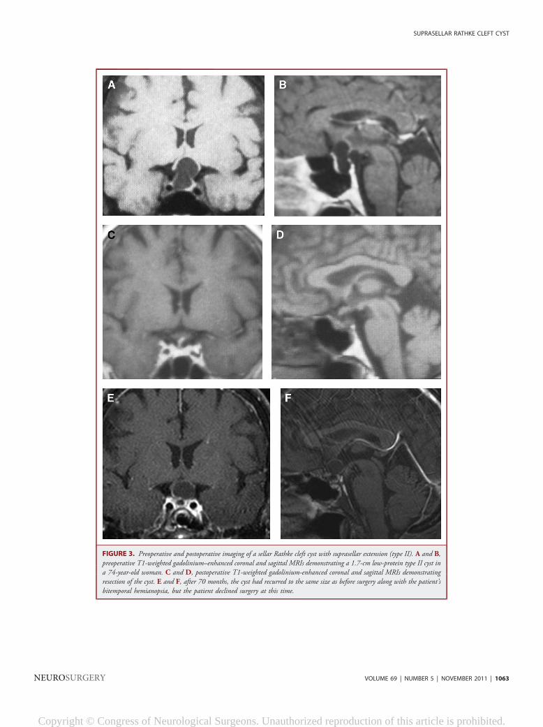
FIGURE 3. Preoperative and postoperative imaging of a sellar Rathke cleft cyst with suprasellar extension (type II). A and B,preoperative T1-weighted gadolinium–enhanced coronal and sagittal MRIs demonstrating a 1.7-cm low-protein type II cyst ina 74-year-old woman. C and D, postoperative T1-weighted gadolinium-enhanced coronal and sagittal MRIs demonstratingresection of the cyst. E and F, after 70 months, the cyst had recurred to the same size as before surgery along with the patient’sbitemporal hemianopsia, but the patient declined surgery at this time.
SUPRASELLAR RATHKE CLEFT CYST
NEUROSURGERY VOLUME 69 | NUMBER 5 | NOVEMBER 2011 | 1063
Copyright © Congress of Neurological Surgeons. Unauthorized reproduction of this article is prohibited.
Copyright © Congress of Neurological Surgeons. Unauthorized reproduction of this article is prohibited.

the diaphragma sella in the suprasellar cistern. It is thereforethought that purely suprasellar RCCs arise from a remnant of theRathke pouch within the pars tuberalis in the suprasellar cistern(Figure 1).26,30,43 In this series of consecutive surgically managedRCCs over a 20-year period at our institution, we report the largestcollection of purely suprasellar RCCs to date (Table 3). We dividedRCCs into 3 distinct anatomical subgroups to distinguish purelysuprasellar RCCs from sellar-based RCCs with and without su-prasellar extension (Figure 2). We show that this classificationsystem can be used both to characterize preoperative symptomsand to prognosticate surgical outcomes.Before this series, the largest series of surgically treated RCCs
was reported by Aho et al6 in 2005, Lillehei et al44 in 2010,and Benveniste et al7 in 2004. Aho et al6 studied 118 RCCs andreported a 49% incidence of preoperative visual dysfunction and53% incidence of endocrinopathy. Complete cyst decompressionwas achieved in 97% of patients in this series, resulting in im-proved vision in 97% of patients with preoperative visual im-pairments. Aho et al did not specify cyst location in their report.Lillehei et al44 reported a series of 82 surgically managed RCCswith presenting symptoms of headache in 68%, visual dys-function in 35%, and endocrinopathy in 56%. They reportedimproved headaches in 71% and improved vision in 83%, as wellas improvement in various endocrinopathies in 33% to 94% ofpatients. They did not characterize the anatomic location of theirRCCs. Benveniste et al7 reported a series of 62 surgically managedRCCs. Among these patients, 71% presented with headache,55% presented with endocrinopathies, and 16% presented withvisual complaints. Complete cyst decompression was achieved in53% of patients in the Benveniste et al series, resulting in 91%
FIGURE 4. Preoperative and postoperative imaging of a purely suprasellarRathke cleft cyst (type III). A and B, preoperative T1-weighted gadolinium-enhanced coronal and sagittal MRIs demonstrating a 7-mm low-protein type IIIcyst in a 64-year-old man. C and D, postoperative T1-weighted gadolinium-enhanced coronal and sagittal MRIs demonstrating drainage of the cyst, asevidenced by a change in signal characteristics. E and F, after 30 months, the cysthad recurred to the same size as before surgery, along with the patient’s visualsymptoms. G and H, the cyst was redrained, and the patient’s visual symptomsagain improved.
FIGURE 5. Recurrence rates of different types of Rathkecleft cysts. Kaplan-Meier analysis of rates of radiographicrecurrence vs time in months of Rathke cleft cysts that weresellar (type I; green), sellar with suprasellar extension (typeII; blue), and suprasellar (type III; red). There was a sig-nificant difference (P , .001) between the recurrence oftype I and II cysts and between the recurrence of type I andIII cysts (P , .001), whereas the difference between therecurrence of type II and III cysts was not statistically sig-nificant (P = .5).
POTTS ET AL
1064 | VOLUME 69 | NUMBER 5 | NOVEMBER 2011 www.neurosurgery-online.com
Copyright © Congress of Neurological Surgeons. Unauthorized reproduction of this article is prohibited.
Copyright © Congress of Neurological Surgeons. Unauthorized reproduction of this article is prohibited.

improvement in headaches and 70% improvement in visualsymptoms. Twenty-seven percent of the RCCs were confined tothe sella, (type I RCCs); 73% were both sellar and suprasellar(type II); and none were purely suprasellar (type III). Surgicaloutcomes based on cyst location were not reported.
In our series, we found a trend toward greater preoperativevisual dysfunction in purely suprasellar (type III) RCCs, a findingthat is consistent with the anatomical location of these lesionsabove the diaphragma sella in close proximity to the optic nerve.Interestingly, 2 of the 76 type I RCCs presented with visualdisturbance. This would not be predicted on the basis of thepurely sellar location of these RCCs, but review of these patients’preoperative MRIs showed displacement of the pituitary glandsuperiorly into the optic chiasm. These patients’ visual dys-function also improved with cyst decompression, further con-firming that their visual dysfunction was related to their RCC.There were no significant differences in the incidence of hypo-pituitarism among these subgroups, perhaps because none of the3 types of RCCs truly arise in the anterior lobe of the pituitarygland and all must therefore exert mass effect adjacent to ratherthan from within the anterior lobe of the gland the way anadenoma does. In other words, suprasellar RCCs may be ascapable of exerting mass effect on the underlying anterior lobe ofthe pituitary gland as sellar RCCs growing in the pars intermediabehind the anterior lobe. Surgically, we found that purelysuprasellar RCCs were more difficult to drain completely thansellar-based RCCs. This coincided with significantly pooreroutcomes with respect to resolution of headaches and visualdysfunction. Interestingly, rates of improvement of laboratoryhypopituitarism did not vary with cyst location, again suggestingthat all RCCs cause mass effect on the anterior lobe by growingadjacent to rather than from within the anterior lobe. Thus,
despite a lesser volumetric reduction, decompressing type IIIRCCs from above the pituitary is as effective at normalizingendocrine function as decompressing type I RCCs from behindthe pituitary.Regardless of the surgical approach chosen, treatment of
suprasellar lesions is more difficult because of the close proximityto vital structures like the optic chiasm and pituitary stalk. Whenusing an endonasal approach, as in this report, the surgeon musttypically traverse the tuberculum sella and sometimes the planumsphenoidale16 to reach the cyst in the suprasellar cistern. Unlikean excision of the contents of sellar-based RCCs, where the opticnerve sits above the surgical corridor and is not seen during theoperation, the optic nerve typically lies in closer proximity tosuprasellar RCCs and must be carefully protected during cystdecompression. There were no incidences of optic nerve injuryfrom decompression of type III RCCs, suggesting that the poorerrates of postoperative visual dysfunction improvement were dueto lower likelihood of complete decompression. Several reports ofpurely suprasellar RCCs have described pterional or frontalcraniotomies to obtain access to the suprasellar region forcyst decompression.33 A craniotomy is more invasive than anendonasal approach and involves retraction of the frontal andtemporal lobes. Furthermore, it is not yet clear if a craniotomyprovides higher success rates for suprasellar RCCs than anendonasal approach. Endonasal endoscopic techniques forsuprasellar lesions like meningiomas have been well describedrecently34,36,37,45 and were applied to 3 of the cases in this series.This technique may allow higher rates of complete removal ofcyst contents while providing a minimally invasive approach, asdemonstrated by Madhok et al16 in a series of 33 RCCs (20 sellar,10 sellar with suprasellar extension, and 3 purely suprasellar) thatwere drained through an endoscopic endonasal approach, with
TABLE 2. Predictors of Rathke Cleft Cyst Recurrence
Univariate Analysis Multivariate Analysis
Variablea Hazard Ratio (95% Confidence Interval) Pb Hazard Ratio (95% Confidence Interval) Pb
Primary variables
Cyst type NA ,.001
Type III vs II 1.2 (0.9-1.3) .5 1.2 (0.6-1.5) .6
Type III/II combined vs I 2.5 (1.6-3.9) ,.001 2.6 (1.8-4.1) .004
Cyst size (per 1-cm diameter) 2.4 (1.1-5.4) .06 2.7 (0.6-11.8) .2
Secondary variables
Squamous metaplasia 1.8 (0.5-7.8) .5
Age (per decade) 1.3 (1.0-1.6) .2
Sex (male) 1.7 (0.8-2.0) .6
Inflammation 1.8 (0.6-5.8) .5
Use of fat graft 1.0 (0.5-2.5) .9
Ethanol irrigation 0.7 (0.4-1.7) .5
aPrimary variables were those that pretest hypotheses suggested would contribute to recurrence risk, whereas secondary variables were not suspected to contribute to
recurrence risk.bSignificant P values (P , .03 for univariate analysis, P , .05 for multivariate analysis) are indicated in bold.
SUPRASELLAR RATHKE CLEFT CYST
NEUROSURGERY VOLUME 69 | NUMBER 5 | NOVEMBER 2011 | 1065
Copyright © Congress of Neurological Surgeons. Unauthorized reproduction of this article is prohibited.
Copyright © Congress of Neurological Surgeons. Unauthorized reproduction of this article is prohibited.

TABLE 3. Summary of Prior Reports of Purely Suprasellar Rathke’s Cleft Cystsa
Study
Age,
y/Sex Presentation
Laboratory
Hypopituitarism
Cyst
Size,
cm Operation
Cyst
Contents
Cyst
Epithelium Outcome Recurrenceb
Frazier and
Alpers,17 1934
52/M HA, VD NR NR Crani Brownish Ciliated columnar Improved HA and vision NR
Eisenberg et al,21 1976 10/M Impaired growth,
frequent urination
Hypothyroid,
hypocortisolemia
2 Crani Opalescent,
mucinous
Ciliated columnar Postoperative DI NR
Palma and Celli,22 1983 17/F VD Decreased LH/FSH NR Crani Clear Cuboidal Some visual
improvement, persistent
low LH/FSH
NR
Rout et al,23 1983 19/F HA, VD, amenorrhea,
polyuria, polydipsia
NR NR Crani Yellowish
pultaceous
Ciliated columnar Improved vision,
persistent amenorrhea
No
Barrow et al,26 1985 35/F HA, VD Hyperprolactinemia NR Crani Thick, yellow-
tinged, white
fluid
Ciliated columnar;
plus squamous
metaplasia
Vision returned
to normal; decreased
prolactin
No
31/M VD, impotence Hypothyroid,
hypocortisolemia,
hypotestosteronism
NR Crani Thick, yellow-
tinged, white
fluid
Squamous
metaplasia
Vision returned;
still on thyroid
and testosterone
replacement
No
13/M VD Panhypopituitarism
with DI
NR TS Thick, yellowish Columnar Improved visual
acuity but
persistent VD and
panhypopituitarism
No
Pangopoulos
et al,28 1989
29/F Panic episodes,
depression
NR NR Crani Mucinous Cuboidal No further
panic episodes,
persistent depression
No
30/F Infertility, galactorrhea Hyperprolactinemia NR TS Mucinous NR Normalized prolactin No
Itoh and Usui,30 1992 21/F HA/VD/fatigue/
menstrual irregularity
NR NR Crani Clear, mucinous Ciliated cuboidal Improved symptoms No
Cavallo et al,31 1993 26/F HA, polydipsia, nocturia,
oligomenorrhea
NR 2.9 NR Milky Ciliated cuboidal and
pseudostratified
columnar
Improved symptoms NR
Graziani et al,32 1995 39/F Amnestic episode NR 1 Crani Clear Columnar NR NR
40/F HA NR 2 Crani Clear Columnar NR NR
Mukherjee et al,11 1997 37/F VD, amenorrhea Hyperprolactinemia NR TS Yellow, mucinousCiliated cuboidal NR No
Rincon et al,33 1999 29/F VD, HA NR 1.2 Crani White, mucinous Columnar Transient DI, resolution
of symptoms
NR
Kim et al,34 2000 41/F VD, HA Normal NR TS NR Ciliated columnar NR NR
Wenger et al,43 2001 58/M Vertigo, decreased libido Normal 1.2 Crani White mucinous Cuboidal Resolution of symptoms NR
Nakahara et al,35 2004 66/F HA Normal NR ETV Cloudy mucinousNR NR No
73/M VD Hypopituitarism NR ETV NR NR NR No
Laufer et al,38 2007 53/F VD NR 1.1 ETS NR NR Transient postoperative
DI, hypocortisolemia,
and blurry vision,
all of which resolved
No
aCrani, craniotomy; DI, diabetes insipidus; ETS, endoscopic transsphenoidal; ETV, endoscopic transventricular; FSH, follicle-stimulating hormone; HA, headache; LH, lutenizing hormone; NR, not recorded;
TS, transsphenoidal; VD, visual deficit. Other cases of purely suprasellar Rathke cleft cysts have been reported as part of a larger series that did not comment on individual patient characteristics.10,13,16,36,37,39,40
bRecurrence within the follow-up period of each individual study.
POTTSET
AL
1066
|VOLU
ME69|NUMBER
5|NOVEM
BER
2011
www.neurosu
rgery-onlin
e.co
m
Copyright © Congress of Neurological Surgeons. Unauthorized reproduction of this article is prohibited.
Copyright © Congress of Neurological Surgeons. Unauthorized reproduction of this article is prohibited.

only 2 recurrences reported. However, the authors did notattempt to correlate recurrences and rate of symptomaticimprovement with cyst location.
We report Kaplan-Meier 3-year recurrence rates of 0%, 16%,and 29% for type I, II, and III RCCs, respectively, withrecurrence occurring in 11% of the total population. Althoughour overall recurrence rate is similar to that in many priorreports,6,7,14,40,44,46-48 it should be noted that the range ofreported recurrence rates in the literature is quite large,11,12,16,49-51
with 1 study reporting none52 and another reporting 42%.15 Themajority of our patients received their first postoperative MRI at6 weeks, raising the possibility that some early recurrences before6 weeks could be designated residual cysts rather than recurrentcysts. However, prior studies have shown that cyst reaccumulationtypically does not occur before this 6-week interval,6,7,41 and noneof our 15 patients who underwent immediate postoperativeimaging exhibited a recurrence at their first subsequent MRI. Ofnote, prior reports have suggested that the use of an abdominal fatgraft for closure has actually been associated with higher rates ofrecurrence.6 It is theorized that an abdominal fat or fascial graftmay prevent marsupialization of a cyst and lead to reaccumulation.We found that use of an abdominal fat graft, which occurred in62 cases in our series, was not associated with recurrence in amultivariate analysis.
Pathologically, RCCs are characterized by a columnar orcuboidal epithelium,53 and findings of cyst wall inflammation orsquamous metaplasia have been reported.7,54 In this series, wealso found that purely suprasellar RCCs were distinct from sellar-based RCCs with regard to histology. Type III RCCs were morelikely to have evidence of squamous metaplasia compared withsellar-based RCCs, although this finding was not associated withincreased recurrence rate in our multivariate analysis. Otherauthors have reported on the significance of such histologicalfindings. Benveniste et al7 showed that both cyst wall in-flammation and squamous cell metaplasia were associated withincreased recurrence rate. Hama et al54 specifically examinedchanges in the epithelium of RCCs and found that the presenceof inflammation led to stratified cyst epithelium and was asso-ciated with hypophysitis and hypopituitarism. Two potentialcauses of RCC epithelial inflammation have been identified:bacterial infection and aseptic irritation. We recently reporteda series of infected RCCs and showed that the surgeon’s suspicionof bacterial infection was a strong predictor of cyst recurrence.41
Conversely, there have been several reports of asepticinflammation thought to be associated with irritation from themucinous contents of a cyst.54 Interestingly, although we founda higher rate of squamous metaplasia in type III RCCs, we alsofound that this subgroup was less likely to have proteinaceousfluid on the basis of preoperative MRI. It is possible that thehigher squamous metaplasia in type III RCCs may reflect theobservation by Harrison et al55 that RCCs and craniophar-yngiomas are part of a continuum of epithelium-lined cysticlesions and the subsequent finding by Aho et al6 that RCCs withsquamous metaplasia have a natural history in terms of recurrence
rates that resembles that of craniopharyngiomas. Although we didnot find squamous metaplasia to be predictive of a higher re-currence rate in this series, given their suprasellar location, it ispossible that the squamous metaplasia we found in type III RCCsmay reflect the fact that type III RCCs are closer to the cra-niopharyngioma side of the spectrum of cystic epithelium-linedsellar and suprasellar lesions. On the other hand, the tendency oftype III RCCs to have less proteinaceous fluid may reflect reducedlevels of infection in these cysts compared with type I RCCs.Although we excluded RCCs in which the surgeon suspectedinfection from this analysis, a review of our previously reportedRCC cases in which the surgeon suspected infection41 revealedthat 10% of infected RCCs were type III (data not shown),similar to the frequency reported in this article. Regardless, it ispossible that our series contains some infected RCCs that did notevoke suspicion for infection. Further work is needed to clarifythese findings.
CONCLUSION
Although large symptomatic suprasellar RCCs such as thosedescribed here clearly warrant treatment, there are unique chal-lenges in their neurosurgical treatment owing to their intimateproximity to the optic chiasm and pituitary stalk. In particular,our retrospective review found that, compared with sellar RCCs,RCCs with a suprasellar component are more difficult to removecompletely and to obtain symptomatic resolution. The RCCswith a suprasellar component also carry a higher recurrence rate.These findings suggest that RCCs with a suprasellar componentare best handled by experienced pituitary surgeons and that theexpectations of cure and symptomatic resolution should becarefully discussed with patients.
Disclosure
The authors have no personal financial or institutional interest in any of thedrugs, materials, or devices described in this article.
REFERENCES
1. Prabhu VC, Brown HG. The pathogenesis of craniopharyngiomas. Childs NervSyst. 2005;21(8-9):622-627.
2. Shanklin WM. On the presence of cysts in the human pituitary. Anat Rec.1949;104(4):379-407.
3. Fager CA, Carter H. Intrasellar epithelial cysts. J Neurosurg. 1966;24(1):77-81.4. McGrath P. Cysts of sellar and pharyngeal hypophyses. Pathology. 1971;3(2):
123-131.5. Teramoto A, Hirakawa K, Sanno N, Osamura Y. Incidental pituitary lesions in
1,000 unselected autopsy specimens. Radiology. 1994;193(1):161-164.6. Aho CJ, Liu C, Zelman V, Couldwell WT, Weiss MH. Surgical outcomes in
118 patients with Rathke cleft cysts. J Neurosurg. 2005;102(2):189-193.7. Benveniste RJ, King WA, Walsh J, Lee JS, Naidich TP, Post KD. Surgery for
Rathke cleft cysts: technical considerations and outcomes. J Neurosurg.2004;101(4):577-584.
8. Zada G, Lin N, Ojerholm E, Ramkissoon S, Laws ER. Craniopharyngioma andother cystic epithelial lesions of the sellar region: a review of clinical, imaging, andhistopathological relationships. Neurosurg Focus. 2010;28(4):E4.
9. Voelker JL, Campbell RL, Muller J. Clinical, radiographic, and pathologicalfeatures of symptomatic Rathke’s cleft cysts. J Neurosurg. 1991;74(4):535-544.
SUPRASELLAR RATHKE CLEFT CYST
NEUROSURGERY VOLUME 69 | NUMBER 5 | NOVEMBER 2011 | 1067
Copyright © Congress of Neurological Surgeons. Unauthorized reproduction of this article is prohibited.
Copyright © Congress of Neurological Surgeons. Unauthorized reproduction of this article is prohibited.

10. Ross DA, Norman D, Wilson CB. Radiologic characteristics and results ofsurgical management of Rathke’s cysts in 43 patients. Neurosurgery. 1992;30(2):173-178.
11. Mukherjee JJ, Islam N, Kaltsas G, et al. Clinical, radiological and pathologicalfeatures of patients with Rathke’s cleft cysts: tumors that may recur. J ClinEndocrinol Metab. 1997;82(7):2357-2362.
12. Frank G, Sciarretta V, Mazzatenta D, Farneti G, Modugno GC, Pasquini E.Transsphenoidal endoscopic approach in the treatment of Rathke’s cleft cyst.Neurosurgery. 2005;56(1):124-128.
13. Sade B, Albrecht S, Assimakopoulos P, Vezina JL, Mohr G. Management ofRathke’s cleft cysts. Surg Neurol. 2005;63(5):459-466.
14. Koutourousiou M, Grotenhuis A, Kontogeorgos G, Seretis A. Treatment of Rathke’scleft cysts: experience at a single centre. J Clin Neurosci. 2009;16(7):900-903.
15. Raper DM, Besser M. Clinical features, management and recurrence of symp-tomatic Rathke’s cleft cyst. J Clin Neurosci. 2009;16(3):385-389.
16. Madhok R, Prevedello DM, Gardner P, Carrau RL, Snyderman CH, Kassam AB.Endoscopic endonasal resection of Rathke cleft cysts: clinical outcomes and sur-gical nuances. J Neurosurg. 2010;112(6):1333-1339.
17. Frazier CH, Alpers BJ. Tumors of Rathke’s cleft (hitherto called tumors ofRathke’s pouch). Arch Neurol and Psychiatry. 1934;32(5):973-984.
18. Bayoumi ML. Rathke’s cleft and its cysts. Edinb Med J. 1948;55(12):745-749.19. Naiken VS, Tellem M, Meranze DR. Pituitary cyst of Rathke’s cleft origin with
hypopituitarism. J Neurosurg. 1961;18(5):703-708.20. Fairburn B, Larkin IM. A cyst of Rathke’s cleft. J Neurosurg. 1964;21:223-225.21. Eisenberg HM, Sarwar M, Schochet S Jr. Symptomatic Rathke’s cleft cyst: case
report. J Neurosurg. 1976;45(5):585-588.22. Palma L, Celli P. Suprasellar epithelial cyst. Case report. J Neurosurg. 1983;58(5):
763-765.23. Rout D, Das L, Rao VR, Radhakrishnan VV. Symptomatic Rathke’s cleft cysts.
Surg Neurol. 1983;19(1):42-45.24. Hiramatsu K, Ohnishi H, Nikaido Y, Fujita T, Kawaguchi S. Suprasellar epithelial
cyst associated with cerebral aneurysm: case report [in Japanese]. Neurol Med Chir(Tokyo). 1984;24(5):359-363.
25. Yamamoto M, Takara E, Imanaga H, Jimbo M, Kubo O. Rathke’s cleft cyst:report of two cases [in Japanese]. No Shinkei Geka. 1984;12(5):609-616.
26. Barrow DL, Spector RH, Takei Y, Tindall GT. Symptomatic Rathke’s cleft cystslocated entirely in the suprasellar region: review of diagnosis, management, andpathogenesis. Neurosurgery. 1985;16(6):766-772.
27. Ishii T, Yamasaki T, Tanaka J, Tanaka S, Hori T, Muraoka K. Rathke’s cleft cyst:report of three cases [in Japanese]. No Shinkei Geka. 1987;15(4):451-456.
28. Panagopoulos KP, Jolesz FA, el-Azouzi M, Black PM. Mucinous cysts of thepituitary stalk: report of two cases. J Neurosurg. 1989;71(2):276-278.
29. Yuge T, Shigemori M, Tokutomi T, et al. Entirely suprasellar symptomaticRathke’s cleft cyst [in Japanese]. No Shinkei Geka. 1991;19(3):273-278.
30. Itoh J, Usui K. An entirely suprasellar symptomatic Rathke’s cleft cyst: case report.Neurosurgery. 1992;30(4):581-584.
31. Cavallo AV, Murphy MA, McKelvie PA, Cummins JT. An epithelial cyst of thesuprasellar region. Aust N Z J Surg. 1993;63(6):490-493.
32. Graziani N, Dufour H, Figarella-Branger D, Donnet A, Bouillot P, Grisoli F. Dothe suprasellar neurenteric cyst, the Rathke cleft cyst and the colloid cyst constitutea same entity? Acta Neurochir (Wien). 1995;133(3-4):174-180.
33. Rincon JL, Nunes J, Camuto P, Goodrich I. Intracranial approach to suprasellarRathke’s cleft cyst. Skull Base Surg. 1999;9(1):71-73.
34. Kim J, Choe I, Bak K, Kim C, Kim N, Jang Y. Transsphenoidal supra-diaphragmatic intradural approach: technical note. Minim Invasive Neurosurg.2000;43(1):33-37.
35. Nakahara Y, Koga H, Maeda K, Takagi M, Tabuchi K. Neuroendoscopic trans-ventricular surgery for suprasellar cystic mass lesions such as cystic craniopharyngiomaand Rathke cleft cyst. Neurol Med Chir (Tokyo). 2004;44(8):408-413.
36. Dusick JR, Esposito F, Kelly DF, et al. The extended direct endonasal trans-sphenoidal approach for nonadenomatous suprasellar tumors. J Neurosurg. 2005;102(5):832-841.
37. de Divitiis E, Cavallo LM, Cappabianca P, Esposito F. Extended endoscopicendonasal transsphenoidal approach for the removal of suprasellar tumors, part 2.Neurosurgery. 2007;60(1):46-58.
38. Laufer I, Anand VK, Schwartz TH. Endoscopic, endonasal extended trans-sphenoidal, transplanum transtuberculum approach for resection of suprasellarlesions. J Neurosurg. 2007;106(3):400-406.
39. Wen L, Hu LB, Feng XY, et al. Rathke’s cleft cyst: clinicopathological and MRIfindings in 22 patients. Clin Radiol. 2010;65(1):47-55.
40. Nishioka H, Haraoka J, Izawa H, Ikeda Y. Headaches associated with Rathke’scleft cyst. Headache. 2006;46(10):1580-1586.
41. Tate MC, Jahangiri A, Blevins L, Kunwar S, Aghi MK. Infected Rathke cleft cysts:distinguishing factors and factors predicting recurrence. Neurosurgery. 2010;67(3):762-769
42. Kleinschmidt-DeMasters BK, Lillehei KO, Stears JC. The pathologic, surgical,and MR spectrum of Rathke cleft cysts. Surg Neurol. 1995;44(1):19-26.
43. Wenger M, Simko M, Markwalder R, Taub E. An entirely suprasellar Rathke’scleft cyst: case report and review of the literature. J Clin Neurosci. 2001;8(6):564-567.
44. Lillehei KO, Widdel L, Arias Astete CA, Wierman ME, Kleinschmidt-DemastersBK, Kerr JM. Transsphenoidal resection of 82 Rathke cleft cysts: limited value ofalcohol cauterization in reducing recurrence rates. J Neurosurg. 2010;114(2):310-317.
45. Dehdashti AR, Ganna A, Witterick I, Gentili F. Expanded endoscopic endonasalapproach for anterior cranial base and suprasellar lesions: indications and limi-tations. Neurosurgery. 2009;64(4):677-687.
46. Billeci D, Marton E, Tripodi M, Orvieto E, Longatti P. Symptomatic Rathke’scleft cysts: a radiological, surgical and pathological review. Pituitary. 2004;7(3):131-137.
47. Shin JL, Asa SL, Woodhouse LJ, Smyth HS, Ezzat S. Cystic lesions of thepituitary: clinicopathological features distinguishing craniopharyngioma,Rathke’s cleft cyst, and arachnoid cyst. J Clin Endocrinol Metab. 1999;84(11):3972-3982.
48. Kim JE, Kim JH, Kim OL, et al. Surgical treatment of symptomatic Rathke cleftcysts: clinical features and results with special attention to recurrence. J Neurosurg.2004;100(1):33-40.
49. Eguchi K, Uozumi T, Arita K, et al. Pituitary function in patients with Rathke’scleft cyst: significance of surgical management. Endocr J. 1994;41(5):535-540.
50. Cohan P, Foulad A, Esposito F, Martin NA, Kelly DF. Symptomatic Rathke’s cleftcysts: a report of 24 cases. J Endocrinol Invest. 2004;27(10):943-948.
51. Dusick JR, Fatemi N, Mattozo C, et al. Pituitary function after endonasal surgeryfor nonadenomatous parasellar tumors: Rathke’s cleft cysts, craniopharyngiomas,and meningiomas. Surg Neurol. 2008;70(5):482-490.
52. el-Mahdy W, Powell M. Transsphenoidal management of 28 symptomaticRathke’s cleft cysts, with special reference to visual and hormonal recovery.Neurosurgery. 1998;42(1):7-16.
53. McLendon RE, Bigner DD, Bigner SH, Provenzale JM. Pathology of Tumors of theNervous System: A Guide to Histologic Diagnosis. London, UK: Arnold; 2000.
54. Hama S, Arita K, Nishisaka T, et al. Changes in the epithelium of Rathke cleft cystassociated with inflammation. J Neurosurg. 2002;96(2):209-216.
55. Harrison MJ, Morgello S, Post KD. Epithelial cystic lesions of the sellar andparasellar region: a continuum of ectodermal derivatives? J Neurosurg.1994;80(6):1018-1025.
COMMENTS
T his large, well-documented series adds worthwhile information tothe literature. I am particularly pleased to see the breakdown of
these Rathke cleft cysts (RCCs) into the 3 types. As the authors dem-onstrate, there are significant differences in presentation and resultsamong the different types. This information is important when dis-cussing potential results with patients and when we review our ownseries. Primarily suprasellar RCCs, I believe, fit into a continuum ofectodermal derivatives, including squamous metaplasia and cranio-pharyngioma, as we published in 1994.1 They may require moreaggressive resections than we have previously advised for predominatelysellar-based RCCs when aggressive resection leads to higher incidence ofpostoperative hypopituitarism.
Kalmon D. PostNew York, New York
POTTS ET AL
1068 | VOLUME 69 | NUMBER 5 | NOVEMBER 2011 www.neurosurgery-online.com
Copyright © Congress of Neurological Surgeons. Unauthorized reproduction of this article is prohibited.
Copyright © Congress of Neurological Surgeons. Unauthorized reproduction of this article is prohibited.

1. Harrison MJ, Morgello S, Post KD. Epithelial cystic lesions of the sellar andparasellar region: a continuum of ectodermal derivatives? J Neurosurg.1994;80(6):1018-1025.
T his large series of colloid cysts of the third ventricle adds usefulinformation about the management of these relatively rare entities
and tailoring treatment to their anatomical position. It is worth notingthat, for whatever reason, there has always seemed to be a rather higher
recurrence rate in North American series than in European series.Although I have no direct experience with the use of absolute alcohol inthis setting, I have seen serious problems related to its use in trigeminalnerve ablation in the distant past and thus add serious cautions aboutits use.
Michael PowellLondon, United Kingdom
SUPRASELLAR RATHKE CLEFT CYST
NEUROSURGERY VOLUME 69 | NUMBER 5 | NOVEMBER 2011 | 1069
Copyright © Congress of Neurological Surgeons. Unauthorized reproduction of this article is prohibited.
Copyright © Congress of Neurological Surgeons. Unauthorized reproduction of this article is prohibited.
![Case Report Malignant Trigeminal Nerve Sheath Tumor and ...(including glioblastoma) Suh et al. [ ] Suprasellar chordoid glioma and Rathke scle cyst Johnson et al. [ ] Oligodendroglioma](https://img.pdfslide.us/doc/110x75/60f6a026ed06d422737c4388/case-report-malignant-trigeminal-nerve-sheath-tumor-and-including-glioblastoma.jpg)