Embed Size (px)
Citation preview

CIRCHF/201/963769-R3
1
SUPPLEMENTARY MATERIAL
Supplementary Methods
Experimental Model
BALB/cJ (BALB/c) (stock#000651), C57BL/6J (stock#000664), B6.129SF2/J (stock#101045),
type I IL-1R deficient (B6.129S7-Il1r1tm1Imx
/J, stock#003245), IL-6 deficient (B6;129S2-
Il6tmKopf
/J, stock#002254) and IL1RAcP deficient (B6;129S1-Il1raptm1Roml
/J, stock# 003284)
mice were obtained from The Jackson Laboratory (Bar Harbor, ME). Mice were maintained
under pathogen-free conditions in the animal facility at Johns Hopkins School of Medicine, and
approval was obtained from the Animal Care and Use Committee of the Johns Hopkins
University for all procedures. Eight to ten week old male mice were inoculated intraperitoneally
(ip) with 103 plaque forming units (PFU) of heart-passaged CVB3 containing infectious virus
and heart tissue diluted in sterile saline.1 Recombinant (r)IL-33 (1 g/0.1mL/mouse, Cat#3626-
ML), rST2 (5 g/0.1mL/mouse, Cat#1004MR-050) from R&D Systems (Minneapolis, MN), or
sterile PBS were injected ip on day 1, 3, 5, 7 and 9 pi following CVB3 injection on day 0. The
dose and time-course for rIL-33 and rST2 administration were based on previously published
studies that found significantly increased or decreased inflammation using similar doses and
time-course.2-4
Mice were harvested on day 10 pi during acute myocarditis or day 35 pi during
DCM. rIL-33, rST2 or PBS was injected ip using the same protocol as above for healthy
undiseased experiments and mice were examined on the equivalent of day 10 (one day after the
5th
injection) or day 35 post inoculation.

CIRCHF/201/963769-R3
2
Histology
Hearts were fixed in 10% buffered formalin and stained with H&E to assess inflammation.
Sections were examined by two independent investigators and myocarditis assessed as the
percentage of the heart section with inflammation compared to the overall size of the heart
section using an eyepiece grid, as previously.5-7
The development of DCM was assessed by gross
observation of histology sections at low magnification and by echocardiography or pressure-
volume relationships.5-7
Cardiac Function
Cardiac function was assessed by pressure-volume catheter (1.2F Scisense Inc., London,
Ontario) placed in the left ventricle via the apex in open-chest mice anesthetized with 3%
isoflurane, as previously described.7-9
Previously we demonstrated that cardiac dysfunction
observed during CVB3 myocarditis produces similar results using open-chest ventricular
catheterization and closed-chest echocardiography methods.7 Ventricular dilation was assessed
by trans-thoracic echocardiography (Acuson Sequoia C256, 15 MHz linear transducer; Siemens,
Malvern, PA) in conscious mice, as previously.6,7
M-mode left ventricular end-systolic or
diastolic cross-sectional diameters (LVESD or LVEDD) were determined from an average of 3-5
cardiac cycles. To assess β-adrenergic sensitivity, an isoproterenol dose-response protocol was
adapted from previously described protocols.10,11
Left ventricular pressure-volume measurements
were recorded continuously. Baseline hemodynamic values were obtained followed by 10 L
intravenous bolus doses of vehicle, 0.2, 0.6, 2, 6, or 20 ng of isoproterenol in 3 minute

CIRCHF/201/963769-R3
3
increments. The response to isoproterenol was determined by subtracting the maximum response
obtained within 1 min following injection from values obtained immediately preceding that dose.
Plaque Assay
Hearts were homogenized at 10% weight/volume in 2% minimal essential medium (MEM) and
individual supernatants used in plaque assays to determine the level of infectious virus (a
measure of viral replication). The plaque assay method has been described in detail previously.1,5
Briefly, homogenates were incubated for 1h on Vero cells (ATCC) at 37◦C for viral attachment
and covered with 2% MEM in methyl cellulose for three days to allow plaque formation. Viral
plaques were quantitated using a light microscope and normalized to the size of the heart
according to tissue wet weight. Viral replication is shown as the number of PFU/g of heart. The
limit of detection for the assay was 10 PFU/g of tissue.
Cytokine Measurements
Hearts were homogenized at 10% weight/volume in 2% MEM and individual supernatants or
sera used in ELISA kits (R&D Systems) to measure cytokines according to the manufacturer’s
instructions and as previously described.5,7
The limits of detection for the ELISA were as
follows: IL-33, 6.9 pg/mL; sST2, 140 pg/mL; IL-4, 2.0 pg/mL; TNF- , 5.1 pg/mL; IL-1 , 2.3
pg/mL and IL-6, 1.6 pg/mL. To control for differences in heart size individual samples were
converted to pg/g of heart tissue.

CIRCHF/201/963769-R3
4
Flow Cytometry
Mice were anesthetized with avertin and the aorta cannulated to perfuse the hearts, which were
then digested with 600 /mL collagenase II (Worthington) + 60 /mL DNAse I (Sigma)
according to the manufacturer’s instructions for the GentleMACSTM
isolation of cardiac cells
(Miltenyi), as previously described.12-14
Isolated immune cells were washed and FcγRII/III
blocked with anti-CD16/32 (eBiosciences) then stained with fluorochrome-conjugated antibodies
against CD45, CD3, CD4, CD19, Fc R1 , CD117, CD11b, F4/80, Ly6G or SiglecF (BD
Pharmingen or eBiosciences). Samples were acquired on a four-color dual-laser FACScalibur
cytometer running CellQuest or the LSR II quadlaser cytometer running FACSDiva (BD
Immunocytometry). Data were analyzed with FlowJo 7 (Treestar).
RT-PCR
Trizol Reagent and the PureLink Micro-to-Midi system (Invitrogen, Carlsbad, CA) were used for
extraction and purification of RNA, as previously.6,7
The hearts were homogenized in 2 mL
Trizol and 1 mL of the homogenate was processed according to the manufacturer's protocol
(Invitrogen). Following elution of purified RNA, quantification was performed using a
NanoDrop spectrophotometer. For each treatment group, processing and analysis were
performed in triplicate. cDNA was generated using a Multiscribe reverse transcriptase (Applied
Biosystems, Foster City, CA). Gene expression was measured using assay-on-demand probe sets
and the ABI 7000 Taqman system according to the manufacturer’s instructions (Applied
Biosystems), as previously.6,7
Hypoxanthine phosphoribosyltransferase (HPRT) was used for
normalization.

CIRCHF/201/963769-R3
5
Statistical Analysis
The Mann-Whitney rank sum test was used to evaluate two groups (p<0.05). Comparisons
involving three groups were analyzed using Kruskal-Wallis tests. When three groups were
significant (p<0.05) then pairwise comparisons were made using Mann-Whitney rank tests with
a Bonferroni correction (p<0.013). Repeated measures data (i.e. isoproterenol treatments) were
evaluated using generalized estimating equations (GEE) and linear mixed effects models with
dose as a covariate. An omnibus test for group differences was used at the first stage (p<0.05)
and if significant then pairwise comparisons were assessed (p<0.013). Dose was converted to a
logarithmic scale for linear mixed model analysis because dose-response curves were log-linear
during preliminary analysis. A small number (equivalent to the next lower dilution, 0.06) was
added to allow for log transformation of a 0 dose. For sensitivity analyses, we modeled dose non-
linearity with a quadratic non-linear term to assess group differences.
The authors had full access to and take full responsibility for the integrity of the data. All
authors have read and agree to the manuscript as written.
Supplementary Results
rIL-33 exacerbates cardiac dysfunction during CVB3 myocarditis/DCM but
rST2 improves cardiac function
To determine the effect of IL-33 or ST2 administration on cardiac function, we treated male
BALB/c mice with rIL-33, rST2 (to block IL-33 released during the disease process) or PBS ip

CIRCHF/201/963769-R3
6
every other day from day 1 to 9 pi and examined acute myocarditis at day 10 pi and progression
to DCM at day 35 pi using pressure volume relationships. Evaluating several cardiac functional
parameters over time revealed that rIL-33 significantly decreased cardiac function while rST2
improved function (Figure 2).
During acute CVB3 myocarditis at day 10 pi, mice treated with rIL-33 developed
impaired systolic function (Table 1). In rIL-33 treated mice end systolic pressure (ESP) was
significantly depressed when compared to PBS treated controls (83±2 mmHg vs. 94±3 mmHg, p
= 0.008) (Figure 2). dP/dT Max in IL-33 treated mice was 7141 ±312 mmHg/s which was 21%
lower than PBS treated mice, 9071 ±546 (p = 0.01). Ejection fraction (EF) was also depressed to
49±5% in IL-33 treated vs. 63±3% in PBS treated mice (Table 1). Cardiac geometry was
significantly different in IL-33 treated mice compared to PBS controls. End diastolic volume
(EDV) increased by 32% (rIL-33 22 ±7 L, PBS 15 ±0.5 L, p = 0.01) (Figure 2). IL-33 treated
mice also demonstrated impaired diastolic function. dP/dT Min was 27% lower in IL-33 treated
mice than in PBS treated mice (IL-33 -6224 ±477 mmHg/s vs. PBS -8502 ±424mmHg/s, p =
0.003).
In contrast, rST2 treatment was either similar to PBS treated mice or displayed
significantly improved systolic/ diastolic ventricular function during acute CVB3 myocarditis
(day 10 pi) compared to PBS controls (Table 1). dP/dT Max in ST2 treated mice was 11570
±350 mmHg/s vs. 9071 ±546 mmHg/s in PBS treated mice (p = 0.002). Maximum ventricular
power (PMX) was 40% higher in rST2 treated mice as compared to PBS treated controls (p =
CIRCHF/201/963769-R3
7
0.003) and the power output in the left ventricles of rST2 treated mice was double that of rIL-33
treated mice (IL-33 7±0.4 mW, ST2 14±0.6 mW, PBS 10±0.7 mW, p < 0.00001).
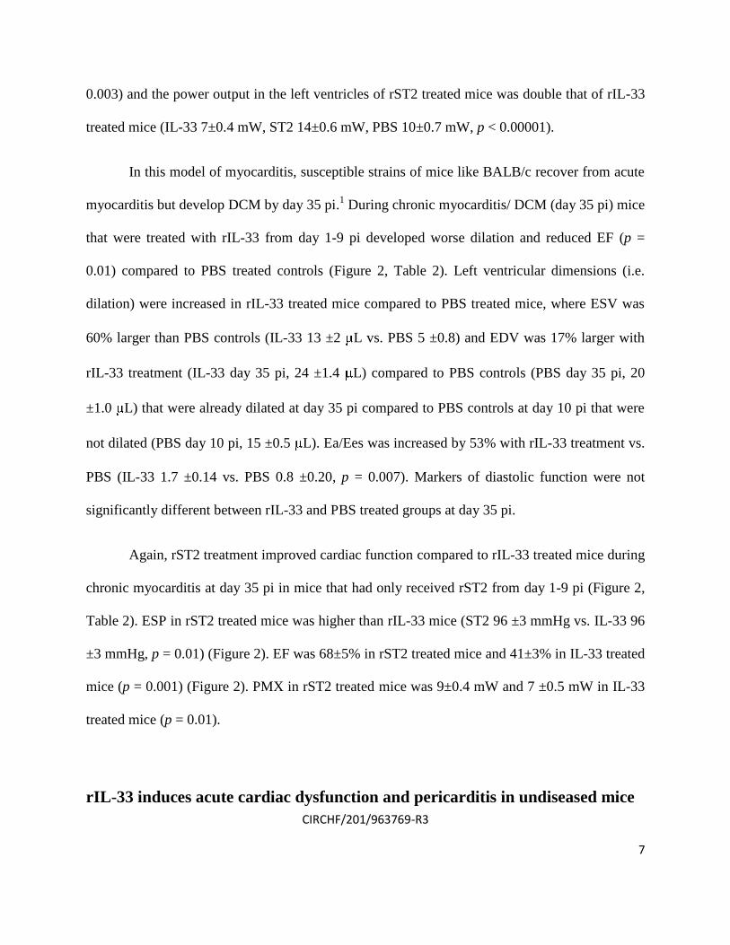
In this model of myocarditis, susceptible strains of mice like BALB/c recover from acute
myocarditis but develop DCM by day 35 pi.1 During chronic myocarditis/ DCM (day 35 pi) mice
that were treated with rIL-33 from day 1-9 pi developed worse dilation and reduced EF (p =
0.01) compared to PBS treated controls (Figure 2, Table 2). Left ventricular dimensions (i.e.
dilation) were increased in rIL-33 treated mice compared to PBS treated mice, where ESV was
60% larger than PBS controls (IL-33 13 ±2 L vs. PBS 5 ±0.8) and EDV was 17% larger with
rIL-33 treatment (IL-33 day 35 pi, 24 ±1.4 L) compared to PBS controls (PBS day 35 pi, 20
±1.0 L) that were already dilated at day 35 pi compared to PBS controls at day 10 pi that were
not dilated (PBS day 10 pi, 15 ±0.5 L). Ea/Ees was increased by 53% with rIL-33 treatment vs.
PBS (IL-33 1.7 ±0.14 vs. PBS 0.8 ±0.20, p = 0.007). Markers of diastolic function were not
significantly different between rIL-33 and PBS treated groups at day 35 pi.
Again, rST2 treatment improved cardiac function compared to rIL-33 treated mice during
chronic myocarditis at day 35 pi in mice that had only received rST2 from day 1-9 pi (Figure 2,
Table 2). ESP in rST2 treated mice was higher than rIL-33 mice (ST2 96 ±3 mmHg vs. IL-33 96
±3 mmHg, p = 0.01) (Figure 2). EF was 68±5% in rST2 treated mice and 41±3% in IL-33 treated
mice (p = 0.001) (Figure 2). PMX in rST2 treated mice was 9±0.4 mW and 7 ±0.5 mW in IL-33
treated mice (p = 0.01).
rIL-33 induces acute cardiac dysfunction and pericarditis in undiseased mice

CIRCHF/201/963769-R3
8
Administration of certain cytokines, like TNF- or IL-1 , is capable of causing acute cardiac
dysfunction in normal mice.15,16
To assess whether rIL-33 administration could induce cardiac
dysfunction in the absence of myocarditis, normal uninfected male BALB/c mice were injected
ip with PBS, rIL-33 or rST2 every other day for a total 5 injections (day 1, 3, 5, 7 and 9) and
cardiac function assessed the day after the final treatment (the equivalent of day 10) using
pressure-volume relationships or at day 35 by echocardiography. We found that rIL-33 treatment
was able to impair cardiac function in the absence of myocarditis compared to PBS treated mice
at day 10 (Table 3) but not at day 35 (Supplemental Table 1). Although LVEDD and LVESD
were significantly increased in rIL-33-treated mice compared to PBS controls at day 35, the
increases were within the normal physiologic range and EF and FS were not different between
groups indicating that the hearts were not dilated (Supplemental Table 1). In contrast, rST2
treatment from day 1-9 had no significant overall effect on heart function compared to PBS
controls at day 10 (Table 3). For this reason, rST2 was not examined at day 35 in undiseased
mice.
Specifically, rIL-33 treatment impaired systolic ventricular function and induced
bradycardia (IL-33 533 ±3 bpm vs. PBS 570 ±10 bpm, p = 0.003) (Table 3). ESP was unchanged
by rIL-33 administration. dP/dT Max was decreased by 20% in rIL-33 treated mice (IL-33 8055
±395 mmHg/s vs. PBS 10156 ±346 mmHg/s, p = 0.002). rIL-33 treatment placed these mice at a
risk for heart failure by reducing EF by 44% from 68±5% in PBS treated mice to 38±7% in IL-
33 treated mice (Table 3). With rIL-33 treatment ESV significantly increased to 14 ±2.1 L from
6 ±1.1 L in PBS treated mice (p = 0.005). rIL-33 treatment induced dilation by changing EDV

CIRCHF/201/963769-R3
9
from 16 ±1.1 L in PBS treated mice to 23 ±1.8 L in rIL-33 treated mice. Ventricular-arterial
coupling was significantly impacted by rIL-33 treatment (IL-33 2.31 ±0.29, PBS 0.96 ±0.09, p =
0.007). Diastolic function was also impaired by rIL-33 treatment (Table 3). dP/dT Min was -
7138 ±551 mmHg/s in rIL-33 treated mice and -9460 ±83 mmHg/s in PBS treated mice (p =
0.001). Tau was 7 ±0.7 ms with rIL-33 treatment and 5 ±0.2 ms with PBS (p = 0.01). There were
no significant differences between PBS and ST2 treated mice for any hemodynamic parameter
(Table 3).
Histological examination revealed that rIL-33-treatment induced pericarditis in
undiseased mice (Supplemental Figure 1) similar in appearance to that observed during acute
CVB3 myocarditis (Figure 1D). The pericardium is a single cell layer in normal mice
(Supplemental Figure 1A) but was severely inflamed with numerous eosinophils following rIL-
33 administration (Supplemental Figure 1B). However, a single injection of rIL-33 at day 0 was
not capable of inducing pericarditis, myocarditis or eosinophilia at day 10 in undiseased mice (n
= 10/group) (data not shown). In contrast, pericardial damage in rST2-treated mice appeared
“lacey” with few inflammatory cells and no eosinophilia (Supplemental Figure 1C). Myocardial
inflammation was not present in PBS, rIL-33 or rST2-treated mice that had not received CVB3.
rIL-33 increases cardiac IL-33, IL-4, IL-1 IL-6 and serum sST2, and alters
remodeling during acute CVB3 myocarditis

CIRCHF/201/963769-R3
10
To examine whether rIL-33 treatment affected cardiac remodeling, we conducted RT-PCR
during acute myocarditis on mice that had been treated with rIL-33 from day 1-9, as previously,
or PBS controls and examined whether changes occurred in the level of the remodeling genes
Mmp3 or Mmp9 in the heart. We, and others, have found that reduced levels of Mmp3 and
Mmp9 during CVB3 myocarditis are associated with cardiac remodeling and fibrosis.17-19
In this
study we found that rIL-33 reduced Mmp3 (PBS 11.2±1.3 vs. rIL-33 7.4±1.6, p = 0.04) and
Mmp9 (PBS 11.0±1.3 vs. rIL-33 6.8±1.6, p = 0.04) expression in the heart compared to PBS
controls.
rIL-33 treatment directly decreases heart function
Because rIL-33 treatment induced cardiac dysfunction and β-adrenergic insensitivity in diseased
and undiseased mice at day 10, we wanted to determine whether IL-33 could be independently
responsible for these effects.15,16
However, since rIL-33 treatment increased IL-1 and IL-6
levels in the heart it was possible that IL-33 worked indirectly to alter heart function via these
cytokines. To test this possibility we treated undiseased male IL-1R, IL-6 or IL-1RAcP (this
receptor is required for IL-1R and IL-33R-mediated signaling)20
deficient mice with PBS or rIL-
33, using the same protocol as before, and examined heart function by pressure-volume
relationships. We found that rIL-33 treatment significantly decreased most cardiac functional
parameters compared to PBS treated IL-1R or IL-6 deficient mice (IL-1R-/- ESP p = 2.63x10⁻⁵
,
CO p = 0.02, SW p = 0.02, PMX p = 2.63x10⁻⁵
; IL-6-/- ESP p = 2.74x10⁻⁵
, CO p = 0.003, SW p
= 0.0003, PMX p = 2.74x10⁻⁵
) using the Mann-Whitney rank test (Figure 5A and 5B). These
CIRCHF/201/963769-R3
11
data indicate that IL-1 and IL-6 were not responsible for the effect of rIL-33. In contrast,
cardiac dysfunction following rIL-33 treatment was not significantly different from PBS controls
for any parameter examined in IL-1RAcP deficient mice (ESP p = 0.37, CO p = 0.38, SW p =
0.30, PMX p = 0.39) (Figure 5C), demonstrating that the IL-33R is required for the cardiac
dysfunction observed in rIL-33 treated mice.
Because IL-1 has been shown to be critical for acute CVB3 myocarditis,21-23
we
examined whether rIL-33 could increase myocarditis indirectly via IL-1 by treating IL-1R
deficient mice with rIL-33 from day 1-9 pi. We found that although rIL-33 significantly
increased myocarditis compared to PBS control C57BL/6 mice (p = 0.01), there was no
significant difference in myocarditis between WT and IL-1R deficient mice in response to rIL-33
(p = 0.42) (Supplemental Figure 4). These results indicate that rIL-33 does not increase acute
CVB3 myocarditis via IL-1 .
Supplementary References
1. Fairweather D, Rose NR. Coxsackievirus-induced myocarditis in mice: a model of
autoimmune disease for studying immunotoxicity. Methods. 2007; 41:118-122.
2. Schmitz J, Owyang A, Oldham E, Song Y, Murphy E, McClanahan TK, Zurawski G,
Moshrefi M, Qin J, Li X, Gorman DM, Bazan JF, Kastelein RA. IL-33, an interleukin-1-
like cytokine that signals via the IL-1 receptor-related protein ST2 and induces T helper
type 2-associated cytokines. Immunity. 2005; 23:479-490.

CIRCHF/201/963769-R3
12
3. Fagundes CT, Amaral FA, Souza ALS, Vieira AT, Xu D, Liew FY, Souza DG, Teixeira
MM. ST2, an IL-1R family member, attenuates inflammation and lethality after intestinal
ischemia and reperfusion. J Leukoc Biol. 2007; 81:492-499.
4. Miller AM, Xu D, Asquith DL, Denby L, Li Y, Sattar N, Baker AH, McInnes IB, Liew
FY. IL-33 reduces the development of atherosclerosis. J Exp Med. 2008; 205:339-346.
5. Fairweather D, Frisancho-Kiss S, Yusung SA, Barrett MA, Gatewood SJL, Davis SE,
Njoku DB, Rose NR. IFN- protects against chronic viral myocarditis by reducing mast
cell degranulation, fibrosis, and the profibrotic cytokines TGF- , IL-1 , and IL-4 in the
heart. Am J Pathol. 2004; 165:1883-1894.
6. Onyimba JA, Coronado MJ, Garton AE, Kim JB, Bucek A, Bedja D, Gabrielson KL,
Guilarte TR, Fairweather D. The innate immune response to coxsackievirus B3 predicts
progression to cardiovascular disease and heart failure in male mice. Biol Sex Differ.
2011; 2:2.
7. Abston ED, Coronado MJ, Bucek A, Bedja D, Shin J, Kim JB, Kim E, Gabrielson KL,
Georgakopoulos D, Mitzner W, Fairweather D. Th2 regulation of viral myocarditis in
mice: different roles for TLR3 vs. TRIF in progression to chronic disease. Clin Dev
Immunol. 2011; 2012:129486.
8. Georgakopoulos D, Kass DA. Protocols for hemodynamic assessment of transgenic mice
in vivo. Methods Mol Biol. 2003; 219:233-243.
9. Pacher P, Nagayama T, Mukhopadhyay P, Batkai S, Kass DA. Measurement of cardiac
function using pressure-volume conductance catheter technique in mice and rats. Nat
Protocols. 2008; 3:1422-1434.

CIRCHF/201/963769-R3
13
10. Lorenz JN, Robbins J. Measurement of intraventricular pressure and cardiac performance
in the intact closed-chest anesthetized mouse. Am J Physiol. 1997; 272:H1137-H1146.
11. Gyurko R, Kuhlencordt P. Fishman MC, Huang PL. Modulation of mouse cardiac
function in vivo by eNOS and ANP. Am J Physiol Heart Circ Physiol. 2000; 278:H971-
H981.
12. Frisancho-Kiss S, Davis SE, Nyland JF, Frisancho JA, Cihakova D, Rose NR,
Fairweather D. Cutting Edge: Cross-regulation by TLR4 and T cell Ig mucin-3
determines sex differences in inflammatory heart disease. J Immunol. 2007; 178:6710-
6714.
13. Barin JG, Talor MV, Baldeviano GC, Kimura M, Rose NR, Cihakova D. Mechanisms of
IFN regulation of autoimmune myocarditis. Exp Mol Pathol. 2010; 89:83-91.
14. Baldeviano GC, Barin JG, Talor MV, Srinivasa S, Bedja D, Zheng D, Gabrielson K,
Iwakura Y, Rose NR, Cihakova D. Interleukin-17A is dispensable for myocarditis but
essential for progression to dilated cardiomyopathy. Circ Res. 2010; 106:1646-1655.
15. Hedayat M, Mahmoudi MJ, Rose NR, Rezaei N. Proinflammatory cytokines in heart
failure: double-edged swords. Heart Fail Rev. 2010; 15:543-562.
16. Prabhu SD. Cytokine-induced modulation of cardiac function. Circ Res. 2004; 95:1140-
1153.
17. Coronado MJ, Brandt JE, Kim E, Bucek A, Bedja D, Abston ED, Shin J, Gabrielson KL,
Mitzner W, Fairweather D. Testosterone and interleukin-1 increase cardiac remodeling
during acute coxsackievirus B3 myocarditis via serpin A 3n. Am J Physiol Heart Circ
Physiol. 2012; Feb 10, doi:10.1152/ajpheart.00783.2011 [Epub ahead of print].

CIRCHF/201/963769-R3
14
18. Cheung C, Marchant D, Walker EK-Y, Luo Z, Zhang J, Yanagawa B, Rahmani M, Cox J, Overall
C, Senior RM, Luo H, McManus BM. Ablation of matrix metalloproteinase-9 increases severity
of viral myocarditis in mice. Circulation. 2008; 117:1574-1582.
19. Shen D, Tang Q, Huang Z, Chen Y, Xiong R, Wu H, Huang J, Feng S, Yan L, Bian Z. The
effects of NK4 on viral myocarditis in mice. Cardiovasc Pathol. 2009; 18:323-331.
20. Liew FY, Pitman NI, McInnes IB. Disease-associated functions of IL-33: the new kid in the IL-1
family. Nat Rev Immunol. 2010; 10:103-110.
21. Fairweather D, Yusung S, Frisancho(-Kiss) S, Barrett M, Gatewood S, Steele R, Rose
NR. IL-12R 1 and TLR4 increase IL-1 and IL-18-associated myocarditis and
coxsackievirus replication. J Immunol. 2003; 170:4731-4737.
22. Fairweather D, Frisancho-Kiss S, Rose NR. Viruses as adjuvants for autoimmunity:
evidence from coxsackievirus-induced myocarditis. Rev Med Virol. 2005; 15:17-27.
23. Blyszczuk P, Kania G, Dieterle T, Marty RR, Valaperti A, Berthonneche C, Pedrazzini T,
Berger CT, Dirnhofer S, Matter CM, Penninger JM, Lüscher TF, Eriksson U. Myeloid
differentiation factor-88/interleukin-1 signaling controls cardiac fibrosis and heart failure
progression in inflammatory dilated cardiomyopathy. Circ Res. 2009; 105:912-920.

CIRCHF/201/963769-R3
15
Supplemental Table 1. In vivo hemodynamics of undiseased rIL-33-treated BALB/c mice at
day 35 post inoculation based on echocardiography
Parameter PBS rIL-33 p value
Heart rate, bpm 682±10 670±12 0.61
LVEDD, mm 2.91±0.04 3.14±0.02 5.46x10⁻⁵
LVESD, mm 1.09±0.01 1.17±1.0 0.003
IVSD, mm 0.82±0.02 0.82±0.02 1.0
LV PWTED, mm 0.78±0.01 0.78±0.01 1.0
EF, % 85.42±0.47 86.10±0.34 0.28
FS, 61.85±0.61 62.75±.46 0.26
RWT 0.54±0.01 0.49±0.01 0.03
LVmass, mg 72.32±2.44 81.51±1.92 0.008
LVEDD, left ventricular end diastolic dimension; LVESD, left ventricular end systolic dimension; IVSD,
interventricular septal thickness at diastole; LV PWTED, left ventricular posterior wall thickness at end
diastole; EF, ejection fraction; FS, fractional shortening; RWT, relative wall thickness. Male BALB/c mice
received rIL-33 or PBS ip every other day from day 1 to 9 post inoculation and echocardiography was
performed on day 35 post inoculation. Data show mean ±SEM for 10 mice/ group. PBS vs. rIL-33
treatment groups were assessed using the Mann-Whitney rank test.

CIRCHF/201/963769-R3
16
Supplemental Table 2. Generalized estimating equation (GEE) analysis of isoproterenol
treatments for three groups followed by Mann-Whitney rank tests of two groups.*
Parameter Group p value PBS vs. rIL-33 PBS vs. rST2 rIL-33 vs. rST2
Day 10 pi CVB3
Heart rate <0.0001 0.0004 0.25 <0.0001
dP/dT Max 0.0001 0.003 0.20 <0.0001
CO 0.0001 0.0003 0.90 0.0003
PMX 0.002 0.03 0.24 0.0006
Day 10 Undiseased
Heart rate <0.0001 <0.0001 0.08 0.0001
dP/dT Max 0.0004 0.0001 0.04 0.04
CO 0.005 0.001 0.17 0.05
PMX 0.006 0.001 0.17 0.06
Day 35 pi CVB3
Heart rate 0.64 0.43 0.92 0.40
dP/dT Max 0.18 0.06 0.47 0.32
CO 0.003 0.0007 0.18 0.07
PMX 0.13 0.05 0.52 0.22
*Data was log10 transformed, with a small number corresponding to the next dilution (0.06) added to the
zero dose. Sensitivity analyses without this addition, GEE models without transformation but with
nonlinear quadratic dose-response relationship, were also evaluated and remained significant for all
groups that were significant in previous analysis. CO, cardiac output (mL/min); Heart rate (bpm); dP/dT Max,
peak rate of pressure rise (mmHg/s); PMX, maximum ventricular power (mW).

CIRCHF/201/963769-R3
17
Supplementary Figures
Supplemental Figure 1. rIL-33 and rST2 induce pericarditis in undiseased mice. Male BALB/c
mice received PBS (A), rIL-33 (B) or rST2 (C) every other day from day 1 to 9 and myocardial

CIRCHF/201/963769-R3
18
inflammation/ pericarditis was assessed at day 10 post inoculation (n = 10/ group). None of the
mice developed myocardial inflammation, but rIL-33 (B) and rST2 (C) treated mice developed
pericarditis (B) or pericardial damage (C). Representative histology sections, magnification
x260. The infiltrate of rIL-33 treated mice had abundant eosinophils (arrow and insert) (B),
which were absent in rST2-treated mice (C).

CIRCHF/201/963769-R3
19
Supplemental Figure 2. rIL-33 treatment from day 1-9 pi does not induce β-adrenergic
insensitivity during chronic CVB3 myocarditis at day 35 pi. Male BALB/c mice received CVB3
at day 0 and PBS, rIL-33 or rST2 every other day from day 1 to 9 pi and heart function was
assessed at day 35 pi. There were no significant differences between groups. Data show the mean
±SEM of 10 to 12 mice per group. HR, heart rate; dP/dT Max, peak rate of pressure rise; CO,
cardiac output; PMX, maximum ventricular power.

CIRCHF/201/963769-R3
20
Supplemental Figure 3. Comparison of cytokine levels in the heart in normal mice (no
myocarditis) and during acute CVB3 myocarditis. Male BALB/c mice received either PBS or
CVB3 ip at day 0 and PBS, rIL-33 or rST2 every other day from day 1 to 9 pi. Cytokine levels in
the heart were assessed by ELISA one day after the final injection (day 10). Mice with or without
myocarditis were compared for each treatment (e.g. PBS, rIL-33 or rST2) using the Mann-

CIRCHF/201/963769-R3
21
Whitney rank test. Data represent the mean ±SEM of three separate experiments using 10 mice/
group.

CIRCHF/201/963769-R3
22
Supplemental Figure 4. IL-1 via the IL-1R is not responsible for elevated CVB3 myocarditis
following rIL-33 treatment. Male C57BL/6 mice received CVB3 ip at day 0 and PBS or rIL-33
every other day from day 1 to 9 pi and acute myocarditis was assessed histologically at day 10
pi. a, BL/6 PBS vs. BL/6 rIL-33 p = 0.01; b, no significant difference between BL/6 rIL-33 vs.
IL-1R-/- rIL-33, p = 0.42; n = 10 mice/ group. Data analyzed using Kruskal-Wallis test and
Mann-Whitney rank test.


![Slamf1 -/- [ BALB/c.129]](https://img.pdfslide.us/doc/110x75/56815051550346895dbe5296/slamf1-balbc129.jpg)