Embed Size (px)
Citation preview
STUDIES ON DIGESTIBILITY OF PROTEINS IN VITRO
V. RATE OF LIBERATION OF CYSTINE ON HYDROLYSIS OF CASEIN. SOME OBSERVATIONS ON COLORIMETRIC TESTS FOR
CYSTINE WHEN APPLIED TO PEPTIC AND ACID DIGESTS OF CASEIN*
BY D. BREESE JONES AND CHARLES E. Ii‘. GERSDORFF
(From the Protein and Nutrition Division, Bureau of Chemistry and soils, United States Department of Agriculture, Washington)
(Received for publication, May 20, 1933)
This paper is a continuation of a series of studies carried on in this laboratory some time ago on the digestibility of proteins in vitro (l-3). The data now presented are the results of some pre- liminary experiments carried out in connection with proposed studies on the rate of liberation of amino acids from different proteins on digestion. It is known that on enzymic hydrolysis different amino acids are liberated at different rates. According to Abderhalden (4) gastric digestion does not liberate from pro- teins and peptones free amino acids. So called a-biuret products are formed which contain high percentages of proline and phenyl- alanine. On the other hand, certain amino acids are readily lib- erated from proteins on pancreatic digestion, others are liberated more slowly, and still others are liberated from their combination with great difficulty. Tyrosine, tryptophane, and cystine are liberated very early in tryptic digestion. Proline and phenyl- alanine were found to be difficultly liberated, if at all, and glutamic acid was liberated fairly readily, but not so rapidly as tyrosine.
Fiirth and Lieben (5), on the other hand, claim that tryptophane is not set free during the first stages of tryptic digestion. Ragins (6) found that after 1 hour’s digestion with pancreatin, casein
* Read before the Division of Biological Chemistry at the 85th meeting of the American Chemical Society at Washington, March 27-31, 1933.
The authors acknowledge the technical assistance given in this inves- tigation by Mr. S. Phillips of this laboratory.
657
by guest on July 9, 2018http://w
ww
.jbc.org/D
ownloaded from
658 Digestibility of Proteins in Vz’tro
yielded three-fourths of its tryptophane, edestin little less than one-half, and squash seed globulin two-thirds of this amino acid. That proline is difficultly liberated from casein on tryptic digestion was shown by Hunter (7), who isolated an indigestible fraction having a high proline content. Dauphinee and Hunter (8) found that arginine was rapidly liberated from gelatin and from casein on tryptic digestion, but with edestin the rate was much slower. Histidine was found by Boone (9) to be readily split off from casein, edestin, and Witte’s peptone when they were boiled for 1 hour with 5 per cent sodium hydroxide in absolute alcohol. He concludes accordingly that histidine probably occupies a terminal position in the molecule.
Notwithstanding the vast amount of literature on enzymic digestion of proteins, comparatively little is known on the rate of liberation of individual amino acids, and not all the statements are in agreement. Differences in the rate of liberation of different amino acids are very likely connected with peculiarities of molec- ular structure. Some amino acids may be more readily liberated than others because of differences in the way they are linked in the protein molecule, or because some may occupy a more exposed position in the molecule than others. The fact that certain amino acids in ‘a given protein are liberated more readily than others does not justify the conclusion that these same amino acids can be equally readily liberated from all proteins. Conflicting statements in the literature as to the relative rate of liberation of different amino acids on digestion can well be explained on the ground that the observations were made on different proteins.
The methods which have been generally used in following the progress of the digestion of proteins have been limited largely to observations of physical changes in the substrate. The following are some methods which have been used: dyeing the protein and comparing the solution calorimetrically at different intervals as the protein dissolves; estimating the nitrogen which escapes precipitation with protein precipitants; electrical conductivity methods; determining nitrogen in the undigested portion obtained by precipitation with acids; noting liquefying changes in coagulated proteins; polarimetric determinations; viscosity determinations; form01 titration; Van Slyke’s amino nitrogen determination; and recently use has been made of the ultracentrifuge. Many of
by guest on July 9, 2018http://w
ww
.jbc.org/D
ownloaded from
Il. B. Jones and C. E. F. Gersdorff
these methods, although suitable for following advanced stages in the degradation of proteins, are not satisfactory for studying initial changes that occur. In general, they reveal only the extent of proteolysis rather than the nature of the structural changes that are taking place.
Data on the liberation of individual amino acids from proteins on hydrolysis have been obtained chiefly by isolation methods. The difficulties of quantitatively isolating amino acids when present in small quantities practically preclude the use of isolation methods for measuring their rates of liberation.
Recently developed calorimetric methods for the determination of certain amino acids appeared to offer a means whereby free amino acids or groups reactive to the calorimetric reagents might be determined in the initial stages of digestion or hydrolysis. Data on this subject would be of value not only nutritionally from the standpoint of getting information on the digestibility of different proteins, but also in connection with problems related to molecular structure.
Because of the great interest which has recently developed in cystine, and because of the calorimetric methods available for its determination, this amino acid was chosen as the first to be investigated in a series of studies on the rate of liberation of amino acids.
The work described in this paper was carried on with casein. The numerous investigations which have been made on the acid and the enzymic hydrolysis of casein show that it is more readily acted upon by hydrolytic agents than most other proteins which have been studied. The remarkable susceptibility of casein to the effect of different reagents, particularly alkali, which is commonly used in its preparation, probably accounts for some of the con- flicting data in the literature on its composition.
Mate&&-The casein used was prepared from fresh skim milk according to the method of Van Slyke and Baker (10). It was precipitated at 15”, and at no stage of its preparation, including the final drying, did the temperature exceed 18 or 20”. The low concentration of acid used, the entire avoidance of alkali, and the low temperature at which the material was held make it improb- able that the protein suffered any material change during its prep- aration. It had the following percentage composition: ash, 0.02;
by guest on July 9, 2018http://w
ww
.jbc.org/D
ownloaded from
Digestibility of Proteins in V&a
moisture, 7.76; nitrogen, 15.84; cystine, 0.33, as determined both by Folin and Marenzi’s method and by that of Sullivan.
The cystine was prepared in the laboratory, and was of estab- lished purity. Fairchild Brothers and Foster’s pepsin was used in the digestion experiments.
Acid Hydrolysis of Casein-Samples of casein were hydrolyzed for periods ranging from 15 minutes to 24 hours, and the hydroly- sates were examined both by the Folin and Marenzi (11) and by the Sullivan (12) methods for t.he calorimetric determination of cystine.
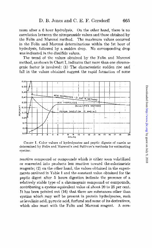
For the determinations by the Folin and Marenzi method samples of casein equivalent to 1 gm. of t’he ash- and moisture-free material were heated in 20 cc. of 20 per cent hydrochloric acid to gentle boiling for the desired length of time. The hydrolysates were diluted at once with distilled water to a volume of 50 cc. and decolorized with kaolin. 5 cc. aliquots were taken and the color developed by the Folin and Marenzi reagents compared against that similarly developed with a standard cystine solution. The color values obtained are shown on Chart I as percentages of cystine calculated on the basis of the ash- and moisture-free casein.
Two striking features of the results are (1) the exceedingly high values obtained on the short periods of hydrolysis, beginning with 0.46 per cent after 15 minutes and reaching a maximum of 0.55 per cent after 45 minut,es, and (2) the abrupt drop to 0.42 per cent after a 1 hour’s hydrolysis. Thereafter the values gradually de- creased, reaching 0.33 per cent at the end of 18 hours, which then remained constant up to 24 hours. This constant value is prac- tically identical with the cystine content of the casein as deter- mined by the Sullivan method. The high values found by the Folin and Marenzi method on the shorter periods of hydrolysis must be attributed to some causative factor other than free cystine.
For the determinations of cystine by the Sullivan method sam- ples of casein equivalent to 1 gm. of ash- and moisture-free material were used when the periods of hydrolysis were 6 hours or longer. Because of the low cystine content of casein it was found neces- sary to take 2 gm. for hydrolyses of shorter duration than 6 hours in order to get a color intensity satisfactory for accurate measure- ments. The results of these determinations are given in Chart I.
by guest on July 9, 2018http://w
ww
.jbc.org/D
ownloaded from
D. B. Jones and C. E. F. Gersdorff
The results obtained on the shorter periods of hydrolysis are in striking contrast to those obtained by the Folin and Marenzi method. Instead of the high values reaching a maximum at the end of 45 minutes and then dropping abruptly, the Sullivan method shows that gradually increasing quantities of cystine are liberated until a maximum of 0.33 per cent is reached after 6 hours hydrolysis. On further hydrolysis this value remains constant up to 24 hours, and is the same as the value obtained by the Folin and Marenzi method after 18 hours hydrolysis.
The values obtained by the Sullivan method show that cystine is early liberated from casein on acid hydrolysis. After a 30 minute hydrolysis, 20 per cent of the total cystine content of the protein had been liberated, and after 3+ hours the value amounted to 50 per cent.
Digestion of Casein with Pepsin-Although it is generally accepted that free amino acids are not liberated when proteins are digested with pepsin, it was thought desirable as preliminary to a study of the rate of liberation of cystine when casein is acted on by digestive ferments, to apply the calorimetric cystine tests to peptic digests. Because of the inadequacy of the methods here- tofore used for estimating cystine when present in small quantities it is not inconceivable that in previous studies some of this amino acid which may have been present escaped detection,
The digestions were made separately on duplicate samples of cascin, each equivalent to 2 gm. of the ash- and moisture-free protein. The casein was added to a mixture of 25 cc. of 0.1 N
hydrochloric acid and 25 cc. of a 0.2 per cent sblution of pepsin in 0.1 N hydrochloric acid. Before they were mixed, all the components of the digest were separately heated to 38”. The mixtures were then placed at once in an incubator and maintained at 37-38” for periods ranging from 5 minutes to 18 hours. At the end of the digestion periods the digests were heated to 80” in order to inactivate the pepsin, and then diluted with distilled water to 100 cc. Duplicate 5 cc. portions were taken and examined accord- ing to both Folin and Marenzi’s and Sullivan’s methods for the estimation of cystine. The color developed was compared with that similarly produced in a standard cystine solution.
The tests made by the Sullivan method on the peptic digests after different periods of digestion gave negative results in every
by guest on July 9, 2018http://w
ww
.jbc.org/D
ownloaded from
662 Digestibility of Proteins in Vitro
case, even after 18 hours digestion. These results are in agree- ment with the views generally held that free amino acids are not liberated on peptic digestion.
In every determination made by the Folin and Marenzi method a blank test was run on the pepsin by incubating in separate flasks simultaneously and parallel with the casein-pepsin mixtures, 25 cc. of a 0.2 per cent solution of pepsin in 0.1 N hydrochloric acid, and determining the color developed with the Folin and Marenzi reagents. The color values thus obtained were subtracted as a blank from those obtained for the casein digests. The corrected cystine equivalent values found for the casein digests are given in Chart I.
The values obtained by the Folin and Marenzi method for both the peptic digests and the acid hydrolysates follow the same gen- eral trend. They are in striking contrast to those obtained by the Sullivan method. There is first a rapid rise in values, followed by a sudden drop, then a decrease until a constant value is reached. The peptic digests starting with a cystine equivalent value of 0.21 per cent after 5 minutes digestion reached a maximum of 0.40 per cent after digestion for 1 hour, then dropped to 0.25 per cent in 5 hours. On further digestion this value remained constant. With the more specific Sullivan method, on the other hand, the peptic digests gave negative results, whereas the acid hydroly- sates showed continua~lly increasing values until a maximum was reached which t,hereafter remained constant.
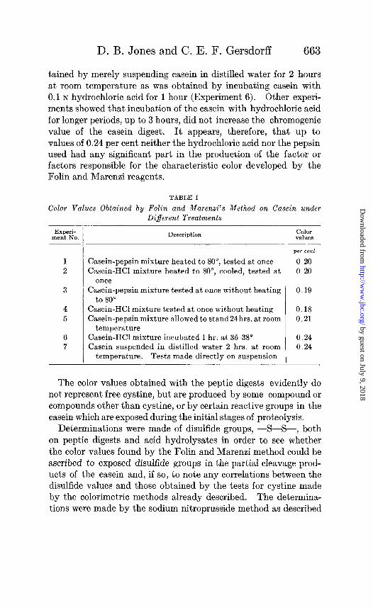
The color values obtained by the Folin and Marenzi method after short periods of digestion raised the question whether we were measuring the effect of enzyme action or that of mild acid hydrolysis. The experiments outlined in Table I were designed to get information on this question, and also to throw light on the nat.ure and manner of formation of the chromogenic factor or factors responsible for the color values obtained. The expression “casein-pepsin mixture” as used in Table I refers to a mixture of 2 gm. of casein in 25 cc. of 0.1 N hydrochloric acid and 0.2 per cent solution of pepsin in 0.1 N hydrochloric acid. The “casein- hydrochloric acid mixture” consisted of 2 gm. of casein in 50 cc. of the acid. In the first four experiments essentially the same values were obtained, irrespective of whether pepsin was used or not. In Experiment 7, the same value, 0.24 per cent, was ob-
by guest on July 9, 2018http://w
ww
.jbc.org/D
ownloaded from
D. B. Jones and C. E. F. Gersdorff 663
tained by merely suspending casein in distilled water for 2 hours at room t,emperature as was obtained by incubating casein with 0.1 N hydrochloric acid for 1 hour (Experiment 6). Other experi- ments showed that incubation of the casein with hydrochloric acid for longer periods, up to 3 hours, did not increase the chromogenic value of the casein digest. It appears, therefore, that up to values of 0.24 per cent neither the hydrochloric acid nor the pepsin used had any significant part in the production of the factor or factors responsible for the characteristic color developed by the Folin and Marenzi reagents.
TABLE I
Color Values Obtained by Folin and Marenzi’s Method on Casein under
Experi- ment No.
- Diferent Treatments
Description
Casein-pepsin mixture heated to 80”, tested at once Casein-HCI mixture heated to 80”, cooled, tested at
once Casein-pepsin mixture tested at once without heating
to 80” Casein-HCl mixture tested at once without heating Casein-pepsin mixture allowed to stand 24 hrs. at room
tempera.ture Casein-HCl mixture incubated 1 hr. at 3538” Casein suspended in distilled water 2 hrs. at, room
temperature. Tests made directly on suspension
Color VdlU3S
per cent 0.20 0.20
0.19
0.18 0.21
0.24 0.24
The color values obtained with the peptic digests evidently do not represent free cystine, but are produced by some compound or compounds other than cystine, or by certain reactive groups in the casein which are exposed during the initial stages of proteolysis.
Determinations were made of disulfide groups, -S-S-, both on peptic digests and acid hydrolysates in order to see whether the color values found by the Folin and Marenzi method could be ascribed to exposed disulfide groups in the partial cleavage prod- ucts of the casein and, if so, to note any correlations between the disulfide values and those obtained by t’he tests for cystine made by the calorimetric methods already described. The determina- tions were made by the sodium nitroprusside method as described
by guest on July 9, 2018http://w
ww
.jbc.org/D
ownloaded from
664 Digestibility of Proteins in Vitro
by Walker (13). The color developed was compared with an empirical standard consisting of a mixture of Bordeaux red and methyl orange in the proportions given by Abderhalden and Wertheimer (14). This mixture had been previously standardized calorimetrically against a cystine solution of known conccntjration containing a few drops of sodium nitroprusside to which 5 drops of a 10 per cent sodium cyanide solution had been added. The em- pirical dye standard was used instead of a standard cystine solution because the latter gives with sodium nitroprusside a color which is so unstable as to make an accurate reading rather difficult.
TABLE II
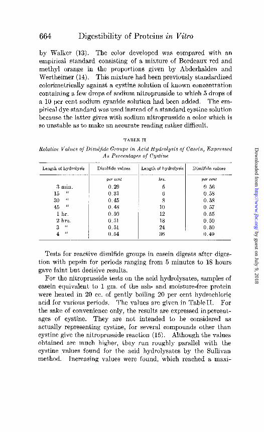
Relative Values of DisulJide Groups in Acid Hydrolysis of Casein, Expressed As Percentages of Cyst&e
Length of hydrolysis
3 min. 15 “ 30 “ 45 “
1 hr. 2 hrs. 3 “ 4 “
Disulfide values 1 Length of hydrolysis Disulfide values
per cent bs. per cent
0.29 5 0.56 0.31 6 0.58 0.45 8 0.58 0.48 10 0.57 0.50 12 0 55 0.51 18 0.50 0.51 24 0.50 0.54 36 0.49
-
Tests for reactive disulfide groups in casein digests after diges- tion with pepsin for periods ranging from 5 minutes to 18 hours gave faint but decisive results.
For the nitroprusside tests on the acid hydrolysates, samples of casein equivalent to 1 gm. of the ash- and moisture-free protein were heated in 20 cc. of gently boiling 20 per cent hydrochloric acid for various periods. The values are given in TableII. For the sake of convenience only, the results are expressed in percent,- ages of cystine. They are not intended to be considered as actually representing cystine, for several compounds other than cystine give the nitroprusside reaction (15). Although the values obtained are much higher, they run roughly parallel with the cystine values found for the acid hydrolysates by the Sullivan method. Increasing values were found, which reached a maxi-
by guest on July 9, 2018http://w
ww
.jbc.org/D
ownloaded from
D. B. Jones and C. E. F. Gersdorff 665
mum after a 6 hour hydrolysis. On the other hand, there is no correlation between the nitroprusside values and those obtained by the Folin and Marenzi method. The maximum values occurred in the Folin and Marenzi determinations within the 1st hour of hydrolysis, followed by a sudden drop. No corresponding drop was indicated in the disulfide values.
The trend of the values obtained by the Folin and Marenzi method, as shown in Chart I, indicates that more than one chromo- genic factor is involved: (1) The characteristic sudden rise and fall in the values obtained suggest the rapid formation of some
CIIART I. Color values of hydrolysates and peptic digests of casein as determined by Folin and Marenzi’s and Sullivan’s methods for estimating cystine.
reactive compound or compounds which is either soon volatilized or converted into products less reactive toward the calorimetric reagents; (2) on the other hand, the values obtained in the experi- ments outlined in Table I and the constant value obtained for the peptic digest after 5 hours digestion indicate the presence of a relatively stable type of a chromogenic compound or compounds, contributing a cystine equivalent value of about 20 to 25 per cent. It has been pointed out (16) that there are substances other than cystine which may well be present in protein hydrolysates, such as levulinic acid, pyruvic acid, furfural and some of its derivatives, which also react with the Folin and Marenzi reagent. A com-
by guest on July 9, 2018http://w
ww
.jbc.org/D
ownloaded from
666 Digestibility of Proteins in Vitro
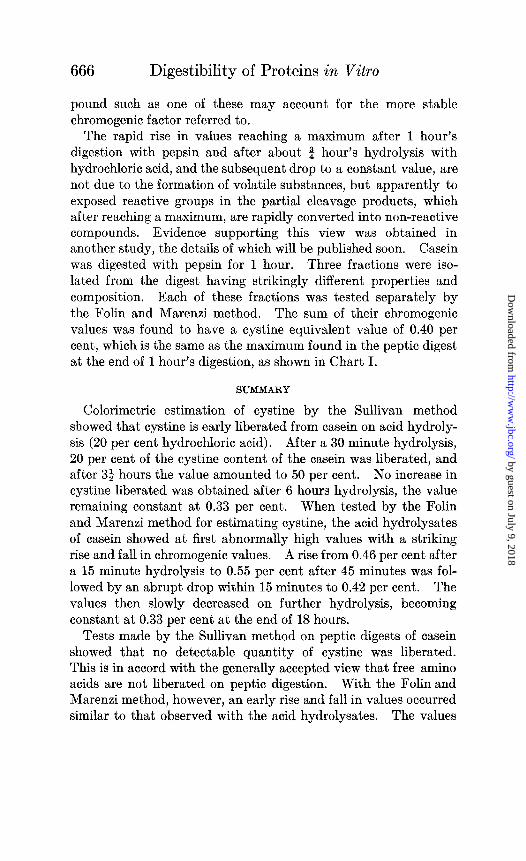
pound such as one of these may account for the more stable chromogenic factor referred to.
The rapid rise in values reaching a maximum after 1 hour’s digestion with pepsin and after about $ hour’s hydrolysis with hydrochloric acid, and the subsequent drop to a constant value, are not due to the formation of volatile substances, but apparently to exposed reactive groups in the partial cleavage products, which after reaching a maximum, are rapidly converted into non-reactive compounds. Evidence supporting t,his view was obtained in another study, the details of which will be published soon. Casein was digested with pepsin for 1 hour. Three fractions were iso- lated from the digest having strikingly different properties and composition. Each of these fractions was tested separately by the Folin and Marenzi method. The sum of their chromogenic values was found to have a cystine equivalent value of 0.40 per cent,, which is the same as the maximum found in the peptic digest at the end of 1 hour’s digestion, as shown in Chart I.
SUMMARY
Calorimetric estimation of cystine by the Sullivan method showed that cystine is early liberated from casein on acid hydroly- sis (20 per cent hydrochloric acid). After a 30 minute hydrolysis, 20 per cent of the cystine content of the casein was liberated, and after 34 hours the value amounted to 50 per cent. No increase in cystine liberated was obtained after 6 hours hydrolysis, the value remaining constant at 0.33 per cent. When tested by the Folin and Marenzi method for estimating cystine, the acid hydrolysates of casein showed at first abnormally high values with a striking rise and fall in chromogenic values. A rise from 0.46 per cent after a 15 minute hydrolysis to 0.55 per cent after 45 minutes was fol- lowed by an abrupt drop within 15 minutes to 0.42 per cent. The values then slowly decreased on further hydrolysis, becoming constant, at 0.33 per cent at the end of 18 hours.
Tests made by the Sullivan method on peptic digests of casein showed that no detectable quantity of cystine was liberated. This is in accord with the generally accepted view that free amino acids are not liberated on peptic digestion. With the Folin and Marenzi method, however, an early rise and fall in values occurred similar to that observed with the acid hydrolysates. The values
by guest on July 9, 2018http://w
ww
.jbc.org/D
ownloaded from
Il. B. Jones and C. E. F. Gersdorff
increased from 0.21 per cent after digestion for 5 minutes to 0.40 per cent at the end of 1 hour. Thereafter the values gradually decreased, becoming constant at 0.25 per cent after 5 hours digestion.
The color values obtained with the peptic digests areproduced by some compound or compounds other than cystine, or by certain reactive groups in the casein which are exposed during the initial stages of proteolysis. Evidence is presented showing that at least two factors are involved.
BIBLIOGRAPHY
1. Jones, D. B., and Waterman, H. C., J. Biol. Chem., 62,357 (1922). 2. Waterman, H. C., and Johns, C. O., J. Biol. Chem., 46,9 (1921). 3. Waterman, H. C., and Jones, D. B., J. Biol. Chem., 47,285 (1921). 4. Abderhalden, E., Lehrbuch der physiologischen Chemie, Berlin, 5th
edition, pt. 1, 468, 471, 483, 485 (1920). 5. Ftirth, O., and Lieben, F., Biochem. Z., 109, 153 (1920). 6. Ragins, I. K., J. Biol. Chem., 60, 551 (1928). 7. Hunter, A., Tr. Roy. Sot. Canada, series 3, sect. 5, 16, 71 (1922). 8. Dauphinee, J. A., and Hunter, A., Biochem. J., 24,1128 (1930). 9. Boone, G. J., Med. Bull. Univ. Cincinnati, 6, 193 (1931).
10. Van Slyke, L. L., and Baker, J. C., New York Agric. Exp. Stat., Tech. Bull. 65 (1918).
11. Folin, O., and Marenzi, A. D., J. Biol. Chem., 83, 103 (1929). 12. Sullivan, M. X., Pub. Health Rep., U. S. P. H. S., suppl. 78 (1929). 13. Walker, E., Biochem. J., 19, 1082 (1925). 14. Abderhalden, E., and Wertheimer, E., Arch. gee. Physiol., 198,122 (1923). 15. Sullivan, M. X., Z’ub. Zieallh Rep., U. S. P. H. S., 41, 1030 (1926). 16. Sullivan, M. X., Pub. Health Rep., U. S. P. H. S., 44, 1421 (1929).
by guest on July 9, 2018http://w
ww
.jbc.org/D
ownloaded from
D. Breese Jones and Charles E. F. GersdorffCASEIN
TO PEPTIC AND ACID DIGESTS OFTESTS FOR CYSTINE WHEN APPLIED OBSERVATIONS ON COLORIMETRIC
HYDROLYSIS OF CASEIN. SOME LIBERATION OF CYSTINE ON
PROTEINS IN VITRO: V. RATE OF STUDIES ON DIGESTIBILITY OF
1933, 101:657-667.J. Biol. Chem.
http://www.jbc.org/content/101/3/657.citation
Access the most updated version of this article at
Alerts:
When a correction for this article is posted•
When this article is cited•
to choose from all of JBC's e-mail alertsClick here
ml#ref-list-1
http://www.jbc.org/content/101/3/657.citation.full.htaccessed free atThis article cites 0 references, 0 of which can be
by guest on July 9, 2018http://w
ww
.jbc.org/D
ownloaded from