Embed Size (px)
Citation preview
1
IMPACT OF LIPID OXIDATION ON DIGESTIBILITY OF DAIRY PROTEINS IN O/W EMULSIONS WITH AUTOXIDIZING LIPIDS
ERIKA LORENA SOTO CHAVARRO Master´s dissertation submitted in partial fulfillment of the requirements for the
degree of Master of Agri-Food Science
Director MÓNICA OBANDO CHAVES
M.Sc Science and technology of Milk
UNIVERSIDAD DEL TOLIMA FACULTAD DE INGENIERÍA AGRONÓMICA
MAESTRÍA EN CIENCIAS AGROALIMENTARIAS IBAGUÉ- TOLIMA
2015
2
3
ACKNOWLEDGEMENTS
I want to express my sincere gratitude to my family and especially my parents Inés
Chavarro Pacheco, Luis Eduardo Soto Suárez and Eduardo Alfonso Soto, who
supported me unconditionally and gave me their love and understanding were key to
finish this stage of study.
I am very grateful for Wilmar Osorio Viana, "My blue sky" Thanks for giving me strength
in the times when he believed fail, for supporting me unconditionally, for giving me
peace and above all for believing in me. My words alone are insufficient to express my
gratitude and my love for you.
I am especially grateful to Bruno De Meulenaer, my promoter and director of the
Nutrifood Chem research group in Ghent University, who allowed me to be part of this
important research project and for leading me to the interesting field of food chemistry
and nutrition. I am very grateful to my tutor Mónica Obando Chaves, for their
unconditional supporting for the planning, execution and writing of this work; a person of
unparalleled professional and human qualities, who taught me to persevere and
overcome obstacles in the academic and research world. Thanks for receive me in
Ghent and for allowing learn and grow professionally by her side.
I am very grateful for the Universidad del Tolima, especially the faculty of agronomy
engineering and its master program in agri-food science, for this type of interinstitutional
agreements, which allowed me to carry out this international internship, broaden my
horizons and learn about other ways of doing research and seeing the world.
To all those who contributed to this work and also to all those who offered me their
human warmth and kindness in Ghent. "This experience was a lovely parenthesis in my
life that allowed me to see the world from another perspective".
4
CONTENT
INTRODUCTION ........................................................................................................... 11
1. LITERATURE REVIEW .......................................................................................... 13
1.1 EMULSIONS ........................................................................................................... 13
1.2 DAIRY PROTEINS .................................................................................................. 14
1.2.1 Casein. ................................................................................................................. 15
1.2.2 Whey proteins.. .................................................................................................... 15
1.3 POLYUNSATURATED FATTY ACIDS (PUFA´s) .................................................... 15
1.4 LIPID OXIDATION ................................................................................................... 16
1.4.1 Mechanisms of lipid oxidation............................................................................... 16
1.4.1.1 Auto-oxidation.. ................................................................................................. 17
1.4.2 Primary lipid oxidation products: Hydroperoxides................................................. 18
1.4.3 Secondary lipid oxidation products.). .................................................................... 19
1.4.3.1 Malondialdehyde (MDA).. .................................................................................. 19
1.4.3.2 Hexanal.. ........................................................................................................... 20
1.5 INTERACTION BETWEEN PROTEINS AND SECONDARY LIPID OXIDATION
PRODUCTS .................................................................................................................. 21
1.6 DIGESTION ............................................................................................................ 22
2 MATERIALS AND METHODS ................................................................................. 24
2.1 MATERIALS ............................................................................................................ 24
2.2 METHODS .............................................................................................................. 25
5
2.2.1 Stripping of soybean and fish oil... ……………………………………………………25
2.2.2 Oxidation. ............................................................................................................. 25
2.2.3 Determination Of Primary Oxidation Products: Peroxide Value (POV) IDF Method.
...................................................................................................................................... 26
2.2.3.1 Principle. ........................................................................................................... 26
2.2.3.2 Preparation of reagents. . ................................................................................. 26
2.2.3.3 Calibration curve for peroxide value (POV) determination. ............................... 27
2.2.3.4 Analysis of sample.. .......................................................................................... 27
2.2.3.5 Data calculations……………………………………………………………………...27
2.2.4 Determination of secondary oxidation products: p-Anisidine value (P-AV) ........... 28
2.2.4.1 Principle………………………………………………………………………………..28
2.2.4.2 Procedure. ......................................................................................................... 28
2.2.4.3 Data calculations. .............................................................................................. 29
2.2.5 Experimental setup: interaction between autoxidized lipids and dairy proteins in
o/w emulsion. ................................................................................................................ 29
2.2.5.1 Preparation of the oil-in-water (O/W) emulsion.. ................................................ 29
2.2.6 Determination of secondary lipid oxidation products ............................................ 30
2.2.6.1 Malondialdehyde (MDA). ................................................................................... 30
2.2.6.2 Hexanal. ............................................................................................................ 30
2.2.7 In-Vitro Digestion of emulsions: static model ........................................................ 31
2.2.7.1 Procedure .......................................................................................................... 31
2.2.7.2 Static gastric phase.. ......................................................................................... 31
2.2.7.3 Gastrointestinal digestion (Duodenal phase). .................................................... 31
2.2.8 Extraction of protein after in vitro digestion and determination of protein content by
kjeldahl. ......................................................................................................................... 32
2.2.8.1 Protocol for extraction of protein in digested and non-digested samples. ......... 32
6
2.2.8.2 Determination of nitrogen content by Kjeldahl.. ................................................. 32
2.2.8.2 Data calculations. The amount of nitrogen was calculated according to the
following equation:......................................................................................................... 33
2.2.9 Sodium Dodecyl Sulphate-Polyacrylamide Gel Electrophoresis.. ........................ 33
2.2.10 Statistical treatment of data.. .............................................................................. 34
3 RESULTS ................................................................................................................ 35
3.1 OIL OXIDATION STATUS OF THE STRIPPED FRESH AND OXIDIZED OILS ..... 35
3.2 SECONDARY LIPID OXIDATION PRODUCTS IN EMULSIONS ........................... 36
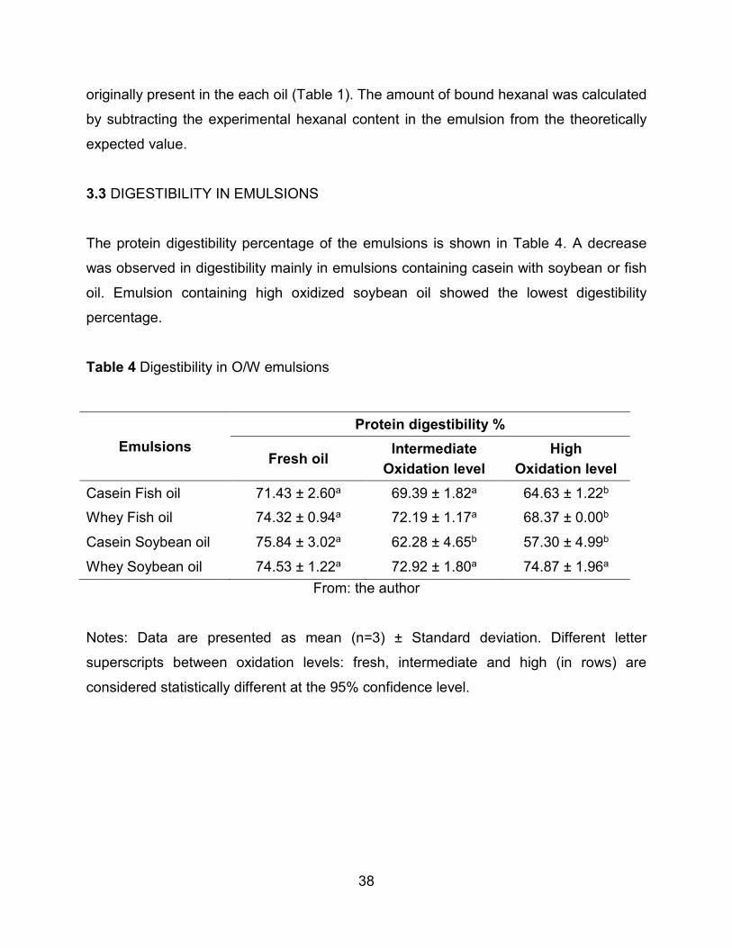
3.3 DIGESTIBILITY IN EMULSIONS ............................................................................ 38
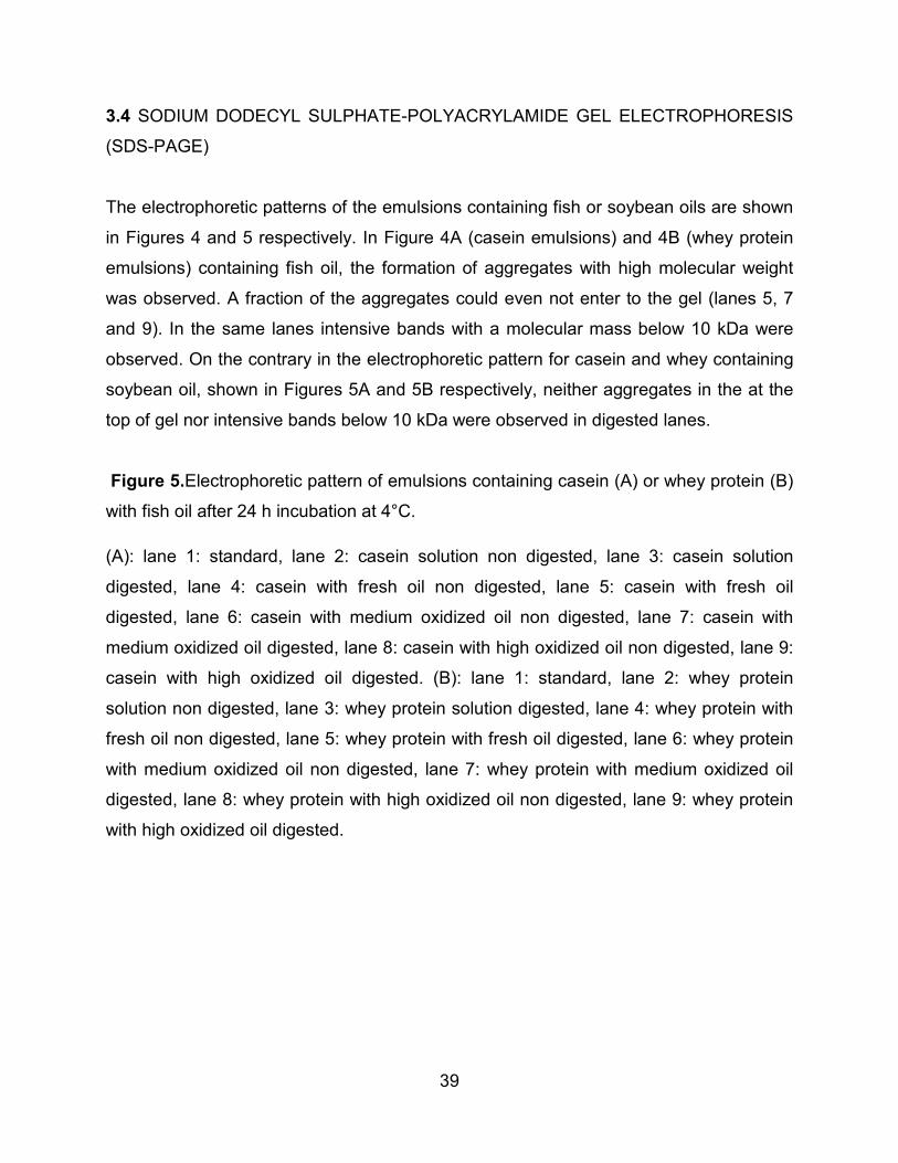
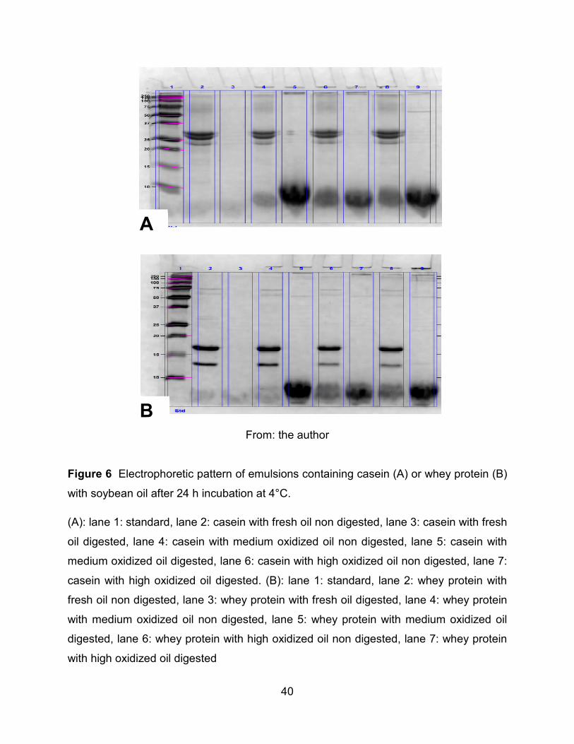
3.4 SODIUM DODECYL SULPHATE-POLYACRYLAMIDE GEL ELECTROPHORESIS
(SDS-PAGE) ................................................................................................................. 38
4 DISCUSSION ........................................................................................................... 42 5. CONCLUSION ........................................................................................................ 50 RECOMMENDATIONS ................................................................................................. 51 REFERENCES .............................................................................................................. 52 APPENDIX……………………………………………………………………………………..63
7
LIST OF TABLES
Table 1 Characterization of oils at three oxidation levels .............................................. 35
Table 2 Experimental and theoretical MDA content in O/W emulsions stabilized with
casein and whey proteins .............................................................................................. 36
Table 3 Experimental and theoretical hexanal content in O/W emulsions stabilized with
casein and whey proteins .............................................................................................. 37
Table 4 Digestibility in O/W emulsions .......................................................................... 38
8
LIST OF FIGURES Figure 1 Schematic representation of simple emulsions, oil in water emulsion (O/W)
and water in oil emulsion (W/O). ................................................................................... 13
Figure 2 Theoretical development of primary and secondary oxidation products as a
function of time in lipid oxidation ................................................................................... 18
Figure 3 Malodialdehyde (MDA) structure. ................................................................... 20
Figure 4 Hexanal structure ........................................................................................... 20
Figure 5 Electrophoretic pattern of emulsions containing casein (A) or whey protein (B)
with fish oil after 24 h incubation at 4°C. ....................................................................... 39
Figure 6 Electrophoretic pattern of emulsions containing casein (A) or whey protein (B)
with soybean oil after 24 h incubation at 4°C. ............................................................... 40
9
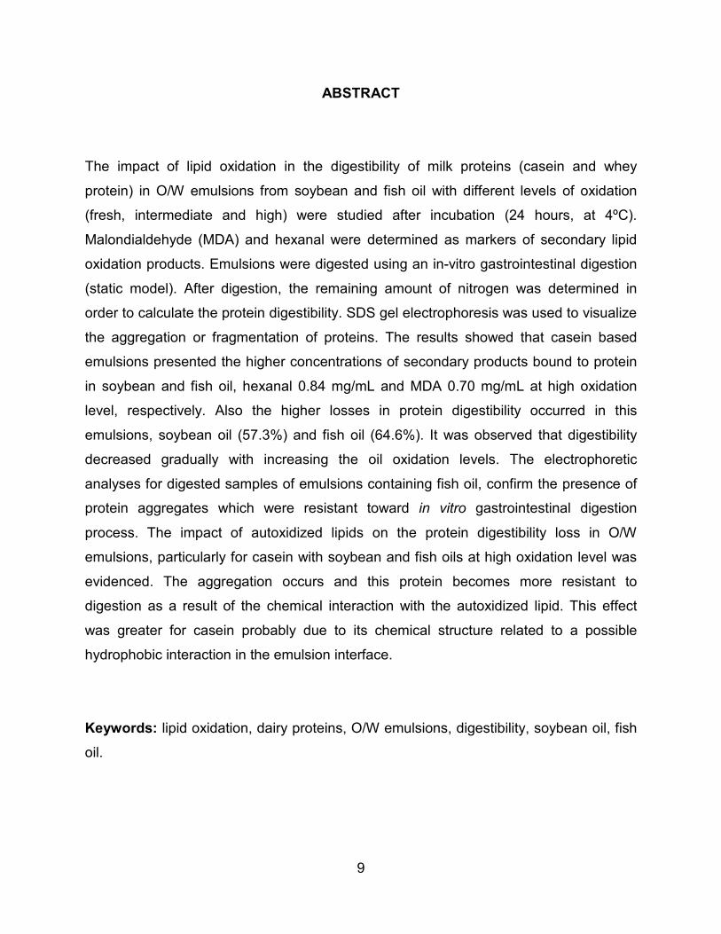
ABSTRACT
The impact of lipid oxidation in the digestibility of milk proteins (casein and whey
protein) in O/W emulsions from soybean and fish oil with different levels of oxidation
(fresh, intermediate and high) were studied after incubation (24 hours, at 4ºC).
Malondialdehyde (MDA) and hexanal were determined as markers of secondary lipid
oxidation products. Emulsions were digested using an in-vitro gastrointestinal digestion
(static model). After digestion, the remaining amount of nitrogen was determined in
order to calculate the protein digestibility. SDS gel electrophoresis was used to visualize
the aggregation or fragmentation of proteins. The results showed that casein based
emulsions presented the higher concentrations of secondary products bound to protein
in soybean and fish oil, hexanal 0.84 mg/mL and MDA 0.70 mg/mL at high oxidation
level, respectively. Also the higher losses in protein digestibility occurred in this
emulsions, soybean oil (57.3%) and fish oil (64.6%). It was observed that digestibility
decreased gradually with increasing the oil oxidation levels. The electrophoretic
analyses for digested samples of emulsions containing fish oil, confirm the presence of
protein aggregates which were resistant toward in vitro gastrointestinal digestion
process. The impact of autoxidized lipids on the protein digestibility loss in O/W
emulsions, particularly for casein with soybean and fish oils at high oxidation level was
evidenced. The aggregation occurs and this protein becomes more resistant to
digestion as a result of the chemical interaction with the autoxidized lipid. This effect
was greater for casein probably due to its chemical structure related to a possible
hydrophobic interaction in the emulsion interface.
Keywords: lipid oxidation, dairy proteins, O/W emulsions, digestibility, soybean oil, fish
oil.
10
RESUMEN
El impacto de la oxidación lipídica en la digestibilidad de proteínas lácteas (caseína y
proteína del suero) en emulsiones O/W con aceites de soya y pescado, con diferentes
niveles de oxidación fueron estudiados después de ser sometidos a incubación (24
horas, 4ºC). Malondialdehido (MDA) y hexanal fueron determinados como marcadores
de productos secundarios de oxidación lipídica. Las emulsiones fueron digeridas
usando un modelo estático in vitro de digestión gastrointestinal. Después de la
digestión la cantidad de nitrógeno remanente fue determinada con el fin de calcular la
digestibilidad proteica. La técnica de electroforesis (SDS-PAGE) fue usada para
visualizar la agregación o fragmentación de proteínas. Los resultados mostraron que
las emulsiones estabilizadas con caseína presentaron altas concentraciones de
productos secundarios de oxidación lipídica enlazados a la proteína tanto en el aceite
de soya, con hexanal (0.84 mg/mL), como en el de pescado, con MDA (0.70 mg/mL),
en altos niveles de oxidación. Asimismo las altas pérdidas en la digestibilidad proteica
se produce en estas mismas emulsiones con caseína en altos niveles de oxidación con
aceite de soya (57.3%) y con aceite de pescado (64.6%). Se observó una disminución
en la digestibilidad proporcional al incremento del nivel de oxidación de los aceites.
Además, se confirmó la presencia de agregados de proteína en las muestras digeridas
de las emulsiones con aceite de pescado los cuales resistieron a la digestión
gastrointestinal. Se evidenció el impacto de los lípidos auto-oxidados sobre la pérdida
de digestibilidad proteica en las emulsiones O/W, especialmente para caseína con
aceite de soya y pescado en altos niveles de oxidación. Como resultado de la
interacción proteínas con lípidos auto-oxidados se producen agregados de proteínas
que se tornan más resistentes a la digestión gastrointestinal. Este efecto fue observado
en gran medida en caseína probablemente debido a su estructura química relacionada
a una posible interacción hidrofóbica en la interfase de la emulsión.
Palabras clave: oxidación lipídica, proteínas lácteas, emulsiones O/W, digestibilidad,
soybean oil, fish oil.
11
INTRODUCTION
There is currently a growing demand for food products which prevent nutrition-related
diseases and improve the physical and mental state of people, in this regard, milk
products have been conducting for new market needs. This trend has created a need in
the market to create and design new dairy products with extra properties which go
further than just nutrition, such as those who contribute to prevent chronic diseases and
cardiovascular problems (Dawczynski, Martin, Wagner and Jahreis, 2010). The intrinsic
quality of dairy foods with added functional fatty acids is influenced by the possible
occurrence of chemical processes such as oxidation of lipids and the consequent
formation of new chemical substances that alter the organoleptic properties of the food,
along with its nutritional value and functional value which may even be harmful to the
body. However, enrichment of foods with omega-3 polyunsaturated fatty acid (PUFA)
should be evaluated carefully, since they are highly susceptible to oxidation (Cucu,
Devreese, Mestdagh, Kerkaert and De Meulenaer, 2011).
This susceptibility claims to have precautions in processing and storage to ensure
product quality and safety (Mestdagh, Cucu and De Meulenaer, 2011). Thus, recent
studies have focused on investigating the effect of lipid oxidation on the nutritional
quality of dairy products (Kolanowski and Weissrodt, 2007; Livney, 2010). Since it is
known that products of lipid oxidation not only interfere with the functionality of proteins
by decreasing their nutritional value, but also the oxidation products are potentially toxic
and lead to a risk for human health (Papastergiadis, 2014; Cucu et al, 2011;. Mestdagh
et al, 2011).
However, understanding the interaction between secondary lipid oxidation products and
dairy proteins that leads to the formation of complexes and their impact on digestibility
of proteins is still poorly understood. For these reason, the purpose of this study was to
study the impact of the secondary lipid oxidation on the digestibility of dairy proteins,
casein and whey protein, in O/W emulsions with the addition of autoxidized lipids, as
12
well as need to contribute to the understanding of the factors involving in these
phenomena which it can be controlled to ensure the nutritional quality of these foods.
13
1. LITERATURE REVIEW
1.1 EMULSIONS
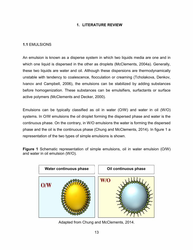
An emulsion is known as a disperse system in which two liquids media are one and in
which one liquid is dispersed in the other as droplets (McClements, 2004a). Generally,
these two liquids are water and oil. Although these dispersions are thermodynamically
unstable with tendency to coalescence, flocculation or creaming (Tcholakova, Denkov,
Ivanov and Campbell, 2006), the emulsions can be stabilized by adding substances
before homogenization. These substances can be emulsifiers, surfactants or surface
active polymers (McClements and Decker, 2000).
Emulsions can be typically classified as oil in water (O/W) and water in oil (W/O)
systems. In O/W emulsions the oil droplet forming the dispersed phase and water is the
continuous phase. On the contrary, in W/O emulsions the water is forming the dispersed
phase and the oil is the continuous phase (Chung and McClements, 2014). In figure 1 a
representation of the two types of simple emulsions is shown.
Figure 1 Schematic representation of simple emulsions, oil in water emulsion (O/W) and water in oil emulsion (W/O).
Adapted from Chung and McClements, 2014.
Water continuous phase Oil continuous phase
14
The emulsifiers have a polar part that dissolves well in the aqueous phase and a non-
polar part that is soluble in the oil phase. Consequently, those molecules are absorbed
in the interphase between water and oil (Hoffmann and Reger, 2014), forming a
protective membrane against the aggregation of the droplets (Dickinson, 1992;
McClements, 2004a), by generating repulsive forces between them. Emulsifiers
commonly used in the food industry are amphiphilic proteins, phospholipids, protein–
carbohydrate conjugates and small molecule surfactants. (McClements and Decker,
2000).
Some of the proteins widely used as emulsifiers in the food industry are whey protein,
casein, soy and egg protein (McClements, 2004b). Proteins are considered as surface
active polymers due to presence of hydrophobic groups and also because multipolar
compounds that confer high chemical reactivity (Hoffmann and Reger, 2014).
The forces involve in the stabilization and destabilization of emulsions are mainly van
der Waals, attractive forces, electrostatic interactions and steric factors. At pH values far
from the isoelectric point of proteins, electrostatic repulsion occurs, preventing the
dispersed droplets in the emulsion to come close together. Furthermore the interactions
between protein-stabilized droplets can be influenced for the presence of certain ions,
especially calcium (Singh and Ye, 2009).
1.2 DAIRY PROTEINS
In food systems, dairy proteins have found many applications due to their functional
properties, including their ability to form stable emulsions (Singh and Ye, 2009). The
main proteins in milk are casein and whey protein. Caseins are phosphoproteins with a
random coil structure and precipitate at their isoelectric point (P.I 4.6). Whey proteins do
not contain phosphorus, are globulins and remain in solution at P.I 4.6. In cow milk,
caseins correspond to 82% of the milk protein approximately, whereas the remaining
18% are whey proteins. (Singh and Ye, 2009).
15
1.2.1 Casein. Casein is produced from skimmed milk by addition of hydrochloric or lactic
acid precipitating caseins near their isoelectric point of pH 4.6. At this pH caseins are at
their lowest point of solubility due to the decrease of intermolecular repulsion. Caseins
have excellent holding water capacity and fat emulsification properties (Singh and Ye,
2009). They have an open tertiary structure due to its high content of proline, a feature
that allows easy access for proteolytic cleavage by gastric proteases (Livney, 2010).
The amphipathic nature of the caseins is defined by both hydrophobic and hydrophilic
residues and also due to positive and negative charges of aminoacid side chain groups
distributed in different areas of the protein. This amphipatic nature contributes to their
ability to stabilize oil in water emulsions (Horne, 2009).
1.2.2 Whey proteins. Represent 18-20% of the total nitrogen content in milk
approximately. β-lactoglobuline is the main whey protein, followed by the α-lactalbumin
that represents 20% of the whey protein fraction. Other fractions are bovine serum
albumin and immunoglobulins. Whey proteins are highly susceptible to heat induced
denaturation leading to coagulation (Jovanović, Barać and Maćej, 2005).
Whey proteins have secondary, tertiary and quaternary structure and in many cases
most of them are globular proteins (Singh and Flanagan, 2006). In emulsions, whey
proteins are rapidly absorbed, unfolding and reorienting at the oil-water interfase and
(Singh and Flanagan, 2006).
1.3 POLYUNSATURATED FATTY ACIDS (PUFA´s)
In recent decades, many nutritional and epidemiological studies have linked the
consumption of polyunsaturated fatty acids (PUFAs) omega-3 (i.e, α-linolenic acid,
eicosapentaenoic acid [EPA] and docosahexaenoic acid [DHA]) to a significant
decrease in some risk factors of cardiovascular disease, diabetes type 2, rheumatoid
arthritis, Crohn disease, and chronic obstructive pulmonary disease (Simopoulos,
1999). Thus, the potential benefits of omega 3 fatty acids has stimulated a growing
16
interest in food fortification, particularly with oils rich in these fatty acids (Betti et al.,
2009; Iafelice et al., 2008; Ye, Cui, Taneja, Zhu and Singh, 2009).
The high susceptibility of polyunsaturated lipids to lipid oxidation has restricted their
incorporation into many food products, however which is unfortunate because greater
consumption of polyunsaturated lipids is beneficial to health (Watkins and German,
1998) and is recommended in dietary guidelines (Kritchevsky, 1998). Progress in the
development of PUFA-enriched foods with desirable nutritional and physical attributes
depends on the availability of improved methods of controlling their oxidative stability,
which in turn relies on a deep understanding of the mechanisms of lipid oxidation
(McClements and Decker, 2000).
PUFA´s are found mainly in fish oil a source of omega-3 fatty acids, especially long-
chain docosahexaenoic acid (DHA, C22:6n-3) and eicosapentaenoic acid (EPA,
C20:5n-3). Fish oil is characterized by its high degree of unsaturation, the high content
of the long-chain omega-3 type PUFAs (Tehrany et al., 2012). While the major source of
fatty acid in soybean oil are PUFAs corresponding to alpha-linolenic acid (C-18: 3), 7-
10% and linoleic acid(C-18: 2), 51% (Dhiman et al., 2000).
1.4 LIPID OXIDATION
The lipid oxidation process consists of a complex sequence of chemical changes that
result from the interaction of lipids with reactive oxygen species (ROS). The mechanism
of lipid oxidation in food depends on the nature of the reactive species present and their
physicochemical environment (McClements and Decker, 2000).
1.4.1 Mechanisms of lipid oxidation. The oxidation process can be initiated by exposure
to light (photo-oxidation); the presence and the activity of lipooxygenases (enzymatic
oxidation) and by the direct reaction with oxygen (auto-oxidation), in this section only
autoxidation process will be addressed.
17
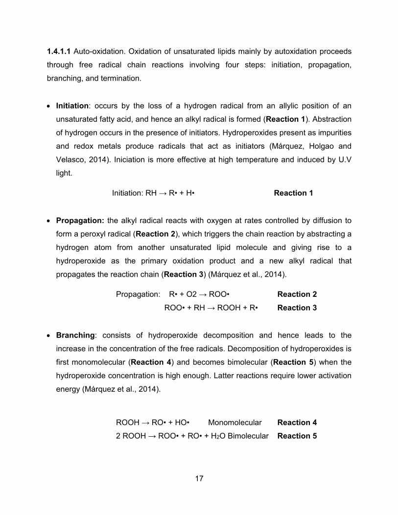
1.4.1.1 Auto-oxidation. Oxidation of unsaturated lipids mainly by autoxidation proceeds
through free radical chain reactions involving four steps: initiation, propagation,
branching, and termination.
• Initiation: occurs by the loss of a hydrogen radical from an allylic position of an
unsaturated fatty acid, and hence an alkyl radical is formed (Reaction 1). Abstraction
of hydrogen occurs in the presence of initiators. Hydroperoxides present as impurities
and redox metals produce radicals that act as initiators (Márquez, Holgao and
Velasco, 2014). Iniciation is more effective at high temperature and induced by U.V
light.
Initiation: RH → R• + H• Reaction 1
• Propagation: the alkyl radical reacts with oxygen at rates controlled by diffusion to
form a peroxyl radical (Reaction 2), which triggers the chain reaction by abstracting a
hydrogen atom from another unsaturated lipid molecule and giving rise to a
hydroperoxide as the primary oxidation product and a new alkyl radical that
propagates the reaction chain (Reaction 3) (Márquez et al., 2014).
Propagation: R• + O2 → ROO• Reaction 2
ROO• + RH → ROOH + R• Reaction 3
• Branching: consists of hydroperoxide decomposition and hence leads to the
increase in the concentration of the free radicals. Decomposition of hydroperoxides is
first monomolecular (Reaction 4) and becomes bimolecular (Reaction 5) when the
hydroperoxide concentration is high enough. Latter reactions require lower activation
energy (Márquez et al., 2014).
ROOH → RO• + HO• Monomolecular Reaction 4
2 ROOH → ROO• + RO• + H2O Bimolecular Reaction 5
18
• Termination: radicals react between each other to yield relatively stable nonradical
species (some reactions are 6 and 7). All stages of oxidation occur simultaneously in
a complex series of sequential and overlapping reactions (Márquez et al., 2014).
ROO• + ROO• → ROOR + O2 Reaction 6
R• + ROO• → ROOR Reaction 7
1.4.2 Primary lipid oxidation products: Hydroperoxides. Hydroperoxides are unstable
compounds and decompose into radicals (mainly alkoxyl and hydroxyl radicals) that
follow different pathways to produce a great variety of secondary oxidation products,
(i.e. ketones, aldehydes, epoxides, alcohols, hydrocarbons, acids). Hydroperoxides
form and decompose simultaneously and constitute the most abundant compounds
under low and moderate temperature conditions. When hydroperoxides are
accumulated at relatively high levels, their decomposition becomes faster than their
formation, and the overall oxidation rate increases exponentially (Márquez et al., 2014).
The decrease in the concentration of hydroperoxides occurs when the decomposition
rate towards secondary lipid oxidation products exceeds the hydroperoxide formation
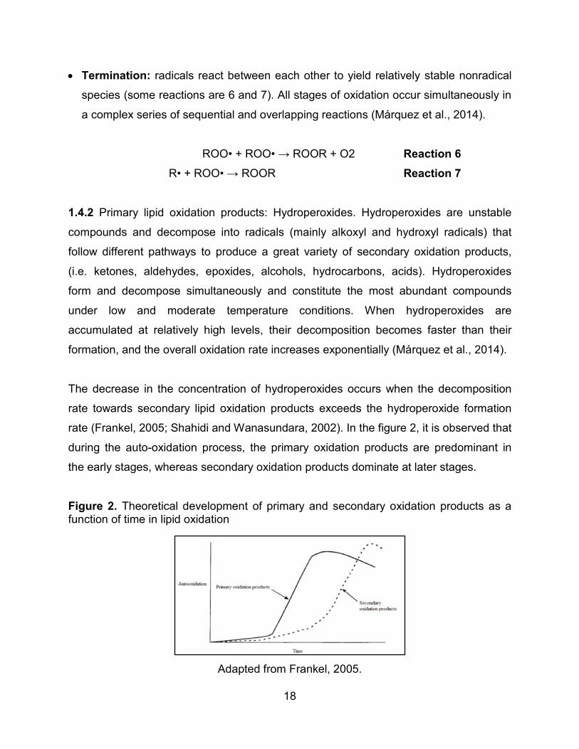
rate (Frankel, 2005; Shahidi and Wanasundara, 2002). In the figure 2, it is observed that
during the auto-oxidation process, the primary oxidation products are predominant in
the early stages, whereas secondary oxidation products dominate at later stages.
Figure 2. Theoretical development of primary and secondary oxidation products as a function of time in lipid oxidation
Adapted from Frankel, 2005.
19
1.4.3 Secondary lipid oxidation products. Secondary lipid oxidation products are low
molecular weight and tipically volatile compounds that cause rancidity. Combinations of
different decomposition compounds give different sensory properties (Fennema, Parkin
and Damodaran, 2007; Frankel, 1985).
From the decomposition of hydroperoxides from ω-3 fatty acids a large variety of
secondary oxidation products such as epoxides, ketones, aldehydes, alkanes etc. are
produced. Particularly, the formation of aldehydes is considered to be relevant because
of their low sensorial threshold and because of their reactivity towards biopolymers such
as protein, RNA, DNA. Thus they are considered as toxic. (Esterbauer, Schaur and
Zollner, 1991).
1.4.3.1 Malondialdehyde (MDA). One of the most studied secondary lipid oxidation
products is malondialdehyde (MDA), which is widely used as a marker of oxidative
stress (Onyango and Baba, 2010). This aldehyde is produced by multiple cleavages of
cyclic hydroperoxides which are formed from fatty acids containing three or more double
bonds (linolenic acid and higher) (Schaich, Shahidi, Zhong and Eskin, 2013).
Hydroperoxy epidioxides of ω-3 and ω-6 PUFAs and bicycloendoperoxides have been
identified as the main precursors of MDA (Papastergiadis, 2014).
MDA is considered as a highly toxic and mutagenic compound able to interact with
macromolecules by binding with proteins, DNA and RNA forming a molecule known
adduct. It has been demonstrated that MDA reacts readily with DNA to form adducts of
deoxyguanosine and deoxyadenosine which can be mutagenic. Adducts have already
been detected in human tissues (Lykkesfeldt, 2007).
20
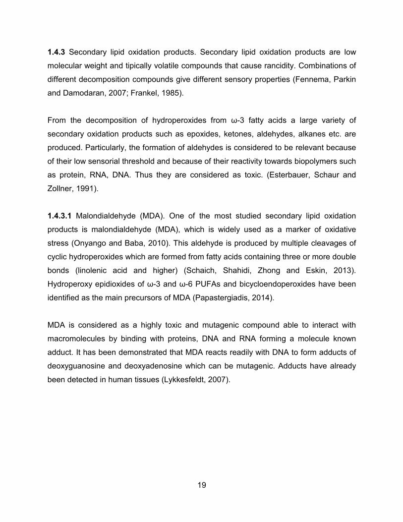
Figure 3. Malodialdehyde (MDA) structure.
Adapted from Pizzimenti et al., 2013.
1.4.3.2 Hexanal. Hexanal is an aldehyde formed by breakdown of hydroperoxides of
omega 3 and 6 fatty acids (Zhou and Decker, 1999). According to Sanchez, Rodriguez,
López and Paseiro, 2004, one of the best known synthetic pathways for hexanal occurs
during oxidation of linoleic acid (linoleic acid) through 13-hydroperoxide.
The hexanal content is related directly with undesirable flavors and compound is readily
detected due to its low odor threshold, below 5 ng g-1(Ha, Seo, Chen, Hwang and
Shim, 2011). Hexanal has been identified as the main volatile aldehydes generated
from lipid peroxidation in human milk (Elisia and Kitts, 2011). This compound has
become a marker of lipid peroxidation, and is considered one of the main products of
lipid oxidation due to the fact that it increases consistently during storage (Panseri,
Soncin; Chiesa and Biondi, 2011).
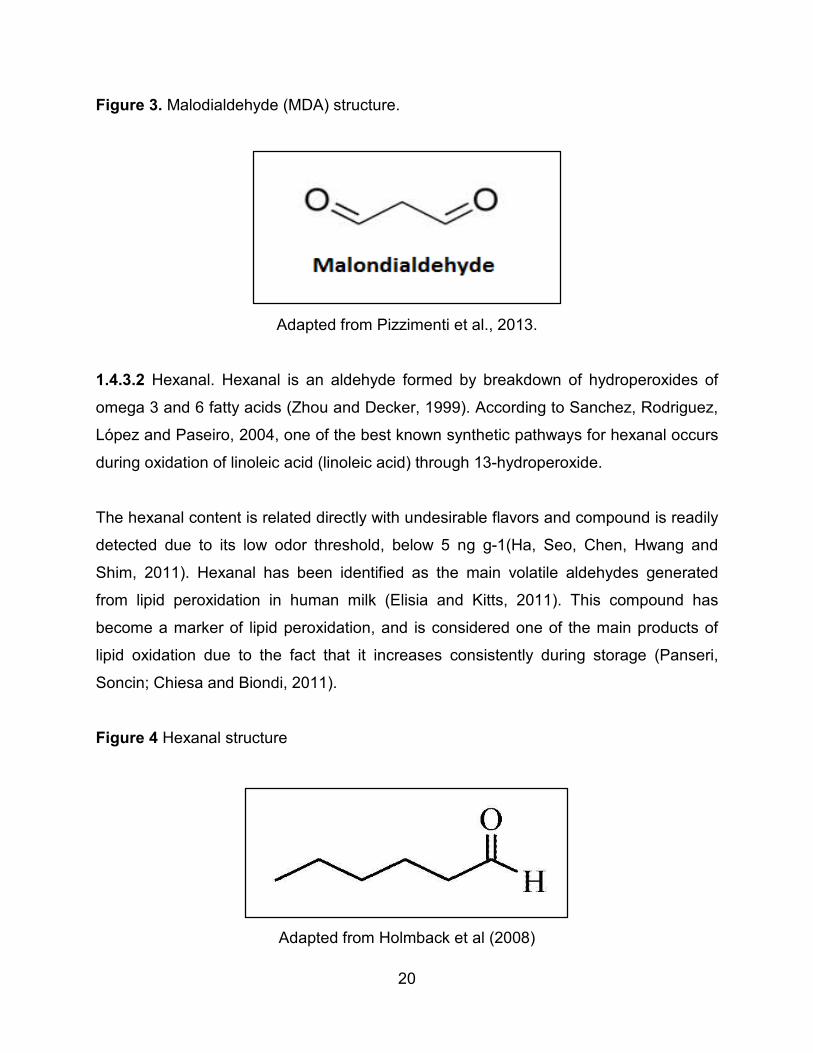
Figure 4 Hexanal structure
Adapted from Holmback et al (2008)
21
1.5 INTERACTION BETWEEN PROTEINS AND SECONDARY LIPID OXIDATION
PRODUCTS
In biological tissues and food systems, proteins are prone to degradation due to
exposure to lipid oxidation products and their decomposition by-products, leading to the
formation of protein-lipid complexes (Gardner, 1979). The strong interaction between
proteins and lipids allow moreover that oxidation reactions can transferred easily from
lipids to proteins (Wu, Zhang and Hua, 2009; Viljanen, 2005).
Moreover proteins can be subjected to fragmentation, aggregation and changes in
hydrophobicity. All this modification leads to a loss of their nutritional value (Wu et al.,
2009; Headlam and Davis, 2004; Tironi, Tomás and Añón, 2007).
Secondary lipid oxidation products, such as aldehydes, can both physically bind or react
covalently to proteins (Gardner, 1979). There are two major pathways for reaction
between unsaturated aldehydes and amino acids from proteins: Michael addition and
Schiff’s base reactions. The formation of a Schiff's base occurs when electrophilic
carbon atoms of aldehydes and ketones are targets of nucleophilic attack by amines, as
a result of this reaction the oxygen of the carbonyl group is replaced with nitrogen of the
amine, resulting in the formation of a Schiff base. In the Michael addition the reaction
occurs between carbon atom of the aldehyde and the nucleophilic amino acid residues
(the e-amino group of lysine, the imidazole fraction of histidine or the sulfhydryls group
of cysteine) (Viljanen, 2005).
Malondialdehyde, a bifunctional aldehyde (containing two functional groups) is a very
reactive compound which reacts with nucleophilic amine groups to form covalent
adducts with lysine residues of proteins. Other amino acids in which MDA may also
react are histidine, tyrosine, arginine and methionine. This reaction lead to a loss of
specific amino acid residues, protein cross-linking and fluorescence formation.
Functional properties of the protein are reduced (Wu et al., 2009).
22
Hexanal, a saturated aldehyde, could potentially react with proteins, peptides, amino
acids, polyamines, and sulfhydryls through Schiff base reactions. The study carried out
by Zhou and Decker, 1999, suggests that the interaction between the carbonyl group of
hexanal and Leu-His occurs with both the free amino acid group and the imidazole
nitrogens.
Meynier, Rapon, Dalgalarrondo and Genot, 2004, carried out a research about the
ability of hexanal and t-2-hexenal to form covalent bonds with whey protein and sodium
caseinate in aqueous solution. The results of the study suggest that covalent binding,
leading to aggregation of proteins, losses in Histidine and Lysine, and the formation of
fluorescent compounds, in the presence of t-2-hexenal and hexanal binding of aldehyde
to proteins and their aggregation was also observed. In both cases, protein
modifications were accompanied by a decrease in Triptophane fluorescence due to
changes in protein conformation and in the environment of the amino acid.
1.6 DIGESTION
Digestion is the process in which the food is transformed into small components which
can be more easily assimilated and absorbed by the body. The human gastrointestinal
tract consist in a set of organs (mouth, stomach, small and large intestine) that interact
and cooperate with each other, playing an important role in digestion and absorption of
food.
Several researchers have studied the digestion using an in vitro static gastrointestinal
digestion model with food matrices like emulsions (Picarello, Mamone, Chiara, Addeo
and Ferranti, 2013; Malinauskyte, 2014; Stuknytė, Cattaneo, Masotti and De Noni,
2015). These in vitro tests exhibited several advantages such as versatility, time
efficiency and costs. Moreover they are more practical compared to bioassays using
experimental animal models (in vivo) (Picarello et al., 2013). The in vitro
gastrointestinal digestion model involves mainly two phases: the gastric and intestinal
phase.
23
In the gastric phase that begins in the stomach, the proteins are metabolized through
proteolitic enzymes and due to the high acidity of the environment. Pepsine, the enzyme
included in this phase, has an affinity for cleaved peptide bonds involving aromatic
amino acids such as phenylalanine, tyrosine and tryptophan and hydrophobic residues
(leucine). Furthermore it has been demonstrated that pepsine can breakdown bonds of
acidic amino acids such as glutamic acid (Nik, Wright and Corredig, 2010). Proteolytic
ability of pepsine depends on the tertiary structure of the protein and its conformation
under hydrophobic or hydrophilic environments. Consequently the casein disordered
structure is readily digested by pepsin; in contrast the β-lactoglobulin shows greater
resistance to proteolysis in its native state, which is related to its highly folded
conformation in solution (Nik et al., 2010).
In the intestinal phase, the partially digested emulsion, that is part of the chyme formed
in the stomach phase is mixed with substances including bile salts, phospholipids, salts,
bicarbonate, proteases (trypsin and chymiotrypsine) and lipases (pancreatic lipase and
phospholipase). After entering the duodenum, chyme is mixed with sodium bicarbonate,
causing an increase in pH (1-3 in the stomach to the duodenum 5.8-6.5) where
enzymes act more efficiently (McClements and Li, 2010).
The protease trypsin catalyzes the peptide chain at the terminal carbon (C-terminal) of
the aliphatic amino acids, especially lysine and arginine; while chymotrypsine acts on
aromatic residues such as phenylalanine, tyrosine and tryptophan. The peptides/protein
hydrolysis does not occur solely because lipase are present (Singh and Ye, 2013).
Phospholipids and bile salts from the liver (via the gall bladder), facilitate the
emulsification of fats.
Lipase and bile salts inducing lipid hydrolysis in the duodenum, generating lipid
digestion products as free fatty acids, cholesterol, monoacylglycerol, etc., that are
solubilized within micelles and then are transported to epithelial cells for absorption. The
function of bile salts is to displace any protein or peptide remaining in the surface of the
oil droplet after protease activity. (McClements and Li, 2010).
24
2 MATERIALS AND METHODS
2.1 MATERIALS
Soybean oil and fish oil were obtained from Carrefour (Belgium N.V.) and Smit Zoon
(The Netherlands) respectively; p-anisidine reagent (99% purity) was purchased from
Acros Organics. Silica gel 60 was obtained from Merck (Germany), aluminum oxide
(Al2O3) was obtained from Sigma-Aldrich (Switzerland), ferrous chloride, barium chloride
(BaCl2.2H2O), ammonium thiocyanate (NH4CNS, 99%), hydrochloric acid (37%) and
isooctane (99.5%) were purchased from Chem-Lab (Zedelgem, Belgium). Glacial acetic
acid (99.7%), petroleum ether, methanol and hexane (99 %) were from Fisher Scientific
(UK). Potassium dihydrogen phosphate a.r. (KH2PO4) from Chem-Lab (Zedelgem,
Belgium) and potassium phosphate dibasic (HK2P O4, ≥ 99.9% pure) from Sigma-
Aldrich (St. Louis, MO, USA), hydrochloric acid (HCl 25%), sulfuric acid (H2SO4 98%)
and NaOH (99.8%) were obtained from Chem-Lab (Zedelgem, Belgium). Trichloroacetic
acid (CCl3COOH, TCA) (99+ %) was obtained from Acros organics (New Jersey, USA).
Kjeltab CX tablets made up of 5 g potassium sulphate (K2SO4) and 0.5 g copper (II)
sulphate (CuSO4.5H2O) were obtained from Thompson & Capper Ltd. (Cheshire, UK).
Boric acid (H3BO3) was purchased from VWR (Leuven, Belgium). Mixed indicator was
obtained from Merck (Darmstadt, Germany). Auto distillator Kjeltec 2200 (Foss
Tecator). All reagents were analytical grade.
Sodium caseinate (Miprodan 30) and whey protein isolate (Lacprodan DI 9224) were
purchased from Acatris food (Belgium).
Pepsin from porcine pancreas (P6867), lipase from porcine pancreas (Type II, 100 –
400 units/mg protein), bile extract porcine (B8631), trypsin from porcine pancreas
(T0303), α-chymotrypsin from bovine pancreas (type lyophilized powder, ≥ 40 units/mg
protein) were purchased from Sigma-Aldrich (St. Louis, MO, USA).
25
2.2 METHODS
2.2.1 Stripping of soybean and fish oils. Soybean and fish oils were purified by stripping
minor components such as pigments, tocopherols and sterols in a two-step procedure.
In the first step, 50 g of oil were mixed with 50 mL of hexane. The solvent/oil mixture
was then passed through a 40 cm (height) x 4 cm (diameter) glass column previously
filled with SiO2 activated was wrapped with aluminum foil to avoid light-induced
oxidations during the purification process. The column was rinsed tree times with 50 mL
of hexane. The oil/solvent mixture was collected in an aluminum-covered round bottom
flask. The solvent was evaporated using a rotavapor, then 40 g of each oil was mixed
with 56 mL of petroleum ether and passed through a glass column (38 cm Height x 4
cm diameter) packed with petroleum ether and activated aluminum oxide (achived at
200 °C overnight in muffle furnace). The glass column was rinsed with 100 mL of
hexane and the stripped oil was collected, rotaevaporated (40°C), poured into a brown
bottle and flushed with nitrogen to remove solvent traces and prevent oxidation during
storage at – 20 °C until use.
2.2.2 Oxidation. The stripped oils were oxidized under accelerated temperature
conditions; the aging was carried out in an oven at 60 °C from 0- 24 hours in the dark
with air in the headspace of the container. The oxidation status for fresh and aged
stripped oils was determined by the peroxide value (POV) and p-anisidine (p-AV) value.
Fresh stripped oil and oils with two different levels of oxidation were selected for the
experiment. Based on studies with oils during frying process by Aladedunye and
Przybylski, 2009, the p-AV values of the oils were used to classify oils oxidation levels
as follows: fresh status is the stripped oil without heat treatment that contain the lowest
concentration of lipid oxidation products, intermediate and high level were obtained
heating the oils at 60°C according to oxidation kinetics on stripped fish and soybean oil
performed on a previously study (Quintero, 2014).
26
2.2.3 Determination Of Primary Oxidation Products: Peroxide Value (POV) IDF Method.
2.2.3.1 Principle. The POV value was determined using the International Dairy
Federation, 1991, spectrophotometric method with modifications according to Shantha
and Decker, 1994, based on the ability of hydroperoxides to oxidize Fe2+ (ferrous) to
Fe+3 (ferric) ions that once formed, react with ammonium thiocyanate in an acidic
medium to form ferric-thiocyanate (chromophores), a red-violet complex with absorption
spectra at 500-510 nm. It comprises a weighted amount of sample in a mixture of
dichloromethane/methanol and addition of iron (II) chloride and ammonium thiocyanate.
After a fixed reaction time, the optical density is measured.
2.2.3.2 Preparation of reagents. The iron (II) chloride stock solution was prepared under
indirect, dimmed light as follows: 0.4 g of barium chloride (BaCl2.2H2O) dissolved in 50
mL of distilled water was gently mixed with a solution of 0.5 g of iron sulphate
(FeSO4.7H2O). Then 2 mL of concentrated hydrochloric acid solution (10 N) was added
to the resulting solution which was precipitated and filtrated until a clear liquid was
obteined. The iron (II) chloride solution was poured in a brown bottle and storage until
further use. The solution of ammonium thiocyanate (NH4SCN) was prepared by
weighting 30 g of NH4SCN in a volumetric flask and diluting until 100 mL with distilled
water.
Iron (III) stock solution was prepared as follows: 0.5 g of iron sulphate (FeSO4.7H2O)
was dissolved in about 50 mL of 10 N hydrochloric acid then 2 mL of 30 % (w/w)
hydrogen peroxide solution were added, the excess of hydrogen peroxide was removed
by boiling for 5 minutes. Afterwards the solution was cooled down at room temperature
(20 °C) and diluted with distilled water until 500 mL. From the previous solution 1 mL
was transferred to a 100 mL volumetric flask and the volume completed with a mixture
of dichloromethane: methanol (70:30) (Fe (III) concentration 10 mg/mL).
27
2.2.3.3 Calibration curve for peroxide value (POV) determination. The Fe (III) standard
was used to prepare a calibration curve with concentrations ranging from 1 – 40 µg. In
order to generate the chromophores, the final step comprised of the addition of 50 μL of
ammonium thiocyanate to each sample. Then the samples were vortexed (2 – 3 s) and
after 5 minutes the optical density was measured in a spectrophotometer at 500 nm.
The solvent blank was made up by the mixture of dichloromethane: methanol (70:30).
The calibration curve was obtained in triplicate, the absorbance of samples were plotted
versus Fe (III) concentrations expressed as µg Fe (III). The best fitting straight line
through the points was used to calculate the slope.
2.2.3.4 Analysis of sample. A reagent blank was prepared (Ereagent blank) as follows: 10
mL of dichloromethane: methanol (70:30) mixture were mixed with 50 μL of ammonium
thiocyanate, then 50 μL of Fe (II) solution was added. This blend was mixed for 2 – 3 s
and the absorbance was recorded after 5 min at 500 nm.
For the determination of POV in either fresh or aged stripped oil samples 20 – 30 mg of
oil depending on oxidation rate was weighted in a 10 mL volumetric flask wrapped with
aluminum foil, then the sample was diluted until mark with dichloromethane: methanol
(70:30). From the diluted solution 100 μL was taken and added to a test tube containing
10 mL of dichloromethane: methanol (70:30). Then 50 μL of ammonium thiocyanate
were added, the mix was vortexed and 50 μL of Fe (II) were added to trigger the
reaction. Finally the absorbance was measured spectrophotometrically after 5 min at
500 nm. All the data were recorded using a 1 cm path length quartz cell.
2.2.3.5 Data calculations. The peroxide value of oils, expressed as milliequivalents/Kg
was calculated according to:
𝑃𝑃𝑃𝑃𝑃𝑃 =𝐶𝐶𝐶𝐶𝐶𝐶𝐶𝐶𝐶𝐶𝐶𝐶𝐶𝐶𝐶𝐶𝐶𝐶 𝑎𝑎𝑎𝑎𝑎𝑎𝐶𝐶𝐶𝐶𝑎𝑎𝑎𝑎𝑎𝑎𝐶𝐶𝐶𝐶 𝑥𝑥 𝑚𝑚
55.48 𝑥𝑥 𝑊𝑊 𝑥𝑥 2
28
Where:
Corrected absorbance = Esample – Ereagent
m = slope of calibration curve
W = mass in grams of the sample
55.84 = atomic weight of Iron
2.2.4 Determination of secondary oxidation products: p-Anisidine value (P-AV)
2.2.4.1 Principle. The p-anisidine value is a measurement of secondary oxidation
products which resulted from decomposition of fatty acid hydroperoxides. The method is
based on the reaction of p-anisidine and aldehydes, principally 2,4-dienals and 2-
alkenals. As a consequence of the aforementioned reaction a yellow-colored compound
is formed and which is detectable spectrophotometrically at 350 nm.
2.2.4.2 Procedure. The determination of secondary oxidation products in oxidized oils
was based on American Oil Chemists Society, 1993, Official Method Cd 18-90. 0.25 g
of p-anisidine reagent was dissolved in 100 mL of glacial acetic acid. The solution was
stored at 4 °C and protected from light.
The sample size was taken depending on the oil oxidation rate as follows: 0.5 g for
fresh and 0.2 g for oxidized oil were weighted in a 25 mL volumetric flask previously
wrapped with aluminum foil to avoid direct exposition of sample to light. Then the
sample was dissolved in isooctane and diluted to the mark.
For the spectrophotometric measurements isooctane was used as a blank was
measured at 350 nm. Blank reagents was prepared by mixing 5 mL of isooctane and 1
mL of p-anisidine in a test tube. The mixture was vortexed and the optical density was
recorded after 10 min.
29
A similar procedure was followed for test solution. 5 ml of dissolved oil was mixed with 1
mL of p-anisidine reagent, and the absorbance was recorded exactly after 10 min of
reaction.
2.2.4.3 Data calculations. All determinations including blanks readings were carried out
in triplicate. p-anisidine value (P-AV) was calculated from the expression:
P − AV =25 ∗ (1.2𝐴𝐴𝑠𝑠 − 𝐴𝐴𝑏𝑏)
𝑊𝑊
Where:
Ab = Absorbance of the solution before addition of p-anisidine reagent at 350 nm.
As= Absorbance of the solution after addition of p-anisidine reagent at 350 nm.
W = mass of oil examinated in test solution in grams (g).
2.2.5 Experimental setup: interaction between autoxidized lipids and dairy proteins in
o/w emulsion.
2.2.5.1 Preparation of the oil-in-water (O/W) emulsion. The oil in water pre-emulsions
were prepared in 0.1M potassium phosphate buffer (pH 7.4) (continuous phase) mixing
6 mg/mL of protein (sodium caseinate or whey protein) and 3% of stripped soybean or
fish oil (fresh, medium or high oxidation level) (disperse phase) with an Ultra-Turrax
equipped with a S 25N–18G dispersion device during 3 min at 9000 rpm. This coarse
emulsion was then homogenized for 1 pass (250 bar) at 50°C using a microfluidizer.
After homogenization, 100 mL of the emulsion was poured into graduated (250 mL)
DURAN® laboratory glass bottles, which were tightly closed making use of screw caps.
Solutions of sodium caseinate and whey protein were prepared as controls. Samples
were prepared in triplicate for each treatment including also the controls and then
incubated at 4 °C during 24 hours under dark.
30
2.2.6 Determination of secondary lipid oxidation products
2.2.6.1 Malondialdehyde (MDA). The method of Papastergiadis, Mubiru, Van
Langenhove and De Meulenaer, 2012, was followed to determine MDA. Trichloroacetic
acid was added until final concentration of 15% to precipitate the protein and samples
were kept on ice for 10 min, after that centrifugation at 13000 g per 30 min was done to
get a clear supernatant. The top layer was discarded and 1 mL of supernatant was
mixed with 3 mL of TBA reagent (40 mM dissolved in 2 M acetate buffer at pH 2.0) in a
closed test tube and heated in a boiling water bath for 40 min. The reaction mixture was
cooled prior to the addition of 1 mL of methanol. After filtration 20 μL of the sample was
injected into a Varian C18 HPLC column (5 μm, 150 × 4.6 mm), held at 30 °C. The
mobile phase consisting of 50 mM KH2PO4 buffer solution, methanol, and acetonitrile
(72:17:11, v/v/v, pH 5.3) was pumped isocratically at 1 mL/min. Fluorometric detector
excitation and emission wavelengths were set at 525 and 560 nm, respectively. For
quantification, standard solutions of MDA in 7.5% TCA were prepared from 1,1,3,3-
tetraethoxypropane (TEP) and calibration curves were prepared at a concentration
ranging from 0.6 to 10 μM.
2.2.6.2 Hexanal. Hexanal formation in emulsions and in fish or soybean oil was
evaluated using headspace solid-phase microextraction (HS-SPME) combined with gas
chromatography–mass spectrometry (GC–MS). A total of 1 mL of emulsion was placed
in a glass vial (size 10 mL, H 22.5 mm × 46 mm) and mixed with 2 mL of 0.02 M
Na2HPO4, 0.02 M KH2PO4 pH 2 buffer. Butylated hydroxyanisol (BHA) dissolved in
methanol was added in the vial at a final concentration of 2.8 M and 15 µL of internal
standard of Hexanal–d12 was incorporated in the sample.
Then, the vial was sealed with a PTFE septum cup and was subjected to HS- SPME
extraction. The SPME fiber (75 μm Carboxen/PDMS, Supelco, Bellefonte, PA,USA) was
inserted into the headspace of the vial and left there for 30 min at 70 °C. Volatile
compounds were desorbed by inserting the fiber into the injection port of an Agilent
7890A chromatograph (Agilent Technologies, Palo Alto, CA) operated in splitless mode
31
for 10 min at 240 °C. Helium was used as carrier gas with a constant flow rate of 1.3
mL/min. The compounds were separated on a DB-624 column (60 × 0.25 mm x 1.4
μm). The oven temperature program began with 5 min at 50 °C for 5 min, increase to 4
°C/min to 140°C, then 30 °C/min increase to 240 °C for 10 minutes. An Agilent 5975C
inert XL mass spectrometry detector was used and detection was carried out on the
total ion current obtained by electron impact at 70 eV. The selected ion was 56 for
hexanal. External calibration curves were prepared using a hexanal standard (Sigma–
Aldrich).
2.2.7 In-Vitro Digestion of emulsions: static model
2.2.7.1 Procedure. For digestion of emulsion samples, the oral phase was considered
negligible due to the fast swallowing thus, static gastric phase and gastrointestinal
digestion (duodenal phase) were carried out as follows, according to the method
described by Wickham, Faulks and Mills, 2009, with some modifications.
2.2.7.2 Static gastric phase. After incubation of the samples (24 hours, 4°C) glass
laboratory bottles were taken out from cold room and immediately was initiated
sampling. For this purpose 6 falcon tubes per treatment (including solutions) were filled
with 5 mL of sample: 3 tubes were disposed for digestion and the rest were identified as
non-digested samples.
First of all, the pH of arranged samples for digestion was adjusted at 2.0 by adding 8 M
HCL, then 10 µL of dissolved enzyme (10 mg pepsine/1 mL of water) was incorporated,
vortexed and incubated at 37 °C during 2 hours with constant shaking.
2.2.7.3 Gastrointestinal digestion (Duodenal phase). Initially the pH was adjusted to 6.5
by adding NaOH 8 M. Then, a mix containing 1 g of lipase (100-400 units/mg L3126)
and 2.5 g of bile salts in 50 mL of buffer phosphate 5mM were prepared and 400 µL
was added to each falcon tube. In addition, 15 µL of CaCl2 was incorporated as well.
32
Afterwards, the trypsin and chymotrypsin were prepared (10 mg/ mL of 0.1 M HCL) and
10 µL of each one was incorporated to the emulsions. Then, samples were incubated at
37 °C for 2.5 hours with constant shaking. After incubation the pH of the samples was
adjusted to 5.0 by adding 8 M HCL.
2.2.8 Extraction of protein after in vitro digestion and determination of protein content by
kjeldahl.
2.2.8.1 Protocol for extraction of protein in digested and non-digested samples. The
protein pellet was obtained according to Cucu et al., 2011, using TCA (trichloroacetic
acid) as follows:
After the in vitro digestion, 5 mL of TCA (30% w/v) were added to each falcon tube
containing 5 mL of emulsion (TCA 15% final concentration). Then, samples were
incubated on ice for 10 min. The tubes were subsequently centrifuged (13000 g x 10
min at 4 °C). The supernatant was discarded and the remaining pellet was dissolved by
adding 2 mL NaOH, 10% w/v. The dissolved pellets were poured in digestion tubes to
determine percentage of nitrogen in samples by Kjeldahl method. The same process
was undertaken for non-digested samples (in triplicate).
2.2.8.2 Determination of nitrogen content by Kjeldahl. The nitrogen content was
determined according with the Kjeldahl method (Association Of Analytical Communities,
1981), that consists in a procedure of catalytically supported mineralization of organic
material in a boiling mixture of sulfuric acid and sulfate salts at temperatures between
340–370 °C. In the digestion process the organically bonded nitrogen is converted into
ammonium sulfate. The digest having been made alkaline with 50% NaOH is steam
distilled with 2% boric acid to release the ammonia which is trapped and titrated with
standard hydrochloric acid.
33
2.2.8.2 Data calculations. The amount of nitrogen was calculated according to the
following equation:
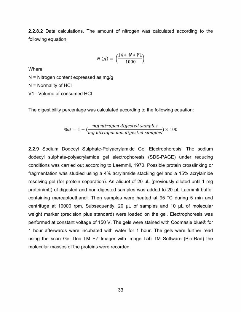
𝑁𝑁 (𝑔𝑔) = �14 ∗ 𝑁𝑁 ∗ 𝑃𝑃1
1000�
Where:
N = Nitrogen content expressed as mg/g
N = Normality of HCl
V1= Volume of consumed HCl
The digestibility percentage was calculated according to the following equation:
%𝐷𝐷 = 1 − (𝑚𝑚𝑔𝑔 𝑎𝑎𝑛𝑛𝐶𝐶𝐶𝐶𝐶𝐶𝑔𝑔𝐶𝐶𝑎𝑎 𝐶𝐶𝑛𝑛𝑔𝑔𝐶𝐶𝑎𝑎𝐶𝐶𝐶𝐶𝐶𝐶 𝑎𝑎𝑎𝑎𝑚𝑚𝑠𝑠𝑠𝑠𝐶𝐶𝑎𝑎
𝑚𝑚𝑔𝑔 𝑎𝑎𝑛𝑛𝐶𝐶𝐶𝐶𝐶𝐶𝑔𝑔𝐶𝐶𝑎𝑎 𝑎𝑎𝐶𝐶𝑎𝑎 𝐶𝐶𝑛𝑛𝑔𝑔𝐶𝐶𝑎𝑎𝐶𝐶𝐶𝐶𝐶𝐶 𝑎𝑎𝑎𝑎𝑚𝑚𝑠𝑠𝑠𝑠𝐶𝐶𝑎𝑎) × 100
2.2.9 Sodium Dodecyl Sulphate-Polyacrylamide Gel Electrophoresis. The sodium
dodecyl sulphate-polyacrylamide gel electrophoresis (SDS-PAGE) under reducing
conditions was carried out according to Laemmli, 1970. Possible protein crosslinking or
fragmentation was studied using a 4% acrylamide stacking gel and a 15% acrylamide
resolving gel (for protein separation). An aliquot of 20 μL (previously diluted until 1 mg
protein/mL) of digested and non-digested samples was added to 20 μL Laemmli buffer
containing mercaptoethanol. Then samples were heated at 95 °C during 5 min and
centrifuge at 10000 rpm. Subsequently, 20 μL of samples and 10 μL of molecular
weight marker (precision plus standard) were loaded on the gel. Electrophoresis was
performed at constant voltage of 150 V. The gels were stained with Coomasie blue® for
1 hour afterwards were incubated with water for 1 hour. The gels were further read
using the scan Gel Doc TM EZ Imager with Image Lab TM Software (Bio-Rad) the
molecular masses of the proteins were recorded.
34
2.2.10 Statistical treatment of data. The data concerning digestibility, MDA and hexanal
were determined in triplicates. One way two factors Analysis of Variance (ANOVA) with
significance levels of P<0.05, was applied to detect differences between samples and
were compared by LSD test. All statistical analyses were performed using SPSS 18
statistics package.
35
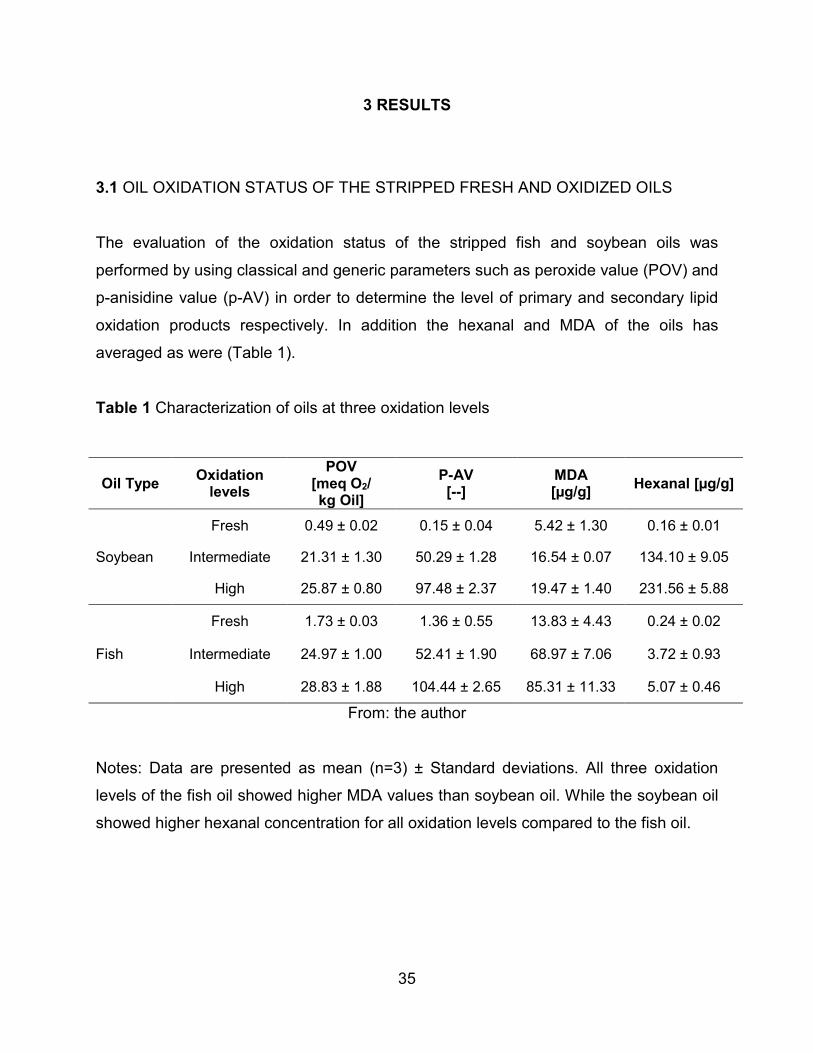
3 RESULTS
3.1 OIL OXIDATION STATUS OF THE STRIPPED FRESH AND OXIDIZED OILS
The evaluation of the oxidation status of the stripped fish and soybean oils was
performed by using classical and generic parameters such as peroxide value (POV) and
p-anisidine value (p-AV) in order to determine the level of primary and secondary lipid
oxidation products respectively. In addition the hexanal and MDA of the oils has
averaged as were (Table 1).
Table 1 Characterization of oils at three oxidation levels
Oil Type Oxidation levels
POV [meq O2/ kg Oil]
P-AV [--]
MDA [µg/g] Hexanal [µg/g]
Soybean
Fresh 0.49 ± 0.02 0.15 ± 0.04 5.42 ± 1.30 0.16 ± 0.01
Intermediate 21.31 ± 1.30 50.29 ± 1.28 16.54 ± 0.07 134.10 ± 9.05
High 25.87 ± 0.80 97.48 ± 2.37 19.47 ± 1.40 231.56 ± 5.88
Fish
Fresh 1.73 ± 0.03 1.36 ± 0.55 13.83 ± 4.43 0.24 ± 0.02
Intermediate 24.97 ± 1.00 52.41 ± 1.90 68.97 ± 7.06 3.72 ± 0.93
High 28.83 ± 1.88 104.44 ± 2.65 85.31 ± 11.33 5.07 ± 0.46
From: the author
Notes: Data are presented as mean (n=3) ± Standard deviations. All three oxidation
levels of the fish oil showed higher MDA values than soybean oil. While the soybean oil
showed higher hexanal concentration for all oxidation levels compared to the fish oil.
36
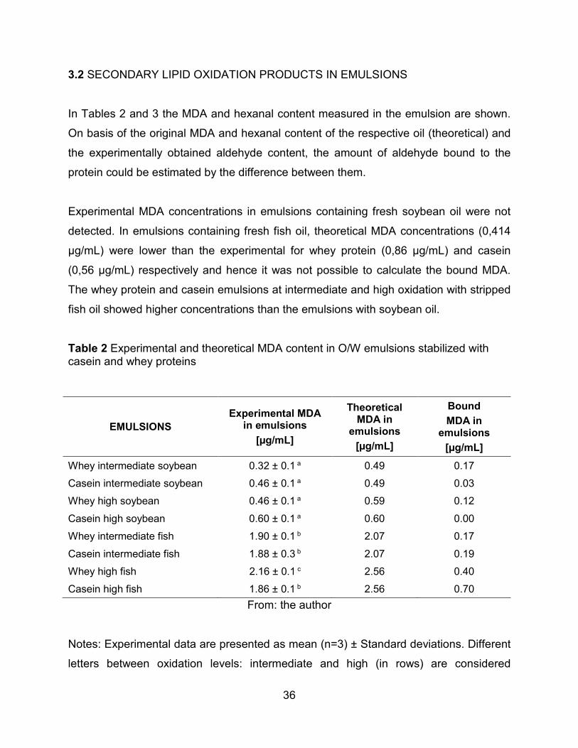
3.2 SECONDARY LIPID OXIDATION PRODUCTS IN EMULSIONS
In Tables 2 and 3 the MDA and hexanal content measured in the emulsion are shown.
On basis of the original MDA and hexanal content of the respective oil (theoretical) and
the experimentally obtained aldehyde content, the amount of aldehyde bound to the
protein could be estimated by the difference between them.
Experimental MDA concentrations in emulsions containing fresh soybean oil were not
detected. In emulsions containing fresh fish oil, theoretical MDA concentrations (0,414
µg/mL) were lower than the experimental for whey protein (0,86 µg/mL) and casein
(0,56 µg/mL) respectively and hence it was not possible to calculate the bound MDA.
The whey protein and casein emulsions at intermediate and high oxidation with stripped
fish oil showed higher concentrations than the emulsions with soybean oil.
Table 2 Experimental and theoretical MDA content in O/W emulsions stabilized with casein and whey proteins
EMULSIONS Experimental MDA
in emulsions [µg/mL]
Theoretical MDA in
emulsions [µg/mL]
Bound MDA in
emulsions [µg/mL]
Whey intermediate soybean 0.32 ± 0.1 a 0.49 0.17 Casein intermediate soybean 0.46 ± 0.1 a 0.49 0.03 Whey high soybean 0.46 ± 0.1 a 0.59 0.12 Casein high soybean 0.60 ± 0.1 a 0.60 0.00 Whey intermediate fish 1.90 ± 0.1 b 2.07 0.17 Casein intermediate fish 1.88 ± 0.3 b 2.07 0.19 Whey high fish 2.16 ± 0.1 c 2.56 0.40 Casein high fish 1.86 ± 0.1 b 2.56 0.70
From: the author
Notes: Experimental data are presented as mean (n=3) ± Standard deviations. Different
letters between oxidation levels: intermediate and high (in rows) are considered
37
statistically different at the 95% confidence level. MDA detection limit: 0.3 µg/mL. The
theoretical MDA content was based on the amount of MDA originally present in the
each oil (Table 1). The amount of bound MDA was calculated by subtracting the
experimental MDA content in the emulsion from the theoretically expected value.
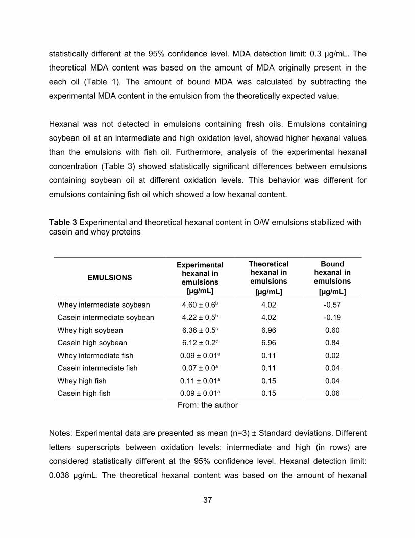
Hexanal was not detected in emulsions containing fresh oils. Emulsions containing
soybean oil at an intermediate and high oxidation level, showed higher hexanal values
than the emulsions with fish oil. Furthermore, analysis of the experimental hexanal
concentration (Table 3) showed statistically significant differences between emulsions
containing soybean oil at different oxidation levels. This behavior was different for
emulsions containing fish oil which showed a low hexanal content.
Table 3 Experimental and theoretical hexanal content in O/W emulsions stabilized with casein and whey proteins
EMULSIONS Experimental
hexanal in emulsions
[µg/mL]
Theoretical hexanal in emulsions
[µg/mL]
Bound hexanal in emulsions
[µg/mL] Whey intermediate soybean 4.60 ± 0.6b 4.02 -0.57 Casein intermediate soybean 4.22 ± 0.5b 4.02 -0.19 Whey high soybean 6.36 ± 0.5c 6.96 0.60 Casein high soybean 6.12 ± 0.2c 6.96 0.84 Whey intermediate fish 0.09 ± 0.01a 0.11 0.02 Casein intermediate fish 0.07 ± 0.0a 0.11 0.04 Whey high fish 0.11 ± 0.01a 0.15 0.04 Casein high fish 0.09 ± 0.01a 0.15 0.06
From: the author
Notes: Experimental data are presented as mean (n=3) ± Standard deviations. Different
letters superscripts between oxidation levels: intermediate and high (in rows) are
considered statistically different at the 95% confidence level. Hexanal detection limit:
0.038 µg/mL. The theoretical hexanal content was based on the amount of hexanal
38
originally present in the each oil (Table 1). The amount of bound hexanal was calculated
by subtracting the experimental hexanal content in the emulsion from the theoretically
expected value.
3.3 DIGESTIBILITY IN EMULSIONS
The protein digestibility percentage of the emulsions is shown in Table 4. A decrease
was observed in digestibility mainly in emulsions containing casein with soybean or fish
oil. Emulsion containing high oxidized soybean oil showed the lowest digestibility
percentage.
Table 4 Digestibility in O/W emulsions
Emulsions Protein digestibility %
Fresh oil Intermediate Oxidation level
High Oxidation level
Casein Fish oil 71.43 ± 2.60a 69.39 ± 1.82a 64.63 ± 1.22b Whey Fish oil 74.32 ± 0.94a 72.19 ± 1.17a 68.37 ± 0.00b
Casein Soybean oil 75.84 ± 3.02a 62.28 ± 4.65b 57.30 ± 4.99b
Whey Soybean oil 74.53 ± 1.22a 72.92 ± 1.80a 74.87 ± 1.96a From: the author
Notes: Data are presented as mean (n=3) ± Standard deviation. Different letter
superscripts between oxidation levels: fresh, intermediate and high (in rows) are
considered statistically different at the 95% confidence level.
39
3.4 SODIUM DODECYL SULPHATE-POLYACRYLAMIDE GEL ELECTROPHORESIS
(SDS-PAGE)
The electrophoretic patterns of the emulsions containing fish or soybean oils are shown
in Figures 4 and 5 respectively. In Figure 4A (casein emulsions) and 4B (whey protein
emulsions) containing fish oil, the formation of aggregates with high molecular weight
was observed. A fraction of the aggregates could even not enter to the gel (lanes 5, 7
and 9). In the same lanes intensive bands with a molecular mass below 10 kDa were
observed. On the contrary in the electrophoretic pattern for casein and whey containing
soybean oil, shown in Figures 5A and 5B respectively, neither aggregates in the at the
top of gel nor intensive bands below 10 kDa were observed in digested lanes.
Figure 5.Electrophoretic pattern of emulsions containing casein (A) or whey protein (B)
with fish oil after 24 h incubation at 4°C.
(A): lane 1: standard, lane 2: casein solution non digested, lane 3: casein solution
digested, lane 4: casein with fresh oil non digested, lane 5: casein with fresh oil
digested, lane 6: casein with medium oxidized oil non digested, lane 7: casein with
medium oxidized oil digested, lane 8: casein with high oxidized oil non digested, lane 9:
casein with high oxidized oil digested. (B): lane 1: standard, lane 2: whey protein
solution non digested, lane 3: whey protein solution digested, lane 4: whey protein with
fresh oil non digested, lane 5: whey protein with fresh oil digested, lane 6: whey protein
with medium oxidized oil non digested, lane 7: whey protein with medium oxidized oil
digested, lane 8: whey protein with high oxidized oil non digested, lane 9: whey protein
with high oxidized oil digested.
40
From: the author
Figure 6 Electrophoretic pattern of emulsions containing casein (A) or whey protein (B)
with soybean oil after 24 h incubation at 4°C.
(A): lane 1: standard, lane 2: casein with fresh oil non digested, lane 3: casein with fresh
oil digested, lane 4: casein with medium oxidized oil non digested, lane 5: casein with
medium oxidized oil digested, lane 6: casein with high oxidized oil non digested, lane 7:
casein with high oxidized oil digested. (B): lane 1: standard, lane 2: whey protein with
fresh oil non digested, lane 3: whey protein with fresh oil digested, lane 4: whey protein
with medium oxidized oil non digested, lane 5: whey protein with medium oxidized oil
digested, lane 6: whey protein with high oxidized oil non digested, lane 7: whey protein
with high oxidized oil digested
A
B
41
From: the author
A
B
42
4 DISCUSSION
Fresh soybean and fresh fish oil presented the lowest peroxide value (POV) as shown
in Table 1. However between the oil at intermediate and high oxidation level a notable
difference was not observed for hydroperoxides. The p-anisidine value (p-AV) for both
oils at the high oxidation level were twice the intermediate level value. These results
suggested the possibility that the speed of decomposition of the hydroperoxides into
secondary products exceeds the formation rate as indicated by the theory (Shahidi and
Wanasundara, 2002).
Malondialdehyde and hexanal were determined in fish and soybean oils as secondary
lipid oxidation products markers. In Table 1, MDA was found at higher concentrations at
the three oxidation levels of fish oil with respect to soybean oil. The major source for the
production of MDA are fatty acids highly unsaturated, with more than three double
bonds in their structure, such as alpha linolenic acid [C 18:3 (n-3)], eicosapentaenoic
acid [EPA 20: 5 (n-3)] and docosahexaenoic acid [DHA 22: 6 (n-3)] (Egert et al, 2012),
EPA and DHA are present in fish oil in an amount of 18.4 g and 7.9 g/100 g fatty acids,
respectively (see Appendix 1).
Analysis of variance (ANOVA) of the experimental concentrations of MDA (Table 2)
showed that emulsions containing soybean oil were not statistically significant different
compared with the intermediate and high oxidation levels, probably because of low
content of MDA precursors in soybean oil. Emulsions containing fish oil had higher MDA
concentration than emulsions containing soybean oil. The highest MDA content could
be related to the high content of this aldehyde in the highly oxidized fish oil.
The experimental and theoretical MDA content presented in Table 2 showed that in the
casein and whey protein based emulsions containing fish oil, the theoretical values
expected for MDA were higher than the experimental ones. This may suggest that part
of the MDA presented in the emulsion was bound to the protein. The casein based
43
emulsion containing fish oil (at high oxidation level) was the system with more bound
MDA (0.70 mg/mL). The ability of MDA to form adducts and crosslinking with
biomolecules such as proteins, DNA and RNA has been extensively reported
(Esterbauer et al., 1991; Del Rio, Stewart and Pellegrini, 2005; Lykkesfeldt, 2007). This
molecule is very prone to reaction with the nucleophilic side chain of cysteine, histidine
and lysine residues due to its chemical structure (a three carbon aldehyde) (Esterbauer
et al., 1991).
In addition, it has been reported that MDA can react with the free amino groups
(especially the ε-amino group of lysine group) of proteins and therefore introduce
carbonyl groups to this biomolecule to form adducts (Wu et al., 2009). Several authors
suggest that the formation of crosslinks between MDA and the amino acids of the
proteins is performed by Schiff base and Michael addition reactions (Refsgaard, Tsai
and Stadtman, 2000; Wu et al., 2009; Gardner, 1979).
While the MDA was detected mainly in fish oil, the hexanal showed high concentrations
in soybean oil, 134 µg/g and 231 µg/g at intermediate and high oxidation level,
respectively (Table 1). Hexanal is a decomposition product of linoleic acid which is
presented mainly in soybean oil (50.55 g/100g fatty acids, Appendix 1).
In emulsions containing soybean oil (at high oxidation level) the theoretical values
expected for hexanal were higher than the experimental ones as a shown in Table 3,
indicating that the system with more bound hexanal was casein based emulsion with
soybean oil at high oxidation level (0.84 mg/mL). Other studies have also demonstrated
that hexanal interact with amino acids, peptides and proteins (Refsgaard et al., 2000;
Meynier et al., 2004; Zhou and Decker, 1999). The ability of hexanal to bind covalently
to dairy proteins leading to protein aggregation and modification was reported by
Meynier et al., 2004. Besides the interaction of proteins that contain sulfhydryl and
amino groups with saturated aldehydes as hexanal has been studied by Zhou and
Decker, 1999, showing that as a result of the interaction of histidine-hexanal, the
hexanal carbonyl group reacts with the free α-amino group and imidazole nitrogen.
44
Moreover, it was observed that in emulsions containing soybean oil the experimental
concentration of this aldehyde at intermediate oxidation level exceeded the expected
concentration (theoretical). Probably due to the favored hexanal production even after
the incubation period of these emulsions (Table 3). The hexanal formation can be
performed through multiple pathways in chain reactions (alkoxyl, peroxyl radicals and
hydroperoxides decomposition) (Shaich, 2013) and therefore with the accumulation of
hexanal it was not possible to calculate the aldehyde bound to protein.
Another possible explanation is that other secondary lipid oxidation products apart from
hexanal such as epoxy, ketones, dimers, alcohols and other aldehydes (not measured
in this study), which could interfere with the hexanal binding sites with protein. Thus, this
compound remained free in the continuous phase of the emulsion and available for its
detection due to its high stability as a saturated aldehyde (Shahidi and Zhong, 2005;
Zhou and Decker, 1999).
In the electrophoretic patterns for non-digested samples in casein and whey protein
based emulsions containing fish and soybean oil (Figures 4 and 5), the formation of
protein aggregates with high molecular weight (> 250 kDa) were observed. These
aggregates were formed probably as a consequence of the interaction protein-protein
and also protein with secondary lipid oxidation products (Cucu et al., 2011). The major
targets for the oxidation are proteins and these reactions can produce modifications in
this biomolecule that include oxidation of the side chain groups, backbone
fragmentation, aggregation and consequently loss of functional properties of the
proteins (Zhu et al., 2009). It has been reported that lipid oxidation-derived aldehyde is
involved in cross-links between protein chains leading to the formation of high mass
(250 kDa) protein aggregates (Sayre, Lin, Yuan, Zhu and Tang, 2006). Also Cucu et al.,
2011, reported the strong influence of the kind of oil and its oxidation status in the
protein oxidation. This study showed that fish oil and the highly oxidized soybean oil
induced oxidation in whey protein, observed mainly in the increase of carbonyl content,
loss of lysine, changes in amino acid composition and protein aggregates formation.
45
According to Schaich, 2013, all lipid oxidation products react in some way with proteins
in parallel oxidation reactions. In this progressive damage known as co-oxidation,
secondary lipid oxidation products, particularly aldehydes, acted in the later stages of
lipid oxidation introducing different types of protein oxidation products. Lipid-protein
complexes (aggregates, adducts and crosslinking formation) in addition to generate
conformational and functional changes, can also interfere with the bioavailability of
proteins and amino acids. As a consequence it may decrease the bioavailability of
amino acid residues and modify the digestibility, which negatively affects the nutritional
values of proteins (Lund, Heinonen, Baron and Estévez, 2010).
Secondary lipid oxidation products are capable to covalently bind to amino acids
(Gardner, 1979) and thus block the action sites of the enzymes involved in the process
of in vitro gastrointestinal digestion. The enzymes are unable to cleavage bonds
involving specific amino acids and lead to a potential reduction in the protein
digestibility.
As reported in the protein digestibility percentage (Table 4), in emulsions containing fish
oil a decrease in the protein digestibility percentage at higher oxidation levels was
observed, particularly in casein based emulsions. It is well known that the high oxidation
susceptibility of fish oil (rich in PUFAs EPA and DHA) leads to the formation of the
hydroperoxides and secondary oxidation products that are able to interact with the side
chain of amino groups and therefore, inducing generation of protein modifications
and/or degradation (Mestdagh et al., 2011). This degradation was even more
pronounced when the lipids in the emulsion were more unsaturated and thus more
prone to oxidation (Frankel, 2005).
Protein digestibility loss with increasing oxidation level in whey protein based emulsions
with soybean oil was not observed (Table 4). These results could be due to a poor
interaction between the hydrophobic amino acid of whey protein with the oxidation
products of soybean oil in the emulsion interface. It has been reported that the polarity
of the oil phase influence strongly on the degree of protein conformational re-
46
arrangement and the way in which amino acid are physically oriented in the O/W
emulsion interface, due to the oil acts as a solvent of hydrophobic amino acids (Zhai,
Day, Aguilar and Wooster, 2013). Also Maldonado, Wilde, Mulholland and Morris, 2012,
reported that in emulsions the lower polarity oil (tetradecane) induced a greater
attraction of the β-lactoglobulin hydrophobic residues towards the oil phase and
therefore their strong interaction compared to olive oil. Consequently, polarity of the oil
phase is one of the factors that might determine the adsorption of the protein at the
interface leading to lipid-protein interaction.
On the contrary, in casein based emulsions containing soybean oil there was a
difference in the digestibility percentage at intermediate and high oxidation levels with
respect to fresh oil. Surprisingly emulsions with soybean oil presented a dramatic
decrease in the protein digestibility loss. This result could be related to soybean oil
generating sufficient secondary oxidation products that lead to the formation of
aggregates. Also its chemical structure consists mainly of triacylglycerols that contain
multiple oxidized fatty acids residues that could interact with nucleophilic amino acid
residues in proteins which induce to protein aggregation (Cucu et al., 2011; Schaich, et
al., 2013).
The protein digestibility loss results (Table 4) showed that the higher losses occurred in
casein based emulsions at high oxidation level for soybean oil (57.3%) and fish oil
(64.6%). It was observed that digestibility decreased gradually with increasing the oil
oxidation levels. This is probably due to the open and unfolded structure of casein in the
O/W emulsion, in which the protein exposes its hydrophobic groups in order to be
absorbed in the emulsion interface covering completely the oil droplet (Aynié, Le Meste,
Colas and Lorient, 1992). This structural conformation facilitates the interaction of
casein with the secondary oxidation products from soybean and fish oils. Fomuso,
Corredig and Akoh, 2002 and Hu, McClements and Decker, 2003, suggested that due
to the flexible structure of caseins, these tend to form crosslinking in the presence of
oxidized lipids. Also Chao and Standman, 1997, established that oxidative modification
47
could simultaneously result in exposure of hydrophobic groups due to protein partial
unfolding and cross-linking hydrophobic interactions, resulting in aggregation.
On the contrary in whey protein based emulsions, significant losses of protein
digestibility percentage were not observed; this can be due to its structure (rigid and
compact) and thermal stability. In this study, temperature below 60°C were used to
prepare emulsions and incubation at -4°C were not enough to unfold the protein
completely, exposing its hydrophobic groups and covering fully the oil droplet. Therefore
the interaction of whey protein with the secondary lipid oxidation products of the
oxidized oils in the emulsion interface was partial and incomplete. Whey protein
digestibility loss percentages were low or imperceptible particularly in emulsions
containing soybean oil with the increase in oxidation level (Table 4). The interaction of
whey protein with secondary lipid oxidation products at 70°C has been reported by
Cucu et al., 2011 and Mestdagh et al., 2011, evidencing the formation of aggregates,
fragmentations, among other protein modifications.
Also, Nicolai, Britten and Schmitt, 2011, suggest that when the β-lactoglobulin is heated,
higher than 60°C, its structure is modified and becomes more mobile. Buried
hydrophobic groups and the thiol group in the native state became exposed and interact
with other molecules.
Several authors have been reviewed other factors involved in the interaction of proteins
and oil droplet in the O/W emulsion interface and the differences between casein and
whey protein behavior as emulsifiers (Hu et al., 2003; McClements, 2004a; Zhai et al.,
2013., Horn et al., 2011; Singh and Ye, 2009). One factor is the unfolding and
rearrangement of protein native structure, which is related with temperature, as
previously discussed. Proteins as emulsifiers reduced the system free energy and
therefore the interfacial tension due to their adsorption at the emulsion interface. In
consequence caseins (flexible proteins) are more effective at reducing the interfacial
tension due to its higher proportion of non-polar groups in contrast to whey protein (rigid
protein) that have fewer non-polar groups (Dickinson and McClements, 1995).
48
Hu et al., 2003, showed that the amino acid composition and its physical orientation
(e.g., toward the lipid or water face) for each type of protein might influence their
interaction with the oil droplet in the O/W emulsion interface and oxidative stability.
However more detailed studies about the role of amino acids in the oxidative stability in
protein-stabilized emulsions are needed.
Another factor that might influence the protein interaction is the droplet size. The smaller
droplet could be easier to oxidize because it has larger contact area on the surface and
consequently it presented greater interaction with oxidized lipids and its products
(McClements and Decker 2000). Hu et al., 2003, and Horn et al., 2011, showed that
smaller droplets were formed in casein than in whey based emulsions. However it was
not reported a direct correlation between the tendency for decreasing droplet size and
increased oxidation. This suggests that other factors besides droplet size may influence
lipid-protein interactions in O/W emulsions.
The SDS-PAGE analysis for digested samples of emulsions containing fish oil, display
the smearing towards the top of gel with molecular mass higher than 250 kDa (Figure 4
A and B) corresponding to protein aggregates. These molecules showed a resistance
towards proteolysis, despite of the enzymatic treatment with proteases in gastric and
duodenal phases in the in-vitro gastrointestinal digestion of emulsions. Zamora and
Hidalgo, 2000, observed in electrophoretic gels that protein aggregates with high
molecular weight were more resistant to digestion.
According to Zhang, Xiao and Ahn, 2013, in general heavily oxidized proteins could
present less digestibility due to protein aggregation, whereas slightly oxidized proteins
are easily digested by proteases. This phenomenon occurs probably due to the
conformational changes that expose some buried peptide bonds for enzyme hydrolysis
and increase degradation susceptibility. Otherwise, strong oxidation environment can
result in the formation of protein cross-linkings, which further leads to protein
49
aggregation. These cross-linkings and aggregates are more resistant to enzymatic
degradation compared to native protein forms (Davies, Lin and Pacifici, 1987.
Figure 4 also showed that the gels of casein (A) and whey protein (B) based emulsions
containing fish oil, had intense bands below the 10 kDa in the digested samples at the
three oxidation levels (fresh, intermediate and high). This small protein fragments could
indicate a backbone fragmentation. Similar result has been reported previously in whey
protein emulsion with fish oil upon photo-oxidation (Mestdagh et al., 2011). Oxidants
such as reactive aldehydes can directly attack the backbone of a protein to cause
fragmentation and conformational changes in the secondary and tertiary structure of the
protein (Zhang et al., 2013).
In contrast, in the electrophoretic patterns of the protein emulsions containing soybean
oil showed in Figure 5 A and B, it was no observed smearing neither towards the top of
the gel (up to 250 kDa), nor below 10 kDa. This may possibly indicate that the protein
binding sites for dye Coomassie blue® (basic and aromatic amino acids) were occupied
by secondary oxidation compounds. In fact, it has been reported that the amino acids
lysine, arginine, histidine and tyrosine (which are the Coomassie blue® binding sites)
are also the main targets for attack by lipid radicals and secondary lipid oxidation
products as hexanal and MDA (Schaich, 2008).
Even though in this study the possible effect of the interaction of MDA and hexanal with
milk proteins (casein and whey) in emulsion were observed, it is not appropriate to
attribute the decrease in protein digestibility only to these two secondary oxidation
products. Lipid oxidation is a dynamic process in which is produced, transform and react
continuously several oxidation compounds via multiple pathways. Thus, it is known that
epoxides are up to 1000 times more reactive with proteins than aldehydes, generating
unidentified products that have no organoleptic changes (Schaich, 2008). Other lipid
oxidation products as dimers and polymers are capable to reduce the digestibility and
can damage the intestinal mucosa (Schaich, 2013).
50
5. CONCLUSION
To conclude, the impact of autoxidized lipids on the protein digestibility loss in O/W
emulsions particularly for casein with soybean and fish oils at high oxidation level was
evidenced. The casein emulsion with soybean oil, at high oxidation level, was the
system with the most hexanal bound to protein and also the greatest protein digestibility
loss. As a result of the chemical interaction of the autoxidized lipid with dairy proteins,
the aggregation occurs and the protein becomes more resistant to digestion. This effect
was greater for casein probably due to its chemical structure related to a possible
hydrophobic interaction at emulsion interface.
51
RECOMMENDATIONS The results of this study provide important evidence of the impact of lipid oxidation on
protein digestibility in emulsions, which affects the nutritional value of dairy foods
enriched with polyunsaturated fatty acids, generating implications for production, shelf
life and consumer welfare. The consumption increase of functional foods where the
matrices are dairy emulsions (e.g. yoghurt, flavored milk, salad dressing, nutritional
beverages, infant formula, etc) should promote studies in order to understand the
physical and chemical processes occurring in this type of emulsions to ensure quality.
52
REFERENCES
Association Of Analytical Communities (1981). Crude protein in meat: block digestion
method. In AOAC official method 98110: Official methods of analysis of association of
official analytical chemists (16th ed.). Gaithersburg, MA, USA: AOCS Press.
Association Of Analytical Communities (1993). p-Anisidine value. In AOCS official
method Cd 18-90: Official methods and recommended practices of the American oil
chemists’ society (4th ed). Campaign, IL, USA: AOCS Press.
American Oil Chemists Society (1993). Official methods and recommended practices of
the American oil chemists society, Method Cd 18-90.Champaign, IL.
Aynié, S.; Le Meste, M.; Colas, B. & Lorient, D. (1992). Interactions between lipids and
milk proteins in emulsion. J Food Sci, 57, 883-886.
Betti, M.; Schneider, B.; Wismer, W.; Carney, V.; Zuidhof, M. & Renema, R. (2009).
Omega-3-enriched broiler meat: 2. Functional properties, oxidative stability, and
consumer acceptance. Poultry Sci, 88, 1085-1095.
Chao, C.; Ya, M. & Standman, E. (1997). Modification of protein surface hydrophobicity
and methionine oxidation by oxidative systems. Proc Natl Acad Sci USA, 94(7), 2969-
2974.
Chung, C. & McClements, D.J. (2014). Review Structure–function relationships in food
emulsions: Improving food quality and sensory perception. Food struct. 1, 106–126.
Cucu, T.; Devreese, B.; Mestdagh, F.; Kerkaert, B. & De Meulenaer, B. (2011). Protein-
lipid interactions during the incubation of whey proteins with autoxidizing lipids. Int Dairy
J, 21(6), 427-433.
53
Davies, K.; Lin, S. & Pacifici, R. (1987). Protein damage and degradation by oxygen
radicals. IV. Degradation of denatured protein. J Biol Chem, 262, 9914-9920.
Dawczynski, C.; Martin, L.; Wagner, A. & Jahreis, G. (2010). n 3 LC-PUFA-enriched
dairy products are able to reduce cardiovascular risk factors: A double-blind, cross-over
study. Clinical Nutrition, 29, 592-599.
Del Rio, D.; Stewart, A. & Pellegrini, N. (2005). A review of recent studies on
malondialdehyde as toxic molecule and biological marker of oxidative stress. Nutr
Metab Cardiovasc Dis, 15, 316-328.
Dickinson, E. (1992). An introduction to food colloids. New York: Oxford University
Press.
Dickinson, E. & McClements, D.J. (1995). Advances in Food Colloids. London: Blackie
Academic and Professional.
Dhiman, T.; Satter, L.; Pariza, M.; Galli, M.; Albright, K. & Tolosa, M. (2000). Conjugated
linoleic acid (CLA) content of milk from cows offered diets rich in linoleic and linolenic
acid. J Dairy Sci, 83,1016-1027.
Egert, S.; Lindenmeier, M.; Harnack, K.; Korme, K.; Erbersdobler, H... Somoza, V.
(2012). Margarines fortified with a-linolenic acid, eicosapentaenoic acid, or
docosahexaenoic acid alter the fatty acid composition of erythrocytes but do not affect
the antioxidant status of healthy adults. J Nutr, 142(9), 1638-1644.
Elisia, I. & Kitts, D. (2011). Quantification of hexanal as an index of lipid oxidation in
human milk and association with antioxidant components. J Clin Biochem. Nutr,
49(3),147-52.
54
Esterbauer, H.; Schaur, R. & Zollner, H. (1991). Chemistry and biochemistry of 4-
hydroxynonenal, malonaldehyde and related aldehydes. Free Radic Biol Med, 11, 81–
128.
Fennema, O.R.; Parkin, K. & Damodaran, S. (2007). Food Chemistry. New York: Taylor
& Francis Group.
Fomuso, L.; Corredig, M. & Akoh, C. (2002). Effect of emulsifier on oxidative properties
of fish oil-based structured lipid emulsions. J Agric Food Chem, 50, 2957-2961.
Frankel E. (1985). Chemistry of autoxidation: mechanism, products and flavor
significance. London: AOCS.
Frankel, E. N. (2005). Lipid oxidation. London: The oily press.
García, M.; Márquez, G.; Fontecha, J. & Gordon, M. (2009). Volatile oxidation
compounds in a conjugated linoleic acid-rich oil. Food Chem, 113, 926-931.
Gardner, H. (1979). Lipid hydroperoxide reactivity with proteins and amino acids: A
review. J Agric Food Chem, 27, 220-229.
Georgiou, C.; Grintzalis, K.; Zervoudakis, G. & Papapostolou, I. (2008). Mechanism of
Coomassie brilliant blue G-250 binding to proteins: a hydrophobic assay for nanogram
quantities of proteins. Anal Bioanal Chem, 391, 391-403.
Genot, C., Meyner, A.; Riaublanc, A. & Chobert, J. (2003). Protein alterations due to
lipid oxidation in multiphase systems. Upsala, Sweden: AOCS Publishing.
Ha, J.; Seo, D.; Chen, X.; Hwang, J. & Shim, Y. (2011). Determination of hexanal as an
oxidative marker in vegetable oils using an automated dynamic headspace sampler
coupled to a gas chromatograph/mass spectrometer. Anal Sci, 24, 873-878.
55
Headlam, H. & Davis, M. (2004). Markers of protein oxidation: different oxidants give
rise to variable yields of bound and released carbonyl products. Free Radic BiolMed,
36,1175–1184.
Hoffmann, H. & Reger M. (2014). Emulsions with unique properties from proteins as
emulsifiers. Adv Colloid Interface Sci, 205, 94–104.
Horne D. (2009). Chapter 5: Casein micelle structure and stability. In Milk Proteins:
From Expression to Food. 321-340. Elsevier Inc. ISBN: 978-0-12-374039-7.
Horn, A.; Nielsen, N.; Andersen, U.; Søgaard, L.; Horsewell, A. & Jacobsen, C. (2011).
Oxidative stability of 70% fish oil-in-water emulsions: Impact of emulsifiers and pH. Eur
J Lipid Sci Technol, 113, 1243-1257.
Hu, M.; McClements, D.J & Decker, E. (2003). Lipid oxidation in corn oil-in-water
emulsions stabilized by casein, whey protein isolate, and soy protein isolate. J. Agric
Food Chem, 51, 1696-1700.
International Dairy Federation. (1991). Anhydrous Fat, Determination of Peroxide Value.
IDF Standard 74A; IDF. Brussels, Belgium.
Iafelice, G.; Caboni, M.; Cubadda, R.D.; Criscio, T.; Trivisonno, M. & Marconi, E. (2008).
Development of functional spaghetti enriched with long chain omega-3 fatty acids.
Cereal Chem, 85, 146-151.
Jovanović, S.; Barać, M. & Maćej, O. (2005). Whey proteins-Properties and Possibility
of Application. Mljekarstvo, 55(3), 215-233.
Kritchevsky, D. (1998). History of Recommendations to the Public about Dietary Fat.
Journal of nutrition, 128(2), 449-452.
56
Laemmli, U. K. (1970). Cleavage of structural proteins during the assembly of the head
of bacteriophage T4. Nature, 227, 680–685.
Livney, Y. (2010). Milk proteins as vehicles for bioactives. Curr Opin Colloid In, 15, 73-
83.
Lund, M.N.; Heinonen, M. & Baron, C.; Estévez, M. (2010). Protein oxidation in muscle
foods: A review. Mol Nutr Food Res, 54, 1-13.
Lund, M.N.; Hviid, M.S. & Skibsted, L.H. (2007). The combined effect of antioxidants
and modified atmosphere packaging on protein and lipid oxidation in beef patties during
chill storage. Meat Science, 76, 226-233.
Lykkesfeldt, J. (2007). Malondialdehyde as biomarker of oxidative damage to lipids
caused by smoking. Clin Chim Acta, 380, 50-58.
Maldonado, J.; Wilde, P.; Mulholland, F. & Morris, V. (2012). Protein unfolding at fluid
interfaces and its effect on proteolysis in the stomach. Soft Matter, 8, 4402-4414.
Malinauskyte, E.; Ramanauskaite, J.; Leskauskaite, D.; Devold, T.; Schüller, R. &
Vegarud, G. (2014). Effect of human and simulated gastric juices on the digestion of
whey proteins and carboxymethylcellulose-stabilised O/W emulsions. Food Chem,
165,104–112.
Márquez, G.; Holgao, F. & Velasco, J. (2014). Mechanisms of Oxidation in Food Lipids.
London: CRC Press Taylor & Francis Group.
Mcclements, D.J. & Decker, E. (2000). Lipid Oxidation in Oil-in-Water Emulsions:
Impact of Molecular Environment on Chemical Reactions in Heterogeneous Food
Systems. J Food Sci, 65(8), 1270-1282.
57
McClements, D.J. (2004a). Food emulsions: Principles, practice, and techniques. CRC
Press: Boco Raton, FL,
McClements, D.J. (2004b). Protein-stabilized emulsions. Curr. Opin. Colloid Interface
Sci, 9, 305-313.
McClements, D.J. & Li, Y. (2010). Review of in vitro digestion models for rapid
screening of emulsion-based systems. Food Funct, 1, 32–59.
Mestdagh, K.; Cucu, T. & De Meulenaer, B. (2011). Interaction between whey proteins
and lipids during light-induced oxidation. Food Chem, 126, 1190-1197
Meynier, A.; Rapon, V.; Dalgalarrondo, M. & Genot, C. (2004). Hexanal and t-2-hexenal
form covalent bonds with whey proteins and sodium caseinate in aqueous solution. Int
Dairy J, 14, 681–690.
Nik, A.; Wright, A. & Corredig, M. (2010). Surface adsorption alters the susceptibility of
whey proteins to pepsin-digestion. J Colloid Interface Sci, 344, 372–381.
Nicolai, T.; Britten, M. & Schmitt, C. (2011). β-Lactoglobulin and WPI aggregates:
Formation, structure and applications. Food Hydrocolloid, 25 (8), 1945-1962.
Onyango, A. & Baba, N. (2010). New hypotheses on the pathways of formation of
malondialdehyde and isofurans. Free Radic Biol Med, 49(10), 1594-1600.
Panseri, S.; Soncin; S.; Chiesa, L. & Biondi, O. (2011). A headspace solid-phase
microextraction gas-chromatographic mass-spectrometric method (HS-SPME–GC/MS)
to quantify hexanal in butter during storage as marker of lipid oxidation. Food Chem,
127, 886-889.
58
Papastergiadis, A.; Mubiru, E.; Van Langenhove, H. & De Meulenaer, B. (2012).
Malondialdehyde measurement in oxidized foods: evaluation of the spectrophotometric
thiobarbituric acid reactive substances (TBARS) test in various foods. J Agric Food
Chem. 60(38), 9589-9594.
Papastergiadis, A. (2014). A contribution to the risk assessment in relation to the
formation of toxic aldehydes in foods as a result of lipid oxidation. (PhD dissertation).
Faculty of Bioscience Engineering, Ghent University, Gent.
Picariello, G.; Mamone, G.; Chiara, N.; Addeo, F. & Ferranti P. (2013). Protein
digestomics: Integrated platforms to study food-protein digestion and derived functional
and active peptides. Trends Anal Chem, 52, 120-134.
Pizzimenti, S.; Ciamporcero, E.; Daga, M.; Pettazzoni, P.; Arcaro, A.; Cetrangolo, G…
Barrera, G. (2013). Interaction of aldehydes derived from lipid peroxidation and
membrane proteins. Front. Physiol, 4, 242-258.
Quintero, J. (2014). Impact of lipid oxidation on digestibility of dairy proteins (Master
dissertation). Faculty of bioscience engineering: University of Gent, Gent.
Refsgaard, H.; Tsai, L. & Stadtman, E. (2000). Modifications of proteins by
polyunsaturated fatty acid peroxidation product. Proc Natl Acad Sci USA, 97(2), 611-
616.
Sanchez, A.; Rodríguez, A.; López, J. & Paseiro, P. (2004). Determination of hexanal as
indicator of the lipidic oxidation state in potato crisps using gas chromatography and
high-performance liquid chromatography. J Chromatogr A, 1046, 75-81.
Sayre, L.; Lin, D.; Yuan, Q.; Zhu, X. & Tang, X. (2006). Protein adducts generated from
products of lipid oxidation: Focus on HNE and ONE. Drug Metab Rev, 38(4), 651-675.
59
Schaich, K. M. (2008). Co-oxidations of oxidizing lipids: Reactions with proteins, in Lipid
Oxidation Pathways. Illinois: AOCS Press, Urbana.
Schaich, K. (2013). Challenges in Elucidating Lipid Oxidation Mechanisms: When,
Where, and How do Products Arise? Illinois: AOCS press.
Schaich, K.; Shahidi F.; Zhong Y. & Eskin, N. (2013). Lipid Oxidation. San Diego:
Academic Press.
Shahidi, F. & Wanasundara, U. (2002). Methods for Measuring Oxidative Rancidity in
Fats and Oils. New York: Marcel Dekker.
Shahidi, F. & Zhong, Y. (2005). Lipid Oxidation: Measurement Methods. (6th ed.). New
Jersey: John Wiley & Sons Inc.
Shantha, N. & Decker, N. (1994). Rapid, sensitive, iron-based spectrophotometric
methods for determination of peroxide values of food lipids. J. AOAC Int, 77, 412–424.
Singh, H. & Flanagan, J. (2006). Milk proteins. Boca Raton: CRC Press.
Singh, H. & Ye, A. (2009). Milk Proteins: From Expression to Food. London: Academic
Press Elsevier Inc.
Singh, H. and Ye, A. (2013). Structural and biochemical factors affecting the digestion of
protein-stabilized emulsions. Curr Opin Colloid Interface Sci, 18, 360-370.
Simopoulos, A. (1999). Essential fatty acids in health and chronic disease. Am J Clin
Nutr, 70, 560–569.
60
Stuknytė, M.; Cattaneo, S., Masotti, F. & De Noni, I. (2015). Occurrence and fate of
ACE-inhibitor peptides in cheeses and in their digestates following in vitro static
gastrointestinal digestion. Food Chem, 168, 27-33.
Tcholakova, S.; Denkov, N.D.; Ivanov, I.B. & Campbell, B. (2006). Coalescence stability
of emulsions containing globular milk proteins. Adv Colloid Interface Sci, 123-126, 259-
293.
Tehrany, E.; Jacquot, M.; Gaiani, C.; Imran, M.; Desobry, S. & Linder, M. (2012).
Beneficial effects and oxidative stability of omega-3 long- chain polyunsaturated fatty
acids. Trends Food Sci Tech, 25(1), 24–33.
Tironi, V.; Tomás, M. & Añón, M. (2007). Lipid and protein deterioration during the
chilled storage of minced sea salmon (Pseudopercis semifasciata). J Sci Food Agric,
87, 2239–2246.
Viljanen, K. (2005). Protein oxidation and protein-lipid interactions in different food
models in the presence of berry phenolics. (Academic dissertation). Department of
Applied Chemistry and Microbiology Food Chemistry University of Helsinki, Helsinki.
Watkins, S. M. & German, J.B. (1998). Omega fatty acids. New York: Marcel Dekker.
Wickham, M.; Faulks, R. & Mills, C. (2009) In vitro digestion methods for assessing the
effect of food structure on allergen breakdown. Mol Nutr Food Res, 53, 952–958.
Wu, W.; Zhang, C. & Hua, Y. (2009). Structural modification of soy protein by the lipid
peroxidation product malondialdehyde. J Sci Food Agric, 89, 1416–1423.
Ye, A.; Cui, J.; Taneja, A.; Zhu, X. & Singh, H. (2009). Evaluation of processed cheese
fortified with fish oil emulsion. Food Res Int, 42, 1093-1098.
61
Zamora, R. & Hidalgo, F.J. (2001). Inhibition of proteolysis in oxidized lipid damaged
proteins. J Agric Food Chem., 49(12), 6006-6011.
Zhang, W.; Xiao, S. & Ahn, D. (2013). Protein oxidation: basic principles and
implications for meat quality. Crit Rev Food Sci Nutr, 53(11), 1191-201.
Zhai, J., Day, L.; Aguilar, M. & Wooster, T. (2013). Protein folding at emulsion oil/water
interfaces. Curr Opin Colloid Interface Sci, 18, 257-271.
Zhou, S. & Decker, E. (1999). Ability of amino acids, dipeptides, polyamines, and
sulfhydryls to quench hexanal, a saturated aldehydic lipid oxidation product. J Agric
Food Chem., 47, 1932-1936.
Zhu, Q.; Zheng, Z.; Cheng, K.; Wu, J.; Zhang, S.; Tang, Y…Wang, S. (2009). Natural
polyphenols as direct trapping agents of lipid peroxidation-derived acrolein and 4-
hydroxy-trans-2-nonenal. Chem Res Toxicol., 22, 1721-1727.
62
APPENDIX
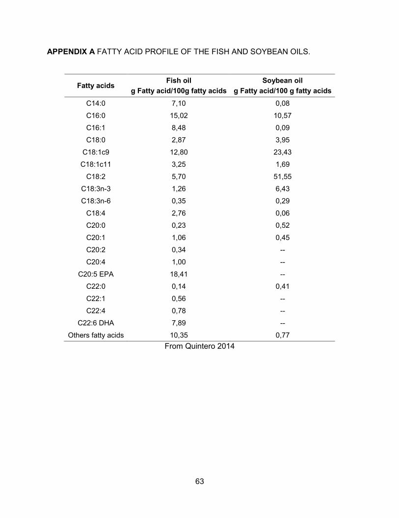
63
APPENDIX A FATTY ACID PROFILE OF THE FISH AND SOYBEAN OILS.
Fatty acids Fish oil g Fatty acid/100g fatty acids
Soybean oil g Fatty acid/100 g fatty acids
C14:0 7,10 0,08 C16:0 15,02 10,57 C16:1 8,48 0,09 C18:0 2,87 3,95
C18:1c9 12,80 23,43 C18:1c11 3,25 1,69
C18:2 5,70 51,55 C18:3n-3 1,26 6,43 C18:3n-6 0,35 0,29
C18:4 2,76 0,06 C20:0 0,23 0,52 C20:1 1,06 0,45 C20:2 0,34 -- C20:4 1,00 --
C20:5 EPA 18,41 -- C22:0 0,14 0,41 C22:1 0,56 -- C22:4 0,78 --
C22:6 DHA 7,89 -- Others fatty acids 10,35 0,77
From Quintero 2014