Embed Size (px)
Citation preview
Studies on antimicrobial and antioxidant properties of phosvitin
hydrolysates produced by high hydrostatic pressure combined with
enzymatic hydrolysis
by
Hee Joo Yoo
A thesis submitted in partial fulfillment of the requirements for the degree of
Master of Science
in
Pharmaceutical Sciences
Faculty of Pharmacy and Pharmaceutical Sciences
University of Alberta
© Hee Joo Yoo, 2016
ii
Abstract
Phosvitin (PV) is a metal binding protein in egg yolk with unique amino
acid composition (more than 55% serine). Due to a high proportion of
phosphorylated serine residues, PV shows metal chelating, antioxidant and
antimicrobial activities. The use of PV, however, in nutra- or pharmaceutical
application has been restricted mainly due to the formation of insoluble
complexes with divalent metal ions in the gastrointestinal tract. The PV
hydrolysates (PVH) that are hydrolyzed by proteases may solve the problem and
increase their applications without compromising the PV’s functional properties.
There are many attempts to produce the PVH. However, the yield of the PVH is
low due to the PV’s negative charges causing the resistance to digestive enzymes.
The present technology of the high hydrostatic pressure combined with enzymatic
hydrolysis (HHP-EH) is a new method to increase the enzyme efficiency. The
present study of the HHP-EH shows a higher degree of hydrolysis containing
more peptides with Mw< 3 kDa compared to atmospheric pressure (AP). PV
Hydrolysis with Alcalase (Alc) under HHP resulted in the highest degree of
hydrolysis (31.3%). The PVH treated by Alc and Trypsin (Try) obtained from
both HHP and AP treatments showed superior iron chelation capacity (69-73%).
Alc-PVH produced by HHP-EH displayed significantly greater reducing power
(γ.5 μM Trolox equivalent/mg) than AP-PVH (1.γ μM Trolox equivalent/mg). In
the second study, a combination of specific IgY (100 μg/mL) and PVH-Alc-HHP
(1 mg/mL) as an anti-microbial agent was found to be the most efficient to control
iii
the foodborne Enterotoxigenic Escherichia coli (ETEC) K88 and K99 in vitro.
The synergistic anti-microbial activities of IgY and PVH may support their
potential application in feed supplementation to prevent microbial contamination
and infectious diseases. In the third study, there is limited information available
on the quantification of PV. The double antibody sandwich ELISA (DAS-ELISA)
and biotinylated DAS-ELISA developed has a PV detection range of 5.6 – 90
µg/mL and 2.5 – 40 ng/mL, respectively. The biotinylated DAS-ELISA is a
superior method for PV quantification regarding accuracy and sensitivity. This
highly efficient PV detection method may recuperate the performance of the
existing protein assay methods as well as facilitate future research on PV
bioactivities and applications.
iv
Acknowledgements
I would like to express deep gratitude to my supervisor, Dr. Hoon Sunwoo,
for his constant guidance and full support throughout my study and research,
without which this work would have been a frustrating and overwhelming pursuit.
I would like to thank Dr. Raimar Loebenberg and Dr. Jianping Wu for
giving advice and guidance in the course of completing the experiments.
In addition, I express my appreciation to the external examiner Dr. Arno
Siraki for willing to spend his precious time in evaluating this thesis.
My special thanks go to Drs. Fatemeh Bamdad and Naiyana Gujral, who
provided many assistance, insights and comments to help me.
I am also grateful to my colleagues and numerous friends for giving helpful
advices throughout this academic exploration.
I would like to thank to NSER-CRD (#485627-15) and Mitacs Accelerate
for financial support.
Finally, my immense gratitude goes to my family for the support they
provided me through my entire life. Without their unconditional love and
encouragement, I would not have been able to complete this thesis.
v
Table of Contents
Abstract .................................................................................................................. ii
Acknowledgements .............................................................................................. iv
Table of Contents .................................................................................................. v
List of Figures ....................................................................................................... xi
List of Tables ....................................................................................................... xv
Chapter 1: Literature review ............................................................................... 1
1.1 Introduction of Egg ....................................................................................... 1
1.2 Structure of Egg ............................................................................................ 1
1.3 Composition of Egg.................................................................................... 3
1.3.1 Egg Shell ................................................................................................ 6
1.3.2 Egg Shell Membrane .............................................................................. 7
1.3.3 Egg White ............................................................................................... 8
1.3.3.1 Major protein .................................................................................. 9
1.3.3.2 Minor proteins of albumen............................................................ 12
1.3.4 Vitelline membrane .............................................................................. 15
1.3.5 Egg Yolk ............................................................................................... 16
1.3.5.1 Lipids of the Egg Yolk ................................................................... 18
1.3.5.1.1 Fatty acids .............................................................................. 18
1.3.5.1.2 Phospholipids ......................................................................... 20
1.3.5.1.3 Fat-soluble Vitamins and Carotenoids .................................. 21
vi
1.3.5.2 Egg Yolk Protein ........................................................................... 22
1.3.5.2.1 Lipovitellin apoproteins ......................................................... 23
1.γ.5.β.β α, -Livetins ........................................................................... 24
1.γ.5.β.γ -Livetin (IgY) ........................................................................ 25
1.4 Phosvitin (PV) .......................................................................................... 30
1.4.1 Phosvitin (PV) Characteristics ............................................................ 32
1.4.2 Phosvitin (PV) Composition ................................................................ 34
1.4.3 Isolation and Purification of Phosvitin ................................................ 35
1.4.4 Functional properties of Phosvitin ...................................................... 38
1.4.4.1 Metal Chelating Activity ............................................................... 38
1.4.4.2 Antimicrobial Activity ................................................................... 40
1.4.4.3 Antioxidant Activity ....................................................................... 41
1.4.4.4 Emulsifying Activity ...................................................................... 43
1.5 Phosvitin Hydrolysates (PVH) .................................................................... 44
1.5.1 Production of Phosvitin Hydrolysates (PVH) ...................................... 45
1.5.2 Functional Properties of Phosvitin Hydrolysates ................................ 47
1.5.2.1 Metal Chelating Activity of Phosvitin Hydrolysates ..................... 47
1.5.2.2 Antioxidant Activity of Phosvitin Hydrolysates ............................ 48
1.5.2.3 Antimicrobial Activity of Phosvitin Hydrolysates ......................... 50
1.5.2.4 Emulsifying Activity of Phosvitin Hydrolysates ............................ 50
1.6 High hydrostatic pressure-enzymatic hydrolysis (HHP-EH) biotechnology
........................................................................................................................... 51
1.7 Rational ....................................................................................................... 54
vii
1.8 Hypothesis ................................................................................................... 55
1.9 Objectives .................................................................................................... 55
1.10 Specific Objectives .................................................................................... 56
1.10.1 Production of PVH ............................................................................. 56
1.10.2 Iron-chelation .................................................................................... 56
1.10.3 Anti-microbial property ..................................................................... 56
1.11 Study Plan ................................................................................................. 57
Chapter 2 (Study 1): Sensitive Double Antibody Sandwich ELISA for the
Quantification of Phosvitin ................................................................................ 58
2.1 Introduction................................................................................................. 58
2.2 Materials and Methods ............................................................................... 60
2.2.1 Materials .............................................................................................. 60
2.2.2 Production of Anti-PV Polyclonal IgY ................................................. 60
2.2.3 Purification of Anti-PV IgY .................................................................. 61
2.2.4 Titer of Anti-PV IgY by Indirect ELISA ............................................... 62
2.2.5 Total IgY Concentration ...................................................................... 62
2.2.6 Total Protein Assay .............................................................................. 63
2.2.7 Electrophoresis and Western Blot ....................................................... 63
2.2.8 Double Antibody Sandwich ELISA (DAS-ELISA)................................ 64
2.2.9 Biotinylated DAS-ELISA ...................................................................... 64
2.2.10 Validation of the Assay ...................................................................... 65
2.2.11 Statistical analysis ............................................................................. 66
2.3 Results ......................................................................................................... 66
viii
2.3.1 Production of Anti-PV IgY ................................................................... 66
2.3.2 Double Antibody Sandwich ELISA ...................................................... 68
2.3.3 Biotinylated Double Antibody Sandwich ELISA .................................. 69
2.3.4 Accuracy .............................................................................................. 70
2.3.5 Intra- and Inter-assay Precision .......................................................... 71
2.4 Discussion ................................................................................................... 72
2.5 Conclusion .................................................................................................. 74
Chapter 3 (Study 2): Phosvitin Hydrolysates with Iron Chelating Capacity
Produced by High Hydrostatic Pressure Combined with Enzymatic
Hydrolysis ............................................................................................................ 75
3.1 Introduction................................................................................................. 75
3.2 Materials and Methods ............................................................................... 75
3.2.1 Materials .............................................................................................. 77
3.2.2 Apparatus ............................................................................................. 77
3.2.3 IgY separation from egg yolk ............................................................... 78
3.2.4 Phosvitin extraction and purification .................................................. 78
3.2.5 Enzymatic hydrolysis ........................................................................... 79
3.2.6 Degree of hydrolysis (DH) ................................................................... 80
3.2.7 Phosphate content determination ........................................................ 80
3.2.8 Sodium dodecyl sulfate polyacrylamide gel electrophoresis (SDS-
PAGE) ........................................................................................................... 81
3.2.9 Size Exclusion High Pressure Liquid Chromatography (SE-HPLC) .. 81
3.2.10 MALDI-TOF ...................................................................................... 81
ix
3.2.11 Iron chelating activity ........................................................................ 82
3.2.12 FRAP assay ........................................................................................ 82
3.2.13 Statistical analysis ............................................................................. 82
3.3 Results ......................................................................................................... 83
3.3.1 Isolation of phosvitin............................................................................ 83
3.3.2 Enzymatic hydrolysis of purified PV .................................................... 84
3.3.3 Degree of PV hydrolysis ...................................................................... 85
3.3.4 Size exclusion high performance chromatography (SE-HPLC) .......... 87
3.3.5 MALDI-TOF Analysis .......................................................................... 87
3.3.6 Iron chelating activity .......................................................................... 88
3.3.7 FRAP assay .......................................................................................... 89
3.4 Discussion ................................................................................................... 90
2.5 Conclusion .................................................................................................. 90
Chapter 4 (Study 3): Growth Inhibitory Effects of Combination of IgY and
Phosvitin to Enterotoxigenic Escherichia coli K88 and K99 in vitro .............. 95
4.1 Introduction................................................................................................. 96
4.2. Materials and Methods .............................................................................. 98
4.2.1 Materials .............................................................................................. 98
4.2.2 Apparatus ............................................................................................. 99
4.2.3 Anti-ETEC IgY Antibody Preparation ................................................. 99
4.2.3.1 Bacteria and Culture Conditions .................................................. 99
4.2.3.2 Immunization of Chickens ........................................................... 100
4.2.3.3 IgY Separation from Egg Yolk .................................................... 100
x
4.2.4 Protein Assay ..................................................................................... 101
4.2.5 Specific IgY Concentration ................................................................ 101
4.2.6 Total IgY Concentration .................................................................... 102
4.2.7 Phosvitin Hydrolysates Preparation .................................................. 103
4.2.7.1 Phosvitin Extraction and Purification ........................................ 103
4.2.7.2 Sodium Dodecyl Sulfate Polyacrylamide Gel Electrophoresis
(SDS-PAGE) ........................................................................................... 103
4.2.7.3 Phosvitin Enzymatic Hydrolysis ................................................. 104
4.2.8 Degree of Hydrolysis (DH) ................................................................ 104
4.2.9 Lowry Protein Assay .......................................................................... 105
4.2.10 Growth Inhibition of PV and IgY ..................................................... 105
4.2.10.1 Preparation of Bacteria ............................................................ 105
4.2.10.2 ETEC K88 K99 Growth Inhibition ........................................... 106
4.2.10.3 Statistical Analysis .................................................................... 106
4.3 Results ....................................................................................................... 107
4.3.1 Concentrations of Protein, total and specific IgY in the WSF ........... 107
4.3.2 Phosvitin Extraction........................................................................... 109
4.3.3 Enzymatic Hydrolysis of Phosvitin .................................................... 109
4.3.4 Growth Inhibition Assay of IgY and PV ............................................. 110
4.4 Discussion ................................................................................................. 116
4.5 Conclusions ............................................................................................... 120
Chapter 5: Conclusions and future work ....................................................... 121
Bibliography ...................................................................................................... 126
xi
Appendix ............................................................................................................ 145
List of Figures
Figure 1.1: Schematic drawing of egg .................................................................... 2
Figure 2.1: The anti-PV IgY antibody activities in the WSF of egg yolk (at 1:1000
dilution) measured by Indirect ELISA and expressed as absorbance at 405
nm. Values are the mean of quadruple samples. Vertical bars indicate the
standard deviation. X axis indicates the weeks of immunization. Arrows
indicate immunization time points. ............................................................... 67
Figure 2.2: SDS-PAGE analysis of Sigma PV (A); Western blot of Sigma PV
developed by anti-PV IgY as primary antibody (B). Lanes 1: standard
molecular marker; Lane 2: Sigma PV. .......................................................... 68
Figure 2.3: Standard curve for PV detection by double antibody sandwich ELISA
with a working linear range of 5.6 – 90 µg/mL. (Y=0.0971+0.0067X) ....... 69
Figure 2.4: Standard curve for PV detection by biotinylated double antibody
sandwich ELISA with a working linear range of 2.5 – 40 ng/mL.
(Y=0.1302+0.0189X) .................................................................................... 70
Figure 3.1: SDS-PAGE pattern of commercial and purified PV stained with
regular Coomassie Blue (1a) and Coomassie Blue + Aluminum nitrate
staining (1b). Lanes; M; molecular weight marker. A, Sigma PV; B, purified
PV from whole egg yolk; C, purified PV from IgY pre-isolated egg yolk. .. 84
xii
Figure 3.2: SDS-PAGE pattern of PVH produced by different enzymes under
atmospheric pressure (0.1 MPa, 2a) and high hydrostatic pressure (90 MPa,
2b). Conditions of PV hydrolysis include E:S (1:50), 24 h incubation time, at
optimal pH and temperature of enzymes. Lanes: M, molecular weight
marker; A, purified PV; B, Ela; C, Alc; D, Sav; E, Try; F, Ther.................. 85
Figure 3.3: Degree of hydrolysis of PVH produced under high pressure and
atmospheric condition. Values with different lowercase letters in similar
hydrolysis condition differ significantly (p < 0.05). Statistically significant
differences between HHP-EH and AP-EH for each enzyme are indicated by
stars( ★ p < 0.05). ......................................................................................... 86
Figure 3.4: Size exclusion chromatograms of PV hydrolysates using BiosuiteTM
HR-SEC column (7.8 × 300 mm). ................................................................ 87
Figure 3.5: MALDI-TOF spectra of PV hydrolysates. PV hydrolyzed by: Alc
under atmospheric (5a) and high pressure (5b)............................................. 88
Figure 3.6: Iron chelation capacity of intact PV and PVH at the same phosphate
content. Values with different lowercase letters in the same hydrolysis time
differ significantly (p < 0.05). ...................................................................... 89
Figure 3.7: FRAP values of intact PV and PVH at the same phosphate content.
Values with different lowercase letters in the same hydrolysis time differ
significantly (p < 0.05). Statistically significant differences between HHP-
EH and AP-EH for each enzyme are indicated by stars ( ★ p < 0.05). ........ 90
Figure 4.1: Specific IgY activity in the WSF obtained from chickens immunized
with Enterotoxigenic Escherichia coli (ETEC) K88 and K99 during the
xiii
immunization period measured by Indirect ELISA method. Values are the
mean of quadruple samples. Vertical bars indicate the standard deviation.
Arrows indicate time points of immunization. ........................................... 108
Figure 4.2: SDS-PAGE stained with aluminum nitrate added Coomassie blue
added. Lanes: M, molecular weight marker; A, Sigma PV; B, purified PV
from whole egg yolk; C, purified PV from IgY pre-isolated egg yolk. ...... 109
Figure 4.3: Enterotoxigenic Escherichia coli (ETEC) K88 growth inhibition
effects of IgY (a); PV (b); PVH (c). Values are the mean of quadruple
samples. Vertical bars indicate the standard deviation. .............................. 112
Figure 4.4: Enterotoxigenic Escherichia coli (ETEC) K99 growth inhibition
effects of IgY (a); PV (b); PVH (c). Values are the mean of quadruple
samples. Vertical bars indicate the standard deviation. .............................. 113
Figure 4.5: The effects of IgY, PV, PVH and their combinations on the growth of
Enterotoxigenic Escherichia coli (ETEC) K88 (a), ETEC K8 (b). Values are
the mean of quadruple samples. Vertical bars indicate the standard deviation.
..................................................................................................................... 115
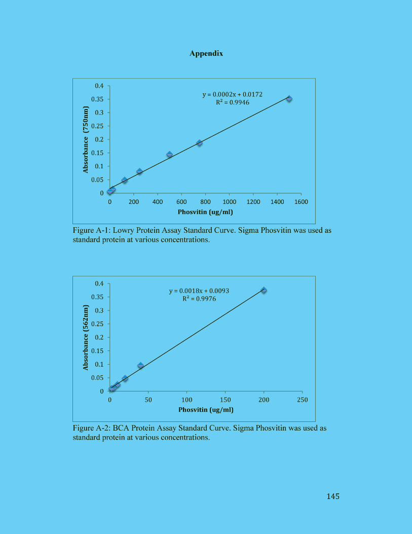
Figure A-1: Lowry Protein Assay Standard Curve. Sigma Phosvitin was used as
standard protein at various concentrations. ................................................. 145
Figure A-2: BCA Protein Assay Standard Curve. Sigma Phosvitin was used as
standard protein at various concentrations. ................................................. 145
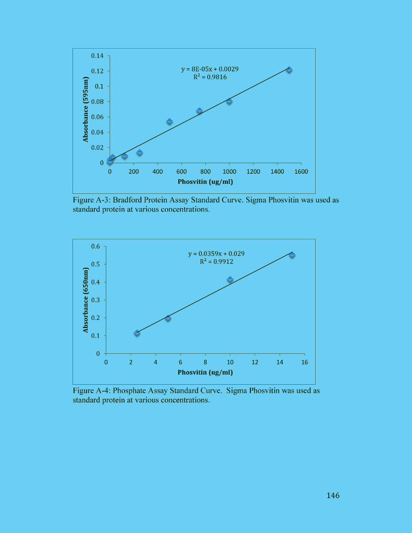
Figure A-3: Bradford Protein Assay Standard Curve. Sigma Phosvitin was used as
standard protein at various concentrations. ................................................. 146
xiv
Figure A-4: Phosphate Assay Standard Curve. Sigma Phosvitin was used as
standard protein at various concentrations. ................................................. 146
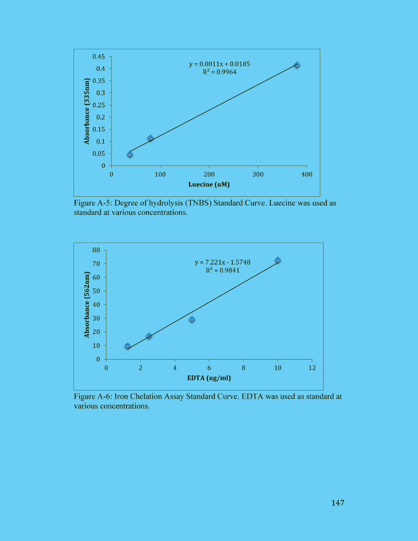
Figure A-5: Degree of hydrolysis (TNBS) Standard Curve. Luecine was used as
standard at various concentrations. ............................................................. 147
Figure A-6: Iron Chelation Assay Standard Curve. EDTA was used as standard at
various concentrations. ............................................................................... 147
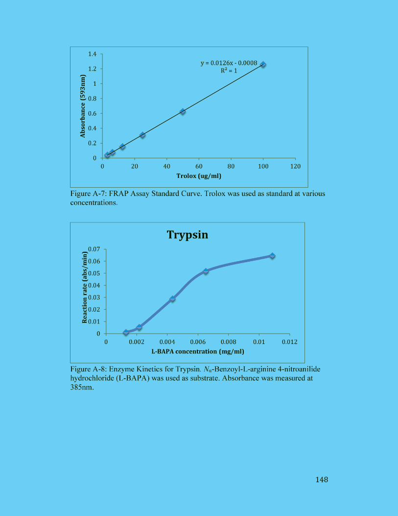
Figure A-7: FRAP Assay Standard Curve. Trolox was used as standard at various
concentrations. ............................................................................................ 148
Figure A-8: Enzyme Kinetics for Trypsin. Nα-Benzoyl-L-arginine 4-nitroanilide
hydrochloride (L-BAPA) was used as substrate. Absorbance was measured
at 385nm...................................................................................................... 148
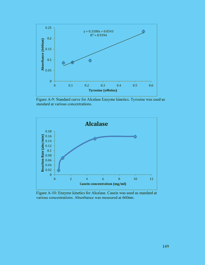
Figure A-9: Standard curve for Alcalase Enzyme kinetics. Tyrosine was used as
standard at various concentrations. ............................................................. 149
Figure A-10: Enzyme kinetics for Alcalase. Casein was used as standard at
various concentrations. Absorbance was measured at 660nm. ................... 149
xv
List of Tables
Table 1.1 Mineral content of edible egg portion and their approximate proportion
in egg white and yolk ...................................................................................... 4
Table 1.2 Vitamin content of egg edible portion and their approximate proportion
in egg white and yolk ...................................................................................... 5
Table 1.3 Proteins in egg albumen .......................................................................... 8
Table 1.4 Proteins in egg yolk .............................................................................. 22
Table 1.5 Comparison of chicken IgY and mammalian IgG ................................ 27
Table 1.6 Functional properties of Phosvitin and Phosvitin Hydrolysates(PVH).53
Table 2.1 Concentration of total IgY and specific IgY purified from the
hyperimmunized and non-immunized egg yolks. Values are the mean of
quadruple samples ± SD. .............................................................................. 69
Table 2.2 Recovery (%) of PV from saline solution sample spiked with 1 – 100
µg/ mL Sigma PV ......................................................................................... 71
Table 2.3 Intra- and inter-assay variances (%CV) of biotinylated DAS ELISA for
PV quantification in 5 different saline solutions........................................... 72
Table 3.1: Enzymes applied in HHP-EH and atmospheric hydrolysis and
operational conditions .................................................................................... 80
Table 4.1 The concentrations of protein, total IgY and specific IgY in WSF
powder containing Enterotoxigenic Escherichia coli (ETEC) K88 and K99-
specific and non-specific IgY. Values are the mean standard deviation. 111
xvi
Table 4.2 Degree of hydrolysis of PVH produced under high pressure and
atmospheric pressure conditions over a period of 12 hr. Values with different
lowercase letters in similar hydrolysis condition differ significantly (p <
0.05). Statistically significant differences between HHP-EH and AP-EH for
each enzyme are indicated by stars (*, p < 0.05). ....................................... 113
xvii
List of Abbreviation
Alc, Alcalase
AP, Atmospheric pressure
Arg, Arginine
Asn, Asparagine
Asp, Aspartic acid
BSA, Bovine serum albumin
CFU, Colony-forming units
DH, Degree of hydrolysis
DHA, Docosahexaenoic acids
DNA, Deoxyribonucleic acid
DPPH, 2,2-diphenyl-1-
picrylhydrazyl
EDTA, Ethylenediaminetetraacetic
acid
Ela, Elastase
ETEC, Escherichia coli
FRAP, Ferric (Fe ) reducing ability
of plasma
GHS, Glutathione
GIT, Gastrointestinal tract
Glu, Glutamine
Gly, Glycine
H, Heavy chains
HDL, High density lipoprotein
HHP-EH, High hydrostatic pressure-
enzymatic hydrolysis
HIC, Hydrophobic interaction
chromatography
His, Histidine
HP, Horseradish peroxidase
HPLC, High performance liquid
chromatography
HRPO, Horseradish peroxidase
IgA, Immunoglobulin A
IEC, Ion exchange chromatography
IgG, Immunoglobulin G
IgM, Immunoglobulin M
IMAP, Immobilized metal affinity
electrophoresis
IgY, Immunoglobulin Y
IL-8, Interleukin 8
L, Light chains
LDL, Low density lipoprotein
LOD, The limit of detection
xviii
LPC, Lysophosphatidylcholine
LPS, Lipopolysaccharides
LPE, Lysophosphatidylethanolamine
Lue, Leucine
Lys, Lysine
MALDI-TOF, Matrix-assisted laser
desorption/ionization
MUFA, Monounsaturated fatty acids
Mw, Molecular weights
NHS-PEO4-Biotin,
Biotinaimdohexanoic acid-3-sulfo-
N-hydroxysuccinimide ester
PBS, Phosphate buffered saline
PE, Phosphatidylethanolamine
PEG, Polyethylene glycol
PC, Phohatidylcholine
PL, Phospholipids
PLA2, Phospholipase A2
PMSF, Phenylmethylsulfonyl
fluoride
ROS, Reactive oxygen species
PUFA, Polyunsaturated fatty acids
PV, Phosvitin
PVH, Phosvitin Hydrolysates
SAFA, Saturated fatty acids
SCWL, Single Comb White Leghorn
SDS, Sodium dodecylsulfate
SE-HPLC, Size exclusion high
performance chromatography
Ser-P, Phosphoryl serine blocks
Ther, Thermolysin
TMB, Tetramethylbenzidine
TNBSA, 2,4,6-trinitrobenzene
sulfonic acid
TPTZ, 2,4,6-tripyridyl-s-triazine
Try, Trypsin
WSF, Water-soluble fractions
1
Chapter 1: Literature review
1. 1 Introduction of Egg
1.2 Structure of Egg
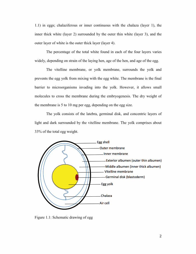
The egg is composed of five distinct structures such as egg shell, egg shell
membrane, egg white (or albumen), vitelline membrane, and egg yolk. The parts
of the egg are shown schematically in Figure 1.1.
The eggshell is formed in a matrix component of calcium and proteins
involving in the eggshell calcification to form egg shapes. The main biological
function of the eggshell as a first line of defense is to prevent bacterial penetration
and to protect the embryo from external aggression during its development. The
chicken eggshell is a natural porous matrix formed in the hen’s oviduct over a
predetermined period (Hamilton 1986; Nys et al., 2004; Tullet 1987). The egg
shell has a distinct pattern of pores to allow gas exchange of carbon dioxide.
However, the pores may permit bacterial penetration as far as the shell
membranes.
The next layers of the eggshell membrane are composed of the inner and
outer shell membranes. The relatively thin keratin-like membranes play a role in
the second line of defenses against possible bacterial invasion. The inner
membrane is thinner than the outer membrane, but together they are only 0.01-
0.02 mm thick.
The egg white, or albumen, represents approximately 60% of total egg
weight. The main function of egg white plays a role in the third line of the defense
during the development of embryo. The egg white consists of 4 layers (see Figure
2
1.1) in eggs; chalaziferous or inner continuous with the chalaza (layer 1), the
inner thick white (layer 2) surrounded by the outer thin white (layer 3), and the
outer layer of white is the outer thick layer (layer 4).
The percentage of the total white found in each of the four layers varies
widely, depending on strain of the laying hen, age of the hen, and age of the egg.
The vitelline membrane, or yolk membrane, surrounds the yolk and
prevents the egg yolk from mixing with the egg white. The membrane is the final
barrier to microorganisms invading into the yolk. However, it allows small
molecules to cross the membrane during the embryogenesis. The dry weight of
the membrane is 5 to 10 mg per egg, depending on the egg size.
The yolk consists of the latebra, germinal disk, and concentric layers of
light and dark surrounded by the vitelline membrane. The yolk comprises about
33% of the total egg weight.
Figure 1.1: Schematic drawing of egg
3
1.3 Composition of Egg
The nutritional value of whole egg protein is considered to be 100. That is
used as standard for measuring nutritional quality of other food proteins. The egg
white contains more than 40 different kinds of proteins that make up 11% of its
entire composition of egg. Due to their functional and pharmacological properties,
egg proteins are desirable ingredients in the pharmaceutical industry.
The major egg white proteins include ovalbumin, ovotransferrin,
ovomucoid, ovomucin and lysozyme. They account for > 83% of total egg white
proteins. Other minor proteins include ovoinhibitors, ovomacroglobulin, cystain,
avidin and ovoglycoprotein at low concentration which accounts for < 17% of
total egg white proteins.
The egg proteins distributed in the yolk exists as lipoproteins, of which
there are low density and high density. The low density lipoprotein (LDL) is the
major protein, accounting for up to 65% of the total yolk proteins. The high
density lipoprotein (HDL) includes a phosphoprotein known as phosvitin. About
80% of phosphorus in eggs is contained in phosvitin, which is derived from
vitellogenin formed in the liver (Sugino et al., 1997). Other yolk proteins include
a water-soluble livetin, non-lipid glycoprotein, and riboflavin-binding protein.
The fat in the egg is exclusively in the yolk, and comprises 5.5 to 6 g in an
average 60 g egg. Almost all lipids are present in lipoprotein complexes within
the yolk. Trace levels of lipids have been observed in the egg white. The lipids in
eggs have attracted attention both at scientific and consumer level due to the link
between high dietary fat consumption and coronary heart diseases.
4
Yolk lipids are classified into triglycerol, phospholipid, and free
cholesterol. Triglycerol and phospholipids are the major components of yolk
lipids, comprising up to 65% and 32%, respectively. Fatty acids are also found in
egg yolk with saturated fatty acids (SAFA), monounsaturated fatty acids
(MUFA), and polyunsaturated fatty acids (PUFA).
Minerals are contained within the egg yolk. The egg yolk contains 1%
mineral that contains with phosphorous as the most abundant mineral component.
More than 60% of the total phosphorous in egg yolk is found in phospholipids.
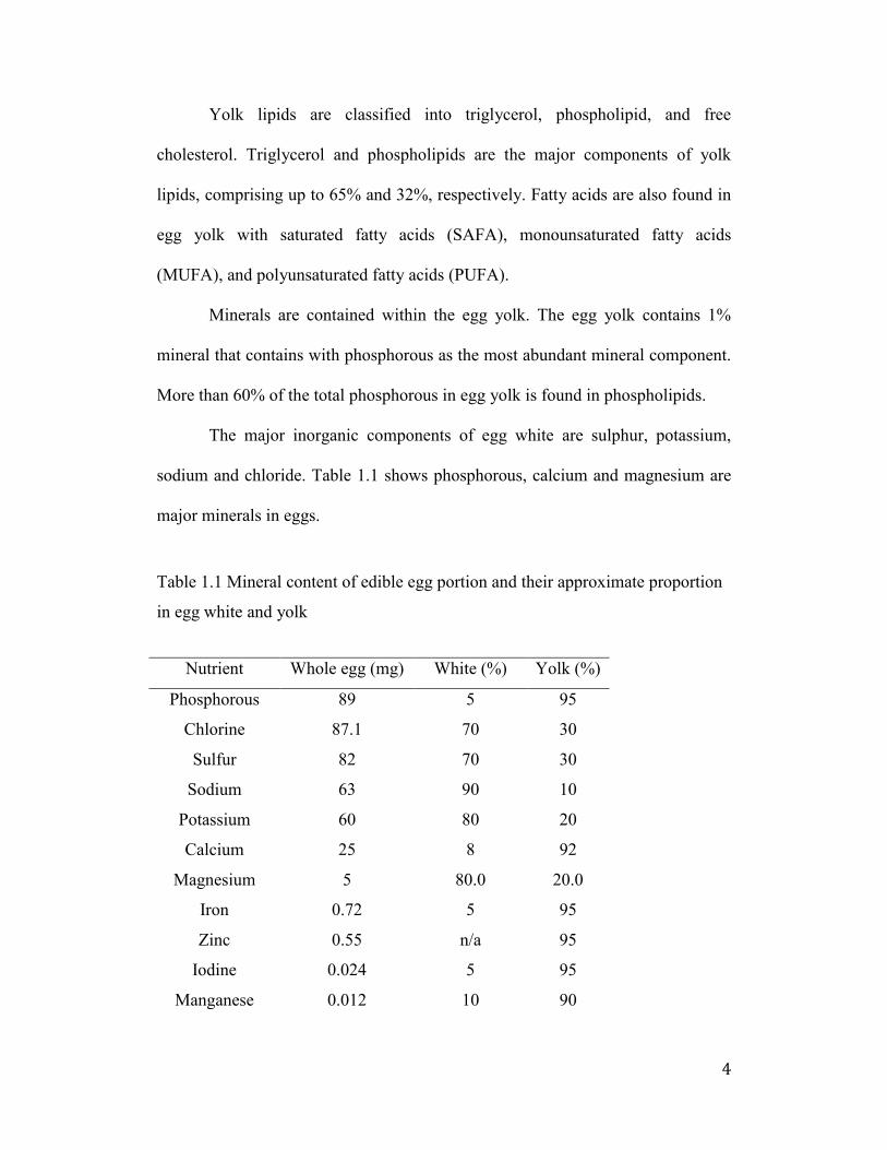
The major inorganic components of egg white are sulphur, potassium,
sodium and chloride. Table 1.1 shows phosphorous, calcium and magnesium are
major minerals in eggs.
Table 1.1 Mineral content of edible egg portion and their approximate proportion
in egg white and yolk
Nutrient Whole egg (mg) White (%) Yolk (%)
Phosphorous 89 5 95
Chlorine 87.1 70 30
Sulfur 82 70 30
Sodium 63 90 10
Potassium 60 80 20
Calcium 25 8 92
Magnesium 5 80.0 20.0
Iron 0.72 5 95
Zinc 0.55 n/a 95
Iodine 0.024 5 95
Manganese 0.012 10 90
5
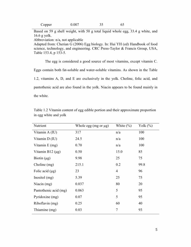
Copper 0.007 35 65
Based on 59 g shell weight, with 50 g total liquid whole egg, 33.4 g white, and
16.6 g yolk.
Abbreviation: n/a, not applicable
Adapted from: Cherian G (2006) Egg biology. In: Hui YH (ed) Handbook of food
science, technology, and engineering. CRC Press-Taylor & Francis Group, USA,
Table 153.4, p 153-5.
The egg is considered a good source of most vitamins, except vitamin C.
Eggs contain both fat-soluble and water-soluble vitamins. As shown in the Table
1.2, vitamins A, D, and E are exclusively in the yolk. Choline, folic acid, and
pantothenic acid are also found in the yolk. Niacin appears to be found mainly in
the white.
Table 1.2 Vitamin content of egg edible portion and their approximate proportion
in egg white and yolk
Nutrient Whole egg (mg or μg) White (%) Yolk (%)
Vitamin A (IU) 317 n/a 100
Vitamin D (IU) 24.5 n/a 100
Vitamin E (mg) 0.70 n/a 100
Vitamin B1β (μg) 0.50 15.0 85
Biotin (μg) 9.98 25 75
Choline (mg) 215.1 0.2 99.8
Folic acid (μg) 23 4 96
Inositol (mg) 5.39 25 75
Niacin (mg) 0.037 80 20
Pantothenic acid (mg) 0.063 5 95
Pyridoxine (mg) 0.07 5 95
Riboflavin (mg) 0.25 60 40
Thiamine (mg) 0.03 7 93
6
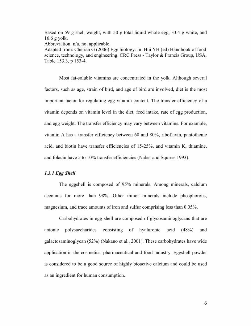
Based on 59 g shell weight, with 50 g total liquid whole egg, 33.4 g white, and
16.6 g yolk.
Abbreviation: n/a, not applicable.
Adapted from: Cherian G (2006) Egg biology. In: Hui YH (ed) Handbook of food
science, technology, and engineering. CRC Press - Taylor & Francis Group, USA,
Table 153.3, p 153-4.
Most fat-soluble vitamins are concentrated in the yolk. Although several
factors, such as age, strain of bird, and age of bird are involved, diet is the most
important factor for regulating egg vitamin content. The transfer efficiency of a
vitamin depends on vitamin level in the diet, feed intake, rate of egg production,
and egg weight. The transfer efficiency may vary between vitamins. For example,
vitamin A has a transfer efficiency between 60 and 80%, riboflavin, pantothenic
acid, and biotin have transfer efficiencies of 15-25%, and vitamin K, thiamine,
and folacin have 5 to 10% transfer efficiencies (Naber and Squires 1993).
1.3.1 Egg Shell
The eggshell is composed of 95% minerals. Among minerals, calcium
accounts for more than 98%. Other minor minerals include phosphorous,
magnesium, and trace amounts of iron and sulfur comprising less than 0.05%.
Carbohydrates in egg shell are composed of glycosaminoglycans that are
anionic polysaccharides consisting of hyaluronic acid (48%) and
galactosaminoglycan (52%) (Nakano et al., 2001). These carbohydrates have wide
application in the cosmetics, pharmaceutical and food industry. Eggshell powder
is considered to be a good source of highly bioactive calcium and could be used
as an ingredient for human consumption.
7
The matrix protein identifies desmosine and isodesmosine, similar of
elastin-like proteins (Leach 1982). Immunohistochemistry using antibodies to
collagen types I, V, and X showed that the matrix may contain collagens. The
proteins also contain hydroxylysine (Wong et al., 1984; Arias et al., 1997; Wang
et al., 2002). However, the bulk of the amino acid composition differs from
collagen and suggests that collagen is not predominant but that a unique protein
containing lysine-derived cross links may be present (Leach 1982).
The outer shell largely consists of calcium carbonate (94%), with other
components including magnesium carbonate (1%), calcium phosphate (1%), and
organic matter that are mostly protein (4%).
The shell color of colored eggs is due to pigments (ooporphins) deposited
on the shell surface. The shell is formed in a distinct pattern with pores for gas
exchange. Even though the pores are partially sealed by keratin, they allow carbon
dioxide and moisture to escape from the egg. Under some conditions, the pores
also permit bacterial penetration as far as the shell membranes.
1.3.2 Egg Shell Membrane
The egg shell membrane is composed of collagen-like proteins (collagen
type I and V), in a ratio of 100 of type I to 1 of type V. Coarse fibers (β.5 μm in
diameter) contain more type I collagen, while type V collagen predominates in the
fine fibres (0.6 μm in diameter), and is largely located in the inner membrane.
Other components identified in eggshell membranes are glycosaminoglycans,
such as dermatan sulfate and chondroitin sulfate (Baker and Balch 1962),
hyaluronic acid (Long et al., 2005), sialic acid (Nakano et al., 2003), desmosine
8
and isodesmosine (Starcher and King 1980), ovotransferrin (Gautron et al., 2001),
lysyl oxidase (Akagawa et al., 1999), lysozyme (Hincke et al., 2000) and –N
acetylglucosaminidase (Ahlborn et al., 2006).
The egg shell membrane contains several bacteriolytic enzymes, such as
lysozyme and N-acetyl glucosaminidase and other membrane proteins that have
been through to have beneficial effects in treating injuries. The peptides derived
from the membrane were shown to stimulate skin fibroblasts in vitro (Suguro et
al., 2000). The egg shell membrane proteins are currently utilized as a cosmetic
ingredient for their emollient properties.
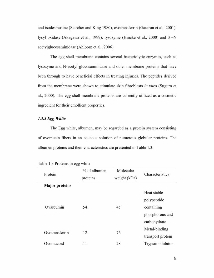
1.3.3 Egg White
The Egg white, albumen, may be regarded as a protein system consisting
of ovomucin fibers in an aqueous solution of numerous globular proteins. The
albumen proteins and their characteristics are presented in Table 1.3.
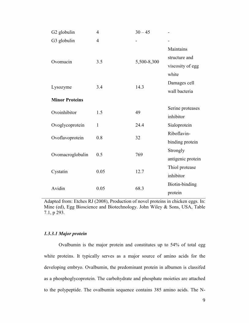
Table 1.3 Proteins in egg white
Protein % of albumen
proteins
Molecular
weight (kDa) Characteristics
Major proteins
Ovalbumin 54 45
Heat stable
polypeptide
containing
phosphorous and
carbohydrate
Ovotransferrin 12 76 Metal-binding
transport protein
Ovomucoid 11 28 Trypsin inhibitor
9
G2 globulin 4 30 – 45 -
G3 globulin 4 - -
Ovomucin 3.5 5,500-8,300
Maintains
structure and
viscosity of egg
white
Lysozyme 3.4 14.3 Damages cell
wall bacteria
Minor Proteins
Ovoinhibitor 1.5 49 Serine proteases
inhibitor
Ovoglycoprotein 1 24.4 Sialoprotein
Ovoflavoprotein 0.8 32 Riboflavin-
binding protein
Ovomacroglobulin 0.5 769 Strongly
antigenic protein
Cystatin 0.05 12.7 Thiol protease
inhibitor
Avidin 0.05 68.3 Biotin-binding
protein
Adapted from: Etches RJ (2008), Production of novel proteins in chicken eggs. In:
Mine (ed), Egg Bioscience and Biotechnology. John Wiley & Sons, USA, Table
7.1, p 293.
1.3.3.1 Major protein
Ovalbumin is the major protein and constitutes up to 54% of total egg
white proteins. It typically serves as a major source of amino acids for the
developing embryo. Ovalbumin, the predominant protein in albumen is classifed
as a phosphoglycoprotein. The carbohydrate and phosphate moieties are attached
to the polypeptide. The ovalbumin sequence contains 385 amino acids. The N-
10
terminal amino acid is glycine, and the C-terminal amino acid is proline. The
molecular weight of the polypeptide is 43.6 kDa. Ovalbumin contains two
phosphate residues on serines 68 and 344 (Kinoshita and Koike 2012).
Purified ovalbumin is made up of three components. These are A1, A2 and
A3, all of which differ in phosphorous content. Ovalbumin A1, A2, and A3
containing two, one and no phosphate groups per molecule, respectively, are
present in albumen fraction in relative portions of about 85:12:3. The molecule
contains a carbohydrate chain attached at asparagine 292. Ovalbumin is the only
albumen protein to contain free sulfhydryl groups. Each ovalbumin molecule
contains four sulfhydryl groups, three of which are reactive to p-
chloromercuribenzoate in native protein and the fourth in the denatured protein.
Ovotransferrin is a monomeric glycoprotein consisting of a single
polypeptide chain of 686 amino acids. The molecular weight of ovotransferrin is
about 78 kDa; this constitutes 13% of total proteins in egg white. This protein
consists of two lobes, each containing a specific binding site for iron, although
ovotransferrin does not contain iron in the egg. Copper, zinc or aluminum may
also bind to this site. Inhibition of gram-negative bacteria occurs by depriving the
iron source which is essential for their growth and survival (Lock and Board
1992). The antimicrobial activity can result from a direct effect on the
membranes: interaction of the cationic ovotransferrin with the anionic outer
membrane of gram-negative bacteria (Valenti et al., 1986).
Ovomucin is a macromolecule and heavily glycosylated glycoprotein,
consisting of peptide-rich α-subunit and a carbohydrate–rich -subunit. Ovomucin
11
is a major egg white glycoprotein (3.5%) with a molecular mass of approximately
254 kDa. It contains O-linked carbohydrate moieties that, upon formation of
extensive hydrogen bonds with water, can give rise to a characteristic gel-like
structure. Ovomucin serves physical functions to maintain the structure and
viscosity of egg white albumen, thus serving to prevent the spread of
microorganisms (Ibrahim et al., 1994), and to characterize foaming and
emulsifying properties.
Ovomucoid is a highly glycosylated protein (20 to 25% carbohydrates,
w/w) of 28 kDa, 11% of egg white proteins. Ovomucoid consists of three domains
of the amino acid sequences of 1-68, 69-130 and 131-186. Each domain is cross-
linked by three disulfide bridges (Kato et al., 1987). The carbohydrate moiety
consists of three oligosaccharide units bound to the protein through asparagine
residues (Montgomery and Wu 1963). The polypeptide chain is composed of 26%
α-helix, 46% -structure, 10% -turns and 18% random coil (Watanabe et al.,
1981).
Lysozymes can be found in not only egg white but also the shell and the
vitelline membrane. It belongs to a class of enzyme that lyses the cell walls of
gram-positive bacteria. The lysozyme also known as muramidase or N-
acetylmuramic hydrolase is a relatively small secretory enzyme that catalyzes the
hydrolysis of beta 1,4 bonds between N-acetylmuramic acid and N-
acetylglycosamine in cell walls of bacteria.
Lysozyme is a polypeptide of 129 amino acid residues having a molecular
weight of 14.3 kDa. With the isoelectric point (pI) of 10-11, it is a strongly basic
12
protein in egg white, well known for bacteriostatic, bacteriolytic and bacteriocidal
activity, particularly against gram-positive bacteria. The protein represents only
3.4% of total egg proteins. It is a good example of naturally occurring enzymes
used in the food industry as a preservative to maintain product quality and reduce
the incidence of spoilage.
The molecular weight of penalbumin is 61.6 kDa which is larger than
ovalbumin (47.1 kDa). This protein has several features related to ovalbumin.
Regarding the composition of penalbumin, it has more carbohydrate and lacks
phosphate. The amino acid compositions are significantly different, but the
differences could be explained if penalbumin is an extended form of ovalbumin.
The composition of ovoglobulin contains 13.6% of hexose, 13.8% of
hexosamine and 3% of sialic acid. Hexose occurs as mannose and galactose in the
ratio of 2:1, hexosamine as glucosamine and sialic acid as N-acetylneuraminic
acid. It has a minimum molecular weight, calculated from the tryptophan content,
of 24.4 kDa. The term ovoglobulin refers to ovoglobulin G2 and G3, each
constituting about 4% of egg albumen proteins. Ovoglobulin G2 and G3 are
similar in many properties, including their molecular weight (49 kDa).
Ovoglobulins are also of interest for commercial applications of albumen
because they denature rapidly and may, therefore, have more effect on the initial
foaming of the albumen than the more plentiful albumen proteins.
1.3.3.2 Minor proteins of albumen
Ovoinhibitor is a glycoprotein in egg white, composed of 447 amino acids
with a molecular weight of 48 kDa. Like the ovomucoid, this protein is a
13
proteinase inhibitor. It inhibits the activities of trypsin, chymotrypsin and some
proteinases of microbial origin.
The ovomacroglobulin is the largest globular protein in eggs white. This
protein, also known as ovostatin, is composed of four subunits, each having a
molecular weight of 175 kDa, with pairs of the subunits joined by disulfide bonds.
Ovomacroglobulin inhibits hemagglutination, possesses anti-collagenase activity,
and has inhibitory activity against diverse proteolytic enzymes including serine
proteases, cysteine proteases, thiol proteases, and metalloproteases (Molla et al.,
1987; Li-Chan and Nakai 1989).
As a member of a “superfamily” of cystatins, egg white cystatin belongs
to the Type 2 cystatin, which has about 115 amino acids and two disulfide bonds,
but no carbohydrates. Secreted cystatin has a theoretical molecular weight of 13.2
kDa. Egg white cystatin has been shown to possess anti-bacterial activity,
preventing the growth of group A streptococcus (Bjork et al., 1989), Salmonella
typhimurium (Nakai et al., 2008), and the periodontitis-causing Porphyromonas
gingivitis (Travis et al., 1997).
Chicken egg white riboflavin-binding proteins are the prototype of a
family that includes other riboflavin and folate binding proteins. Ovoflavoprotein
binds riboflavin at pH above 4.3 with an association constant of 7.9 × 10⁸ M. It is
composed of 219 amino acids (Hamazume et al., 1984) with a molecular weight
of 32 kDa. The carbohydrate content is about 15%, consisting of mannose,
galactose, glucosamine and sialic acid. Ovoflavoprotein also referred to as
flavoprotein or riboflavin-binding protein, is a phosphor-glycoprotein that is
14
responsible for binding most of the riboflavin (vitamin B2) in egg white.
Flavoprotein is considered to have the highest selenium content (1800 ng/g)
among egg white proteins.
The avidin constitutes a maximum of 0.05% of the total protein content of
egg white. Avidin is an alkaline (pI 10.5), highly stable, tetrameric glycoprotein
that is best known for its biotin-binding properties. Each of the four monomers
binds one molecule of biotin and the avidin-biotin interaction, with dissociation
constant of 10-15 M, is the strongest non-covalent interaction reported between
protein and ligand. Each avidin chain, composed of 128 amino acid residues, is
arranged in an eight-stranded anti-parallel -barrel, whose inner region defines the
D-biotin binding site. A fairly rigid binding site is readily accessible in the
apoprotein structure, making it sterically complementary to the shape and polarity
of biotin.
Both chicken egg white avidin and its bacterial relative streptavidin are
widely used as tools in some affinity-based separations, diagnostic assays, and a
variety of other applications. Other applications include the potential of avidin as
an insecticide and antimicrobial agent. Due to its high proportion of tryptophan
residues, avidin is unstable under oxidizing conditions in strong light.
Thiamin-binding protein can be isolated from egg white by using affinity
chromatography. Regarding function, the protein binds thiamine (vitamin B1) in a
1:1 ratio and is similar with avidin in that it is a vitamin scavenger. In terms of
structure, the protein has a molecular weight of 38 kDa and does not contain
15
carbohydrate. It is not usual in that it forms a stoichiometric complex with the
albumen riboflavin-binding protein. An identical protein is present in egg yolk.
Vitamin B2-binding protein has a molecular weight 98 kDa. The vitamin-
binding ability of this protein was heat-labile in 2 h at 80 °C, but the complex was
stable for 6 h at this temperature. Due to this difference, this albumen protein is
distinguished from a B12-binding protein in egg yolk.
Ovoglycoprotein is a protein of lipocalin family present in egg white. It
represents about 1% of egg white protein. It is an acidic glycoprotein (pI 3.9) with
a theoretical molecular weight of 20.3 kDa and a sugar content of 30%. Despite
the information just previously given, very little is known about this protein.
Albumen contains minor enzymes. These enzymes include phosphatase,
catalase, and glycosidase, aminopeptidase (Sugino et al., 1997).
1.3.4 Vitelline membrane
The vitelline membrane is a protein matrix surrounding the yolk (Mann
2008). It is a thick structure of considerable strength separating the yolk form the
egg white and forms the last barrier to microbial infection. Subsequently, it was
found that it consists of three distinct layers (Kido et al., 1995). A thin continuous
layer sandwiched between two fibrous layers, the inner layer in front of the
oocyte, and the outer layer facing the albumen. The components of the inner
layer are glycoproteins, GP-I, GP-II, GP-III, while the outer layer contains
ovomucin, lysozyme, VMO-I, and VMO-II (Kido et al., 1995).
The inner layer, which is the avian parallel to the mammalian zona
pellucida, is secreted by the granulosa cells surrounding the oocyte in the follicle
16
or in the liver from where it is transported to the follicle via blood circulation.
(Kerver et al., 2002) The inner layer forms a diffusion barrier between egg white
and egg yolk and so affects the properties of the shape and development of the
embryo. The main proteins of the inner layer are the glycoproteins GPI (43 kDa),
GPII (110 kDa) and GPIII (300 kDa). Some research showed that inner layer
contains ZP proteins ZPC/ZP3, ZP1 (ZPB1), and ZPD (Zhang et al., 2011)
The outer layer possibly contains ovomucin, lysozyme C, and the vitelline
membrane outer proteins, VMO-I and VMO-II (Kerver et al., 2002). The VMO-I
was found to have 163 amino acids with molecular weight of 18 kDa (Kido et al.,
1995). VMO-I is a simple protein and some research showed that it inhibits the
hemagglutinating activity elicited by wheat germ agglutinin and has a glycan
activity similar to the transferase activity of lysozyme (Mann 2008). On the other
hand, VMO-II is small protein with 53 amino acids and a molecular mass of 6
kDa (Xiao et al., 2004).
1.3.5 Egg Yolk
The egg yolk is composed of about 17% proteins, 35% lipids, and 1%
carbohydrates. The rest of the constituents include cholesterol, vitamins, minerals
and water. Egg yolk is constituted of an aqueous phase (called plasma) and an
insoluble denser phase (granules).
The granules represent about 19-23% of yolk dry matter, accounting for
about 50% of yolk proteins and 7% of yolk lipids (Anton and Gandemer 1997;
Dyer-Hurdon and Nnanna 1993). The granules are mainly composed of high-
density lipoproteins (HDL) (70%), phosvitin (16%) and 12% Low density
17
lipoproteins (LDL) (Burley and Cook 1961; Saari et al., 1964). High-density
lipoprotein (HDL) consists of α- and -lipovitellins, which differ in amino acid
composition and bound phosphorous and carbohydrates. The proportion of α- and
-lipovitellins in HDL appears to be similar. The LDL is a structural constituent
of the granules. HDL-phosvitin complex is the basic unit of granules linked by
phosphocalcic bridges between the phosphate groups of their phosphoseryl
residues (Causeret et al., 1991).
The VLDL, yolk lipoprotein, consists of apoVLDL II and apolipoprotein-
B (Burley et al., 1984). ApoVLDL II is the only apoprotein from blood
lipoproteins to be transferred to the yolk without any modification and is called
apovitellenin I (Dugaiczyk et al., 1981). The source of yolk LDLs is VLDL.
During the transfer from blood to the yolk, apolipoprotein-B is cleaved into
several fragments, referred to as apovitellenin I-VI (Burley et al., 1993).
Vitellogenin consists of three species designated as vitellogenin I, II, and III
(Wang et al., 1983; Wang and Williams 1980). Vitellogenin is cleaved into the
yolk granule proteins lipovitellin I and II and the phosphoprotein phosvitin.
Plasma forms the aqueous phase in which yolk particles are in suspension.
It contains a large quantity of lipoproteins (LDL). Plasma corresponds to about
78% of yolk dry matter and is composed of 85% LDL and15% livetins (Burley
and Cook 1961). It accounts for about 90% of yolk lipids (including nearly all the
carotenoids) and 50% of yolk proteins. Yolk proteins consist of lipoproteins
(30%) and soluble proteins (8%). The protein contents of lipovitellins are about
80%; the lipid contents are about 20%. The lipids include phospholipids (60% of
18
the lipid, primarily lecithin), triacylglycerols, and small amounts of cholesterol,
sphingomyelin, and other lipids. The lipovitellins include glycoconjugates with
mannose, galactose, glucosamine, and sialic acid. However, α-lipovitellin has a
much higher sialic acid content than does -lipovitellin, which explains α-
lipovitellin’s relatively acidic nature (Seko et al., 1997).
Livetin is a water-soluble protein that accounts for 30% of plasma proteins
and is composed of α-livetin (serum albumin), -livetin (αβ-glycoprotein), and -
livetin [ -globulin immunoglobulin Y (IgY)] (Sugino et al., 1997). The mean
molecular weights of α-, -, and -livetins are reported to be 80 kDa, 45 kDa, and
150 kDa, respectively. The relative proportion of the three livetins is 2:5:3,
respectively. Chicken serum albumin (α-livetin) has been implicated as the
causative allergen of the bird-egg syndrome. -livetin has been identified as a 45
kDa αβ-glycoprotein. Chemically, its composition includes 14.3% nitrogen and
7% hexose.
1.3.5.1 Lipids of the Egg Yolk
Lipids are the main components (32 to 36%) of the egg yolk solids. The
composition of yolk lipid is generally about 65% triglyceride, 28 to 30%
phospholipids, and 4 to 5% cholesterol. However, the composition of yolk lipids
can be affected by various factors including hen age, genotype, and changes in the
diet of the hens.
1.3.5.1.1 Fatty acids
The predominant saturated fatty acids in eggs are palmitic (C16:0) and
stearic (C18:0). The content of these two fatty acids in chicken eggs may range
19
from 22 to 26% and 8 to 10%, respectively. In addition to these two fatty acids,
there are also other minor amounts of C14 and C20. The total saturated fatty acids
may constitute up to 30 to 35% of total fatty acids in egg yolks.
Monounsaturated fatty acids (MUFA) in eggs are C16:1 and C18:1, which
constitutes 42-46% of total fatty acids. Oleic acid (C18:1) is the major
monounsaturated fatty acid in chicken eggs. The contents of long-chain (20 and
22) omega-6 and omega-3 polyunsaturated fatty acids (PUFA) were 20% and
25%, respectively, in egg yolk.
There are two families of PUFA in egg, namely n-6 and n-3 fatty acids.
The predominant n-6 PUFA in egg lipids is C18:2n-6 (linoleic acid). Other n-6
fatty acids in eggs may include C20:4n-6, C22:4n-6, and C22:5n-6. The content
of n-3 fatty acids in eggs is made of α-linoleic acids (18:3n-3) and
docosahexaenoic acids (DHA, 22:6n-3). Among these, DHA is the major n-3 fatty
acid in the egg. The α-linoleic content in regular eggs is under 1% of the total
lipids; DHA may constitute between 1 and 3%. The content of n-3 PUFA is a
reflection of dietary fat. Addition of flax, fish oil, and marine algae in laying hen
diet leads to significant increases in α-linoleic acid and DHA in eggs (Cherian and
Sim 1991)
Products enriched with PUFA are prone to oxidation and the enrichment
with antioxidants is necessary in order to prevent the risk of oxidative damage.
Grune et al. (2001) suggested supplementation of feed with at least 80 IU vitamin
E/kg to prevent increase in cytotoxic aldehydic lipid peroxidation during
production and storage of omega-3 PUFA-enriched eggs. Dietary vitamin E
20
resulted in a decrease of PUFA, SFA, and total lipids in fresh yolk lipids, whereas
monounsaturated fatty acids (MUFA) did not change. Also, dietary vitamin E
supplement slowed down the process of oxidation of egg yolk fatty acid during
storage.
1.3.5.1.2 Phospholipids
The major components of egg yolk phospholipids (PL) are
phohatidylcholine (PC) and phosphatidylethanolamine (PE), which may make up
~81% and 12% of egg yolk lecithin; lysophosphatidylcholine (LPC),
lysophosphatidylethanolamine (LPE), and sphingomyelin are also minor
components of yolk PL. The major fatty acids in egg PC are palmitic, oleic,
stearic and linoleic acids, represented 32%, 26%, 16% and 13%, respectively;
arachidonic and docosahexanoic acids (4.8% and 4%, respectively) are also
present in significant amounts.
Kivini et al. (2004) studied the influence of oil-supplemented feeds
(containing 15% vegetable-based or fish oils) on the concentration of the
phospholipid content and their composition in hen eggs. Also, the total
phospholipid contents and proportions of PC, PE and sphingomyelin were similar
for all feeding groups. The supplemented feeds had a significant (p < 0.05) effect
on the fatty acid composition of phosphatidylcholines. Furthermore, supplements
decreased the proportion of saturated fatty acids in total fat, but not in the
phospholipids.
Many studies have been conducted on methods for extraction and
separation of phospholipids or lecithins from egg, as well as preparation of
21
lysolecithin by the enzymatic action of phospholipase A2 (PLA2), including
immobilized PLA2. In addition to providing sources of purified phospholipids for
basic research, these methods have been established to meet the demand to
produce purified egg lecithin for pharmaceuticals, nutraceutical, and food
applications. Examples of beneficial properties of yolk phospholipids are potential
industrial applications as nutraceuticals and functional food ingredients (Sugino et
al., 1997)
1.3.5.1.3 Fat-soluble Vitamins and Carotenoids
Most egg yolk vitamins, especially the fat-soluble vitamins, are contained
in the yolk. Hen egg is considered a source of most vitamins necessary for human
nutrition, except vitamin C. One egg may supply almost 12% vitamin A, more
than 6% of vitamin D, 9% riboflavin, and 8% pantothenic acid of the
recommended daily allowance in the United States. Only fish contains more
vitamin D than eggs.
The color of the yolk is an important factor for the consumer acceptability
of commercial eggs. The natural pigments in egg yolk are carotenoids that are
conjugated isoprene derivatives. Among carotenoids, lutein and zeaxanthin are
incorporated to a larger extent than -carotene and astaxanthin. A large proportion
of the yolk pigments is transported through the blood from the intestine by
lipoproteins, which are normally deposited in the yolk. The functions of pigments
are not known clearly, but the health of the chick after hatching may be improved
by these carotenoids.
22
1.3.5.2 Egg Yolk Protein
Yolk lipoprotein precursors such as very low-density lipoproteins (VLDL)
and vitellogenin are synthesized in laying hen’s liver and are transported in the
blood to the oocyte.
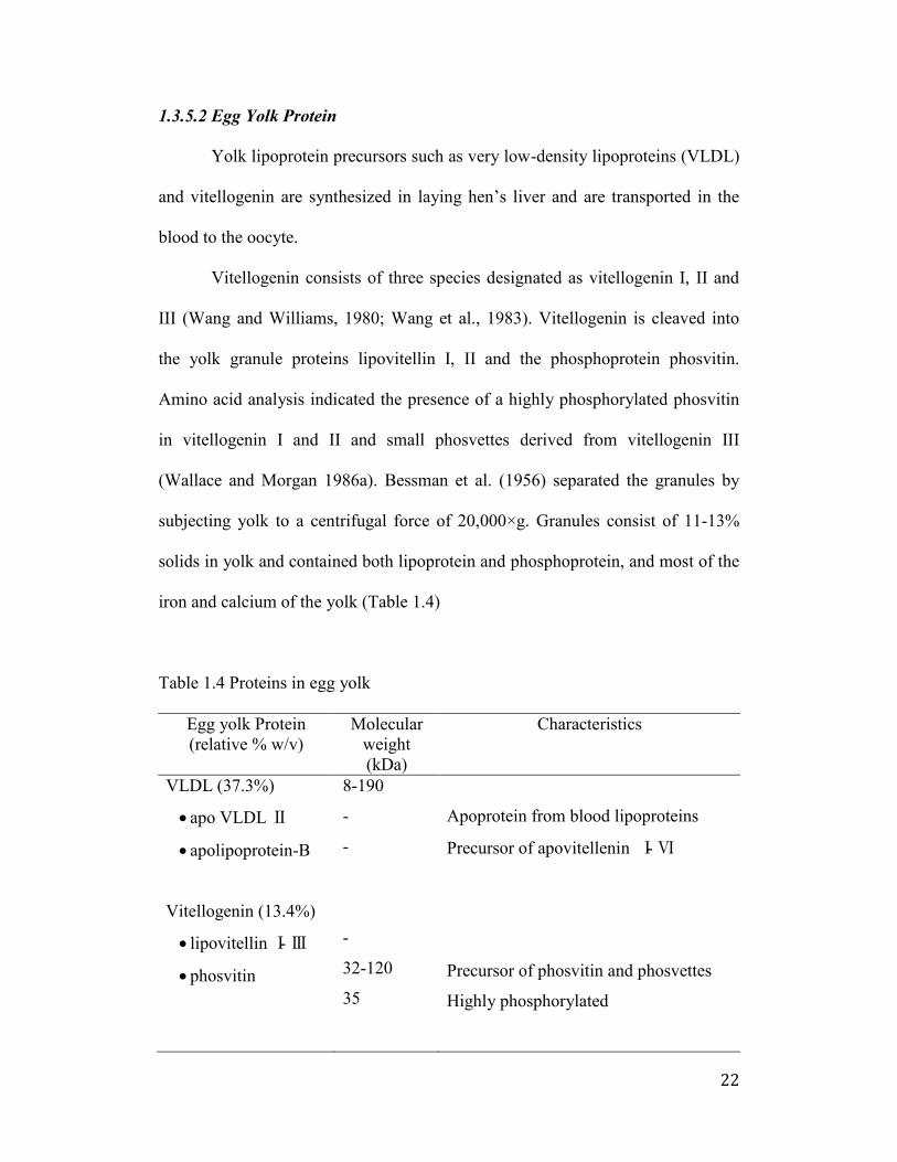
Vitellogenin consists of three species designated as vitellogenin I, II and
III (Wang and Williams, 1980; Wang et al., 1983). Vitellogenin is cleaved into
the yolk granule proteins lipovitellin I, II and the phosphoprotein phosvitin.
Amino acid analysis indicated the presence of a highly phosphorylated phosvitin
in vitellogenin I and II and small phosvettes derived from vitellogenin III
(Wallace and Morgan 1986a). Bessman et al. (1956) separated the granules by
subjecting yolk to a centrifugal force of 20,000×g. Granules consist of 11-13%
solids in yolk and contained both lipoprotein and phosphoprotein, and most of the
iron and calcium of the yolk (Table 1.4)
Table 1.4 Proteins in egg yolk
Egg yolk Protein
(relative % w/v)
Molecular
weight
(kDa)
Characteristics
VLDL (37.3%)
apo VLDL
apolipoprotein-B
Vitellogenin (13.4%)
lipovitellin -
phosvitin
8-190
-
-
-
32-120
35
Apoprotein from blood lipoproteins
Precursor of apovitellenin - Ⅵ
Precursor of phosvitin and phosvettes
Highly phosphorylated
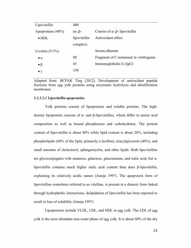
23
Lipovitellin
Apoproteins (40%)
HDL
Livetins (9.3%)
α
400
(α-, -
lipovitellin
complex)
80
45
150
Consist of α-, - lipovitellin
Antioxidant effect
Serum albumin
Fragment of C-termninal in vitellogenin
Immunoglobulin G (IgG)
Adapted from: BCPAK Ting (2012). Development of antioxidant peptide
fractions from egg yolk proteins using enzymatic hydrolysis and ultrafiltration
membranes
1.3.5.2.1 Lipovitellin apoproteins
Yolk proteins consist of lipoproteins and soluble proteins. The high-
density lipoprotein consists of α- and -lipovitellins, which differ in amino acid
composition as well as bound phosphorous and carbohydrates. The protein
content of lipovetellin is about 80% while lipid content is about 20%, including
phospholipids (60% of the lipid, primarily a lecithin), triacylglycerols (40%), and
small amounts of cholesterol, sphingomyelin, and other lipids. Both lipovitellins
are glycoconjugates with mannose, galactose, glucosamine, and sialic acid, but α-
lipovitellin contains much higher sialic acid content than does -lipovitellin,
explaining its relatively acidic nature (Juneja 1997). The apoprotein form of
lipovitellins sometimes referred to as vitelline, is present in a dimeric form linked
through hydrophobic interactions; delipidation of lipovitellin has been reported to
result in loss of solubility (Juneja 1997).
Lipoproteins include VLDL, LDL, and HDL in egg yolk. The LDL of egg
yolk is the most abundant non-water phase of egg yolk. It is about 60% of the dry
24
weight of egg yolk. The LDL can be separated by high-speed centrifuging, gel
chromatography and ion-exchange chromatography. Yolk low-density lipoprotein
from egg yolks contains about 12% of protein, the rest being neutral and
phospholipid.
The VLDL consists of apoVLDL II and apolipoprotein-B (Burley et al.,
1984). ApoVLDL II is the only apoprotein from blood lipoproteins to be
transferred to yolk without any modification and is called apovitellenin I
(Dugaiczyk et al., 1981).
Apovitellenin I is a small protein of low molecular weight that lacks
histidine with a small homodimer with disulfide-linked subunits of 9 kDa.
Apovitellenin II is also isolated from egg yolk low-density lipoprotein. The
protein’s molecular weight is β0 kDa with polysaccharide residues of
glucosamine, hexose and sialic acid. The functions of both apovitellenins are not
clearly understood, but their properties appear to be an essential part of the
lipoprotein structure. Apovitellenin III and IV with molecular weight more than
60 kDa are isolated from the total apoprotein mixture of egg yolk low-density
lipoprotein by gel and hydrophobic chromatography.
1.γ.5.β.β α, -Livetins
Livetin is a water-soluble protein that accounts for 30% of the plasma
proteins and is composed of α-livetin (serum albumin), -livetin (αβ-
glycoprotein), and -livetins [ -globulin immunoglobulin Y (IgY) (Sugino et al.,
1997). The mean molecular weights of α-, -, Υ-livetins are reported to be 80, 45
25
and 170 kDa, respectively. The proportion of the three livetins in yolks is 2:5:3,
respectively (Sugino et al., 1997).
Egg yolk α-livetin and chicken serum albumin are identical. It has a
molecular weight of 70 kDa and isoelectric point of 4.3 and 5.7. Chicken serum
albumin (α-livetins) has been implicated as the causative allergen of the bird egg
syndrome. Chicken serum albumin is partially heat labile inhalant. IgE reactivity
to chicken serum albumin was reduced by nearly 90% by heating for 30 minutes
at 90 ºC.
-livetin has been identified as a 45 kDa αβ-glycoprotein. Chemically, its
composition includes 14.3% nitrogen and 7% hexose. Unfortunately, not much
information is available about this protein and the existing data available is
ambiguous.
1.γ.5.β.γ -Livetin (IgY)
The γ -livetins in yolk are transported from the blood serum of hens. Of
the three immunoglobulins (IgM, IgA and IgG) found in the serum, the laying
hens transfer IgG to yolk at concentration of ~25 mg/ml, whereas IgM and IgA,
are transferred to egg white at concentrations of 0.15 and 0.7 mg/ml, respectively.
Morrison et al. (2001) identified several regions within the antibody molecule
important for its uptake into the egg yolk. Intact Fc and hinge regions, but not the
Fc-associated carbohydrate, are required for transport.(Morrison et al., 2001)
The basic structure of all immunoglobulin molecules is a unit consisting of
two identical light polypeptide chains and two identical heavy polypeptide chains.
These chains are linked together by disulfide bonds. Typically avian plasma
26
contains IgY plus IgA and IgM, which are evolutionary different from five
distinct classes of mammalian immunoglobulins: IgG, IgA, IgM, IgD, and IgE.
Although IgY antibody is functionally equivalent to mammalian IgG, they have
profound structural differences.
The Υ-globulins or γ -livetins in yolk are referred to as immunoglobulin Y
(IgY) to distinguish them from mammalian IgG. Although IgY is derived from
hen serum IgG, it differs in many chemical and structural features from
mammalian IgG (Kovacs-Nolan et al., 2005).
Both IgG and IgY contain Asn-linked oligosaccharides, although their
compositions differ. Like IgG, yolk IgY contains two heavy chains (H) and two
light chains (L), but the molecular weight of the IgY’s H chains is greater than
that of the mammalian IgG’s H chains, yielding an overall molecular weight of
180 kDa compared to 150-160 kDa for mammalian IgG. Furthermore, IgY H
chains lack a hinge region and composed of four constant regions and one
variable domain, whereas the IgG H chain contains a hinge region between the
first two of three constant domains, which lead to the flexibility of the Fab
fragments. The average molecular weights of IgY, heavy-chain, and Fab
fragments are 167, 65 and 45 kDa, respectively.
Peptic digestion degrades IgY into Fab fragments, in contrast to disulfide-
linked F(ab’)β fragments generated from IgG. IgY is relatively heat-stable even
after 30 minutes at 65 ºC. It remains stable over the pH 5–11 range, but the
antigen binding activity was rapidly lost at a pH of 2–3 or lower, probably
because of conformational changes (Table 1.5).
27
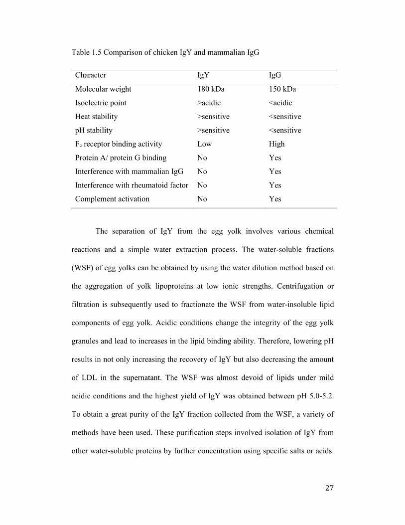
Table 1.5 Comparison of chicken IgY and mammalian IgG
Character IgY IgG
Molecular weight 180 kDa 150 kDa
Isoelectric point >acidic <acidic
Heat stability >sensitive <sensitive
pH stability >sensitive <sensitive
Fc receptor binding activity Low High
Protein A/ protein G binding No Yes
Interference with mammalian IgG No Yes
Interference with rheumatoid factor No Yes
Complement activation No Yes
The separation of IgY from the egg yolk involves various chemical
reactions and a simple water extraction process. The water-soluble fractions
(WSF) of egg yolks can be obtained by using the water dilution method based on
the aggregation of yolk lipoproteins at low ionic strengths. Centrifugation or
filtration is subsequently used to fractionate the WSF from water-insoluble lipid
components of egg yolk. Acidic conditions change the integrity of the egg yolk
granules and lead to increases in the lipid binding ability. Therefore, lowering pH
results in not only increasing the recovery of IgY but also decreasing the amount
of LDL in the supernatant. The WSF was almost devoid of lipids under mild
acidic conditions and the highest yield of IgY was obtained between pH 5.0-5.2.
To obtain a great purity of the IgY fraction collected from the WSF, a variety of
methods have been used. These purification steps involved isolation of IgY from
other water-soluble proteins by further concentration using specific salts or acids.
28
Further purification by gel permeation chromatography, ultracentrifugation, and
ultrafiltration, resulted in a 95% pure IgY diagnostic agent.
Microbial food-borne diseases are responsible for serious health problems
in humans and animals due to pathogens such as Escherichia coli O157:H7,
Salmonella spp., Listeria spp., Campylobacter spp., enteropathogenic E. coli,
viruses and parasites. IgY studies have demonstrated that specific IgY against E.
coli O157:H7 and Salmonella is able to inhibit the growth of pathogens,
eventually resulting in bacterial death. This research offers many advantages over
traditional antibiotics and provides the basis of a highly effective means of
producing inexpensive antibodies in egg yolks as functional food and
nutraceutical ingredients for the prophylactic treatment of humans and animals
against enteric diseases. Among these, oral passive immunotherapy may be of
value due to the advantages of reduced cost, ease of administration, and potential
to treat localized conditions in the gastrointestinal tract (GIT). Chicken egg yolk
IgY is ideal for passive immunotherapy, as it may be readily obtained in large
quantities from egg yolk, presenting a more cost-effective, convenient, and
hygienic alternative to mammalian antibodies. IgY antibody has been proved to
neutralize disease causing pathogens i.e., Rotavirus, E. coli O157:H7, Salmonella
enteritis, Clostridium perfringens and toxic gluten for Celiac disease (Gujral et
al., 2012).
Using chicken as an antibody producer brings a number of advantages
over conventional mammalian antibody and recombinant antibody production and
serves as an alternative to antibody sources. Combined with the egg industry’s
29
capacity to produce thousands of eggs per day and an existing technology for the
efficient fractionation and purification of IgY, it is conceivable that kilogram
quantities of antibodies could be produced on a daily basis. Maintenance of a
large flock of laying hens is inexpensive and practical, because large-scale
feeding of hens and the collection of eggs are less labor intensive and well
integrated. Eggs as the source of IgY can be collected from laying hens by the
non-invasive method, which is compatible with animal protection regulations, as
compared to mammal’s sera from which IgG is separated.
Also, immunization of hens (vaccination) has long been applied to prevent
hens from infectious diseases, indicating that immunization of hens is much more
systematized to be effective than doing it for animals. The immune response of
chickens could be maintained for a long period of more than 20 weeks with two
injections. On the contrary to the conventional method of sacrificing animals to
collect blood, using chicken is simple; eggs laid by super immunized hens. Thus,
IgY has been widely used as a passive immunization therapy to treat enteric
infections in humans and animals. A laying hen produces an average of 285 eggs
in a year with a yolk of approximately 15 g whereas an immunized rabbit
provides about 40 ml of sera. One gram of egg yolk contains about 10 mg of IgY
whereas 1 ml of rabbit serum yields about 35 mg of IgG. An immunized hen
produces about 43 g of antibodies per year. As egg yolk is known as a perfect
food package, the isolation of IgY from the yolk is much easier than that of IgG
from animal blood sera. For separation of IgY, a large-scale method is now
applicable by automatic separation of the egg yolk with a machine.
30
Another application is the use of IgY as an immunological tool in the field
of diagnostics as well as biomedical research.
1.4 Phosvitin (PV)
Phosvitin (PV) is a phosphor-glycoprotein that contains about 10%
phosphorus, with α- and - phosvitin containing about 2% to 9% phosphorus,
respectively. It is, therefore, one of the most highly phosphorylated proteins
occurring in nature. About 80% of protein-bound phosphorous in egg yolk is
located in phosvitin. Serine residues are predominant in the protein, many of
which are phosphorylated and occur consecutively in the primary sequence of the
molecule.
The relative abundance of phosphoseryl groups in the phosvitin amino
acid sequence confers to the protein a large central hydrophilic portion
surrounded by two small hydrophobic parts at the N-terminal and C-terminal
(Chay Pak Ting et al., 2011). Due to its polyanionic character (pI = 4), phosvitin
possesses a strong metal-chelating property (Castellani et al., 2004). The major
site of phosvitin binding to carbohydrate is the Aspargine (Asn) residue at
position 169 and the carbohydrate moiety is a branched oligosaccharide,
consisting of mannose, galactose, N-acetylneuraminic acid and N-
acetylglucosamine and linked to protein by the N-glycosidic bond (Shainkin and
Perlmann 1971). Fourier transform infrared spectroscopy showed that the
secondary structure of phosvitin is composed of 0% α-helix, 50% -sheet, 7% -
turns and 43% random coil (Losso et al., 1993). However, factors such as pH can
affect its secondary structure and shift the percentages of -sheets to α-helices and
31
random coils (Prescott et al., 1986; Renugopalakrishnan et al., 1985) The -turn is
located between the α-helix and -sheet, closer to the o-phosphoserine residues.
Due to its structure, phosvitin is resistant to heat treatments. No precipitate
was observed after heating phosvitin solutions at different pHs (4-7) for several
hours at 100°C (Mecham and Olcott 1949). Since native phosvitin has a very
stable conformation, once iron is bound it is not easily released. After a heat
treatment at 90 °C for 60 min, no decrease in the iron binding capacity could be
detected (Castellani et al., 2004).
The unique chemical characteristics of phosvitin conferred by its high
proportion of ionizable phosphorylated serine residues are accompanied by
properties such as high water solubility and resistance to heat denaturation (Anton
et al., 2000) and proteolytic attack (Juneja 1997). Because of the phosphate
groups, phosvitin is one of the strongest naturally occurring metal binding
biomolecule. Under low ionic strength and acidic conditions, phosvitin forms
soluble complexes with Ca2+, Mg2+, Mn2+, Co2+, Fe2+ and Fe3+. Heating to 90 ºC
and high pressure up to 600 MPa did not lead to a loss of iron binding capacity
(Castellani et al., 2004). Nielsen et al (2000) reported that the addition of ascorbic
acid and ascorbic acid 6-palmitate gave rise to an increase in amount of free iron
(Fe2+) in egg yolk dispersions, possibly owing to reaction with phosvitin- Fe (III),
which subsequently propagated lipid oxidation.
The iron chelating activity of phosvitin has been associated with
protection against oxidative damage. Katayama et al (2006) reported that
oligophosphopeptides from hen egg yolk phosvitin have novel anti-oxidative
32
activity against oxidative stress in intestinal epithelial cells. The protective effect
of phosphopeptide structure against H2O2-induced oxidative stress was almost the
same as that of glutathione, and egg phosvitin phosphopeptide with a high content
of phosphorous exhibited higher protective activity than those without
phosphorus. Yet, phosphoserine itself did not show any significant anti-oxidant
activity. These studies suggested that both phosphorus and peptide structure have
key roles in the antioxidant activity.
Khan et al (2000) reported that phosvitin and its peptides exhibited anti-
bacterial and deoxyribonucleic acid (DNA) leakage effects against E. coli under
thermal stress at 50 °C, and suggested that phosvitin peptides disrupt the bacterial
cells by chelating with metals in the outer cell membranes. Anti-bacterial activity
was dramatically reduced by treatment with α-chymotrypsin, although the
chelating effect remained.
1.4.1 Phosvitin (PV) Characteristics
Phosvitin (PV) is consists of two highly unfolded flexible polypeptides, α-
and - PV (Abe et al., 1982; Itoh et al., 1983; Vogel 1983). Taborsky and Mok
(1967) first reported the molecular weights of minor and major PV as 36 and 40
kDa (Taborsky and Mok 1967). Later, Wallace and Morgan (1986 a,b)
determined that un-fractionated phosvitin consists of five major components
including B, C, E1, E2 and F and that their molecular weights are 40, 33, 15, 18,
and 13 kDa, respectively. PV has been shown as a heterogeneous protein with
various ranges of molecular weights composed of 7 components and in which α-
and -PV account for 80% and 15%, respectively (Culbert and Mcindoe 1971).
33
On electrophoresis, PV has been separated into two subunits, namely α– phosvitin
and –phosvitin. Under denaturing conditions in the presence of 0.5% sodium
dodecylsulfate (SDS) and Tris-glycine buffer, α-phosvitin dissociates into three
subunits with a molecular weight of γ7.5, 4β.5 and 45 kDa and -phosvitin into
only one subunit of 45 kDa. These two components differ in their amino acid
composition, their phosphorus contents, their carbohydrate content and their
precipitation in the presence of calcium (Itoh et al., 1983).
The secondary structure of PV is mainly composed of -sheet, and random
coil (Losso et al., 1993). However, PV’s secondary structure could be changed
due to pH. At mild acidic pH condition, the conformation is predominant and
phosphoryl groups can be changed from amide to ester linkages. But, the structure
undergoes a large conformational change in strong acidic conditions (pH < 2) by
shifting the proportion of -sheets to α-helices and random coils (Yasui et al.,
1990). These conformational changes can influence solubility (Castellani et al.,
2003). In alkaline conditions, free amino groups become masked and release acid
from phosvitin (Connelly and Taborsky 1961; Taborsky and Allende 1962).
Taborsky (1970) evaluated the ways in which freezing and thawing affect
PV’s conformation and determined that the primary change is from an unordered
conformation to a -structure (Taborsky 1970). Increasing acidity has been shown
to intensify this effect.
PV can behave as a polyanion in a liquid state and has hydrophilicity with
a small percentage of nonpolar hydrophobic side chains (Dickinson et al., 1997;
Khan et al., 2000). PV takes on an elongated shape at low ionic strengths due to
34
high electrostatic repulsion between the charged phosphate groups. However, in a
narrow range of ionic strength (0.02 to 0.1), it takes a compact shape.
1.4.2 Phosvitin (PV) Composition
PV accounts for 4% of egg yolk dry matter (Wallace and Morgan 1986b).
PV is comprised of 217 amino acid residues that make up a core region of 99
amino acids, grouped in runs of maximally 80 serine residues interspersed by
arginine (Arg), lysine (Lys) and aspartic acid (Asp) (Byrne et al., 1984; Nardelli
et al., 1987). PV is a phosphoglycoprotein that contains about 10% phosphorus
(80% of total yolk phosphorus), 12.3% nitrogen, and 6.5% carbohydrates
(Prescott et al., 1986; Xu et al., 2007; Burley and Vadhera 1989).
The α- and - PV contain about 2% to 9% phosphorus, respectively. It is,
therefore, one of the most highly phosphorylated proteins occurring in nature
(Hatta et al., 1988). The amino acids sequence analysis of PV determined that -
PV includes more serine and histidine residues than α-PV. α-Phosvitin is rich in
glycine, alanine, lysine, glutamine, and threonine. While -PV is rich in histidine
(Itoh et al., 1983).
Due to PV’s distinct amino acid sequence, it contains 56% serine
residues, 15% basic and 10% acidic amino acid residues (Byrne et al., 1984;
Losso et al., 1993; Xu et al., 2007).
Serine residues are predominant in the protein (>55%), many of which are
phosphorylated and occur consecutively in the primary sequence of the molecule
(Allerton and Perlmann 1965).
35
Also, the α-PV molecule contains 6% carbohydrate, 2.5% hexose, 1%
hexosamine, and 2% sialic acids attached to the N-acetyl derivatives.
Carbohydrate components show a triple-branched antenna-like structure
(Brockbank and Vogel 1990). However, -PV includes only 2% carbohydrate.
Unlike many of the other yolk proteins, PV does not contain any lipid (Shainkin
and Perlmann 1971).
1.4.3 Isolation and Purification of Phosvitin
PV was first purified by Mecham and Olcotte (1949) using dilute
magnesium sulfate. Major (α-PV) and minor ( -PV) PV showed two bands in
electrophoresis (Mecham and Olcott 1949). Sigma-Aldrich Company adopted a
more simplified method that has been used for commercial phosvitin
(Sundararajan et al., 1960). In this approach, egg yolks are centrifuged and
collected the precipitate. The precipitate is dissolved in 10% NaCl, and extracted
with butanol and centrifuged. This precipitate is re-dissolved in MgSO4 to
precipitate once more. The process is repeated to obtain a purer protein extract.
Other approaches have simplified Mecham and Olcott’s procedure. Clark
(1970) modified this process by using the buffer stage at pH 4.5, with an ionic
strength of 0.1 and 0.01M ethylenediaminetetraacetic acid (EDTA) (Clark 1970).
Later, Wallace and Morgan (1986) isolated egg yolk PV by modification of the
procedure of Wallace et al., 1966. Egg yolks were mixed with
phenylmethylsulfonyl fluoride (PMSF) and centrifuged, with the precipitate
resuspended in PMSF. This step was repeated several times; then the granules
were dissolved in NaCl and PMSF and centrifuged. The supernatant was extracted
36
with ammonium sulfate and centrifuged, and the supernatant of this mixture
filtered to obtain purified PV.
Isolation of PV has been studied by using size-exclusion, anion-exchange
chromatography, hydrophobic-interaction chromatography, and gel
electrophoresis. A study developed a method with food-grade reagents to isolate
PV for application in the food industry (Losso and Nakai 1994). In this procedure,
egg yolks were diluted in water and spun down, and the precipitate extracted with
hexane and then 10% NaCl. PV was then obtained from the supernatant.
Castellani et al.(2003) carried out chromatographic methods to isolate PV
without using organic solvents. Egg yolk granules were dissolved in 0.17M NaCl
and then centrifuged. The precipitate was collected and mixed with 1.74M NaCl.
The solution was dialyzed against distilled water and centrifuged. The supernatant
was pooled and diluted with Mg2SO4 to obtain crude PV in the precipitate.
Hydrophobic interaction chromatography (HIC) and ion exchange
chromatography (IEC) were applied to fractionate phosvitin. IEC at pH 5.0
allowed for a relatively high yield of purified PV. However, HIC produced a low
yield.
Another study showed that immobilized metal affinity electrophoresis
(IMAP) is also considered a potential method of isolating phosphoproteins. In this
method, different metal ions are immobilized in native polyacrylamide gels and
protein mixtures are electrophoresed in the gel. (Lee et al., 2002). Lee et al.
(2002) have found that Al3+, Ti3+, Fe3+, Fe2+, and Mn2+ were all capable of
catching phosphoproteins, with Fe3+ the most efficient at capturing phosvitin.
37
Ko et al. (2011) worked to develop a method of isolating PV that could be
used for large-scale applications. Egg yolks were homogenized with distilled
water, and the pH (4 to 8) was adjusted. After centrifugation, the precipitate was
mixed with ethanol to remove any lipids. This step was repeated, and this time
after centrifugation, the precipitate was homogenized with either 10% NaCl or 5
to 25% (NH4)2SO4. This solution was adjusted to pH 4.0 and centrifuged, and the
supernatant was filtered and then concentrated (Ko et al., 2011). Ko et al. (2011)
research results show that a higher pH (6, 7, or 8) produces a higher yield of PV
and the best ammonium sulfate concentration was found to be 10%. PV isolated
using ammonium sulfate had a higher level of purity than PV isolated using NaCl,
although the PV isolated with NaCl had a higher recovery rate.
Zhang et al. (2011) attempted to develop a protocol to isolate PV in large
scale mass production using polyethylene glycol (PEG). Egg yolk granules were
isolated, dissolved in NaCl, and then homogenized with PEG6000 at varying pHs
and concentrations. These samples were then used for anion exchange
chromatography to obtain pure, metal-free PV. Zhang et al. (2011) found that at
pH 4.0 (around the isoelectric point of PV) and 3% PEG6000, they were able to
isolate a good yield of PV (47%) containing a high level of purity (99%). This
protocol proved to be simple, efficient, and free of organic solvents, allowing it to
be a potential candidate for large-scale PV production for food products.
Lately, the anion-exchange chromatography was further studied by Lei
and Wu (2012). Egg yolks were diluted with the appropriate amount of water and
spun down, and the precipitate re-suspended in a carbonate-bicarbonate buffer.
38
This solution was filtered, then loaded onto an anion exchange column. PV was
recovered at a yield of 35.4% from the yolk or 1.9% from total dry egg matter.
This protocol had only three simple steps and demonstrated a recovery
comparable to that of previous, more convoluted methods.
1.4.4 Functional properties of Phosvitin
1.4.4.1 Metal Chelating Activity
PV shows a strong metal binding capacity for bivalent cations such as
calcium, iron, and magnesium due to highly negatively charged phosphate
molecules in serine residues (Grizzuti and Perlmann 1973; Taborsky 1963). Most
of the iron ions (95%) in egg yolk are bound to PV (Greengard et al., 1964).
Each PV molecule contains 136 phosphate groups. Approximately two
phosphate groups can bind with one iron ion. PV from fish eggs has a lower iron-
binding capacity than that from hen's eggs because it is smaller and has less
phosphorus and serine (Guerin-Dubiard et al., 2002).
It has about 60 molecules of iron-binding capacity. Therefore, the iron-
binding capacity is about 5.96 mg Fe/g PV and the percentage of Fe-bound in PV
is 0.3% (Greengard et al., 1964; Taborsky 1963). Binding Fe to PV does not cause
a conformational change in the ordered structure (Mecham and Olcott 1949;
Taborsky 1963; Webb et al., 1973; Albright et al., 1984).
PV can bind to both forms of Fe: Fe (II) and Fe (III) (Osaki et al., 1975).
Due to the iron binding capacity, it showed the effect of decreasing the generation
of hydroxyl radical (•OH) from H2O2 in the Fenton reaction system (Ishikawa et
39
al., 2004). PV was also found to protect DNA against oxidative damage induced
by Fe (II) and hydroxyl radical (•OH) in vivo (Ishikawa et al., 2004).
Equilibrium dialysis experiment results suggest that clusters of PV’s di-O-
phosphoryl serine residues play a significant role in PV’s iron chelating activity
(Hegenauer et al., 1979). Research has also shown that PV binds more tightly
with ferric irons than weak complexes bind with ferrous iron (Taborsky 1980).
According to Albright et al. (1984), PV was heated at 110°C for 20 and 40
min. However, it was not able to release the bound Fe from PV, indicating that
PV has a high iron binding ability. Also, they studied the stability of Fe-bound in
the presence of NaCl, citric acid, and EDTA and only EDTA can release bound
Fe from PV because EDTA forms stronger ligand with iron than PV.
Other researchers also determined the stability of PV-Fe complex in the
presence of o- phenanthroline as a strong chelating agent. They found that Fe was
released from phosvitin in the presence of o-phenanthroline (Castellani et al.,
2004). Furthermore, high pressure at 300 and 600 Mpa for 10 min had no effect
on PV’s structure and did not affect the Fe-binding activity (Castellani et al.,
2004). The reason for the strong stability of PV and Fe complex is due to
tetrahedral stoichiometry (once the Fe ion is bound with one phosphate molecule,
another phosphate molecule completes the binding process) or octahedral
stoichiometry (two other phosphate molecules complete the process) (Castellani
et al., 2004).
This PV-Fe complex can increase the activation energy required for the
dissociation when iron ions are released from phosvitin (Castellani et al., 2004).
40
PV’s Fe-binding capacity is influenced by the interaction of pH and ionic
strength. The best Fe-binding activity showed at pH 6.5 and ionic strength 0.15
(Castellani et al., β004), resulting in 115 μg iron/mg PV.
PV can also bind to Mn2+ and Ca2+ (Grizzuti and Perlmann 1973). Grizzuti
and Perlmann (1973) found that phosvitin binds with 103 Mg2+ ions and 127 Ca2+
ions using dialysis equilibrium at pH 6.5 (25 °C). However, they observed that
bound Mg2+ and Ca2+ to PV was released at pH 4.5, which then decreased to 40
and 32 Mg2+and Ca2+ ions, respectively. Choi et al. (2005) observed the solubility
of 0.1 % CaCl2 under ileum conditions (pH 7.0, 37oC) and determined that 1%
phosvitin can increase the solubility of Ca2+ by 29% compared to the control
(without phosvitin). This observation is inconsistent with evidence showing that
PV forms insoluble salt complexes in the presence of bivalent cations.
1.4.4.2 Antimicrobial Activity
PV with a high iron-chelating capacity contributes to its antimicrobial
properties (Taborsky 1963). Because iron is an essential element for all living
organisms, PV is considered as an antimicrobial agent.
Most pathogenic bacteria are gram-negative with a lipopolysaccharide
(LPS) present in the outer membrane that acts as a permeability barrier against
external agents (Vaara 1992). LPS is a good target for antibacterial agents such as
surfactants or chelators. PV with a strong metal-binding ability affects the outer
membrane of bacteria and act as a chelator to inhibit bacterial growth. Although
this ability has not been extensively studied, PV is one of the promising proteins
that can be used as an antimicrobial agent.
41
PV exhibited a strong chelating ability with LPS and eliminated
Escherichia coli in the presence of 0.1% of phosvitin under 50 °C for 20 minutes
(Khan et al., 2000). The antibacterial effect of PV is due to the leakage of
bacterial DNA by PV damages the cell membrane (Khan et al., 2000). Under
thermal stress, PV has the potential to be used as an antibacterial agent (Khan et
al., 2000). Wang et al. (2011) evaluated the antimicrobial effects of zebrafish
(Danio rerio) PV. They found that both the embryos and protein extracts from the
embryos showed antibacterial activity and that this activity could be highly
reduced by adding anti-fish PV antibodies. They also studied that recombinant PV
could directly inhibit the growth of Escherichia coli, Aeromonas hydrophilia, and
Staphylococcus aureus.
The antimicrobial activity of chitosan/PV-layer by layer (LBL) coated
mats was measured (Zhou et al., 2014). The chitosan/PV-LBL-coated mats had a
greater inhibition effect against Escherichia coli and Staphylococcus aureus under
37°C and 50°C than cellulose mats (control). Also, antimicrobial activity was
improved when bilayer numbers increased proportionally to the amount of PV
(Zhou et al., 2014). This study indicates that chitosan/PV composite mats have
potential applications in the areas of the antibacterial coating, tissue engineering,
and wound dressing.
1.4.4.3 Antioxidant Activity
Assessments of antioxidant properties of natural compounds are important
because of their uses in medicine, food and cosmetics (Halliwell 1997; Liu 2003;
Sánchez-Moreno 2002). In living systems various metabolic processes and
42
environmental stresses generate various reactive oxygen species (ROS). The ROS
can damage effect on biological functions. It leads to cellular dysfunction and cell
death. The increase of ROS can trigger oxidative stress in systemic level, and it is
manifested in the form of a variety of health problems such as cancer, age-related
disease and cardiovascular diseases (Grune et al., 2001; Noguchi and Niki, 2000).
Cellular ROS are regulated by the interplay of complex antioxidant machineries
in living systems. Nature has bestowed living systems with numerous antioxidant
molecules. These natural antioxidants are known to minimise the adverse effects
of free radicals in living system. PV is a strong metal chelating agent that can
capture the free iron ions Fe ( ) and Fe ( ) to inhibit the production of reactive
oxygen species (ROS). Although iron is an essential nutrient for living organisms,
Fe ( ) ions can react with H2O2 and produce toxic hydroxyl radicals (∙OH) via the
Fenton reaction. This reaction also produces Fe ( ) ions, which in turn react with
H2O2, causing a continuous build-up of ROS (Ishikawa et al., 2004).
PV’s antioxidant activity was first observed by Lu and Baker (1986). They
evaluated the activity in the phospholipid emulsion system with different
concentrations of nonorganic and organic metals such as Fe2+, Cu2+ and, hemin.
PV can efficiently inhibit Fe2+- and Cu2+-mediated phospholipid oxidations by
chelating metals. PV was not effective at inhibiting hemin-mediated phospholipid
oxidation. It showed a higher inhibitory effect on phospholipid oxidation in the
presence of Fe2+ (up to 30:1 Fe2+-to-PV molar ratio) than in the presence of
copper (1:1 molar ratio).
43
PV’s antioxidant activity was high at pH 6.1, but was not effective at
inhibiting the oxidation induced by Cu2+ at pH 7.8 (Lu and Baker 1986).
Antioxidant ability did not change when PV was pasteurized (61.1°C); however,
autoclaving (121.1°C, 10 min) decreased its inhibitory capacity on iron catalysis
(Lu and Baker 1986). Also, PV inhibited lipid peroxidation induced by UV
irradiation in a dose-dependent manner in the presence of excess iron (Ishikawa et
al., 2004).
These results suggest that PV might be useful to prevent other iron-
mediated oxidative stress-related diseases such as colorectal cancer (Ishikawa et
al., 2009; Katayama et al., 2006).
1.4.4.4 Emulsifying Activity
The emulsifying activities of phosvitin are better than that of other
proteins such as bovine serum albumin, -casein, and soy protein at pH 7 (Chung
and Ferrier 1991; Khan et al., 1998). Chung and Ferrier (1991) observed that the
PV’s emulsifying activity is affected by several factors, such as increasing protein
concentration, oil volume, and mixing speed. The mixing speed range of 10,000-
22,000 rpm showed the greatest influence on emulsifying activity (Chung and
Ferrier 1991).
Dephosphorylated PV using phosphatase with alkaline treatment
drastically reduced the emulsifying activity (Kato et al., 1987). This indicates that
electrostatic and repulsive forces of phosphate moieties play a significant role in
the emulsifying property (Kato et al., 1987). Dephosphorylation and enzymatic
hydrolysis of PV with pepsin, trypsin, and α-chymotrypsin reduced phosvitin’s
44
emulsifying ability and stability. The hydrophobic N- and C- terminal portions are
essential for emulsifying activity (Khan et al., 1998). PV forms a better emulsion
at a low ionic strength; for example, at 0.05 M NaCl, PV has formed a finer
emulsion than that at 1.5 M NaCl (Castellani et al., 2005).
The optimal pH range for the emulsifying activity of PV was pH 3 to 9.
Emulsifying activity was significantly reduced upon pH increasing to 10.
Castellani et al., (2005) concluded that charge and ionic strength could affect
PV’s emulsifying properties. Examining the way in which heat affects PV
emulsions, Chung and Ferrier (1995) found that emulsifying activity decreased
when PV was subjected to heat above 70oC for 1 h. The emulsion stability,
however, was not affected by heating above 67.5oC for 1 h (Chung and Ferrier
1995).
1.5 Phosvitin Hydrolysates (PVH)
PV is known as a potential source of bioactive phosphopeptides
because it contains highly phosphorylated serines. PV is resistant to proteolytic
digestion in vitro due to its extraordinary primary structure, which is composed of
long oligo-phosphoserine blocks containing negatively charged phosphate groups
(Byrne et al., 1984). Developing innovative ways to more efficiently enhance the
yield of phosvitin hydrolysates (PVH) could be challenging. Solutions to increase
PV’s enzymatic susceptibility include enzymatic hydrolysis after treatment in
acidic or alkaline conditions, thermal treatment, and high-pressure treatment. The
use of very effective proteases also plays a role in optimal conditions. However,
these strategies have yet to be studied.
45
Novel hen egg phosvitin phosphopeptide with molecular masses of 1-3
kDa was effective for enhancing calcium binding capacity and inhibiting the
formation of insoluble calcium phosphate. Jiang and Mine (2001) reported that 1-
3 kDa fragments of these peptides derived from partially dephosphorylated
phosvitin by tryptic digestion showed a higher ability than did commercial casein
phosphopeptides to solubilize calcium in a calcium phosphate precipitate, while
Feng and Mine (2006) reported that phosvitin phosphopeptide from partially
dephosphorylated phosvitin increased iron uptake in a Caco2 cell monolayer
model. Choi et al. (2005) demonstrated high Ca solubilization in the presence of
phosvitin or its tryptic peptides when incubated under conditions simulated those
of the ileum, while Choi et al. (2005) found that phosvitin peptides are improved
the bioavailability of Ca and thus increased incorporation of Ca into the bones of
rats.
1.5.1 Production of Phosvitin Hydrolysates (PVH)
The production of PVH was studied with various enzymes such as pepsin,
trypsin, and α-chymotrypsin (Goulas et al., 1996). Pepsin produced three peptides
of Glycine (Gly) 4-Glutamine (Glu) 41 (38 residues), Asparagine (Asn) 44–
Leucine (Leu) 193 (150 residues), and a C–terminal fragment of Leu 193-Glu 214
(21 residues). Trypsin produced two major peptides: Ala 1-Arg 35 (35 residues)
and Gln 49- Arg β1β (164 residues). α-chymotrypsin digestion produced two
major peptides: Gly 4-Gln 49 (46 residues) and Ala 50-Trp 210 (161 residues).
These peptides were evaluated by SDS-PAGE.
46
All three enzymes more efficiently digested the N-terminus than the C-
terminus of the protein. Shainkin and Perlmann (1971) attempted phosvitin
hydrolysis using pronase from Streptomyces griseus to isolate the peptide bound
to the carbohydrate chain (Shainkin and Perlmann 1971). This study was
successful at isolating the glycopeptides, but phosvitin digestion was not
completed, because a significant amount of phosphoserine residues blocked the
enzyme’s access to the cleavage sites. Although the phosvitin was saturated with
iron to neutralize the effect of the negative charge, fragmentation was not
improved. It anticipated that polynuclear complexes could block the formation of
the enzyme-substrate complex (Gray 1972).
The dephosphorylation of phosvitin by alkaline treatment and incubation
of phosvitin at γ7 ºC improved the PV’s digestibility. This treatment made
phosvitin more susceptible to enzyme accessibility and generated short peptides
(1 to 3 kDa) after trypsin treatment, and these phosphopeptides still showed
calcium-binding ability (Jiang and Mine 2000). Some researchers have attempted
to isolate and enrich PVH without first purifying phosvitin. One such study by
Zhang et al., (2012) utilized immobilized metal ion affinity nanoparticles
(IMANs) to separate phosphopeptides without using organic solvents or ion
exchange chromatography. Sun et al., (2013) showed similar results using
magnetite carboxymethyl chitosan nanoparticles and demonstrated the purity of
the PVH.
Czernick et al., (2013) were able to generate a significant number of PVH.
Phosvitin was digested with trypsin for six hours, followed by a second addition
47
of trypsin and a second incubation of 18 hours. This led to approximately 25%
complete digestion. The partially digested fragments could be further digested by
an additional 24-hour trypsin digest. Mass spectrometric analysis was used to
identify the PVH sequences and the precise phosphorylation sites. Although the
process was slow, Czernick et al., (2013) were able to generate 68 PVH 1 and
map 89 phosphorylation sites, and 35 PVH 2 and map 62 phosphorylation sites.
PV could be valuable to develop new functional bioactive peptides derived from
egg yolk by controlling the phosphate residues and peptides chain length.
1.5.2 Functional Properties of Phosvitin Hydrolysates
1.5.2.1 Metal Chelating Activity of Phosvitin Hydrolysates
PVH that was partially dephosphorylated by tryptic digestion showed a
higher ability than did commercial casein phosphopeptides to solubilize calcium
in a calcium phosphate precipitate (Jiang and Mine 2001). Jiang and Mine (2001)
found that small peptides (1-3 kDa) containing about 35% of the phosphorus
content of native PV showed the highest inhibition effect on calcium phosphate
precipitate. This result implies that the PVH has a higher potential to be used as
mineral binding agents than native phosvitin (Jiang and Mine 2001). The native
PV decreased mineral absorption and digestibility in in vitro and in vivo
experiments (Ishikawa et al., 2007).
Phosphoryl serine blocks (Ser-P)n with n ≥ 4 were isolated from PV under
an acid hydrolysis condition (Donella et al., 1976) and found to have Fe3+
binding activity but with less activity than phosvitin. However, these findings
48
indicated that PV-derived phosphoryl serine blocks played a significant role in the
Fe-binding activity.
Choi et al. (2005) studied the effect of the PVH tryptic digest in enhancing
the Ca absorption and accumulation in Sprague-Dawley rat bones. The PVH-
ingestion group showed higher levels of Ca/bone weight and Ca/bone ash ratios in
rat bones compared to the control group. It indicated that a greater amount of Ca
was absorbed in intestinal tract and Ca accumulation in the bones significantly
increased (p < 0.05) compared to the control group.
Feng and Mine (2006) observed that at a low concentration of PVH (0.78-
3.125 ug/ml) prevent the formation of iron- PVH precipitation and improved iron
uptake in Caco-2 cells, however, an excess amount of PVH would bind iron too
tightly and reduce absorption (Feng and Mine 2006).
1.5.2.2 Antioxidant Activity of Phosvitin Hydrolysates
PVH can be exploited as a potential inflammatory response-suppressing
agent, oxidative stress, and lipid peroxidation inhibitor in living cells (Katayama
et al., 2006). In a study conducted by Xu et al., (2007), PVH was produced from
the tryptic digestion of partially dephosphorylated PV. This PVH showed stronger
antioxidant activity in the linoleic acid system and radical-scavenging activity on
2,2-diphenyl-1-picrylhydrazyl(DPPH) free radicals compared to intact PV. Xu et
al., (2007) reported that PVH showed significantly higher antioxidant activity
even at very low concentrations (10 µg/ml) than vitamin E at higher concentration
(100 µg/ml).

49
Although the PVH possesses only 35% phosphorus content of intact PV,
the result indicated greater antioxidant activity. This result implied that not only
the number of phosphorus groups is important in antioxidant activity, but some
other factors also play roles. These factors include other amino acid residues (e.g.
histidine, methionine, and tyrosine) and peptides structure (Hegenauer et al.,
1979; Xu et al., 2007; Yamashoji 1979).
Recently, antioxidant phosphopeptides were produced from egg yolk
using a combination of alcalase and protease N hydrolysis (Young et al., 2011).
These peptides exhibited antioxidative properties in an in vitro hydrogen
peroxide-induced Caco-2 cell culture (Young et al., 2011). In vivo, egg yolk
phosphopeptides induced glutathione synthesis increased antioxidative enzymes’
activities and reduced lipid and protein oxidation (Young et al., 2011). Further
studies have been conducted to elucidate and sequence pepsin- and pancreatin-
digested PVH. Limited proteolysis of PVH was observed after simulated
gastrointestinal digestion. The contiguous serine and phosphoserine sequences
may influence bioactivity (Young et al., 2011).
The PVH antioxidant activity in biological systems has also been
investigated. The three ion-exchange chromatographic fractions of partially
dephosphorylated PVH treated by trypsin have been studied in the Caco-2 cells to
relieve the H2O2-induced oxidative stress in vitro (Katayama et al., 2006).
Katayama et al., (2006) found that PVH showed significantly reduced production
of oxidative stress indicators, such as Interleukin 8 (IL-8), a proinflammatory
mediator, compared to the control and intact phosvitin-treated group. Also, those
50
PVH remarkably suppressed lipid peroxidation and improved the production of
glutathione (GHS) by Caco-2 cells treated with H2O2. These PVH fractions
consist of more than 50% serine residues and basic amino acids such as Arg, Lys,
and His. It implies the importance of phosphorylated serine moieties in PVH that
play a significant role in mitigating oxidative stress.
1.5.2.3 Antimicrobial Activity of Phosvitin Hydrolysates
The lower antibacterial activity of PV hydrolyzed by α-chymotrypsin
compared to native phosvitin was reported by Khan et al., (2000). When the
hydrophobic N- and C-terminus were degraded, phosvitin lost its bactericidal
activity. This result suggests that both the hydrophilic phosphate groups and
hydrophobic termini are necessary for antibacterial activity because the
hydrophobic termini are possibly the anchoring parts to the lipopolysaccharides
(LPS) (Khan et al., 2000). Lactoferrin is a well-known source of antimicrobial
bioactive peptide through its Fe-binding activity (Kontoghiorghes 1986; Rainard
1986; Samuelsen et al., 2005). Since phosvitin’s Fe-binding capacity is higher
than that of lactoferrin, it could be a better candidate than lactoferrin to produce
the antimicrobial bioactive phosphopeptides.
1.5.2.4 Emulsifying Activity of Phosvitin Hydrolysates
Partially dephosphorylated phosvitin digested by different enzymes such
as pepsin, trypsin, and α-chymotrypsin showed lower emulsifying ability and
stability compared to that of intact PV (Khan et al., 1998). Thereby, Khan et al.,
(1998) concluded that N- and C–terminal moieties of phosvitin are essential for its
emulsifying properties to anchor the oil droplets. Nakamura et al. (1998) found
51
that conjugating PV with galactomannan can remarkably increase phosvitin’s
emulsifying activity and stability (Nakamura et al., 1998).
1.6 High hydrostatic pressure-enzymatic hydrolysis (HHP-EH) biotechnology
High hydrostatic pressure-enzymatic hydrolysis (HHP-EH) Biotechnology
is a combination of cold isostatic pressure (100 MPa) and enzymatic hydrolysis.
The high hydrostatic pressure technology known as a ‘cold isostatic pressure over
γ00 MPa’ is widely used in the food industry to extend shelf life of food products
(Aertsen et al., 2009). Recently this system was modified to control pressure and
temperature (Toyo Koatsu Co Ltd, Hiroshima Japan), making it applicable to
changing functionality of food components (Sunwoo et al., 2013; Sunwoo et al.,
2014). High pressure processing changes protein structure by altering non-
covalent molecular interactions (Zeece MG 2008) and partially unfolding the
protein.
Of particular importance, partially unfolded proteins under high pressure
conditions are susceptible to enzyme cleavage at more sites along the protein
chain than under ambient conditions. Depending upon the enzymes, pressure
conditions and substrates involved, HHP-EH can increase the rate of protein
hydrolysis dramatically (Aertsen et al., 2009; Sunwoo et al., 2014). Not only
protein but also plant structures can be hydrolyzed by HHP-EH, depending on use
of various commercial enzymes. In our preliminary study, HHP-EH process (100
MPa, 50oC, cellulase enzyme) effectively hydrolyzed carbohydrates in cellulose,
pectin, hemicellulose that are tightly linked with ginsenosides from fresh ginseng
roots. This was demonstrated for α- and -amylase isolated from barley malt; their
52
catalytic activities are notably higher (25 and 16%, respectively) at an optimum of
60°C and 100 MPa when compared with the control at ambient pressure (Sunwoo
et al., 2014).
HHP-EH technology is unique in applying pressure and enzyme
hydrolysis simultaneously. Partially unfolded proteins under HHP-EH are more
susceptible to enzyme cleavage. Moderate hydrostatic pressure (50–200 MPa)
promotes enzyme activity by conformational changes in substrate proteins and
breakdown of cellular compartments (Hendrickx M 1998). For some enzymes,
pressures and substrates, HHP-EH dramatically increases the rate of protein
hydrolysis through more effective enzyme–substrate contact (Aertsen et al., 2009;
Buckow et al., 2007; Hendrickx M 1998; Vanhoutte et al., 2009; Zeece MG 2008).
Most published research uses high pressure as a protein denaturing pre-treatment
before hydrolysis (Chicon R 2008; Dong X 2011; Hoppe A 2013; Sunwoo et al.,
2014). With HHP-EH we apply moderate hydrostatic pressure (100 MPa) during
hydrolysis to improve enzyme efficiency without fully denaturing enzymes or
protein substrates.
Enzymatic hydrolysis of proteins is the starting point for producing
bioactive peptides from diverse sources of animal and plant proteins. The major
drawbacks of enzymatic hydrolysis include high enzyme cost, low digestibility of
some proteins, and enzyme instability, restricting large-scale industrial
applications. The main advantages of HHP-EH are higher enzyme efficiencies,
lower production costs, and improved functionality of hydrolysates. Our
preliminary trials show that half the amount of enzyme is needed in HHP-EH
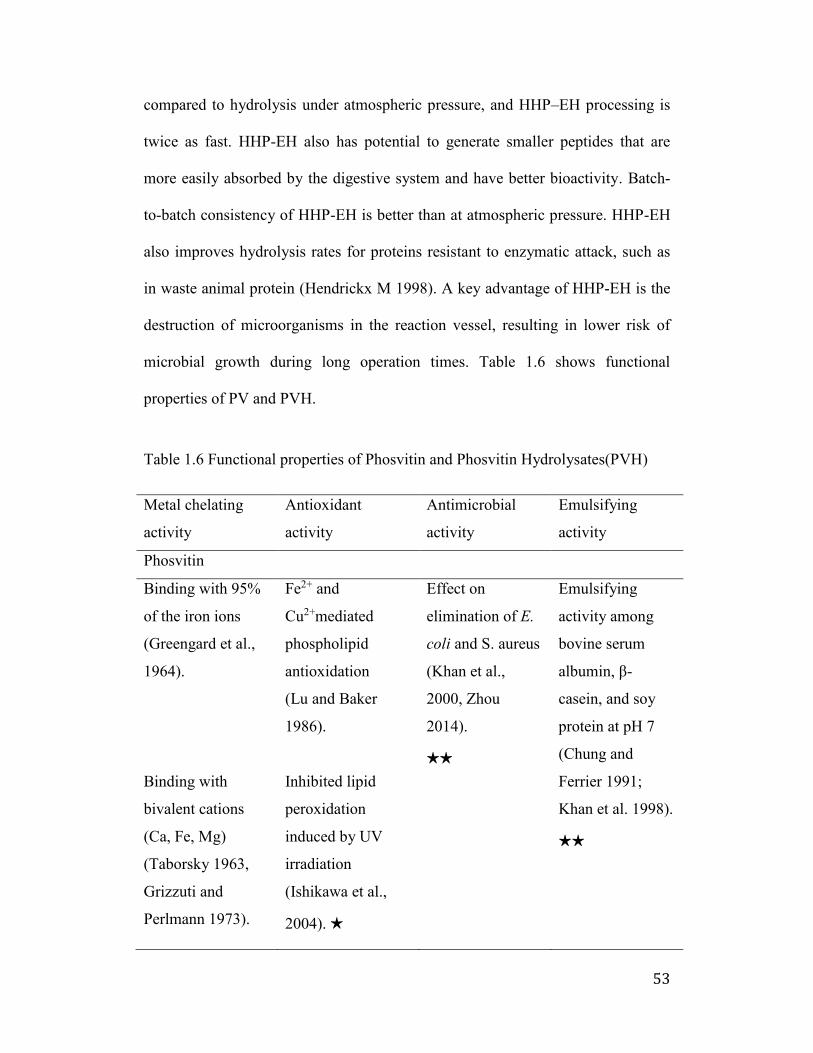
53
compared to hydrolysis under atmospheric pressure, and HHP–EH processing is
twice as fast. HHP-EH also has potential to generate smaller peptides that are
more easily absorbed by the digestive system and have better bioactivity. Batch-
to-batch consistency of HHP-EH is better than at atmospheric pressure. HHP-EH
also improves hydrolysis rates for proteins resistant to enzymatic attack, such as
in waste animal protein (Hendrickx M 1998). A key advantage of HHP-EH is the
destruction of microorganisms in the reaction vessel, resulting in lower risk of
microbial growth during long operation times. Table 1.6 shows functional
properties of PV and PVH.
Table 1.6 Functional properties of Phosvitin and Phosvitin Hydrolysates(PVH)
Metal chelating
activity
Antioxidant
activity
Antimicrobial
activity
Emulsifying
activity
Phosvitin
Binding with 95%
of the iron ions
(Greengard et al.,
1964).
Binding with
bivalent cations
(Ca, Fe, Mg)
(Taborsky 1963,
Grizzuti and
Perlmann 1973).
Fe2+ and
Cu2+mediated
phospholipid
antioxidation
(Lu and Baker
1986).
Inhibited lipid
peroxidation
induced by UV
irradiation
(Ishikawa et al.,
2004). ★
Effect on
elimination of E.
coli and S. aureus
(Khan et al.,
2000, Zhou
2014).
★★
Emulsifying
activity among
bovine serum
albumin, -
casein, and soy
protein at pH 7
(Chung and
Ferrier 1991;
Khan et al. 1998).
★★
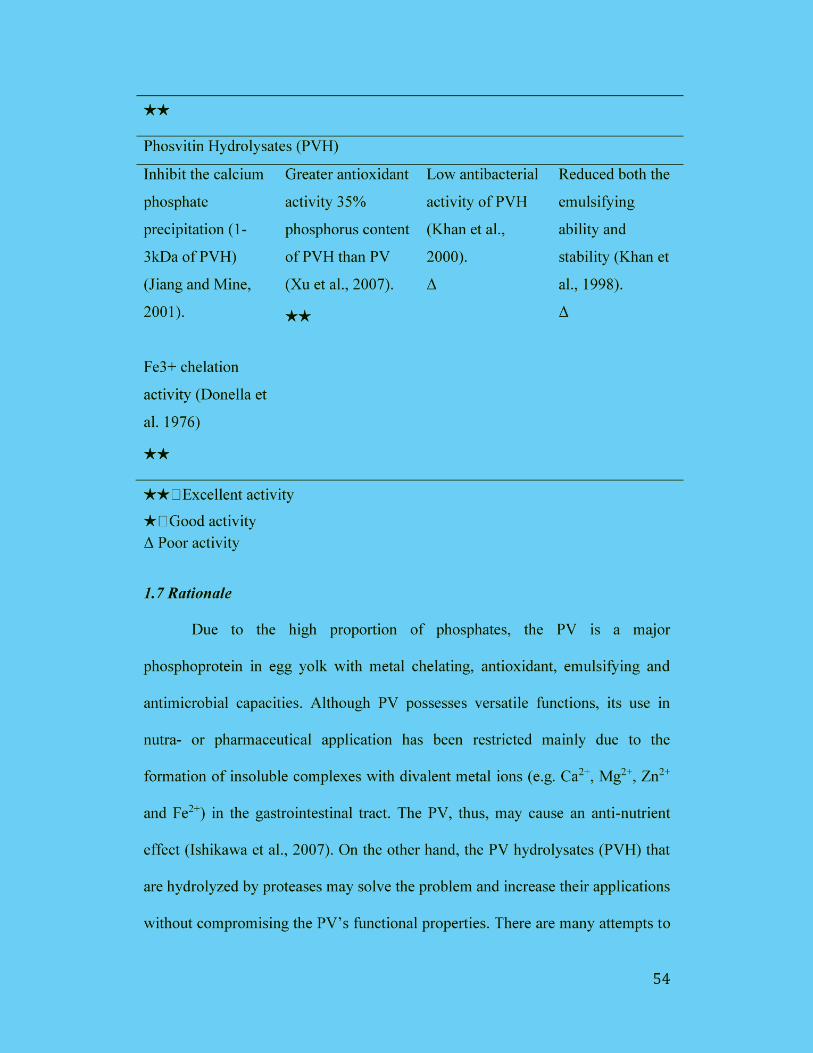
55
produce the PVH. However, the yield of the PVH is low due to the PV’s negative
charges causing the resistance to digestive enzymes. Further studies are needed to
increase the yield of PVH and enhance their functional properties. As an
alternative processing procedure, high hydrostatic pressure combined with
enzymatic hydrolysis (HHP-EH) is a new method to augment enzyme efficiency.
However, there is limited information available for the production of PVH by
HHP-EH.
1.8 Hypothesis
We hypothesize that the application of HHP-EH increases the degree of
hydrolysis (DH) of PV. The PVH produced by HHP-EH shows stronger
antioxidant and antimicrobial properties compared to the intact PV, and PVH
treated under atmospheric pressure (AP).
1.9 Objectives
The objectives of this study are to 1) to increase the yield of PVH by using
an HHP-EH under optimal conditions of pressures, enzyme:substrate (E:S) ratios,
temperatures and incubation times, 2) to determine the effect of iron-chelation and
antimicrobial properties of PVH, and 3) to quantitate the PV in eggs and liquid
media by DAS-ELISA.
56
1.10 Specific Objectives
1.10.1 Production of PVH
The aim of this study was 1) to determine optimum condition of pressures,
enzyme: substrate (E:S) ratios, various temperature and pH for producing PVH; 2)
to measure degree of hydrolysis of PVH with 2,4,6-trinitrobenzene sulfonic acid
(TNBSA) method; 3) to measure the molecular weight of the PVH by SDS-
PAGE, matrix-assisted laser desorption/ionization (MALDI-TOF); 4) to monitor
PVH by high performance liquid chromatography (HPLC).
1.10.2 Iron-chelation
The aim of this study was 1) to determine the iron binding (Fe ) capacity
of PVH; 2) to measure the ferric (Fe ) reducing ability of plasma (FRAP) assay
of PVH.
1.10.3 Anti-microbial property
The aim of this study was 1) to evaluate the PVH’s antimicrobial activity
in vitro; 2) to develop antimicrobial formulation for the prevention of diarrhea in
the animal by a combination of PVH and/or IgY antibodies.
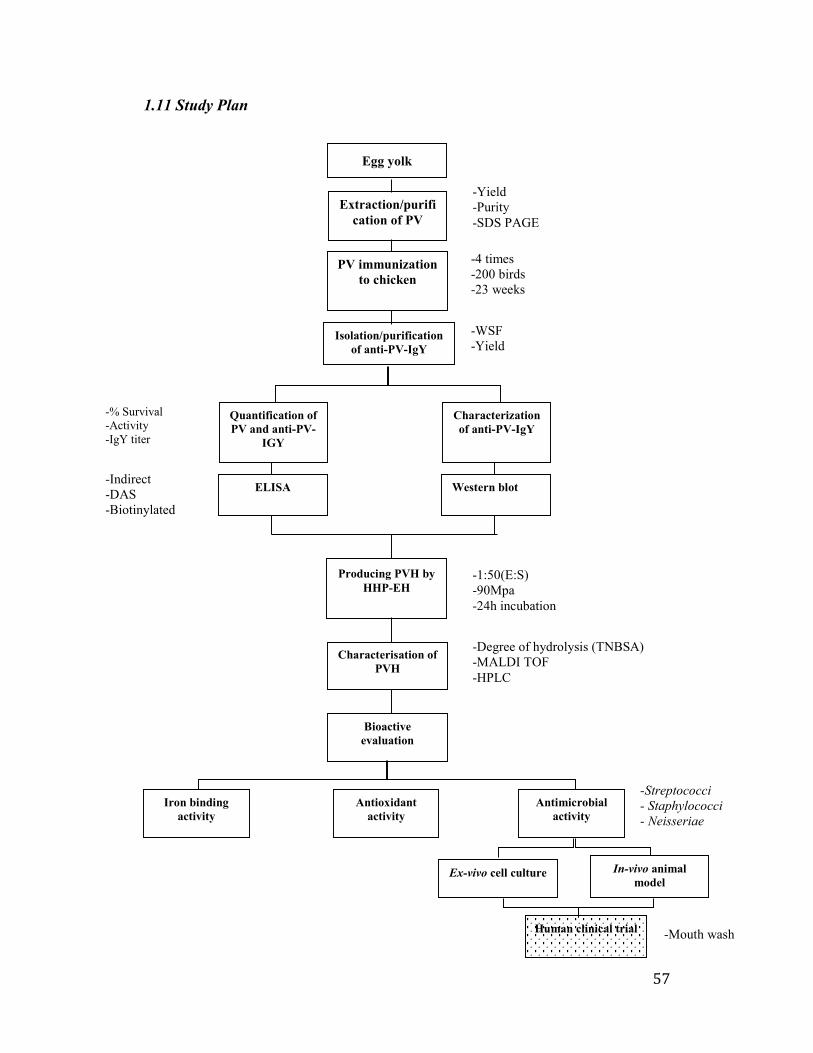
57
1.11 Study Plan
-% Survival
-Activity
-IgY titer
Producing PVH by
HHP-EH
Characterisation of
PVH
Bioactive
evaluation
Antioxidant
activity
-Yield
-Purity
-SDS PAGE
-WSF
-Yield
ELISA Western blot
Isolation/purification
of anti-PV-IgY
Egg yolk
Quantification of
PV and anti-PV-
IGY
Characterization
of anti-PV-IgY
Extraction/purifi
cation of PV
PV immunization
to chicken
-4 times
-200 birds
-23 weeks
-Mouth wash
-Streptococci
- Staphylococci
- Neisseriae
Human clinical trial
-Degree of hydrolysis (TNBSA)
-MALDI TOF
-HPLC
-1:50(E:S)
-90Mpa
-24h incubation
-Indirect
-DAS
-Biotinylated
Iron binding
activity
Antimicrobial
activity
Ex-vivo cell culture In-vivo animal
model
58
Chapter 2 (Study 1): Sensitive Double Antibody Sandwich ELISA for the
Quantification of Phosvitin
2.1 Introduction
Phosvitin (PV), one of the most highly phosphorylated proteins in nature, is
produced by proteolytic cleavage of chicken vitellogenin (a metal-binding
protein) which is disseminated in the hen blood and picked up by developing
oocytes (Richards 1997). Vitellogenin is cleaved into lipovitellin and PV yolk
proteins, sometime during or after transport into the oocyte (Yamamura et al.,
1995).
PV accounts for 60% of total egg yolk phosphoproteins and bears about
90% of the egg yolk phosphorus (Xu et al., 2007). The numerous phosphorylated
serine residues in PV exhibits strong affinity for divalent metals such as iron,
calcium, and magnesium (Grizzuti and Perlmann 1973). The PV prevents lipid
oxidation in lipid-containing systems such as food and biological systems also by
its ability to bind iron and copper (Lu and Baker 1986). PV has also displayed
strong antimicrobial and emulsifying properties (Khan et al., 2000; Khan et al.,
1998). It has been considered that the emulsifying activity of PV is better than
that of bovine serum albumin, -casein and soy protein (Chung and Ferrier 1992;
M. S. Khan et al., 1998).
In recent years PV has drawn attention of researchers scientists and
industries due to its phosphopeptides content for food and nutraceutical
formulations (Samaraweera et al., 2011). PV is considered to be an efficient
59
source of bioactive phosphopeptides but the high cost of PV along with
complicated detection and production procedures hinders its application efforts
particularly in the food industry. Also, there is a huge potential of PV application
in the cosmetics industry as naturally derived antioxidant and anti-aging copper-
binding peptides. To the best of our knowledge, there is no well-established
method to detect or quantify PV. Accordingly, an efficient, precise and
reproducible method for quantitation of PV is necessary for the development of
extraction and purification techniques.
Antibodies, being highly specific in nature, are persuasive candidates for
PV protein identification (Jensenius et al., 1981). Antibodies are typically
produced by immunization of animals, mostly mammals. However, mammalian
antibodies are produced by invasive means in limited quantities and their
purification is time-consuming as well as expensive. The uses of avian antibodies
have increased spectacularly to replenish mammalian antibodies. A major lead of
avian antibodies is ease of harvest from their eggs instead of serum. Also, an egg-
laying hen antibody productivity is much higher than that of a similar sized
mammal (Hau and Hendriksen 2005). Today, hens are recognized as an expedient
and economical source of antibodies.
Therefore, in this study, we aim to develop an accurate ELISA based on
monoclonal and chicken egg yolk IgY polyclonal antibodies for the quantitation
of microgram to nanogram levels of PV. This quantification method developed,
may be applied in studies on PV for its accurate quantification and further
improved for detection of other challenging proteins.
60
2.2 Materials and Methods
2.2.1 Materials
Phosvitin (from chicken egg yolk), phosphatase, Freund's incomplete
adjuvant, purified chicken IgG, rabbit anti-chicken IgG, rabbit anti-chicken IgG
conjugated with horseradish peroxidase and streptavidin-HRPO were purchased
from Sigma (St. Louis, MO, USA). Biotinaimdohexanoic acid-3-sulfo-N-
hydroxysuccinimide ester (NHS-PEO4-Biotin), Slide-A-Lyzer® and
Tetramethylbenzidine (TMB) were purchased from Thermo Fisher Scientific
(Burlington, ON, Canada) and KPL (Gaithersburg, MD, USA), respectively. Anti-
PV (PPP-6) monoclonal antibody was obtained from Myungji University
(Yongin, Korea). Bio-Rad protein assay kit, glycine, Precision Plus Protein
Standard, sodium dodecyl sulfate (SDS), Mini-PROTEAN® TGXTM Any kD
precast gels Precast Gels in Tris-Glycine-SDS buffer, anion exchange
chromatography, Gel DocTM EZ Imager and BioLogic LP Chromatography
system were purchased from Bio-Rad Laboratory (Mississauga, ON, Canada).
Sephacryl S-300 gel filtration column was purchased from GE Healthcare Bio-
Sciences Corp (Piscataway, NJ, USA). The ELISA Vmax kinetic microplate reader
was obtained from Molecular Devices Corp (Sunnyvale, CA, USA). All analytical
grade chemicals and solvents were from commercial sources.
2.2.2 Production of Anti-PV Polyclonal IgY
Production of polyclonal IgY directed against PV was followed by chicken
immunization. The Sigma PV was dephosphorylated with acid phosphatase as
described by Miller et al. (1982) (Miller et al., 1982). The dephosphorylated PV
61
was suspended in sterilized PBS at a concentration of 100 μg/mL and emulsified
with an equal volume of Freund's incomplete adjuvant. Twelve 23-weeks-old
Single Comb White Leghorn chickens were intramuscularly injected with the
emulsified saline with or without PV at four different sites (0.25 mL per site) of
breast muscles (two sites per left or right breast muscle). Two booster
immunizations were given after 2 and 8 weeks of the initial immunization. Eggs
were collected daily and stored at 4°C until the extraction of the antibodies.
2.2.3 Purification of Anti-PV IgY
The egg yolk from immunized hens was physically separated from egg
white by mixing gently with 8 volumes of cold distilled water (acidified with 0.1
M HCl to give a pH of 4.0) to avoid possible disruptions of egg yolk granules due
to the presence of high concentrations of acid. Cold acidified distilled water (pH
2.0) was then added to make the final dilution of 1:10. After mixing well, the
mixture was adjusted to a pH of 5.0 – 5.2 and incubated at 4°C for 12 h. Water
soluble fraction (WSF) was obtained by centrifugation (3125 × g) at 4°C for 20
min. The supernatant was collected as the IgY rich WSF and titrated by indirect
ELISA (described in the following section) using Sigma PV as coating antigen.
The fractions found to have high titers, particularly the fractions collected during
weeks 4 to 10 of the immunization period were further purified by ammonium
sulfate (40%) followed by Sephacryl S-300 gel filtration column (Ishikawa et al.,
2007). Control IgY antibody was extracted from eggs obtained from non-
immunized chickens.
62
2.2.4 Titer of Anti-PV IgY by Indirect ELISA
Microtiter plates were coated with 100 μL per well of PV in carbonate-
bicarbonate buffer (0.05 M, pH 9.6) at a concentration of 8 μg/mL and incubated
at 37°C for 1 h. Throughout the experiment, the plates were then washed four
times with PBS containing 0.05% Tween 20 (PBS-Tween) between each step.
After washing, 100 μL of 1% BSA solution (w/v) in PBS-Tween was added to
each well, and the plates were incubated at 37°C for 30 min. The BSA solution
was then discarded, and the wells were washed. Egg yolk WSF (diluted 1:1000 in
PBS-Tween) containing specific IgY against PV or non-immunized egg yolk IgY
antibody (as a control) was added to each well (100 μL per well), and the plates
were further incubated at γ7°C for 1 h. After washing the plates, 100 μL of rabbit
anti-chicken IgG conjugated with horseradish peroxidase (HRPO, diluted 1:5000
in PBS-Tween) was added to each well and incubated at 37°C for 1 h. The plates
were washed with PBS-Tween, followed by the addition of 100 μL of freshly
prepared substrate solution, tetramethylbenzidine (TMB). OD650 was taken after
30 min using an ELISA Vmax kinetic microplate reader. The optical density value
of antibody activity was determined by subtracting the value of the control
antibody from that of specific antibody.
2.2.5 Total IgY Concentration
To quantify the total IgY concentration purified from egg yolks, ELISA was
performed as mentioned above. A microtiter plate was coated with 100 μL per
well of rabbit anti-chicken IgG at a final concentration of β μg. Samples of the
PV-specific IgY were diluted 1:10,000 in PBS. Specific and non-specific IgY
63
were reconstituted and serially diluted in PBS. Two-fold serial dilutions of
purified chicken IgG in PBS (0.5 to 0.0γ1 μg/mL) were used as the reference
antibody to prepare a standard curve. Total and specific IgY concentrations were
determined using the standard curve.
2.2.6 Total Protein Assay
The Bio-Rad protein assay, based on the method of Bradford, was
performed using purified chicken IgY (1 mg protein/mL) as the reference protein.
Serial dilutions of the purified IgY, and the reference protein in PBS (0.5 to
0.0625 mg/mL) were analyzed using the microtiter plate. OD595 was taken after 5
min using an ELISA Vmax kinetic microplate reader.
2.2.7 Electrophoresis and Western Blot
Sigma PV samples were run on Mini-PROTEAN® TGXTM Any kD Precast
Gels in Tris-Glycine-SDS buffer and stained with a solution of 0.05% Coomassie
Blue R-250, 0.1M aluminum nitrate, 25% isopropanol, 10% acetic acid, and 1%
TritonX-100. Gels were destained with 7% acetic acid. Bio-rad Precision Plus
Protein Standards were loaded as a molecular weight marker at a constant voltage
(100 V). Images of gels were scanned in the Bio-Rad Gel DocTM EZ Imager.
For Western blot assay, the gel was electrophoretically transferred to
nitrocellulose membrane in Tris-borate electrophoresis buffer without sodium
dodecyl sulfate at 40 V for 2 h. Nitrocellulose membrane was then soaked in 2%
BSA in PBS (pH 7.2) for 1 h at room temperature. After washing with PBS (three
times for 5 min each), the nitrocellulose membrane was incubated with anti-PV
IgY antibodies (in PBS containing 1% BSA) for 1 h at room temperature. The
64
incubation was followed by washing in PBS and 1 h incubation with rabbit anti-
chicken IgG conjugated with HRPO (diluted 1:3000 in PBS-Tween). Color was
developed by incubation in 0.05% diaminobenzidine tetra-hydrochloride in PBS
containing hydrogen peroxide (0.01% w/v) and cobalt chloride (0.033% w/v) for
5 min. Stained blots were then washed several times with water and dried.
2.2.8 Double Antibody Sandwich ELISA (DAS-ELISA)
A microtiter plate was coated with 100 µL of anti-PV mAb (1 µg/mL) in
0.05M carbonate buffer, pH 9.6, at 4°C overnight. Nonspecific binding sites were
blocked with 120 µL of 3% BSA for 45 min. The PV was serially diluted from
0.7 to 180 µg/mL in PBS, pH 7.0 (containing 0.05% Tween 20) and added to each
well, and then the plate was incubated at 37°C for 1 h. The plate was washed, and
100 μL of the anti-PV IgY (1 µg/mL) was added to each well and incubated at
37°C for 1 h. After incubation, the plate was washed, and streptavidin-HRPO was
added and incubated at 37°C for 45 min. TMB substrate was added and OD650
was taken after 10 min using an ELISA Vmax kinetic microplate reader. The
antibody titre was determined by subtracting the value of the control antibody
from that of specific antibody. Throughout the experiment, four times washing
was conducted by PBS-Tween between each step.
2.2.9 Biotinylated DAS-ELISA
Anti-PV IgY polyclonal antibodies were biotinylated by using long arm
NHS-PEO4-Biotin. One mg of anti-PV IgY in PBS (pH 7.β) were added to β0 μL
of long chain biotin (3 mg/mL) and then incubated at room temperature for 1 h. A
10 μL glycine (100 μg/μL) was added and the solution kept on a shaker for 10
65
min. The solution was then dialyzed in a Slide-A-Lyzer against PBS (pH 7.2)
overnight at 4°C.
Except otherwise indicated, four times washing was conducted by PBS-
Tween between each step. A microtiter plate was coated with 100 µL of anti-PV
mAb (PPP-6, 1 µg/mL) in 0.05M carbonate buffer, pH 9.6, at 4oC overnight.
Nonspecific binding sites were blocked with 120 µL of 3% BSA for 45 min. The
Sigma PV was serially diluted from 0.3 to 160 ng/mL in PBS, pH 7.0, containing
0.05% Tween 20 and added to each well then the plate was incubated at 37°C for
1 h. The plate was washed, and 100 μL of the biotinylated anti-PV IgY (1 µg/mL)
was added to each well and incubated at 37°C for 1 h. After incubation, the plate
was washed and streptavidin-HRPO was added and incubated at 37°C for 45 min.
TMB substrate was added and OD650 was taken after 10 min using an ELISA Vmax
kinetic microplate reader. The antibody titre was determined by subtracting the
value of the control antibody from that of specific antibody.
2.2.10 Validation of the Assay
The accuracy of the two ELISA methods was evaluated by performing
recovery studies. A saline solution was spiked with Sigma PV Standard at levels
of 1 µg, 10 µg, 50 µg and 100 µg/mL sample in a total volume of 10 mL. For the
determination of recovery rates, the extractions were done in triplicates and
analyzed by the DAS-ELISA and biotinylated DAS-ELISA. Intra-assay (within
plate) precision was determined as the mean coefficients of variation (CV) based
on 10 replicates. Inter-assay (between plates) precision was also determined as the
mean CV by triplicate analyses on 10 plates.
66
The limit of detection (LOD) was calculated from 3.3 times standard error
of the predicted Y-value for each X in a regression divided by slope. Additionally,
the LOD in three saline solutions was determined in the same way, on the basis of
five experiments. Linearity and range of detection were determined from serially
diluted Sigma PV standard curve plot.
2.2.11 Statistical analysis
The student t-test (one-tailed t-test) was used to analyze the significant
difference (p < 0.05) between the groups.
2.3 Results
2.3.1 Production of Anti-PV IgY
The Anti-PV IgY titer of hyperimmunized egg yolks against PV was
determined by indirect ELISA method during the immunization period (Figure
2.1). A primary antibody response was determined by slight IgY titer increase
after the first PV immunization. Following the first booster a rapid secondary
response was expressed with high PV specific IgY activity at a peak of week 5 (p
< 0.05), thereafter the titer decreased, but reached at a peak again after the second
booster at week 8 (p < 0.05). As a result, a high antigen-binding specificity of
anti-PV IgY could be obtained from the egg yolks collected from immunized eggs
during weeks 4 to 10 of the immunization period.
The crude WSF of egg yolk was isolated, further purified by ammonium
sulfate precipitation, followed by Sephacryl S-300 gel chromatography. The
content of total IgY in the egg yolk was found to be constant (9.0 ± 1.1 mg/mL)
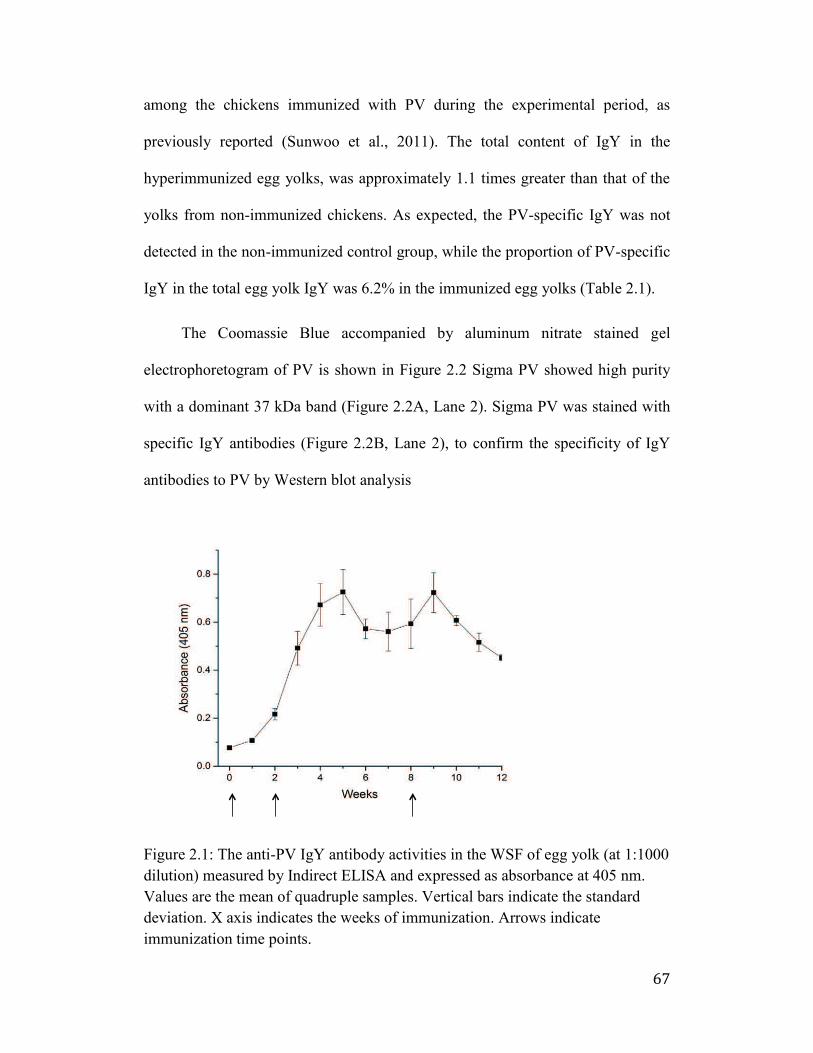
67
among the chickens immunized with PV during the experimental period, as
previously reported (Sunwoo et al., 2011). The total content of IgY in the
hyperimmunized egg yolks, was approximately 1.1 times greater than that of the
yolks from non-immunized chickens. As expected, the PV-specific IgY was not
detected in the non-immunized control group, while the proportion of PV-specific
IgY in the total egg yolk IgY was 6.2% in the immunized egg yolks (Table 2.1).
The Coomassie Blue accompanied by aluminum nitrate stained gel
electrophoretogram of PV is shown in Figure 2.2 Sigma PV showed high purity
with a dominant 37 kDa band (Figure 2.2A, Lane 2). Sigma PV was stained with
specific IgY antibodies (Figure 2.2B, Lane 2), to confirm the specificity of IgY
antibodies to PV by Western blot analysis
Figure 2.1: The anti-PV IgY antibody activities in the WSF of egg yolk (at 1:1000
dilution) measured by Indirect ELISA and expressed as absorbance at 405 nm.
Values are the mean of quadruple samples. Vertical bars indicate the standard
deviation. X axis indicates the weeks of immunization. Arrows indicate
immunization time points.
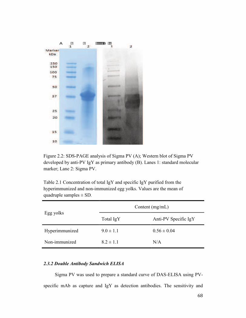
68
Figure 2.2: SDS-PAGE analysis of Sigma PV (A); Western blot of Sigma PV
developed by anti-PV IgY as primary antibody (B). Lanes 1: standard molecular
marker; Lane 2: Sigma PV.
Table 2.1 Concentration of total IgY and specific IgY purified from the
hyperimmunized and non-immunized egg yolks. Values are the mean of
quadruple samples ± SD.
Egg yolks
Content (mg/mL)
Total IgY Anti-PV Specific IgY
Hyperimmunized 9.0 ± 1.1 0.56 ± 0.04
Non-immunized 8.2 ± 1.1 N/A
2.3.2 Double Antibody Sandwich ELISA
Sigma PV was used to prepare a standard curve of DAS-ELISA using PV-
specific mAb as capture and IgY as detection antibodies. The sensitivity and
A B
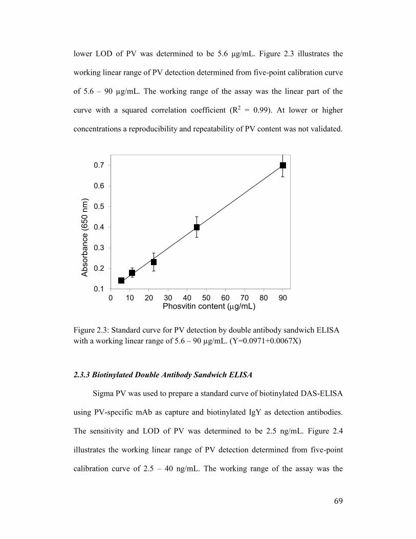
69
lower LOD of PV was determined to be 5.6 μg/mL. Figure 2.3 illustrates the
working linear range of PV detection determined from five-point calibration curve
of 5.6 – 90 µg/mL. The working range of the assay was the linear part of the
curve with a squared correlation coefficient (R2 = 0.99). At lower or higher
concentrations a reproducibility and repeatability of PV content was not validated.
Figure 2.3: Standard curve for PV detection by double antibody sandwich ELISA
with a working linear range of 5.6 – 90 µg/mL. (Y=0.0971+0.0067X)
2.3.3 Biotinylated Double Antibody Sandwich ELISA
Sigma PV was used to prepare a standard curve of biotinylated DAS-ELISA
using PV-specific mAb as capture and biotinylated IgY as detection antibodies.
The sensitivity and LOD of PV was determined to be 2.5 ng/mL. Figure 2.4
illustrates the working linear range of PV detection determined from five-point
calibration curve of 2.5 – 40 ng/mL. The working range of the assay was the
0.1
0.2
0.3
0.4
0.5
0.6
0.7
0 10 20 30 40 50 60 70 80 90
Ab
so
rba
nce
(65
0 n
m)
Phosvitin content (mg/mL)
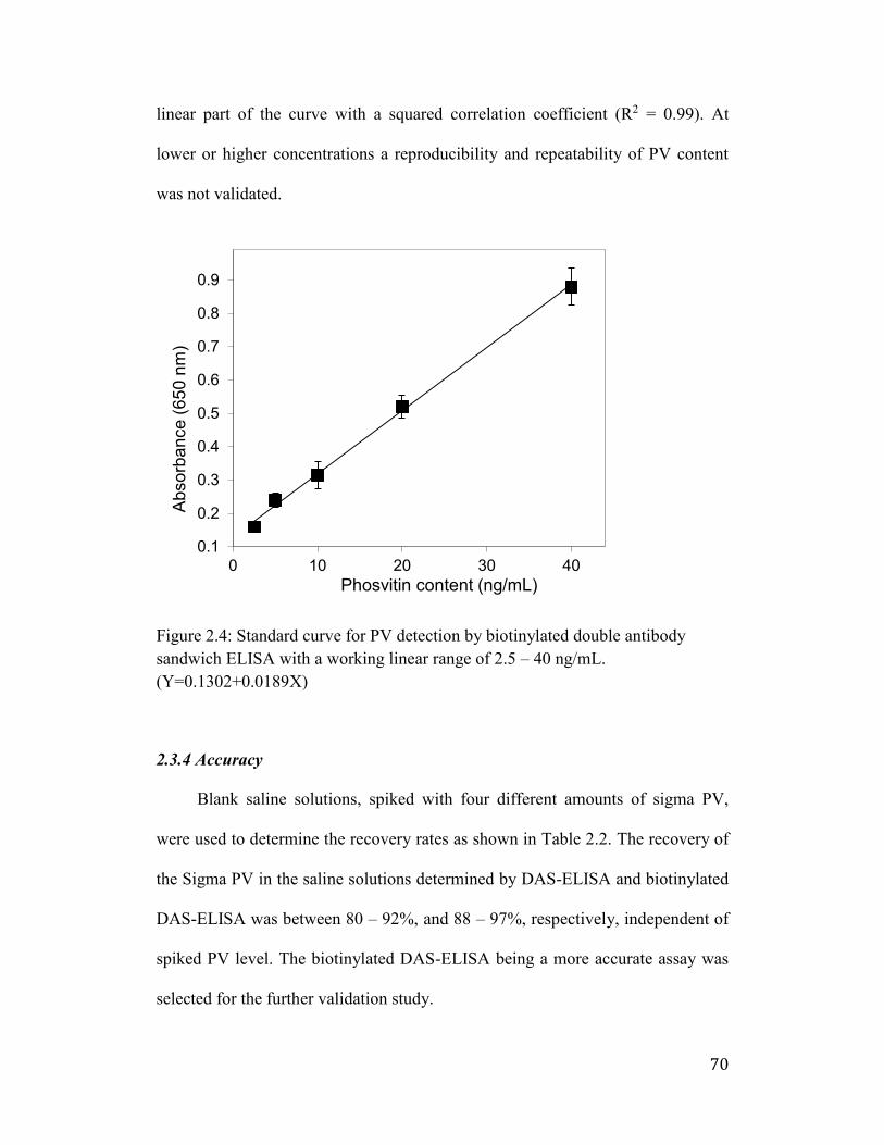
70
linear part of the curve with a squared correlation coefficient (R2 = 0.99). At
lower or higher concentrations a reproducibility and repeatability of PV content
was not validated.
Figure 2.4: Standard curve for PV detection by biotinylated double antibody
sandwich ELISA with a working linear range of 2.5 – 40 ng/mL.
(Y=0.1302+0.0189X)
2.3.4 Accuracy
Blank saline solutions, spiked with four different amounts of sigma PV,
were used to determine the recovery rates as shown in Table 2.2. The recovery of
the Sigma PV in the saline solutions determined by DAS-ELISA and biotinylated
DAS-ELISA was between 80 – 92%, and 88 – 97%, respectively, independent of
spiked PV level. The biotinylated DAS-ELISA being a more accurate assay was
selected for the further validation study.
0.1
0.2
0.3
0.4
0.5
0.6
0.7
0.8
0.9
0 10 20 30 40
Ab
so
rba
nce
(6
50
nm
)
Phosvitin content (ng/mL)
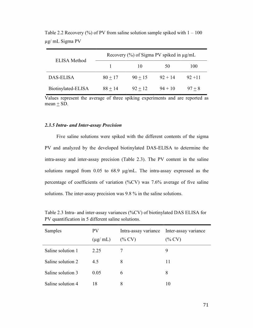
71
Table 2.2 Recovery (%) of PV from saline solution sample spiked with 1 – 100
µg/ mL Sigma PV
ELISA Method Recovery (%) of Sigma PV spiked in µg/mL
1 10 50 100
DAS-ELISA 80 + 17 90 + 15 92 + 14 92 +11
Biotinylated-ELISA 88 + 14 92 + 12 94 + 10 97 + 8
Values represent the average of three spiking experiments and are reported as
mean + SD.
2.3.5 Intra- and Inter-assay Precision
Five saline solutions were spiked with the different contents of the sigma
PV and analyzed by the developed biotinylated DAS-ELISA to determine the
intra-assay and inter-assay precision (Table 2.3). The PV content in the saline
solutions ranged from 0.05 to 68.9 µg/mL. The intra-assay expressed as the
percentage of coefficients of variation (%CV) was 7.6% average of five saline
solutions. The inter-assay precision was 9.8 % in the saline solutions.
Table 2.3 Intra- and inter-assay variances (%CV) of biotinylated DAS ELISA for
PV quantification in 5 different saline solutions.
Samples PV
(µg/ mL)
Intra-assay variance
(% CV)
Inter-assay variance
(% CV)
Saline solution 1 2.25 7 9
Saline solution 2 4.5 8 11
Saline solution 3 0.05 6 8
Saline solution 4 18 8 10
72
Saline solution 5 68.9 9 11
2.4 Discussion
Egg yolk is a mixture of lipids, lipoproteins, PV, and water-soluble proteins
(WSP). The WSP fraction, including α-, ß-, and -livetins, was separated from the
lipids and granular proteins using the acidified water (pH 5.0-5.2) dilution method
(S. K. Lee et al., 2002). The remaining granules includes yolk micelles that
contain lipovitellin and PV (Ren et al., 2015). PV is one of the most highly-
phosphorylated proteins in nature which contains more than 80% of the
phosphorous present in the egg yolk (Joubert and Cook 1958). The high density of
polar residues results in a substantial number of charged side chains in
physiological pH which leads to different functionalities such as metal chelating,
antioxidant, and antimicrobial activities (Li-Chan 2008).
There is limited information available on the quantification of PV.
Since PV is a highly phosphorylated protein it cannot be accurately assessed by
normal colorimetric assays such as Bradford, BCA and Lowry protein assays due
to the interference of their phosphate groups (Hatta et al., 1997). Therefore, this
study introduces, for the first time, a simple and reliable method to obtain protein-
specific IgY antibodies for quantification of PV in combination with mAb
antibodies.
Chicken was used to generate antibody employed in development of PV
detection system. It has been reported that the avian maternal antibodies are
transferred from serum to egg yolk to confer passive immunity to their offsprings
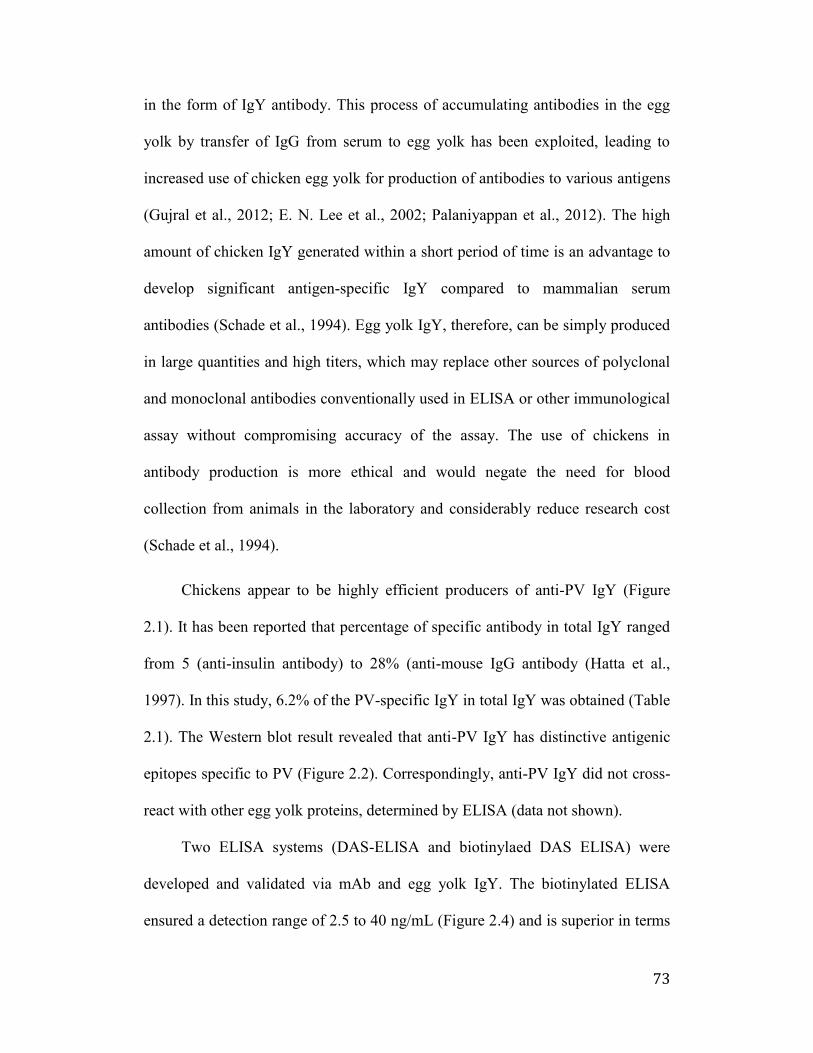
73
in the form of IgY antibody. This process of accumulating antibodies in the egg
yolk by transfer of IgG from serum to egg yolk has been exploited, leading to
increased use of chicken egg yolk for production of antibodies to various antigens
(Gujral et al., 2012; E. N. Lee et al., 2002; Palaniyappan et al., 2012). The high
amount of chicken IgY generated within a short period of time is an advantage to
develop significant antigen-specific IgY compared to mammalian serum
antibodies (Schade et al., 1994). Egg yolk IgY, therefore, can be simply produced
in large quantities and high titers, which may replace other sources of polyclonal
and monoclonal antibodies conventionally used in ELISA or other immunological
assay without compromising accuracy of the assay. The use of chickens in
antibody production is more ethical and would negate the need for blood
collection from animals in the laboratory and considerably reduce research cost
(Schade et al., 1994).
Chickens appear to be highly efficient producers of anti-PV IgY (Figure
2.1). It has been reported that percentage of specific antibody in total IgY ranged
from 5 (anti-insulin antibody) to 28% (anti-mouse IgG antibody (Hatta et al.,
1997). In this study, 6.2% of the PV-specific IgY in total IgY was obtained (Table
2.1). The Western blot result revealed that anti-PV IgY has distinctive antigenic
epitopes specific to PV (Figure 2.2). Correspondingly, anti-PV IgY did not cross-
react with other egg yolk proteins, determined by ELISA (data not shown).
Two ELISA systems (DAS-ELISA and biotinylaed DAS ELISA) were
developed and validated via mAb and egg yolk IgY. The biotinylated ELISA
ensured a detection range of 2.5 to 40 ng/mL (Figure 2.4) and is superior in terms
74
of accuracy of 88 – 97% (Table 2.2). This biotinylated ELISA system is also
highly sensitive with over 2000 times lowered LOD as compared to the DAS-
ELISA (Figure 2.3). Precision studies were also carried out to validate the
arrogant assay, revealing remarkably low intra- and inter-assay variation
coefficients (Table 2.3). This expounded detection system has the potential as a
valuable research tool in monitoring PV, a promising protein for use in food,
nutraceutical and pharmaceutical industries.
2.5 Conclusion
The present study indicates that the IgY-based biotinylated DAS-ELISA
developed provides a specific, sensitive, accurate and precise PV detection system
for PV, with a working detection range of 2.5 – 40 ng/mL.
75
Chapter 3 (Study 2): Phosvitin Hydrolysates with Iron Chelating Capacity
Produced by High Hydrostatic Pressure Combined with Enzymatic
Hydrolysis
3.1 Introduction
Phosvitin (PV) is a phosphoprotein produced by proteolytic cleavage of chicken
vitellogenin, a metal-binding protein in the chicken blood. It is utilized by the
developing oocytes as a metal transporter (Richards 1997). This phosphorylated
protein, present in the egg yolk, contains 10% phosphorus (Hatta et al., 1988),
56% serine, 15% basic and 10% acidic amino acid residues (Byrne et al., 1984;
Taborsky 1963; Taborsky and Mok 1967; Xu et al., 2007). Due to the high
proportion of phosphorylated serine residues, PV shows metal chelating (Choi et
al., 2005), antioxidant (Xu et al., 2007; Katayama et al., 2006), anti-inflammatory
(Xu et al., 2012), emulsifying (Chung and Ferrier 1992) and antimicrobial (Khan
et al., 2000) properties. However, PV is considered nutritionally undesirable due
to the formation of insoluble iron complex, hindering iron absorption in the
digestive tract (Guerin-Dubiard and Anton 2007; Sato et al., 1987). Also, poor
dissolution and stability of PV restrict its applications (Lee et al., 2014). PV
hydrolysates (PVH) produced by enzymes (Goulas et al., 1996), had enhanced
functional properties, although the DH of PVH was low due to the resistance of
phosphoserine clusters to enzymatic digestion.
Hydrolysis under harsh condition of pH and temperature increases the degree of
hydrolysis (DH) and generates higher generation of short peptides. However,
76
undesirable and uncontrolled chemical reactions cause side chain modification
which suppresses peptide bioactivities (Tornvall 2010). An efficient hydrolysis
method is required to produce PV–derived peptides without compromising their
bioactivities. Some pre-treatments such as heating (Samaraweera 2014; Sakanaka
and Tachibana 2006) and alkaline dephosphorylation (Ren et al., 2015; Huang et
al., 2014) have been employed to improve the hydrolysis degree of PV. Several
studies have also reported the use of extremely high-pressure, 400–800 MPa
(Toldra et al., 2011; Garcia-Mora et al., 2015) as pre-treatment before enzyme
hydrolysis (Zeece and Kelly 2008; Chicon et al., 2008; Dong et al., 2011; Hoppe
et al., 2013), which resulted in unsatisfactory protein denaturation.
As an alternative PVH processing method, enzymatic hydrolysis under high
hydrostatic pressure (HHP-EH) is introduced. HHP is used to augment enzyme
efficiency and therefore increase the DH. In the HHP-EH method, moderately
high hydrostatic pressure (90 MPa) is applied during hydrolysis to improve
enzyme efficiency without denaturation of enzymes or protein substrates. High-
pressure processing modifies protein structure by alteration of non-covalent
molecular interactions leading to partially unfolded proteins with more
susceptibility to enzymatic hydrolysis (Volk et al., 2012). Furthermore, iso-
hydrostatic pressure (50-200 MPa) promotes enzyme activity due to higher rates
of mass transfer and more effective enzyme-substrate contact (Ludikhuyze et al.,
2003). Although enzyme stability and functionality are uninfluenced by pressures
lower than 100 MPa, elevated pressures above normal atmospheric level may
increase enzyme reaction rates (Weingand-Ziade et al., 2001). A possible
77
explanation for the acceleration of enzymatic activity at high-pressure could be
the breakdown of cellular compartments and compression of solution which
increases the concentration and movement of the solvated species (Schettino and
Bini 2007).
The main objective of this study was therefore to compare the degree of
hydrolysis of PVH obtained from various enzyme treatments under optimal
conditions of HHP and atmospheric pressure (AP). The resulting hydrolysates can
be used in protein-rich readily absorbed food supplement formulas without the
need for further fractionation. Five different enzymes were selected for PV
hydrolysis and determined for optimal enzyme:substrate (E:S) ratio, temperatures
and incubation period. The PVH was then subjected to evaluation of structural
characteristics (i.e., degree of hydrolysis and molecular weights (Mw) distribution
pattern) and functional characteristics (iron-chelating and reducing capacity). The
developed one-step methodology may be applied for other hydrolysis resistant
proteins, in order to reduce the processing time, avoid the use of harsh chemical
or solvents and conserve their biofunctions.
3.2 Materials and Methods
3.2.1 Materials
Eggs were purchased from the local market. Enzymes were obtained from
commercial suppliers (Sigma-Aldrich, MP Biomedicals and Novozymes). Mini-
PROTEAN Tris-Tricine Precast Gels and Precision Plus Dual Xtra Protein
Standard (15-250 kDa and 2-250 kDa) were obtained from Bio-Rad (Mississauga,
ON, Canada). The Greiss reagent, phosphoprotein phosphate assay and Lowry
78
assay kits were obtained from Sigma (St. Louis, MO, USA), Pierce (Rockford, IL,
USA), and Biorad (Mississauga, ON, Canada), respectively. All other reagents
were of analytical grade.
3.2.2 Apparatus
A portable scale high-hydrostatic pressure processor (TFS-2L, Toyo-
Koatsu Co. Ltd., Hiroshima, Japan) was used for HHP-EH treatment.
Chromatographic analysis was performed on a Shimadzu 10 AVP HPLC system
equipped with a BiosuiteTM 125/5 mm HR-SEC column (7.8 × 300 mm, Waters
Corp., Milford, MA, USA). A Mini-Protein III electrophoresis kit and Bio-Rad
Gel DocTM EZ Imager were purchased from BioRad (Mississauga, ON, Canada).
The VersaMax ELISA microplate reader was obtained from Molecular Devices
Corp (Sunnyvale, CA, USA). All mass spectra were obtained using a Bruker
Ultraflex MALDI-ToF/ToF (Bruker Daltonic GmbH, Bremen, Germany).
3.2.3 IgY separation from egg yolk
Eggs were manually broken; yolks were carefully separated from egg
white and chalazae by rolling on a Whatman filter paper (No. 1). The yolk
membrane was punctured with a needle and the content was collected in a beaker
cooled in iced water. The temperature was maintained at 4°C throughout the
process. Egg yolk was added to nine volumes of pre-chilled acidic water (final pH
5.0-5.2) and incubated at 4°C for 12 h without agitation. After centrifugation at
3200 × g for 30 min, the supernatant (containing IgY antibody) was separated
from the precipitate. The precipitate, containing egg yolk granules, was used for
PV isolation.
79
3.2.4 Phosvitin extraction and purification
Phosvitin was isolated from intact egg yolk and from egg yolk residue
after IgY isolation according to the method presented by McBee and Cotterill
with slight modifications. The precipitate was diluted with two volumes of
distilled water and stirred for 1 h. After centrifugation (3200 ×g, 30 min at 4°C),
the pellet (containing egg yolk granules) was collected and dissolved in nine
volumes of 10% (w/v) NaCl solution, then centrifuged at the same condition. The
supernatant was filtered through Whatman No. 1 filter paper to remove floating
particles. NaCl in the solution was removed by dialysis against water using 12
kDa Mw cut-off dialysis membranes. The dialyzed sample was freeze-dried and
then stored at -20°C until analysis. The protein content of phosvitin purified from
egg yolk was determined using modified Biorad Lowry assay kit. The phosphate
content in purified PV was measured by Phosphoprotein Phosphate assay kit.
3.2.5 Enzymatic hydrolysis
Purified PV, dissolved at 10 mg protein/mL in 10% NaCl, was mixed with
enzymes at enzyme/substrate ratio of 1:50. The enzymes used for PV hydrolysis
include Elastase (Ela), Alcalase (Alc), Savinase (Sav), Trypsin (Try) and
Thermolysin (Ther). The enzyme added samples were then subjected to either AP
(0.1 MPa) or HHP (90 ± 2 MPa) treatments for 24 h at optimal pH, and
temperatures of each enzyme (Table 3.1). The above procedures were also
performed in the absence of enzymes to serve as experimental controls. After the
treatment, enzymes were heat-inactivated (90°C, 10 min) and centrifuged (5000
×g, 10 min) to remove the insoluble fractions. The supernatants were filtered
80
through a 0.22 µm filter, followed by all enzymes removal using Amicon
ultracentrifuge filter unit (10 kDa Mw cut-off) and then stored frozen prior to
further analysis.
Table 3.1: Enzymes applied in HHP-EH and atmospheric hydrolysis and
operational conditions
Enzyme
Treatments Sources Unit pH °C
Alcalase (Alc) Protease from Bacillus
licheniformis 2.4 U/g 7 50
Elastase (Ela) Elastase from hog pancreas 1 U/mg 8 37
Thermolysin
(The) Bacillus thermoproteolyticus 14 U/mg 7 50
Trypsin (Try) Protease derived from porcine
pancreas 30 U/g 7 37
Savinase (Sav) Protease from Bacillus sp. 0.32 U/g 7 55
3.2.6 Degree of hydrolysis (DH)
Hydrolysis degree was analyzed by measuring free primary amines using
2,4,6-trinitrobenzenesulfonic acid (TNBSA) method with L-leucine as standard
(Adlernissen 1979). Total content of amine groups expressed as leucine
equivalent (μg/mL), was obtained from a standard curve prepared from different
concentrations of L-leucine. DH was calculated by the following equation:
DH= (h/htot) × 100
Where, hydrolysis equivalents (h) is the amount of peptide bonds cleaved during
hydrolysis, expressed as millimole leucine equivalents per gram protein (mmol/g
protein); htot is the total amount of peptide bonds in the protein substrate
determined from the protein sample totally hydrolyzed with 6 N HCl at 110°C for
24 h.
81
3.2.7 Phosphate content determination
The phosphate content of PV and PVH samples was determined using
phosphoprotein phosphate assay kit. In this assay, phosphate groups are
hydrolyzed from serine and threonine residues under alkaline condition and then,
quantified through reaction with Malachite green and ammonium molybdate
(Ekman and Jager 1993). The absorbance was measured at 650 nm by UV/Vis
spectrophotometer.
3.2.8 Sodium dodecyl sulfate polyacrylamide gel electrophoresis (SDS-PAGE)
Gel electrophoresis was performed according to the method of Schaegger
in Mini-PROTEAN Tris-Tricine Precast Gels (Bio-Rad, Mississauga, ON,
Canada) with 12% polyacrylamide gels. Precision Plus Dual Xtra Protein
Standard (15-250 kDa and 2-250 kDa, Bio-Rad, Mississauga, ON, Canada) was
used as molecular weight marker. The gels were stained with a solution of 0.05%
Coomassie Blue R-250, 0.1M aluminium nitrate, 25% isopropanol, 10% acetic
acid and 1% TritonX-100, destained with 7% acetic acid and imaged.
3.2.9 Size Exclusion High Pressure Liquid Chromatography (SE-HPLC)
The average Mw of the PVH samples was determined by SE-HPLC using
a Shimadzu 10 AVP HPLC system equipped with a BiosuiteTM 125/5 mm HR-
SEC column (7.8×300 mm, Waters Corp., Mass., USA). Phosphate buffer (100
mM) containing 100 mM NaCl was used as mobile phase at a flow rate of 0.5
mL/min at 25 ºC and the protein was monitored at the UV wavelength of 220 nm.
A calibration curve was made from the log Mw of the standard markers and their
respective elution times (R2 = 0.99).
82
3.2.10 MALDI-TOF
MALDI-TOF-MS analysis was performed at the Alberta Proteomics and
Mass Spectrometry Facility (Faculty of Medicine and Dentistry, University of
Alberta, AB, Canada). One µL of sample was mixed with 1 µL of sinapic acid (10
mg/mL in 50% acetonitrile/water + 0.1% trifluoroacetic acid). One µL of the
sample/matrix solution was then spotted onto a stainless steel target plate and
allowed to air dry. Ions were analyzed in positive mode and external calibration
was performed by use of a standard protein mixture.
3.2.11 Iron chelating activity
The chelation of ferrous ions by PVH was estimated according to the
method of Dinis et al. with slight modifications. EDTA, a strong metal chelator,
was used as a positive control. The ferrous ion chelating ability was calculated by
the following equation:
% Iron chelating ability (%) = (Bc – Bs)/ Bc × 100
Where, Bs and Bc represent the absorbances of the sample and the control
(everything except the protein hydrolysate), respectively.
3.2.12 Ferric Reducing Antioxidant Power (FRAP) assay
The antioxidant capacity of PVH samples was estimated according to the
procedure described by Fogarasi, et al. Trolox (1.0-200 µM) were used as positive
control. The results were corrected for dilution and expressed as Trolox
equivalent (TE) (µM TE/mg sample).
83
3.2.14 Statistical analysis
All experiments were performed at least in three independent trials and the
results were reported as mean ± SD. Results were subjected to the analysis of
variance using the SAS software (SAS Institute, Inc., Cary, NC) and statistical
significance of differences (p < 0.05) was evaluated by the least significant
difference procedure.
3.3 Results
3.3.1 Isolation of phosvitin
Figure 3.1 illustrates the electrophoretic pattern of PV staining with
regular Coomassie Blue (Fig 3.1a) and Coomassie Blue accompanied by
aluminum nitrate (Fig 3.1b). Sigma PV and purified PV showed no band with
regular Coomassie Blue staining but strong bands were observed when aluminum
nitrate was added. Purified PV isolated from egg yolk residue after IgY extraction
showed a higher purity with a dominant 48 kDa and a minor 37 kDa bands (Fig
3.1b, Lane C) compared to PV isolated from whole egg yolk which had other
proteins sized 30 kDa (major band) and 55 kDa (minor band) (Fig 3.1b, Lane B).
The result indicates that pre-isolation of IgY from egg yolk removes unwanted
protein during PV purification. The phosphate content of purified PV, measured
by phosphoprotein phosphate assay kit, was 3.47 μmoles/mg protein.
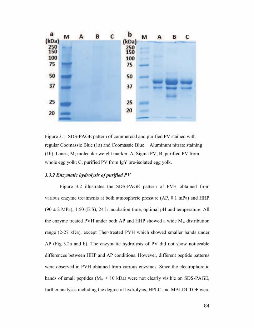
84
Figure 3.1: SDS-PAGE pattern of commercial and purified PV stained with
regular Coomassie Blue (1a) and Coomassie Blue + Aluminum nitrate staining
(1b). Lanes; M; molecular weight marker. A, Sigma PV; B, purified PV from
whole egg yolk; C, purified PV from IgY pre-isolated egg yolk.
3.3.2 Enzymatic hydrolysis of purified PV
Figure 3.2 illustrates the SDS-PAGE pattern of PVH obtained from
various enzyme treatments at both atmospheric pressure (AP, 0.1 mPa) and HHP
(90 ± 2 MPa), 1:50 (E:S), 24 h incubation time, optimal pH and temperature. All
the enzyme treated PVH under both AP and HHP showed a wide Mw distribution
range (2-27 kDa), except Ther-treated PVH which showed smaller bands under
AP (Fig 3.2a and b). The enzymatic hydrolysis of PV did not show noticeable
differences between HHP and AP conditions. However, different peptide patterns
were observed in PVH obtained from various enzymes. Since the electrophoretic
bands of small peptides (Mw < 10 kDa) were not clearly visible on SDS-PAGE,
further analyses including the degree of hydrolysis, HPLC and MALDI-TOF were
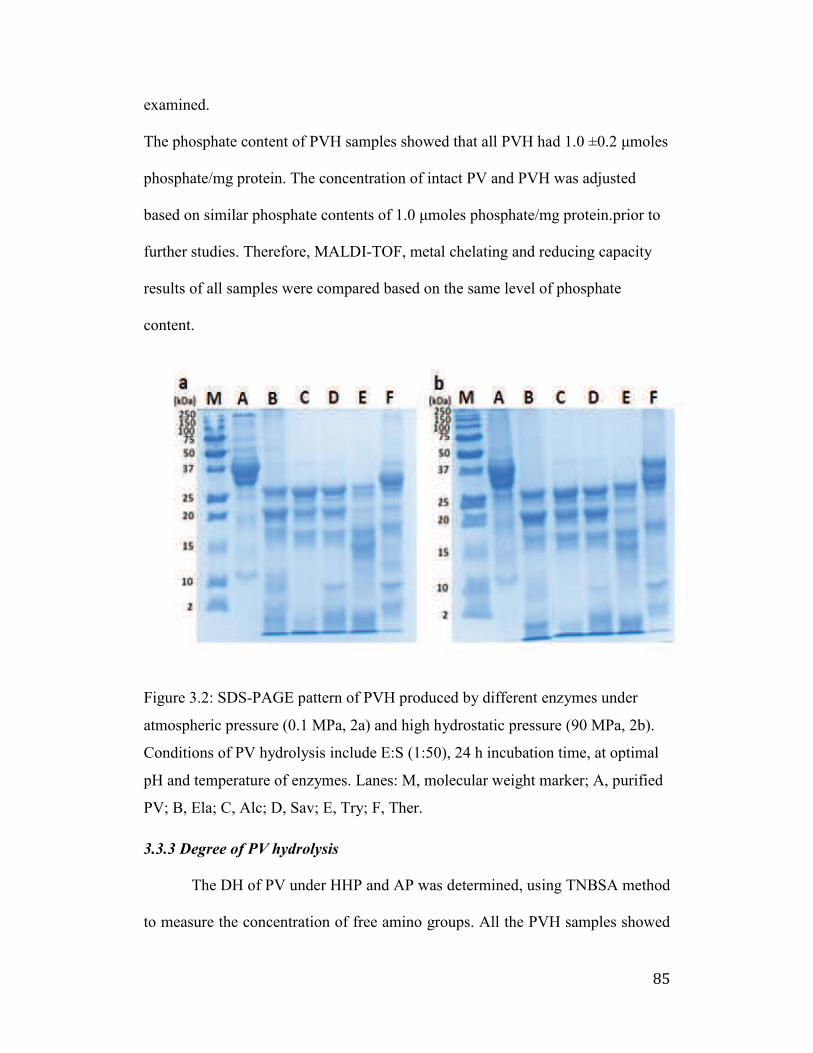
85
examined.
The phosphate content of PVH samples showed that all PVH had 1.0 ±0.2 μmoles
phosphate/mg protein. The concentration of intact PV and PVH was adjusted
based on similar phosphate contents of 1.0 μmoles phosphate/mg protein.prior to
further studies. Therefore, MALDI-TOF, metal chelating and reducing capacity
results of all samples were compared based on the same level of phosphate
content.
Figure 3.2: SDS-PAGE pattern of PVH produced by different enzymes under
atmospheric pressure (0.1 MPa, 2a) and high hydrostatic pressure (90 MPa, 2b).
Conditions of PV hydrolysis include E:S (1:50), 24 h incubation time, at optimal
pH and temperature of enzymes. Lanes: M, molecular weight marker; A, purified
PV; B, Ela; C, Alc; D, Sav; E, Try; F, Ther.
3.3.3 Degree of PV hydrolysis
The DH of PV under HHP and AP was determined, using TNBSA method
to measure the concentration of free amino groups. All the PVH samples showed
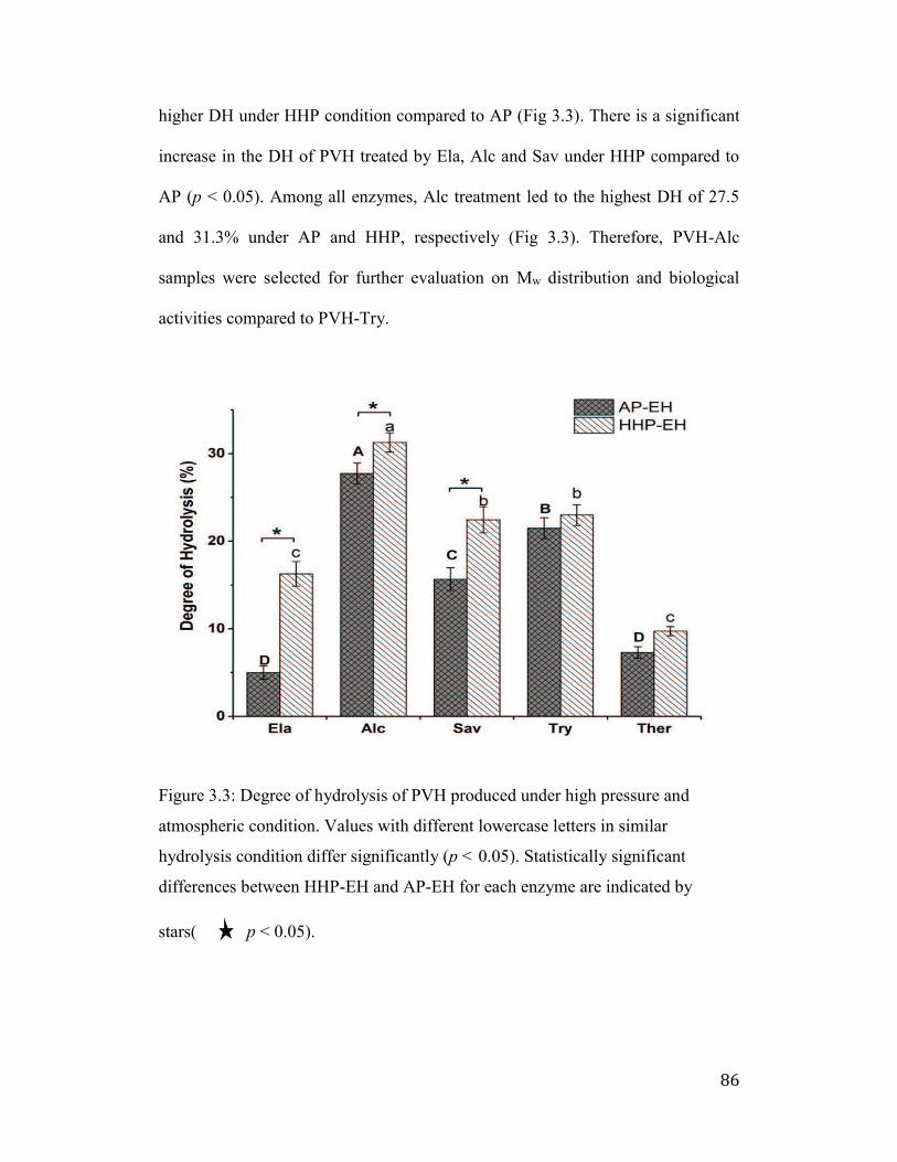
86
higher DH under HHP condition compared to AP (Fig 3.3). There is a significant
increase in the DH of PVH treated by Ela, Alc and Sav under HHP compared to
AP (p < 0.05). Among all enzymes, Alc treatment led to the highest DH of 27.5
and 31.3% under AP and HHP, respectively (Fig 3.3). Therefore, PVH-Alc
samples were selected for further evaluation on Mw distribution and biological
activities compared to PVH-Try.
Figure 3.3: Degree of hydrolysis of PVH produced under high pressure and
atmospheric condition. Values with different lowercase letters in similar
hydrolysis condition differ significantly (p < 0.05). Statistically significant
differences between HHP-EH and AP-EH for each enzyme are indicated by
stars( p < 0.05).
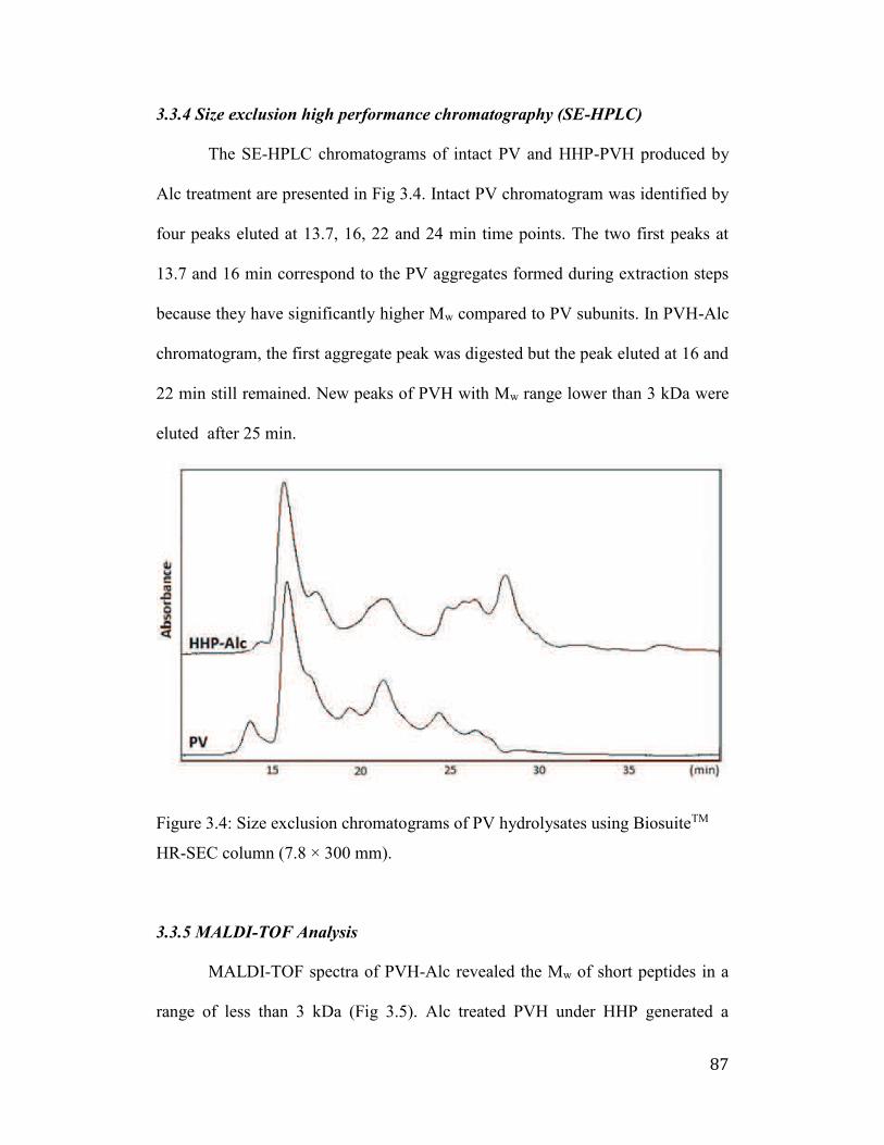
87
3.3.4 Size exclusion high performance chromatography (SE-HPLC)
The SE-HPLC chromatograms of intact PV and HHP-PVH produced by
Alc treatment are presented in Fig 3.4. Intact PV chromatogram was identified by
four peaks eluted at 13.7, 16, 22 and 24 min time points. The two first peaks at
13.7 and 16 min correspond to the PV aggregates formed during extraction steps
because they have significantly higher Mw compared to PV subunits. In PVH-Alc
chromatogram, the first aggregate peak was digested but the peak eluted at 16 and
22 min still remained. New peaks of PVH with Mw range lower than 3 kDa were
eluted after 25 min.
Figure 3.4: Size exclusion chromatograms of PV hydrolysates using BiosuiteTM
HR-SEC column (7.8 × 300 mm).
3.3.5 MALDI-TOF Analysis
MALDI-TOF spectra of PVH-Alc revealed the Mw of short peptides in a
range of less than 3 kDa (Fig 3.5). Alc treated PVH under HHP generated a
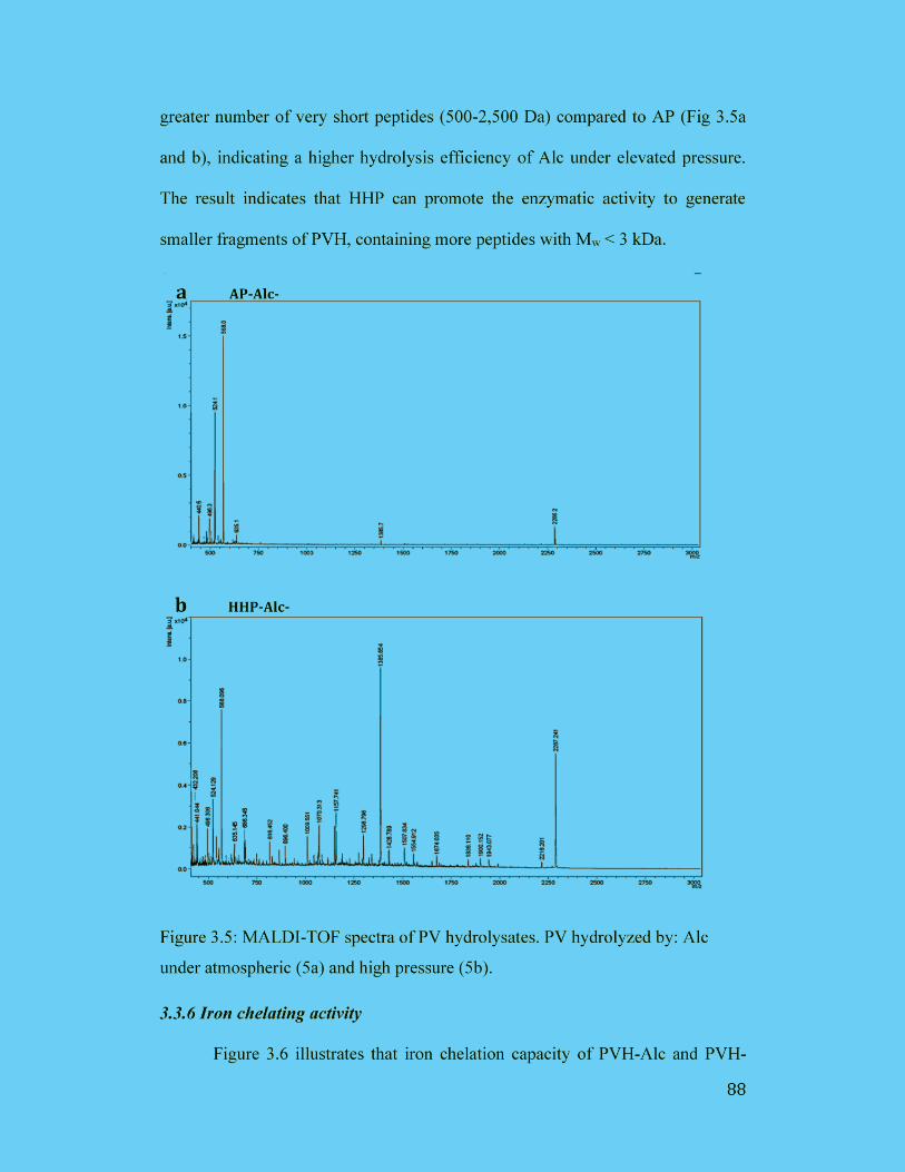
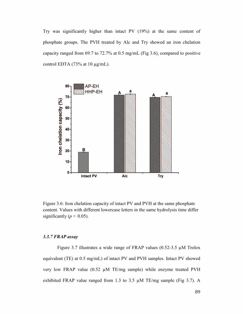
89
Try was significantly higher than intact PV (19%) at the same content of
phosphate groups. The PVH treated by Alc and Try showed an iron chelation
capacity ranged from 69.7 to 72.7% at 0.5 mg/mL (Fig 3.6), compared to positive
control EDTA (73% at 10 µg/mL).
Figure 3.6: Iron chelation capacity of intact PV and PVH at the same phosphate
content. Values with different lowercase letters in the same hydrolysis time differ
significantly (p < 0.05).
3.3.7 FRAP assay
Figure 3.7 illustrates a wide range of FRAP values (0.52-3.5 µM Trolox
equivalent (TE) at 0.5 mg/mL) of intact PV and PVH samples. Intact PV showed
very low FRAP value (0.52 µM TE/mg sample) while enzyme treated PVH
exhibited FRAP value ranged from 1.3 to 3.5 µM TE/mg sample (Fig 3.7). A
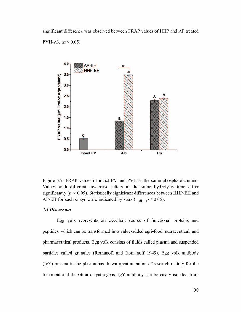
90
significant difference was observed between FRAP values of HHP and AP treated
PVH-Alc (p < 0.05).
Figure 3.7: FRAP values of intact PV and PVH at the same phosphate content.
Values with different lowercase letters in the same hydrolysis time differ
significantly (p < 0.05). Statistically significant differences between HHP-EH and
AP-EH for each enzyme are indicated by stars ( p < 0.05).
3.4 Discussion
Egg yolk represents an excellent source of functional proteins and
peptides, which can be transformed into value-added agri-food, nutraceutical, and
pharmaceutical products. Egg yolk consists of fluids called plasma and suspended
particles called granules (Romanoff and Romanoff 1949). Egg yolk antibody
(IgY) present in the plasma has drawn great attention of research mainly for the
treatment and detection of pathogens. IgY antibody can be easily isolated from
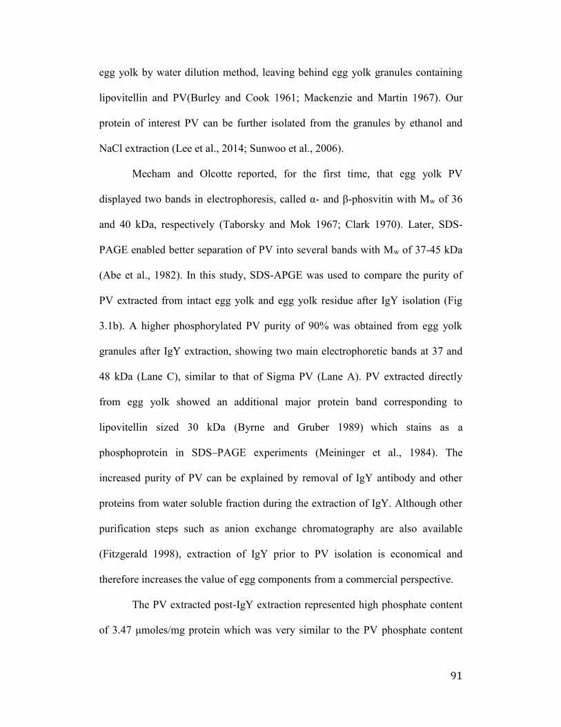
91
egg yolk by water dilution method, leaving behind egg yolk granules containing
lipovitellin and PV(Burley and Cook 1961; Mackenzie and Martin 1967). Our
protein of interest PV can be further isolated from the granules by ethanol and
NaCl extraction (Lee et al., 2014; Sunwoo et al., 2006).
Mecham and Olcotte reported, for the first time, that egg yolk PV
displayed two bands in electrophoresis, called α- and -phosvitin with Mw of 36
and 40 kDa, respectively (Taborsky and Mok 1967; Clark 1970). Later, SDS-
PAGE enabled better separation of PV into several bands with Mw of 37-45 kDa
(Abe et al., 1982). In this study, SDS-APGE was used to compare the purity of
PV extracted from intact egg yolk and egg yolk residue after IgY isolation (Fig
3.1b). A higher phosphorylated PV purity of 90% was obtained from egg yolk
granules after IgY extraction, showing two main electrophoretic bands at 37 and
48 kDa (Lane C), similar to that of Sigma PV (Lane A). PV extracted directly
from egg yolk showed an additional major protein band corresponding to
lipovitellin sized 30 kDa (Byrne and Gruber 1989) which stains as a
phosphoprotein in SDS–PAGE experiments (Meininger et al., 1984). The
increased purity of PV can be explained by removal of IgY antibody and other
proteins from water soluble fraction during the extraction of IgY. Although other
purification steps such as anion exchange chromatography are also available
(Fitzgerald 1998), extraction of IgY prior to PV isolation is economical and
therefore increases the value of egg components from a commercial perspective.
The PV extracted post-IgY extraction represented high phosphate content
of 3.47 μmoles/mg protein which was very similar to the PV phosphate content
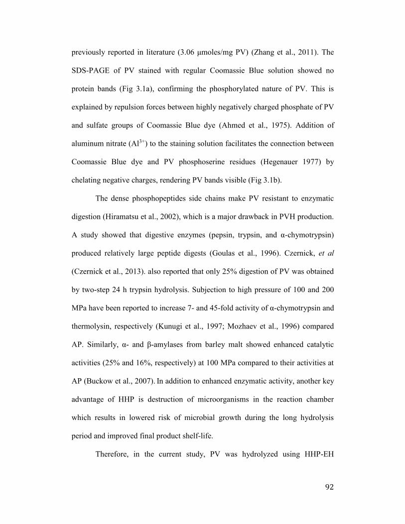
92
previously reported in literature (γ.06 μmoles/mg PV) (Zhang et al., 2011). The
SDS-PAGE of PV stained with regular Coomassie Blue solution showed no
protein bands (Fig 3.1a), confirming the phosphorylated nature of PV. This is
explained by repulsion forces between highly negatively charged phosphate of PV
and sulfate groups of Coomassie Blue dye (Ahmed et al., 1975). Addition of
aluminum nitrate (Al3+) to the staining solution facilitates the connection between
Coomassie Blue dye and PV phosphoserine residues (Hegenauer 1977) by
chelating negative charges, rendering PV bands visible (Fig 3.1b).
The dense phosphopeptides side chains make PV resistant to enzymatic
digestion (Hiramatsu et al., 2002), which is a major drawback in PVH production.
A study showed that digestive enzymes (pepsin, trypsin, and α-chymotrypsin)
produced relatively large peptide digests (Goulas et al., 1996). Czernick, et al
(Czernick et al., 2013). also reported that only 25% digestion of PV was obtained
by two-step 24 h trypsin hydrolysis. Subjection to high pressure of 100 and 200
MPa have been reported to increase 7- and 45-fold activity of α-chymotrypsin and
thermolysin, respectively (Kunugi et al., 1997; Mozhaev et al., 1996) compared
AP. Similarly, α- and -amylases from barley malt showed enhanced catalytic
activities (25% and 16%, respectively) at 100 MPa compared to their activities at
AP (Buckow et al., 2007). In addition to enhanced enzymatic activity, another key
advantage of HHP is destruction of microorganisms in the reaction chamber
which results in lowered risk of microbial growth during the long hydrolysis
period and improved final product shelf-life.
Therefore, in the current study, PV was hydrolyzed using HHP-EH
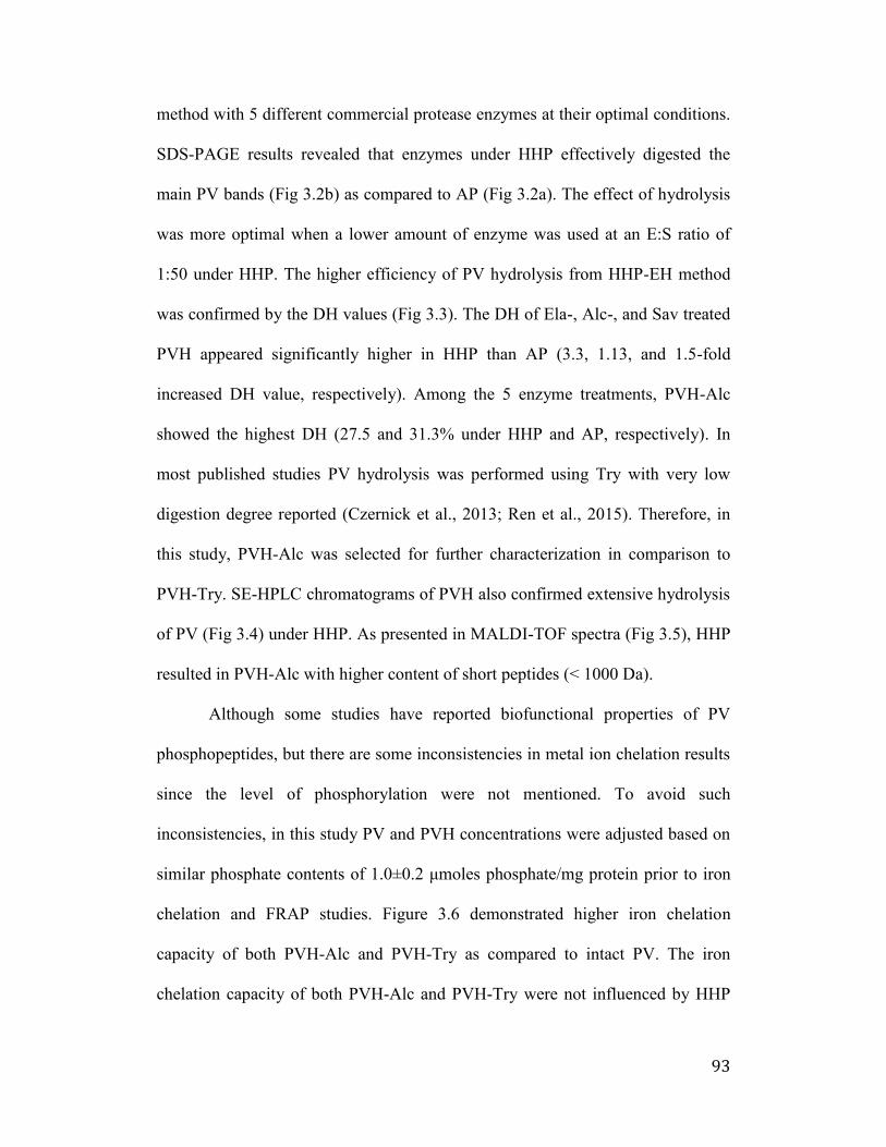
93
method with 5 different commercial protease enzymes at their optimal conditions.
SDS-PAGE results revealed that enzymes under HHP effectively digested the
main PV bands (Fig 3.2b) as compared to AP (Fig 3.2a). The effect of hydrolysis
was more optimal when a lower amount of enzyme was used at an E:S ratio of
1:50 under HHP. The higher efficiency of PV hydrolysis from HHP-EH method
was confirmed by the DH values (Fig 3.3). The DH of Ela-, Alc-, and Sav treated
PVH appeared significantly higher in HHP than AP (3.3, 1.13, and 1.5-fold
increased DH value, respectively). Among the 5 enzyme treatments, PVH-Alc
showed the highest DH (27.5 and 31.3% under HHP and AP, respectively). In
most published studies PV hydrolysis was performed using Try with very low
digestion degree reported (Czernick et al., 2013; Ren et al., 2015). Therefore, in
this study, PVH-Alc was selected for further characterization in comparison to
PVH-Try. SE-HPLC chromatograms of PVH also confirmed extensive hydrolysis
of PV (Fig 3.4) under HHP. As presented in MALDI-TOF spectra (Fig 3.5), HHP
resulted in PVH-Alc with higher content of short peptides (< 1000 Da).
Although some studies have reported biofunctional properties of PV
phosphopeptides, but there are some inconsistencies in metal ion chelation results
since the level of phosphorylation were not mentioned. To avoid such
inconsistencies, in this study PV and PVH concentrations were adjusted based on
similar phosphate contents of 1.0±0.2 μmoles phosphate/mg protein prior to iron
chelation and FRAP studies. Figure 3.6 demonstrated higher iron chelation
capacity of both PVH-Alc and PVH-Try as compared to intact PV. The iron
chelation capacity of both PVH-Alc and PVH-Try were not influenced by HHP
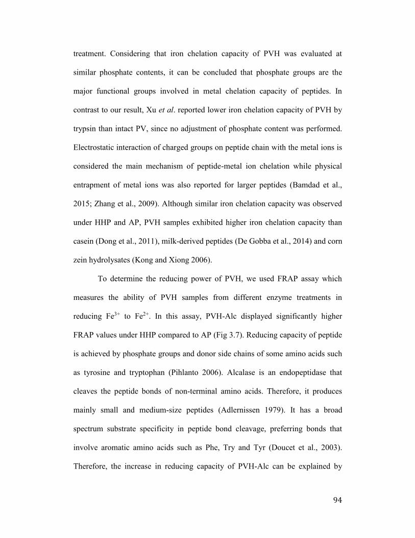
94
treatment. Considering that iron chelation capacity of PVH was evaluated at
similar phosphate contents, it can be concluded that phosphate groups are the
major functional groups involved in metal chelation capacity of peptides. In
contrast to our result, Xu et al. reported lower iron chelation capacity of PVH by
trypsin than intact PV, since no adjustment of phosphate content was performed.
Electrostatic interaction of charged groups on peptide chain with the metal ions is
considered the main mechanism of peptide-metal ion chelation while physical
entrapment of metal ions was also reported for larger peptides (Bamdad et al.,
2015; Zhang et al., 2009). Although similar iron chelation capacity was observed
under HHP and AP, PVH samples exhibited higher iron chelation capacity than
casein (Dong et al., 2011), milk-derived peptides (De Gobba et al., 2014) and corn
zein hydrolysates (Kong and Xiong 2006).
To determine the reducing power of PVH, we used FRAP assay which
measures the ability of PVH samples from different enzyme treatments in
reducing Fe3+ to Fe2+. In this assay, PVH-Alc displayed significantly higher
FRAP values under HHP compared to AP (Fig 3.7). Reducing capacity of peptide
is achieved by phosphate groups and donor side chains of some amino acids such
as tyrosine and tryptophan (Pihlanto 2006). Alcalase is an endopeptidase that
cleaves the peptide bonds of non-terminal amino acids. Therefore, it produces
mainly small and medium-size peptides (Adlernissen 1979). It has a broad
spectrum substrate specificity in peptide bond cleavage, preferring bonds that
involve aromatic amino acids such as Phe, Try and Tyr (Doucet et al., 2003).
Therefore, the increase in reducing capacity of PVH-Alc can be explained by
95
more exposure of polar and charged residues. Amino acid sequence of PV is rich
in serine (55%) and charged residues (24%). Phosphorylation of serine residues
also provides an additional source of electrons to have a high redox potential.
3.5 Conclusion
The present study provides an important insight that the bioactive
properties of PVH is closely related to the enzyme used, due to the effect of
enzymes on amino acid profile and digestibility nature of peptide hydrolysates. To
overcome the challenge of PV hydrolysis, in this study Alcalase enzyme was used
under HHP to prepare bioactive PVH. The HHP-EH method produced PVHs
containing shorter bioactive peptides with strong iron chelation and reducing
capacities. PVH-Alc can used as natural mineral chelating, antioxidant and anti-
inflammatory agents in food, nutraceutical and pharmaceutical products. Further
investigation is required to identify the amino acid sequence of effective peptides
and confirm the correlation between metal chelation and antimicrobial capacity of
these peptides.
Chapter 4 (Study 3): Growth Inhibitory Effects of Combination of IgY and
Phosvitin to Enterotoxigenic Escherichia coli K88 and K99 in vitro
96
4.1 Introduction
Enterotoxigenic Escherichia coli (ETEC) is the main cause of fatal
diarrhea in piglets (Moxley et al., 1998), during the first week of life and over the
time of weaning (Butler 1994; Vaandrager et al., 1992). The strains of ETEC
associated with porcine intestinal colonization are those that express the K88,
K99, P987 and F41 fimbrial adhesions (Garabal et al., 1997). Pathogenesis of
ETEC-causing diarrhea involves intestinal colonization mediated by fimbriae and
hypersecretion of water and electrolytes caused by heat-stable or heat-labile
enterotoxins, or both (Holmgren 1985; Saeed et al., 1983; Parry S H 1984). On
account of the incidence, severity and large economic loss, ETEC has been the
subject of intensive study.
While feeds primarily serve as sources of nutrients for animals, they can
also be important vehicles for the transmission of infectious diseases to and from
animals. Protective strategies for infections are not simple, since they include
hygiene improvement and effective ETEC vaccines development. Although
antibiotics have been routinely used in an animal feed to promote growth and
prevent diseases in pig farms (de Lange 2006) the emerging problem of multiple
antimicrobial resistance affect the efficacy of drugs used. Moreover, due to public
concerns about healthy foods and pressure to discontinue use of antibiotics,
alternatives therapies to achieve similar goals are in dire need.
Antibodies are ideal candidates to replace the use of antibiotics in the
animal feed industries. Chickens have been known as a potent and economic egg
yolk antibody producers, termed as immunoglobulin Y (IgY) (Leslie and Clem
97
1969), in substitute of mammalian antibodies. The IgY accumulated in chicken
egg yolk can be more largely produced and more simply purified in comparison to
the mammalian IgG. In addition to these advantages, IgY is relatively stable under
various conditions, including heat, pressure, acid, alkali, and proteolytic enzymes
(Otani 1991). These characteristics of IgY have made it possible to achieve a
broad range of applications such as passive immunization and diagnosis (Sim
2000).
Both experimental and clinical studies indicate that orally administered
IgY has strong immune modulating properties including anti-toxin (Lee et al.,
2015), anti-microbial (Wang et al., 2014; Sui et al., 2011) and anti-allergen effects
(Gujral et al., 2015). In particular, IgY has been found to be imperative to the
health and function of the gastrointestinal tract, and greatly reduces systemic and
intestinal inflammation (Vega et al., 2011; Sarker et al., 2007).
Another glycoprotein egg yolk component, phosvitin (PV, 36-40 kDa), is
produced by proteolytic cleavage of chicken vitellogenin, and functions as a
metal-binding protein in the egg yolk (Richards, 1997). Due to the high
proportion of phosphorylated serine residues, PV shows iron uptake regulation
(Feng et al., 2006), metal chelation (Choi et al., 2005), antioxidation (Katayama et
al., 2006; Xu et al., 2007), emulsification (Chung et al., 1992) and antimicrobial
(Khan et al., 2000) properties.
Although, both IgY and PV in egg yolk possess anti-microbial activity,
their dual use to prevent feed contamination have not been explored. A
combination of IgY specific for ETEC and metal-chelating PV may have a
98
synergistic effect in reducing the risk of feed contamination with ETEC by
inhibiting bacterial proliferation and stipulating protection against ETEC
infection. To support IgY and PV application in the feed industry, this study is
aimed to produce and investigating their effect on ETEC in vitro growth
inhibition.
4.2. Materials and Methods
4.2.1 Materials
Eggs were purchased from a local market. A local strain of ETEC K88 and
K99 (Em 88-1604) were obtained from Center for Nutraceutical and
Pharmaceutical Materials, Myongji University, Yongin, Korea. Phosvitin,
Freund’s incomplete adjuvant, purified chicken IgG, rabbit anti-chicken IgG and
rabbit anti-chicken IgG conjugated with horseradish peroxidase (HP), Alcalase,
and Savinase (Sigma-Aldrich, Oakville, ON, Canada), Elastase and Thermolysin
(MP Biomedicals, Santa Ana, Ca, USA) and Trypsin (Gibco, Burlington, ON,
Canada) were obtained from commercial sources. Sephadex G-200 column (2.5 x
119 cm) was purchased from GE Healthcare (Piscataway, NJ, USA). Microtiter
96-wells plates were purchased from Costar Inc. (Corning, NY, USA). 2-β’-azino-
bis (3-ethylbenzthiazoline-6- sulphonic acid) (ABTS) substrate was purchased
from KPL (Frederick, MD, USA). Pierce™ modified Lowry protein assay and
Pierce™ phosphoprotein phosphate estimation assay kits were obtained Thermo
scientific (Ottawa, ON, Canada). Precision Plus Dual Xtra Protein Standard,
Bradford protein assay and electrophoresis reagents were obtained from BioRad
(Missisauga, ON, Canada). Membrane filter 0.22 mm-pore-size was obtained from
99
Millipore (Bedford, MA, USA). Columbia agar was from Oxoid (Basingstoke,
Hampshire, England) and TSB agar plates from Difco (Detroit, MI, USA). All
other reagents were of analytical grade.
4.2.2 Apparatus
A portable scale high-hydrostatic pressure processor (TFS-2L, Toyo-
Koatsu Co. Ltd., Hiroshima, Japan) was used to apply HHP during enzyme
treatment. The BioLogic DuoFlow chromatography system and Mini-Protein III
electrophoresis kit (BioRad, Mississauga, ON, Canada) was used for gel
chromatography and electrophoretic techniques, respectively. Bio-Rad Gel DocTM
EZ Imager (BioRad, Mississauga, ON, Canada) was used to image gels. The
VersaMax ELISA microplate reader (Molecular Devices Corp, Sunnyvale, CA,
USA) was used to obtain sample UV/Vis optical reading. Beckman Coulter
centrifuge (Allegra X-30R, Brea, CA, USA) was used in all centrifugation steps.
The Innova 42 incubator (New Brunswick Scientific, Edison, NY, USA) was used
for culture plate incubation. The Nikon Eclipse TS 100 (Nikon Canada Inc.,
Mississauga, ON, Canada) was used to perform microscopic procedures.
4.2.3 Anti-ETEC IgY Antibody Preparation
4.2.3.1 Bacteria and Culture Conditions
Strains of ETEC K88 and K99 bacteria were cultured in E-media broth
(0.1 mM MgSO47H2O, 0.01 M Citric acid, 0.06 M K2HPO4, 0.02 M
NaNH4HPO4H2O, 0.002% chloroform (v/v), 0.5% dextrose (v/v)) at 37C for 48
hr with shaking. After incubation, cells were harvested by centrifugation at 5,000
×g for 15 min and were treated with 3.7% formalin overnight. The inactivated
100
cells were washed three times, suspended in sterile saline and then freeze-dried.
Lyophilized whole cell cultures were stored at -20C until used.
4.2.3.2 Immunization of Chickens
All chickens were cared for in accordance with the Canadian Council on
Animal Care guidelines of animal welfare. Immunization of hens was carried out
as described by Sunwoo et al., (1996). Lyophilized ETEC K88 and K99 whole
cells were suspended in sterilized phosphate buffered saline (PBS, 500 mg of
cell/mL; 15.6 mg of protein/mL) and emulsified with an equal volume of Freund’s
incomplete adjuvant. Single Comb White Leghorn (SCWL) 23-week-old chickens
were divided into 3 groups (n=8) and immunized intramuscularly at four different
sites (0.25 mL per site) of breast muscles with ETEC K88 and K99 or without cell
emulsion as a control. Two booster immunizations of each antigen were given at
week 2 and 4 after the initial immunization in the same manner. Eggs were
collected daily and stored at 4C until used.
4.2.3.3 IgY Separation from Egg Yolk
The water soluble fractions (WSF) containing anti-ETEC K88, anti-ETEC
K99 and control egg yolk antibody (IgY) were prepared from egg yolk using the
water dilution method (Gujral et al., 2015). Eggs were manually broken, and
yolks were carefully separated from egg white and chalazae by rolling on
Whatman filter paper (No. 1). The yolk membrane was punctured with a needle
and the content was collected in a beaker cooled in iced water. Temperature was
maintained at 4°C all through the process. Egg yolks were added to nine volumes
of pre-chilled acidic water a (final pH of 5.0-5.2) and incubated at 4°C for 12 hr
101
without agitation. After centrifugation at 3200 × g for 30 min, supernatant
(containing IgY antibody) was separated from the precipitate. The WSF was
freeze-dried and analyzed for protein content, total IgY, and specific IgY content.
The precipitate, containing egg yolk granules, was also used for the isolation of
PV.
4.2.4 Protein Assay
The Bio-Rad protein assays based on the method of Bradford was
performed using the purified chicken IgG (1 mg/mL of protein) as a reference
protein. The powder of WSF (diluted 1:100 in phosphate buffered saline, PBS)
and two-fold serial dilutions of the reference protein in PBS (0.5 to 0.0625
mg/mL) were analyzed on the microtiter plate. Absorbance at 595 nm after 5 min
reaction was measured using a kinetic microplate reader. The protein
concentrations of reconstituted ETEC-K88, -K99 specific and non-specific IgY
were measured by using the same procedures.
4.2.5 Specific IgY Concentration
The concentration of ETEC K88 and K99-specific IgY in the WSF
powder was measured by using the ELISA method as described by Gujral et al.,
(2012). Unless indicated otherwise, all incubations were performed at 37°C with
four times washing by PBS-Tween 20 (PBST) in each step. Wells of a microtiter
plate were coated with 150 mL of rabbit anti-chicken IgG and lyophilized whole
cells (ETEC K88 or K99) at concentrations of 3.75 mg/mL and 0.5 mg/mL in
carbonate-bicarbonate buffer (0.05 M, pH 9.6), respectively, for 90 min. Non-
specific binding sites were blocked with 200 ml 1% Bovine serum albumin (BSA)
102
in PBS and incubated for 45 min. After washing, three dilutions of reconstituted
specific (25, 12.5 and 6.25 mg/mL) and non-specific (4.5, 2.25, and 1.13 mg/mL)
IgY powder in PBS were added to wells (150 ml/well) coated with ETEC K88
whole cells. Wells coated with rabbit anti-chicken IgG were filled with two-fold
serial dilutions of purified chicken IgG (1 mg/mL) in PBS (0.5 to 0.008 mg/mL).
The plates were subsequently added with rabbit anti-chicken IgG conjugated with
horseradish peroxidase (diluted 1:1,000 in PBS) and incubated for 90 min,
followed by addition of 150 ml of freshly prepared substrate solution, ABTS in
0.05 M phosphate citrate buffer (pH 5.0) containing 30% hydrogen peroxide. The
reaction was continued for 30 min. Absorbance of mixture was read at 405 nm
using a kinetic microplate reader. The ELISA value of antibody activity was
determined by subtracting the value of control antibody from that of specific
antibody. Specific IgY concentration was calculated using the quantitative
standard curve determined by the titration between rabbit anti-chicken IgG and
purified chicken IgG.
4.2.6 Total IgY Concentration
The ELISA was performed as described above, except the microtiter plate
was coated with 150 ml of rabbit anti-chicken IgG at a final concentration of 3.75
mg/mL. The WSF samples were diluted 1:90,000 with PBS. Specific or non-
specific IgY powder was reconstituted and serially diluted with PBS (2 to 0.125
mg/mL). Two-fold serial dilutions of purified chicken IgG (1 mg/mL) in PBS (0.5
to 0.031 mg/mL) were used as the reference antibody to prepare the standard
curves.
103
4.2.7 Phosvitin Hydrolysates Preparation
4.2.7.1 Phosvitin Extraction and Purification
The PV was isolated from egg yolk granule after the IgY extraction
according to the method presented by Lee et al., 2014, with slight modifications.
The precipitate was diluted in two volumes of distilled water and stirred for 1 hr.
After centrifugation at 3,200 × g for 30 min at 4°C, the precipitate was collected
and dissolved in four volumes of 85% ethanol to remove lipids. The fat-free
precipitate was collected and dissolved in nine volumes of 10% (w/v) NaCl
solution at pH 4, then centrifuged at the same condition. The supernatant was
filtered through Whatman No.1 filter paper to remove floating particles, and
desalted by using a membrane filtration of 12 kDa molecular weight cut-off
membrane.
4.2.7.2 Sodium Dodecyl Sulfate Polyacrylamide Gel Electrophoresis (SDS-
PAGE)
SDS-PAGE was carried out on Tris-glycine acrylamide gels with Tris-
glycine buffer system and 12% polyacrylamide gels. Ten µL of PV extracted
directly from egg yolk and PV extracted post-IgY extraction samples (10 mg
protein/mL) and 10 µL of Precision Plus Dual Xtra Protein Standard, as a
molecular weight marker, were loaded and electrophoresed on gels at 100 V. The
gels were stained with a solution of 0.05% Coomassie Blue R-250, 0.1M
aluminum nitrate, 25% isopropanol, 10% acetic acid and 1% TritonX-100 and
destained with 7% acetic acid (Hegenauer et al., 1977). Images of gels were
obtained using Bio-Rad Gel DocTM EZ Imager. The SDS-PAGE was used to
104
estimate the purity of both PV isolation.
4.2.7.3 Phosvitin Enzymatic Hydrolysis
Purified PV, dissolved at 10 mg protein/mL in 10% NaCl, was mixed with
enzymes Alcalase (Alc), Elastase (Ela), Thermolysin (Thr), Trypsin (Try), or
Savinase (Sav) at E:S ratio of 1:50 for 12 hr under atmospheric pressure (AP) or
high hydrostatic pressure (HHP). The treatments of enzymes were performed
under their optimal conditions: Alc (2.4 U/g; pH 7, 50°C), Ela (1 U/mg, pH 8,
37°C), Thr (14 U/mg; pH 7, 50°C), Try (30 U/g, pH 7, 37°C), and Sav (0.32 U/g;
pH 7, 55°C). After the treatment, enzymes were inactivated by heating (90°C, 10
min) and centrifuged at 5000 × g for 10 min to remove the insoluble fractions.
The supernatants were filtered through a 0.22 µm filter and stored frozen before
analysis.
4.2.8 Degree of Hydrolysis (DH)
The DH of PV and phosvitin hydrolysates (PVH) were subjected to
enzymes mentioned above, and were analyzed by measuring free primary amines
using 2,4,6-trinitrobenzenesulfonic acid (TNBSA) method with L-leucine as a
standard (Adlernissen 1979). Briefly, samples of purified PV and PVH were
mixed with phosphate buffer (pH 8.5) and 0.01% TNBS solution, incubated at
37°C for 2 hr. After incubation, reaction was terminated by adding 1 N HCl and
10% SDS. Amine content of samples was measured by reading absorbance at 335
nm. Total content of amine groups, expressed as leucine equivalent (μg/mL), was
obtained from a standard curve prepared from different concentrations of L-
leucine. DH was calculated by the following equation:
105
DH= (h/htot) × 100
Where, h (hydrolysis equivalents) is the amount of peptide bonds cleaved
during hydrolysis, expressed as millimole leucine equivalents per gram protein
(mmol/g protein); htot is the total amount of peptide bonds in the protein substrate
determined from the protein sample totally hydrolyzed with 6N HCl at 110°C for
24 hr.
4.2.9 Lowry Protein Assay
PV samples (40 μL) were mixed with β00 μL of a Modified Lowry
Reagent (Bio-rad) and β0 μL of Folin-Ciocalteu Reagent in 96-well microplates.
The assay mixture was conducted in a dark room and incubated at room
temperature for 30 min followed by absorbance measurement at 750 nm using a
kinetic microplate reader. Standard curve was prepared using Sigma PV as a
reference protein.
4.2.10 Growth Inhibition of PV and IgY
4.2.10.1 Preparation of Bacteria
The same strains of ETEC K88 and K99 used as antigens for immunizing
chickens were separately subcultured on blood agar plates (5% defibrinated sheep
blood in Columbia agar) at 37C overnight and then suspended in E-media broth.
The suspensions were adjusted to an optical density of 0.05 at 600 nm,
corresponding to a cell density of about 2.7 x 10⁷ CFU/mL. The same volume of
20% glycerol in E-media broth were added and stored at -70C until used.
Two milliliters of prepared bacteria culture were mixed with 2 mL of E-
media broth and incubated at 37C with shaking. The turbidity of culture (OD at
106
600 nm) was measured by a spectrophotometer at 1 hr intervals. The growth curve
was plotted until reaching a stationary phase.
4.2.10.2 ETEC K88 K99 Growth Inhibition
The ETEC specific IgY powder was reconstituted to 100, 200, and 400
μg/mL with E-media broth. Purified PV and PVH were diluted in E-media broth
at concentrations of 0.5, 1 and 2 mg protein/mL. Non-specific IgY powder
reconstituted to 0.5 mg/mL was used as a control. The mixtures were centrifuged
at 1,500 × g and 4C for 20 min, and supernatants collected were sterilized using
a 0.22 mm-pore-size membrane filter. Two milliliter of specific IgY, non-specific
IgY, PV or PVH was added to the same volume of prepared ETEC K88 and K99
cultures. The mixture of bacteria and treatment were incubated at 37C with a
continuous gentle shaking. Aliquots of samples (100 mL) were taken at 0, 2, 4,
and 8 hr of incubation time. Plate counts were performed by the spread plate
method on TSB agar plates in duplicate. The inoculated plates were incubated at
37C overnight. The number of colony-forming units (CFU) per plate was
counted to determine the total number of bacteria CFU per mL of sample.
4.2.10.3 Statistical Analysis
All experiments were performed at least in three independent trials, and
the results were reported as mean ± SD. Results were subjected to the analysis of
variance using the SAS software (SAS Institute, Inc., Cary, NC), and statistical
significance of differences (p < 0.05) was evaluated by the least significant
difference procedure.
107
4.3 Results
4.3.1 Concentrations of Protein, total and specific IgY in the WSF
The ETEC K88 and K99 specific IgY activities in the WSF was monitored
by the ELISA method every week during the immunization period (Figure 4.1).
The both ETEC K88 - and K99 - specific IgY increased gradually after the first
immunization followed by an exponential rise after the first booster immunization.
The level of anti-K88 and K99 IgY increased continuously to a maximum of
OD405 0.72 and 0.78, respectively, 2 weeks after the second booster immunization.
Subsequently, the level of IgY remained relatively high showing no considerable
decline throughout the period that the eggs were monitored (up to 8 weeks). Eggs
collected during 6 to 8 weeks expressing relatively high ETEC K88 - and K99 -
specific IgY titre were stored for further use.
Table 4.1 shows that both protein and total IgY concentrations were
relatively constant in ETEC specific and non-specific WSF (p > 0.05). The
percent ratio of total IgY to total protein of all 3 WSF samples was approximately
18%. The antigen specific IgY concentration of the control sample was
significantly lower than the experimental sample containing antigen specific IgY
(p < 0.05). The proportion of ETEC K88- and K99-specific IgY in total IgY
concentration was 7.8% and 8%, respectively.
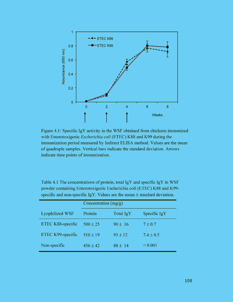
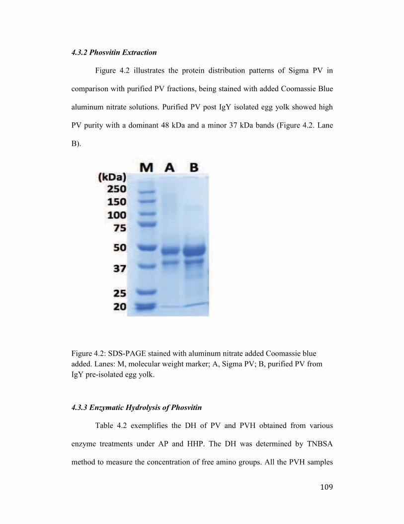
109
4.3.2 Phosvitin Extraction
Figure 4.2 illustrates the protein distribution patterns of Sigma PV in
comparison with purified PV fractions, being stained with added Coomassie Blue
aluminum nitrate solutions. Purified PV post IgY isolated egg yolk showed high
PV purity with a dominant 48 kDa and a minor 37 kDa bands (Figure 4.2. Lane
B).
Figure 4.2: SDS-PAGE stained with aluminum nitrate added Coomassie blue
added. Lanes: M, molecular weight marker; A, Sigma PV; B, purified PV from
IgY pre-isolated egg yolk.
4.3.3 Enzymatic Hydrolysis of Phosvitin
Table 4.2 exemplifies the DH of PV and PVH obtained from various
enzyme treatments under AP and HHP. The DH was determined by TNBSA
method to measure the concentration of free amino groups. All the PVH samples
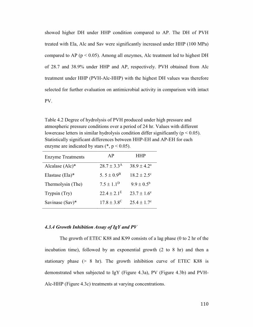
110
showed higher DH under HHP condition compared to AP. The DH of PVH
treated with Ela, Alc and Sav were significantly increased under HHP (100 MPa)
compared to AP (p < 0.05). Among all enzymes, Alc treatment led to highest DH
of 28.7 and 38.9% under HHP and AP, respectively. PVH obtained from Alc
treatment under HHP (PVH-Alc-HHP) with the highest DH values was therefore
selected for further evaluation on antimicrobial activity in comparison with intact
PV.
Table 4.2 Degree of hydrolysis of PVH produced under high pressure and
atmospheric pressure conditions over a period of 24 hr. Values with different
lowercase letters in similar hydrolysis condition differ significantly (p < 0.05).
Statistically significant differences between HHP-EH and AP-EH for each
enzyme are indicated by stars (*, p < 0.05).
Enzyme Treatments AP HHP
Alcalase (Alc)* 28.7 3.3A 38.9 4.2a
Elastase (Ela)* 5. 5 0.9B 18.2 2.5c
Thermolysin (The) 7.5 1.1D 9.9 0.5b
Trypsin (Try) 22.4 2.1E 23.7 1.6e
Savinase (Sav)* 17.8 3.8C 25.4 1.7e
4.3.4 Growth Inhibition Assay of IgY and PV
The growth of ETEC K88 and K99 consists of a lag phase (0 to 2 hr of the
incubation time), followed by an exponential growth (2 to 8 hr) and then a
stationary phase (> 8 hr). The growth inhibition curve of ETEC K88 is
demonstrated when subjected to IgY (Figure 4.3a), PV (Figure 4.3b) and PVH-
Alc-HHP (Figure 4.3c) treatments at varying concentrations.
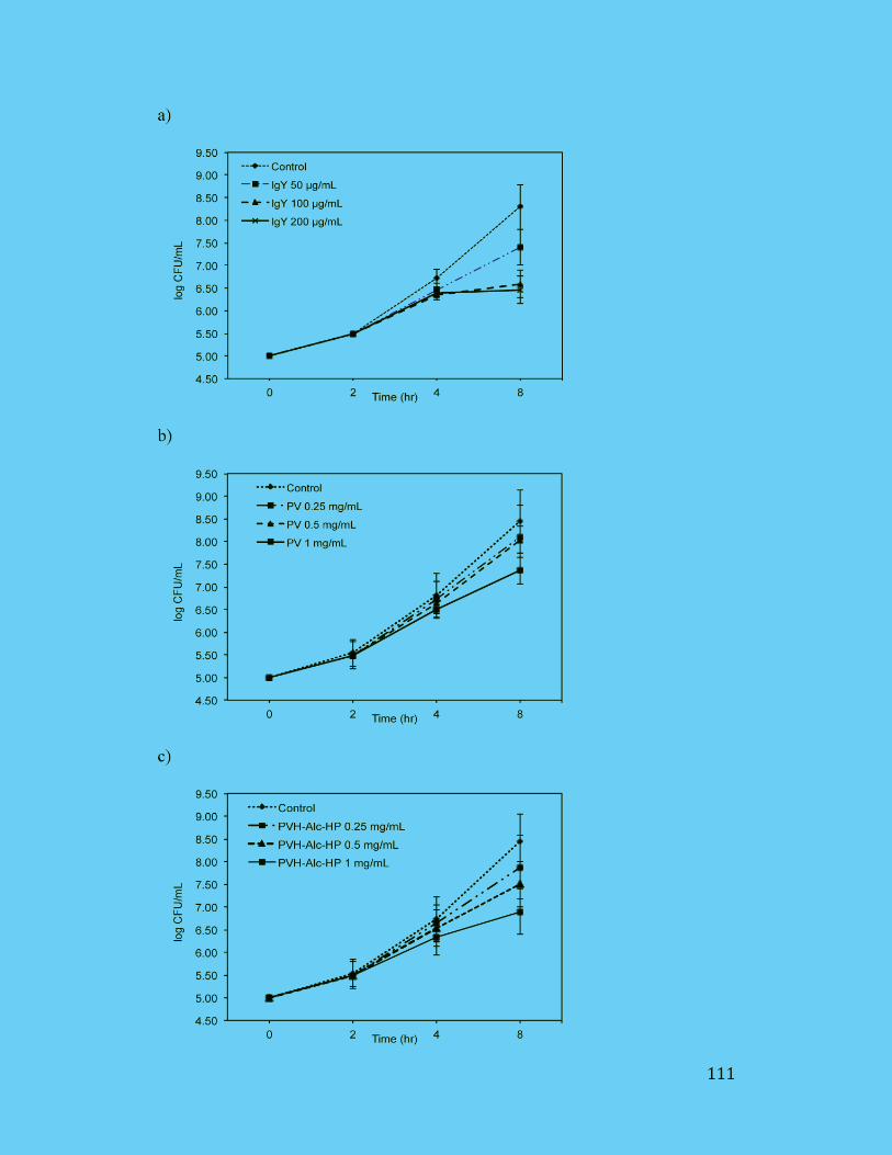
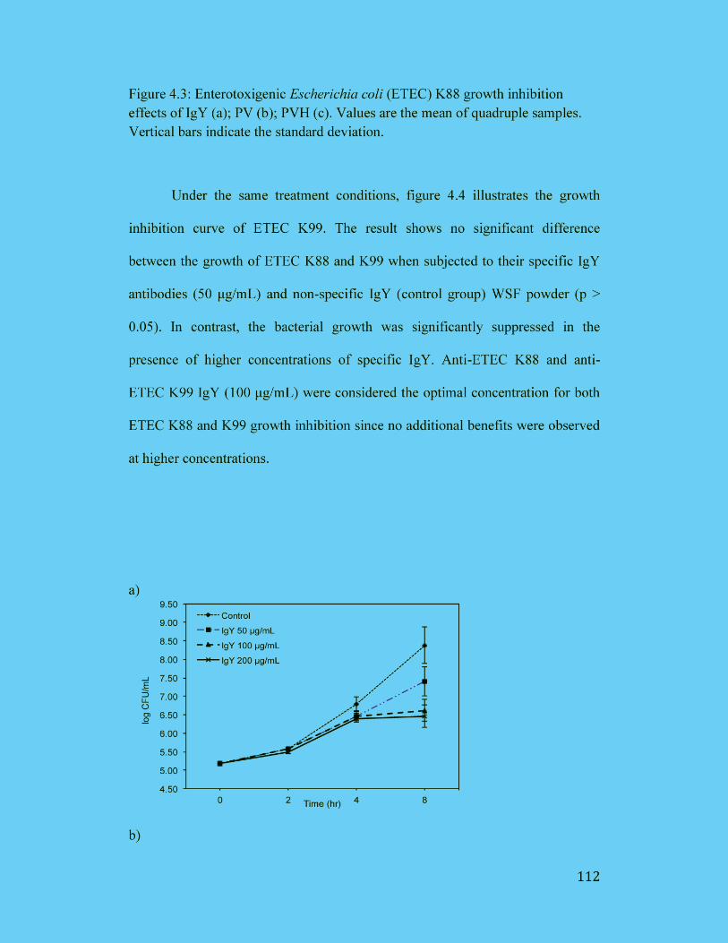
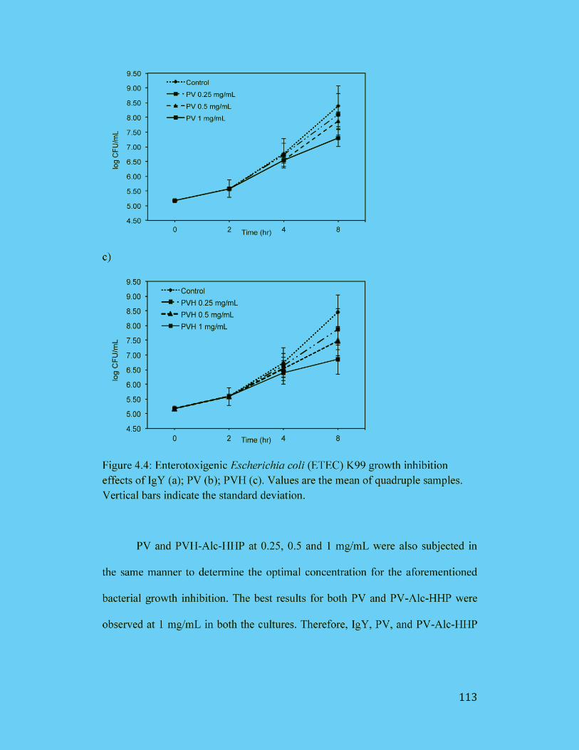
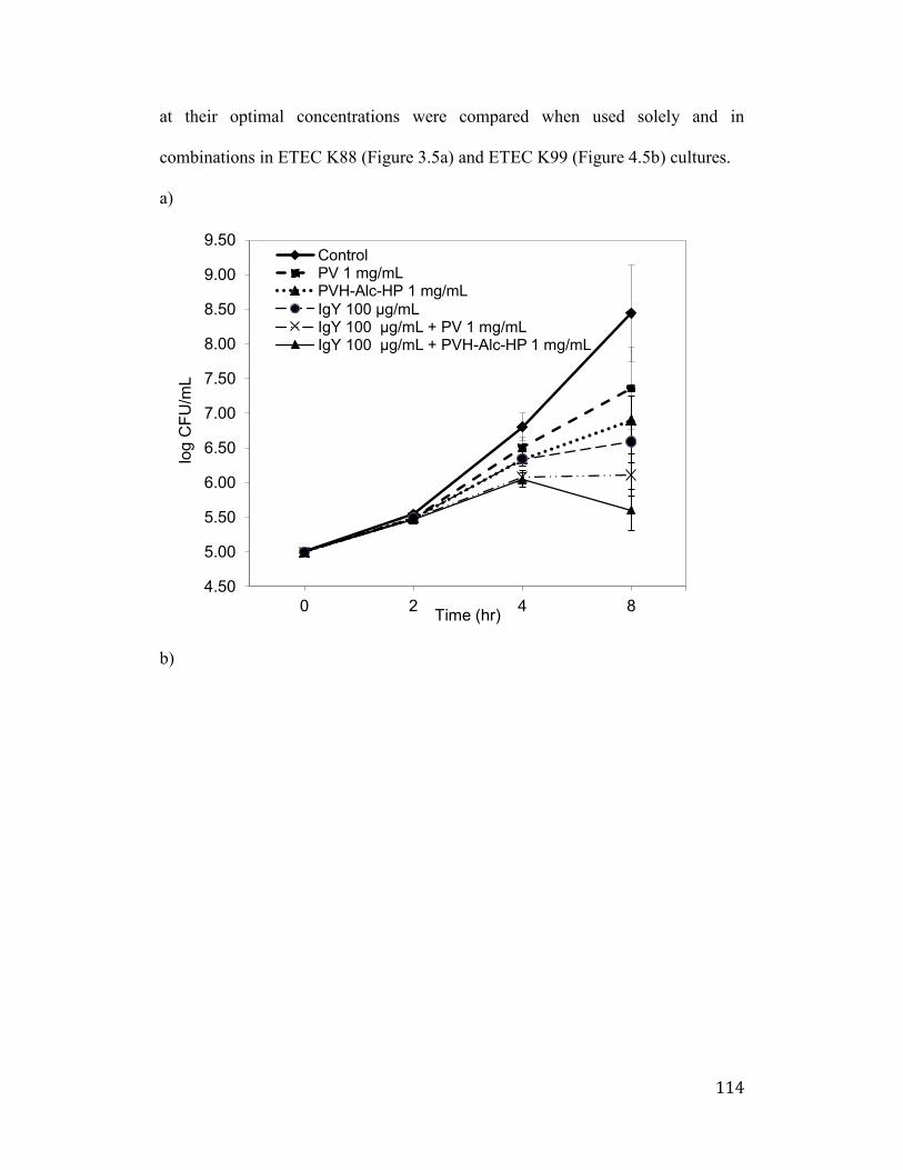
114
at their optimal concentrations were compared when used solely and in
combinations in ETEC K88 (Figure 3.5a) and ETEC K99 (Figure 4.5b) cultures.
a)
b)
4.50
5.00
5.50
6.00
6.50
7.00
7.50
8.00
8.50
9.00
9.50
0 2 4 8
log
CF
U/m
L
Time (hr)
ControlPV 1 mg/mLPVH-Alc-HP 1 mg/mL
IgY 100 μg/mLIgY 100 μg/mL + PV 1 mg/mLIgY 100 μg/mL + PVH-Alc-HP 1 mg/mL
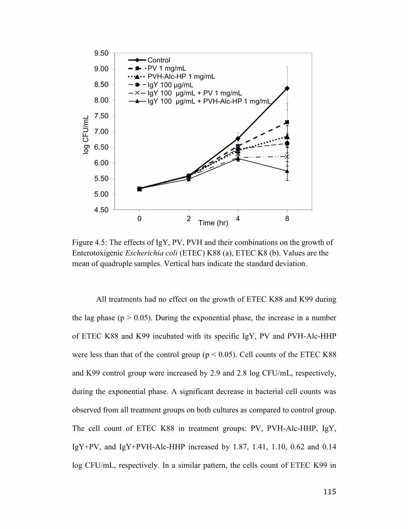
115
Figure 4.5: The effects of IgY, PV, PVH and their combinations on the growth of
Enterotoxigenic Escherichia coli (ETEC) K88 (a), ETEC K8 (b). Values are the
mean of quadruple samples. Vertical bars indicate the standard deviation.
All treatments had no effect on the growth of ETEC K88 and K99 during
the lag phase (p > 0.05). During the exponential phase, the increase in a number
of ETEC K88 and K99 incubated with its specific IgY, PV and PVH-Alc-HHP
were less than that of the control group (p < 0.05). Cell counts of the ETEC K88
and K99 control group were increased by 2.9 and 2.8 log CFU/mL, respectively,
during the exponential phase. A significant decrease in bacterial cell counts was
observed from all treatment groups on both cultures as compared to control group.
The cell count of ETEC K88 in treatment groups: PV, PVH-Alc-HHP, IgY,
IgY+PV, and IgY+PVH-Alc-HHP increased by 1.87, 1.41, 1.10, 0.62 and 0.14
log CFU/mL, respectively. In a similar pattern, the cells count of ETEC K99 in
4.50
5.00
5.50
6.00
6.50
7.00
7.50
8.00
8.50
9.00
9.50
0 2 4 8
log
CF
U/m
L
Time (hr)
ControlPV 1 mg/mLPVH-Alc-HP 1 mg/mL
IgY 100 μg/mLIgY 100 μg/mL + PV 1 mg/mLIgY 100 μg/mL + PVH-Alc-HP 1 mg/mL
116
treatment groups: PV, PVH-Alc-HHP, IgY, IgY+PV, and IgY+PVH-Alc-HHP
increased by 1.72, 1.27, 1.04, 0.62 and 0.26 log CFU/mL, respectively.
4.4 Discussion
In recent years, consumers are in demand of natural food antimicrobials to
extend the shelf life and to enhance the safety of foods. These include
antimicrobial substances from a microbe, plant or animal source (Gálvez et al.,
2014). In this study, we produced and explored the antimicrobial ability of two
egg-derived compounds: IgY and PV in ETEC K88 and K99 media culture
systems.
To implement IgY as an animal feed supplement, modest and practical
antibody production is required. In this study we produced anti-ETEC K88 and
K99 IgY antibodies in SCWL chickens by intramuscular immunization of ETEC
K88 and K99 whole cells, respectively, with Freund’s incomplete adjuvant. Both
antibodies produced contained high titre of specific IgY antibodies during weeks
6-8 of the immunization period (Figure 4.1), indicating that SCWL chickens
expressed relatively strong immune response against ETEC K88 and K99 whole
cells.
IgY (aka -livetin) exists in egg yolk together with two other water-soluble
proteins (-, and -livetin) and lipoprotein (McCully KA et al., 1962). Therefore,
separation of IgY requires extraction of the water-soluble proteins from yolk
lipoprotein, followed by separation from other livetins (Polson et al., 1980). The
WSF containing IgY was isolated from ETEC K88 and K99 hyperimmunized
eggs using the water dilution method. The freeze-drying being a suitable method
117
for preparation of dried concentrated product with high yield and lower moisture
content (Yokoyama et al., 1992), was used to prepare IgY powder of WSF
containing specific and non-specific IgY. The concentration of protein and total
IgY was subsequently determined, as shown in Table 4.1, which is relatively
similar to previous studies (Sunwoo et al., 2010). In literature, the percentage of
specific in total IgY antibody ranged from 5 (anti-insulin antibody) to 28% (anti-
mouse IgG antibody) (Hatta et al., 1997). Our results showed 7.8% and 8% of the
ETEC K88 and ETEC K99-specific IgY concentration in total IgY, respectively,
determined by ELISA.
PV is another attractive egg yolk component with functional
phosphopeptides for metal ion binding, which possesses remarkable bactericidal
property (Khan et al., 2000). In this study, PV purification performed using IgY
extracted egg yolk precipitate, demonstrated higher purity as compared to intact
egg yolk (Figure 4.2). The result was confirmed by gel chromatography indicating
that pre-isolation of IgY from egg yolk removed unwanted proteins. This method
was therefore carried out for PV purification.
Due to the negatively charged phosphorylated PV with serine residues, PV
causes insoluble complex in gastrointestinal tract (Ishikawa et al., 2007).
Hydrolysis of PV into PVH may restrain the intact PV from the formation of
insoluble complexes, therefore assisting the absorption of calcium and iron in the
gut (Choi et al., 2005). However, the enzymatic hydrolysis of PV is extremely
difficult, due to negative charges of the phosphate group surrounding the PV
molecule, which prevents enzymes from access to the peptide bonds (Gray, 1971).
118
HHP is reported to accelerate enzyme activity (Sunwoo et al., 2014), hence, in
this study, HHP treatment is used to ease enzyme access into PV structure during
enzymatic treatment to produce higher yield of smaller PVH fragments. Since
different classes of protease differ in their peptide specificity, several commercial
protease enzymes were used to determine the most appropriate protease for PVH
production. Most published studies on PV hydrolysis used Try and reported very
low digestion degree (Czernick et al., 2013; Ren et al., 2015). It has also been
reported that the activity of α-chymotrypsin and thermolysin increased 7- and 45-
fold at 100 and 200 MPa, respectively (Kunugi et al., 1997; Mozhaev et al.,
1996), compared to their activity at AP. Similarly, α- and -amylases from barley
malt showed enhanced catalytic activities (25% and 16%, respectively) at 100
MPa compared to their activities at optimum catalytic temperatures and AP
(Buckow et al., 2007). Furthermore, a key advantage of using HHP is the
destruction of microorganisms in a reaction chamber which results in lower risk
of microbial growth during long hydrolysis time and improved shelf-life of the
final product.
The DH of PVH revealed that enzymes under HHP digested PV to a
higher extend as compared to AP condition (Table 3.2). The DH of Ela-, Alc-, and
Sav treated PVH appeared significantly higher in HHP than AP (3.3, 1.13, and
1.5-fold increase in DH value, respectively). Among all enzyme treated PVH,
PVH-Alc-HHP showed the highest DH of 38.9%. Therefore, PVH-Alc-HHP is
selected for ETEC growth inhibition study in comparison with intact PV.
119
There have been several reports on the use of IgY in the prevention and
control of diarrhea causing infections in animal. IgY against Salmonella spp.
proved useful in preventing the infection (Chalghoumi et al., 2009; Lee et al.,
2002). IgY against ETEC 987P whole cells resulted in in vitro growth inhibition
of effect ETEC 987P (Sunwoo et al., 2010), and E. Coli O157:H7 (Sunwoo et al.,
2002). Specific IgY against K88 and K99 fimbrial adhesins of ETEC prevented
neonatal piglets from fatal enteric colibacillosis (Yokoyama et al., 1992). Weaned
pigs challenged with F18+ ETEC were fully protected by oral administration of
IgY against the same fimbriae (Zuniga et al., 1997). In vitro studies showed that
the inhibition of ETEC adhesion to piglet intestinal cells or mucosa by IgY
reduced the virulence of the pathogen (Yokoyama et al., 1992; Jin et al., 1998;
Jungling et al., 1991).
Khan et al., (2000) found that PV reduced the growth of E. coli K12 strain
at 50°C for 20 min. The effect of PV is believed to be due to its strong capacity to
bind iron, thereby barring microbial proliferation by limiting the iron supply
(Greengard et al., 1964). As an iron carrier, the role of PV is also the protection
against oxidation of iron-based free radical reactions (Taborsky 1963).
In this study, the dose-effect relationship of specific IgY, PV and PVH-
Alc-HHP in ETEC K88 (Figure 4.4) and K99 (Figure 4.5) cultures was
determined for an incubation period of 8 hr. A non-significant difference in
bacterial growth inhibition pattern was observed between ETEC K88 and K99
under all treatment conditions. After 8 hr incubation in ETEC K88 and K99
cultures, the specific IgY antibodies at the optimal concentration (100 μg/mL)
120
inhibited 62% and 62.7% of bacterial growth, respectively. Intact PV and PVH-
Alc-HHP at a concentration of 1 mg/mL were considered optimal for both ETEC
K88 and K99 growth inhibition. In ETEC K88 and K99 cultures, intact PV
inhibited 35.6% and 38.5%, while PVH-Alc-HHP inhibited 51.5% and 54.8%, of
ETEC K88 and K99, respectively. Even though IgY has higher potency as
compared to PVH-Alc-HHP in terms of lower optimal concentration and higher
bacterial inhibition ability, neither did IgY nor PVH-Alc-HHP provide
satisfactory bacterial growth inhibition when used as a sole. When used in
combination, the synergistic effect of IgY and PVH-Alc-HHP on ETEC K88
(Figure 4.6a) and K99 (Figure 4.6b) growth inhibition was satisfactorily
expressed at 90.6% and 95.2%, respectively.
4.5 Conclusions
The egg-derived antimicrobial compounds: specific IgY (100 μg/mL) and
PVH-Alc-HHP (1 mg/mL) proved to be the most efficient combination for
controlling the foodborne ETEC K88 and K99 pathogens used in the present
study. The synergistic anti-microbial activities of IgY and PV may support their
potential application in feed supplementation to prevent microbial contamination
and infectious diseases.
121
Chapter 5: Conclusions, implications and future work
The phosvitin (PV) has a great potential to produce functional bioactive
peptides with mineral chelating, antioxidant and antimicrobial properties.
However, the challenge to produce phosvitin hydrolysates (PVH) is a resistance
of PV to proteolytic digestion due to the unique amino acids sequence and high
content of phosphate groups. The present results revealed that PV can be
effectively hydrolyzed using high hydrostatic pressure (HHP) combined with
enzymatic hydrolysis (HHP-EH) method to produce PVH containing more short
peptides with strong iron chelation, reducing capacity and antimicrobial activity
compared to PVH hydrolyzed by enzymes under atmospheric pressure (AP).
The objective of the first study was to boost the yield while maintaining
the functional properties of PVH under suitable conditions of HHP and AP. Five
different enzymes were selected for PV hydrolysis to find the optimal
enzyme:substrate (E:S) ratio, temperature and incubation period. The PVH was
then evaluated for iron-chelating and reducing capacity, as well as structural
characteristics (i.e., degree of hydrolysis and molecular weights (Mw) distribution
pattern). Our data indicated that enzymatic digestion produced PV hydrolysates
(PVH) with improved iron-chelating function, although the PVH yield was low
due to resistance of phosphoserine clusters to enzymatic digestion. HHP-EH
produced PVH with higher degree of hydrolysis containing more peptides with
Mw< 3 kDa compared to AP. PV hydrolysis with Alcalase (Alc) under HHP
resulted in the highest degree of hydrolysis (31.3%). Iron chelation and reducing
122
capacity of Alc-PVH were compared to trypsin-PVH at similar phosphate content.
The Alc- and Try-PVH obtained from both HHP and AP treatments showed
superior iron chelation capacity (69-73%). It indicates the critical role of
phosphate groups in metal chelation. Alc-PVH produced by HHP-EH displayed
significantly greater reducing power (γ.5 μM Trolox equivalent/mg) than AP-
PVH (1.γ μM Trolox equivalent/mg). This study revealed that HHP produced
PVH with greater proportion of short peptides, and stronger iron chelating and
reducing capacity. It demonstrates the great potential of HHP-EH in production of
short bioactive peptides from hydrolysis-resistant protein sources. Further studies
need to be conducted to determine identify the amino acid sequence of effective
PVH and confirm the relation between metal binding and antimicrobial capacity.
These peptides could use as food products in nutraceutical and pharmaceutical
applications. PV-derived hydrolysates showed great iron chelation and reduction
capacity which can be used as natural mineral chelating agents for food,
nutraceutical and pharmaceutical applications.
The objective of the second study is to produce and investigate the effect
of PVH or a combination of IgY antibody on Enterotoxigenic Escherichia coli
(ETEC) in vitro growth inhibition. The antimicrobial efficacy of egg-derived IgY
antibodies or phosvitin (PV) alone, or combined against ETEC K88 and K99 in
liquid culture media was investigated. Although, both IgY and PV in egg yolk
possess anti-microbial activity, their dual use to prevent feed contamination have
not been explored. A combination of IgY specific for ETEC and metal-chelating
PV may have a synergistic effect in reducing the risk of feed contamination with
123
ETEC by inhibiting bacterial proliferation and stipulating protection against
ETEC infection. ETEC, a major cause of fatal diarrhea in piglets, is associated
with porcine intestinal colonization expressing K88 and K99 fimbrial adhesions.
Anti-ETEC K88 and K99 IgY were isolated from the egg yolks of 23-week-old
Single Comb White Leghorn chickens immunized with ETEC K88 and K99
whole cells, respectively. High titres of specific IgY against ETEC K88 and K99
were found during 6 to 8 weeks of the immunization period. The proportion of
specific IgY against ETEC K88 and K99 in total IgY concentration was 7.8% and
8%, respectively. During the exponential phase, the increase in a number of
ETEC K88 and K99 incubated with its specific IgY, PV and PVH-Alc-HHP were
less than that of the control group (p < 0.05). Among all treatment conditions,
specific IgY (100 μg/mL) plus PVH-Alc-HHP (1 mg/mL) exhibited the greatest
inhibitory effect against both pathogens with a difference of 2.85 and 2.64 log
CFU/mL, respectively, compare to control. The combination of the two is highly
effective in bacterial growth inhibition and has great potential for improving the
microbial safety in the animal industry. In recent years, consumers are in demand
of natural food antimicrobials to extend the shelf life and to enhance the safety of
foods. These include antimicrobial substances from a microbe, plant or animal
source. The egg-derived antimicrobial compounds: specific IgY (100 μg/mL) and
PVH-Alc-HHP (1 mg/mL) proved to be the most efficient combination for
controlling the foodborne ETEC K88 and K99 pathogens used in the present
study. The synergistic anti-microbial activities of IgY and PV may support their
124
potential application in feed supplementation to prevent microbial contamination
and infectious diseases.
The objective of the third study is to develop a sensitive ELISA based on
monoclonal and chicken egg yolk IgY polyclonal antibodies for the quantitation
of microgram to nanogram levels of PV. An effective double antibody sandwich
ELISA based on monoclonal (mAb) and chicken egg yolk IgY antibodies were
developed to determine phosvitin (PV) content in therapeutic and functional
products. Leghorn laying hens were immunized with the dephosphorylated PV to
produce anti-PV IgY antibody in the egg yolk. High anti-PV IgY titre obtained
from the egg yolks collected during 4 to 10 weeks of the immunization period
contained approximately 6.2% of specific anti-PV IgY in total IgY. The double
antibody sandwich ELISA (DAS-ELISA) and biotinylated DAS-ELISA
developed has a PV detection range of 5.6 – 90 µg/mL and 2.5 – 40 ng/mL,
respectively. However, biotinylated DAS-ELISA is a superior method for PV
quantification regarding accuracy and sensitivity. This highly efficient PV
detection method may recuperate the performance of the existing protein assay
methods as well as facilitate future research on PV bioactivities and applications.
There is limited information available on the quantification of PV. Since PV is a
highly phosphorylated protein it cannot be accurately assessed by normal
colorimetric assays such as Bradford, BCA and Lowry protein assays due to the
interference of their phosphate groups. Therefore, this study introduces, for the
first time, a simple and reliable method to obtain protein-specific IgY antibodies
for quantification of PV in combination with mAb antibodies.
125
In summary of this study is, the purified PV from IgY extracted egg yolk
showed higher purity compare to whole egg yolk. The DAS-ELISA and the
biotinylated DAS-ELISA showed phosvitin quantitation detection ranges of 5.6 –
90 µg/mL and 2.5 – 40 ng/mL, respectively. After quantitation of PV, it was
hydrolyzed to determine optimal parameters of pressures (0.1 vs. 90 Mpa),
proteases, enzyme: substrate (E:S) ratios, incubation time, temperatures and pH.
The result showed that Alc-PVH-HHP showed highest DH (31.3%) when the PV
hydrolyzed under the optimal condition of 90 Mpa, pH 7, 50°C, 24 h incubation,
and 1:50 (E:S). Also, PVH-Alc-HHP showed significantly greater reducing power
(γ.5 μM Trolox equivalent/mg) than Alc-PVH-AP (1.γ μM Trolox equivalent/mg)
while both treatments had similar iron chelation capacity (69-73%). Intact PV
inhibited 35.6% and 38.5% of ETEC K88 and K99 growth, respectively, while
PVH-Alc-HHP inhibited 51.5% and 54.8%. A synergistic effect was observed
when IgY and PVH-Alc-HHP were used in combination, showing the ETEC K88
and K99 growth inhibition of 90.6% and 95.2%, respectively. The combination of
the two has proved to be highly effective in bacterial growth inhibition. The PVH
as natural mineral chelating and the anti-microbial agent may have high potential
nutraceutical and pharmaceutical as well as veterinary biologic values.
126
Bibliography
Abe, Itoh, Adachi (1982). Fractionation and Characterization of Hens Egg-Yolk
Phosvitin. Journal of Food Science, 47(6), 1903-1907.
Adlernissen (1979). Determination of the Degree of Hydrolysis of Food Protein
Hydrolysates by Trinitrobenzenesulfonic Acid. Journal of Agricultural
and Food Chemistry, 27(6), 1256-1262.
Aertsen, Meersman, Hendrickx, Vogel, & Michiels (2009). Biotechnology under
high pressure: applications and implications. Trends in Biotechnology,
27(7), 434-441.
Ahlborn, Clare, Sheldon (2006). Identification of Eggshell Membrane Proteins
and Purification of Ovotransferrin and -NAGase from Hen Egg White.
The Protein Journal, 25-71.
Ahmed, Wilson, Davis (1975). Phosvitin phosphate content. Implications for
protein kinase assay. Biochimica et Biophysica Acta (BBA) - Proteins and
Proteomics, 377(1), 80-83.
Akagawa, Wako, Suyama (1999). The presence of desmosine and isodesmosine
in eggshell membrane protein. Biochimica et Biophysica Acta (BBA), 14,
151-160.
Akita (1992). Immunoglobulins from egg yolk: isolation and purificatio. Journal
of Food Science, 57, 629-634.
Albright, Gordon, Cotteril (1984). Release of Iron from Phosvitin by Heat and
Food Additives. Journal of Food Science, 49(1), 78-81.
Allerton, Perlmann (1965). Chemical characterization of the phosphoprotein
phosvitin. Journal of Biological Chemistry, 240(10), 3892-3898.
Anton, Beaumal, Gandemer (2000). Adsorption at the oil-water interface and
emulsifying properties of native granules from egg yolk: effect of
aggregated state. Food Hydrocolloids, 14(4), 327-335.
Anton, Gandemer (1997). Composition, Solubility and Emulsifying Properties of
Granules and Plasma of Egg Yolk. Journal of Food Science, 62, 484-487.
Arias, Nakaruma, Fernandez, Wu, Knigge, Eyre, et al., (1997). Role of type X
collagen on experimental mineralization of eggshell membranes.
Connective Tissue Research, 36, 21-31.
127
Baker, Balch (196β). A study of the organic material of hen’s eggshell. Journal of
Biochemical and Biophysical Methods, 82, 352–361.
Bamdad, Ahmed, Chen (2015). Specifically designed peptide structures
effectively suppressed oxidative reactions in chemical and cellular
systems. Journal of Functional Foods, 18, 35-46.
Bjork, Alriksson, Ylinenjarvi (1989). Kinetics of binding of chicken cystatin to
papain. Biochemistry, 28, 1568–1573.
Brockbank, Vogel (1990). Structure of the oligosaccharide of hen phosvitin as
determined by two-dimensional 1H NMR of the intact glycoprotein.
Biochemistry, 29(23), 5574-5583.
Buckow, Weiss, Heinz, Knorr (2007a). Stability and catalytic activity of alpha-
amylase from barley malt at different pressure-temperature conditions.
Biotechnology and Bioengineering, 97(1), 1-11.
Burley, Cook (1961). Isolation and Composition of Avian Egg Yolk Granules and
Their Constituent Alpha- and Beta-Lipovitellins. Canadian Journal of
Biochemistry and Physiology, 39(8), 1295-1307.
Burley, Evans, Pearson (1993). Molecular aspects of the synthesis and deposition
of hens' egg yolk with special reference to low density lipoprotein. Poult
Sci, 72(5), 850-855.
Burley, Sleigh, Shenstone (1984). Lipoproteins from the blood and egg yolk of
the hen. The transfer of very-low-density lipoprotein to egg yolk and
possible changes to apoprotein B. European Journal of Biochemistry,
142(1), 171-176.
Butler (1994). Diarrhoea and dysentery in calves. In G. C.L. (Ed.), E. coli in
domestic animals and humans. Wallingford: Cab International, 91-116.
Byrne, Schip, Vandeklundert, Arnberg, Gruber, & Ab (1984). Amino-Acid-
Sequence of Phosvitin Derived from the Nucleotide-Sequence of Part of
the Chicken Vitellogenin Gene. Biochemistry, 23(19), 4275-4279.
Byrne , Gruber (1989). The evolution of egg yolk proteins. Prog. Biophys. Mol.
Biol, 53, 33–69.
Castellani, David-Briand, Guérin-Dubiard, & Anton (2005). Effect of aggregation
and sodium salt on emulsifying properties of egg yolk phosvitin. Food
Hydrocolloids, 19(4) 769-776.
128
Castellani, Guerin-Dubiard, David-Briand, & Anton (2004). Influence of
physicochemical conditions and technological treatments on the iron
binding capacity of egg yolk phosvitin. Food Chemistry, 85(4), 569-577.
Castellani, Martinet, David-Briand, Guerin-Dubiard, & Anton (2003). Egg yolk
phosvitin: preparation of metal-free purified protein by fast protein liquid
chromatography using aqueous solvents. Journal of Chromatogr B Analyt
Technol Biomed Life Sci, 791(1-2), 273-284.
Causeret, Matringe, Lorient (1991). Ionic-Strength and Ph Effects on
Composition and Microstructure of Yolk Granules. Journal of Food
Science, 56(6), 1532-1536.
Chalghoumi, Théwis, Beckers, Marcq, Portetelle, Schneider (2009). Adhesion and
growth inhibitory effect of chicken egg yolk antibody (IgY) on Salmonella
enterica serovars Enteritidis and Typhimurium in vitro. Foodborne Pathog
Dis, 6(5), 593-604.
Chay Pak Ting, Mine, Juneja, Okubo, Gauthier, Pouliot (2011). Comparative
composition and antioxidant activity of Peptide fractions obtained by
ultrafiltration of egg yolk protein enzymatic hydrolysates. Membranes
(Basel), 1(3), 149-161.
Cherian, Sim (1991). Effect of feeding full fat flax and canola seeds to laying
hens on the fatty acid composition of eggs, embryos, and newly hatched
chicks. Poult Sci, 90, 917-922.
Chicon, Alonso, Lopez-Fandino (2008). Immunoreactivity and digestibility of
high-pressure-treated whey proteins. International Dairy Journal, 18, 367-
376.
Choi, Lee, Sunwoo, Lee, Lee, Lim (2005). Effectiveness of real-time quantitative
PCR compare to repeat PCR for the diagnosis of Charcot-Marie-Tooth
Type 1A and hereditary neuropathy with liability to pressure palsies.
Yonsei Med J, 46(3), 347-352.
Chung, Ferrier (1991). Partial Lipid Extraction of Egg Yolk Powder: Effects on
Emulsifying Properties and Soluble Protein Fraction. Journal of Food
Science, 56(5), 1255-1258.
Chung, Ferrier (1992). pH and Sodium-Chloride Effects on Emulsifying
Properties of Egg-Yolk Phosvitin. Journal of Food Science, 57(1), 40-42.
Chung, Ferrier (1995). Heat Denaturation and Emulsifying Properties of Egg-
Yolk Phosvitin. Journal of Food Science, 60(5), 906-908.
129
Clark (1970). Isolation and Composition of 2 Phosphoproteins from Hens Egg.
Biochemical Journal, 118(3), 537-542.
Connelly, Taborsky (1961). Chromatographic fractionation of phosvitin. Journal
of Biological Chemistry, 236, 1364-1368.
Culbert, Mcindoe (1971). Comparison of Heterogeneity of Phosvitin of Egg-Yolk
and Blood-Plasma of Domestic Fowl (Gallus-Domesticus). International
Journal of Biochemistry, 2(12), 617-622.
Czernick, Liu, Serge, Salih (2013). Topographical distribution of phosphorylation
sites of phosvitins by mass spectrometry. Journal of Proteomics, 83, 76-
98.
De Gobba, Tompa, Otte (2014). Bioactive peptides from caseins released by cold
active proteolytic enzymes from Arsukibacterium ikkense. Food
Chemistry, 165, 205-215.
Dickinson, Pinfield, Horne (1997). On the ''anomalous'' adsorption behavior of
phosvitin. Journal of Colloid and Interface Science, 187(2), 539-541.
Dinis, Maderia, Almeida (1994). Action of phenolic derivatives (acetaminophen,
salicylate, and 5-aminosalicylate) as inhibitors of membrane lipid
peroxidation and as peroxyl radical scavengers. Arch Biochem Biophys,
315(1), 161-169.
Donella, Pinna, Moret (1976). Involvement of polyphosphorylserine blocks in the
Fe(III) binding by phosvitin. Chem Biol Interact, 15(2), 165-171.
Dong, Wei, Chen, Mcclements, Decker (2011). Chemical and Antioxidant
Properties of Casein Peptide and Its Glucose Maillard Reaction Products
in Fish Oil-in-Water Emulsions. Journal of Agricultural and Food
Chemistry, 59(24), 13311-13317.
Dong X et al., (2011). Effect of combined high-pressure homogenization and
enzymatic treatment on extraction yield, hydrolysis and function
properties of peanut proteins. Innovative Food Science and Emerging
Technologies, 12, 478-483.
Doucet, Otter, Gauthier, Foegeding (2003). Enzyme-induced gelation of
extensively hydrolyzed whey proteins by Alcalase: peptide identification
and determination of enzyme specificity. Journal of Agricultural and
Food Chemistry, 51(21), 6300-6308.
Dugaiczyk, Inglis, Strike, Burley, Beattie, Chan (1981). Comparison of the
nucleotide sequence of cloned DNA coding for an apolipoprotein (apo
130
VLDL-II) from avian blood and the amino acid sequence of an egg-yolk
protein (apovitellenin I): equivalence of the two sequences. Gene, 14(3),
175-182.
Dyer-Hurdon, Nnanna (1993). Cholesterol content and functionality of plasma
and granules fractionated from egg yolk. Journal of Food Science, 58
1277-1281.
Ekman, Jager (1993). Quantification of subnanomolar amounts of phosphate
bound to seryl and threonyl residues in phosphoproteins using alkaline
hydrolysis and malachite green. Analytical Biochemistry, 214(1), 138-141.
FitzGerald (1998). Potential uses of caseinophosphopeptides. International Dairy
Journal, 8(5-6), 451-457.
Feng, Mine (2006). Phosvitin phosphopeptides increase iron uptake in a Caco-2
cell monolayer model. Journal of Food Sci Technol, 41(4), 455-458.
Fogarasi, Kun, Tanko, Stefanovits-Banyai, Hegyesne-Vecseri (2015). A
comparative assessment of antioxidant properties, total phenolic content of
einkorn, wheat, barley and their malts. Food Chem,167,1-6.
Gálvez, López, Pulido (2014). Natural Antimicrobials for Food Biopreservation.
In: Food Biopreservation, Springer, 3-14.
Garabal, Vázquez, Blanco et al., (1997). Colonization antigens of enterotoxigenic
Escherichia coli isolated from piglets in Spain. Vet Microbiol, 54, 321-328.
Garcia-Mora, Penas, Frias, Gomez, Martinez-Villaluenga (2015). High-pressure
improves enzymatic proteolysis and the release of peptides with
angiotensin I converting enzyme inhibitory and antioxidant activities from
lentil proteins. Food Chem, 171, 224-232.
Gray (1971). Structural models for iron and copper proteins based on
spectroscopic and magnetic properties. Adv Chem Ser, 100, 365-389.
Gautron, Hincke, PANH́LEUX, Garcia-Ruiz, Boldicke, NYS (2001).
Ovotransferrin is a matrix protein of the hen eggshell membranes and
basal calcified layer. Connective Tissue Research, 42, 225-267.
Goulas, Triplett, Taborsky (1996). Oligophosphopeptides of varied structural
complexity derived from the egg phosphoprotein, phosvitin. Journal of
Protein Chemistry, 15(1), 1-9.
131
Gray (1972). End-group analysis using dansyl chloride. Methods in Enzymology,
26, 121-138.
Greengard, Sentenac, Mendelsohn (1964). Phosvitin, the Iron Carrier of Egg
Yolk. Biochimica et Biophysica Acta (BBA) - Proteins and Proteomics,
90, 406-407.
Grizzuti, Perlmann (1973). Binding of Magnesium and Calcium-Ions to
Phosphoglycoprotein Phosvitin. Biochemistry, 12(22), 4399-4403.
Guerin-Dubiard, Anton, Dhene-Garcia, Martinet, Brule (2002). Hen egg and fish
egg phosvitins: composition and iron binding properties. European Food
Research and Technology, 214, 460–464.
Guerin-Dubiard (2007). Egg compounds with antioxidant and mineral binding
properties (Bioactive compounds). Springer, 223-238.
Gujral, Lobenberg, Suresh, Sunwoo (2012a). In-vitro and in-vivo binding activity
of chicken egg yolk immunoglobulin Y (IgY) against gliadin in food
matrix. Journal of Agricultural and Food Chemistry, 60(12), 3166-3172.
Gujral, Suresh, Sunwoo (2012b). Quantitative Double Antibody Sandwich Elisa
for the Determination of Gliadin. Journal of Immunoassay &
Immunochemistry, 33(4), 339-351.
Gujral, Suh, Sunwoo (2015). Effect of anti-gliadin IgY antibody on epithelial
intestinal integrity and inflammatory response induced by gliadin. BMC
Immunol, 16-41.
Hamazume, Mega, Ikenaka (1984). Characterization of hen egg white- and yolk-
riboflavin binding proteins and amino acid sequence of egg white-
riboflavin binding protein. J Biochem, 95(6), 1633-1644.
Hamilton (1986). The Microstructure of the Hens Eggshell - a Short Review.
Food Microstructure, 5(1), 99-110.
Hatta, Sim, Nakai (1988). Separation of Phospholipids from Egg-Yolk and
Recovery of Water-Soluble Proteins. Journal of Food Science, 53(2), 425-
427.
Hatta, Tsuda, Ozeki, Kim, Yamamoto, Otake, et al., (1997). Passive
immunization against dental plaque formation in humans: effect of a
mouth rinse containing egg yolk antibodies (IgY) specific to
Streptococcus mutans. Caries Research, 31(4), 268-274.
132
Hau, Hendriksen (2005). Refinement of polyclonal antibody production by
combining oral immunization of chickens with harvest of antibodies from
the egg yolk. ILAR J, 46(3), 294-299.
Hegenauer, Ripley, Nace (1977). Staining Acidic Phosphoproteins (Phosvitin) in
Electrophoretic Gels. Analytical Biochemistry, 78(1), 308-311.
Hegenauer, Saltman, Nace (1979). Iron(III)--phosphoprotein chelates:
stoichiometric equilibrium constant for interation of iron(III) and
phosphorylserine residues of phosvitin and casein. Biochemistry, 18(18),
3865-3879.
Hendrickx (1998). Effects of high pressure on enzymes related to food quality.
Trends in Food Science & Technology, 9, 197-203.
Hincke, Gautron, Panheleux, Garcia-Ruiz, McKee, Nys (2000). Identification and
localization of lysozyme as a component of eggshell membranes and
eggshell matrix. Matrix Biology, 19, 443-453.
Hiramatsu, Hiramatsu, Hirano, Hara (2002). Vitellogenin-derived yolk proteins in
a hybrid sturgeon, bester (Huso huso x Acipencer ruthenus):
Identification, characterization and course of proteolysis during
embryogenesis. Comparative Biochemistry and Physiology a-Molecular
and Integrative Physiology, 131(2), 429-441.
Hoppe, Patnaik, Zeece (2013). Effect of high pressure treatment on egg white
protein digestibility and peptide products. Innovative Food Science and
Emerging Technologies, 17, 54-62.
Huang, Li, Liu, Wu, Mu, Zeng (2014). Purification of a Novel
Oligophosphopeptide with High Calcium Binding Activity from Carp Egg
Hydrolysate. Food Science and Technology Research. 20(4), 799-807.
Ibrahim, Yamada, Matsushita, Kobayashi, Kato (1994). Enhanced bactericidal
action of lysozyme to Escherichia coli by inserting a hydrophobic
pentapeptide into its terminus. Journal of Biological Chemistry, 269,
5059–5063.
Ishikawa, Asano, Takenoshita, Nozawa, Arihara, Itoh (2009). Egg yolk proteins
suppress azoxymethane-induced aberrant crypt foci formation and cell
proliferation in the colon of rats. Nutr Res, 29(1), 64-69.
Ishikawa, Tamaki, Arihara, Itoh (2007). Egg yolk protein and egg yolk phosvitin
inhibit calcium, magnesium, and iron absorptions in rats. Journal of Food
Science, 72(6), 412-419.
133
Ishikawa, Yano, Arihara, Itoh (2004). Egg yolk phosvitin inhibits hydroxyl
radical formation from the fenton reaction. Biosci Biotechnol Biochem,
68(6), 1324-1331.
Itoh, Abe, Adachi (1983). Comparative Studies on the Alpha-Phosvitin and Beta-
Phosvitin from Hens Egg-Yolk. Journal of Food Science, 48(6), 1755-
1757.
Jensenius, Andersen, Hau, Crone, Koch (1981). Eggs - Conveniently Packaged
Antibodies - Methods for Purification of Yolk Igg. Journal of
Immunological Methods, 46(1), 63-68.
Jiang, Mine (2000). Preparation of novel functional oligophosphopeptides from
hen egg yolk phosvitin. Journal of Agricultural and Food Chemistry,
48(4), 990-994.
Jiang, Mine (2001). Phosphopeptides derived from hen egg yolk phosvitin: effect
of molecular size on the calcium-binding properties. Biosci Biotechnol
Biochem, 65(5), 1187-1190.
Joubert, Cook (1958). Preparation and characterization of phosvitin from hen egg
yolk. Can J Biochem Physiol, 36(4), 399-408.
Juneja (1997). Egg yolk lipids. In T. Yamamoto, L. Juneja, H. Hatta, & M. Kim
(Eds.), Hen eggs,their basic and applied science, CRC Press, New York,
73-98.
Jungling, Wiedemann, Kuhlmann et al., (1991) Chicken egg antibodies for
prophylaxis and therapy of infectious intestinal diseases. IV. In vitro studies
on protective effects against adhesion of enterotoxogenic Escherichia coli to
isolated enterocytes. Zentralbl Veterinarmed. 38(5), 373-381.
Jin, Baidoo, Marquardt et al., (1998) In vitro inhibition of adhesion of
enterotoxigenic Escherichia coli K88 to piglet intestinal mucus by egg-yolk
antibodies. FEMS Immunol Med Microbiol. 21(4), 313-321.
Katayama, Xu, Fan, Mine (2006). Antioxidative stress activity of
oligophosphopeptides derived from hen egg yolk phosvitin in Caco-2
cells. Journal of Agricultural and Food Chemistry, 54(3), 773-778.
Kato, Schrode, Kohr, Laskowski (1987). Chicken ovomucoid: determination of its
amino acid sequence, determination of the trypsin reactive site, and
preparation of all three of its domains. Biochemistry, 26(1), 193-201.
Kerver, Park, Song (2002). The role of eggs in American diets: health
implications and benefits, Iowa State Press: Ames, IA.
134
Khan, Babiker, Azakami, Kato (1998). Effect of protease digestion and
dephosphorylation on high emulsifying properties of hen egg yolk
phosvitin. Journal of Agricultural and Food Chemistry, 46(12), 4977-
4981.
Khan, Nakamura, Ogawa, Akita, Azakami, Kato (2000). Bactericidal action of
egg yolk phosvitin against Escherichia coli under thermal stress. Journal
of Agricultural and Food Chemistry, 48(5), 1503-1506.
Kido, Doi, Kim, Morishita, Narita, Kanaya, et al., (1995). Characterization of
vitelline membrane outer layer protein I, VMO-I: amino acid sequence
and structural stability. Journal of Biochem, 117(6), 1183-1191.
Ko, Nam, Jo, Lee, Ahn (2011). A simple and efficient method for preparing
partially purified phosvitin from egg yolk using ethanol and salts. Poult
Sci, 90(5), 1096-1104.
Kong, Xiong (2006). Antioxidant activity of zein hydrolysates in a liposome
system and the possible mode of action. Journal of Agricultural and Food
Chemistry, 54(16), 6059-6068.
Kontoghiorghes (1986). Iron mobilisation from lactoferrin by chelators at
physiological pH. Biochimica et Biophysica Acta (BBA) - Proteins and
Proteomics, 882(2), 267-270.
Kovacs-Nolan, Phillips, Mine (2005). Advances in the value of eggs and egg
components for human health. journal of agricultural & food Chemistry,
53(22)8421–8431.
Kunugi, Kitayaki, Yanagi, Tanaka, Lange, Balny (1997). The effect of high
pressure on thermolysin. European Journal of Biochemistry, 248(2), 567-
574.
Leach (1982). Biochemistry of the organic matrix of the eggshell. Poult Sci, 61,
2040-2047.
Lee, Abeyrathne, Choi, Suh, Ahn (2014). Sequential separation of
immunoglobulin Y and phosvitin from chicken egg yolk without using
organic solvents. Poult Sci, 93(10), 2668-2677.
Lee, Giordano, Zhang (2012). Autophagy, mitocondria and oxidative stress: cross
talk and redox signaling. Journal of Biochemical, 441 (2), 523-540.
Lee, Han, Decker (2002). Antioxidant activity of phosvitin in phosphatidylcholine
liposomes and meat model systems. Journal of Food Sci, 67(1), 37-41.
135
Lee, Sunwoo, Menninen, Sim (2002). In vitro studies of chicken egg yolk
antibody (IgY) against Salmonella enteritidis and Salmonella
typhimurium. Poult Sci, 81(5), 632-641.
Lei, Wu (2012). Purification of egg yolk phosvitin by anion exchange
chromatography. J Chromatogr A, 1223, 41-46.
Leslie, Clem (1969). Phylogen of immunoglobulin structure and function. 3.
Immunoglobulins of the chicken. Journal of Experimental Medicine,
130(6), 1337-1352.
Li-Chan (2008). Struture and chemical compositions of eggs (Egg Bioscience and
Biotechnology). Wiley & Sons, Inc.
Li-Chan, Nakai (1989). Biochemical basis for the properties of egg white. Poult
Sci, 2, 21-58.
Lock, Board (199β). Persistence of contamination of hen’s egg albumen in vitro
with Salmonella serotypes. Epidemiology & Infection, 108, 389-396.
Long, Adams, DeVore (2005). Preparation of hyaluronic acid from eggshell
membrane.
Losso, Bogumil, Nakai (1993). Comparative studies of phosvitin from chicken
and salmon egg yolk. Comp Biochem Physiol B, 106(4), 919-923.
Lu, Baker (1986). Characteristics of Egg-Yolk Phosvitin as an Antioxidant for
Inhibiting Metal-Catalyzed Phospholipid Oxidations. Poultry Science,
65(11), 2065-2070.
Ludikhuyze L, Van Loey A, Indrawati, Smout C, Hendrickx M (2003). Effects of
combined pressure and temperature on enzymes related to quality of fruits
and vegetables: From kinetic information to process engineering aspects.
Critical Reviews in Food Science and Nutrition, 43(5), 527-586.
MacKenzie, Martin (1967). The macromolecular composition of hen’s egg yolk at successive stages of maturation. J Biochem, 45, 591–601.
Mann (2008). Proteomic analysis of the chicken egg vitelline membrane.
Proteomics, 8(11), 2322-2332.
Mcbee, Cotterill (1979). Ion-Exchange Chromatography and Electrophoresis of
Egg-Yolk Proteins. Journal of Food Science, 44(3), 656-660.
136
McCully, Mok, Common (1962). Paper electrophoretic characterization of
proteins and lipoproteins of hen’s egg yolk. Journal of Biochem Phys, 40,
937-952.
Mecham, Olcott (1949). Phosvitin, the Principal Phosphoprotein of Egg Yolk.
Journal of the American Chemical Society, 71(11), 3670-3679.
Meininger, Raag, Roderick, Banaszak (1984). Preparation of single crystals of a
yolk lipoprotein, Journal of Mol. Biol, 179, 759–764.
Miller, Benore-Parsons, White (1982). Dephosphorylation of chicken riboflavin-
binding protein and phosvitin decreases their uptake by oocytes. Journal
of Biological Chemistry, 257(12), 6818-6824.
Molla, Matsumura, Yamamoto, Okamura, & Maeda (1987). Pathogenic capacity
of proteases from Serratia marcescens and Pseudomonas aeruginosa and
their suppression by chicken egg white ovomacroglobulin. Infection and
Immunity, 55(10), 2509-2517.
Montgomery, Wu (1963). The Carbohydrate of Ovomucoid. Isolation of
Glycopeptides and the Carbohydrate-Protein Linkage. Journal of
Biological Chemistry, 238, 3547-3554.
Morrison, Mohammed, Wims, Trinh, Etches (2001). Sequences in antibody
molecules important for receptor-mediated transport into the chicken egg
yolk. Molecular Immunology, 38, 619-625.
Moxley, Berberov, Francis, Xing, Moayeri, Welch, et al., (1998). Pathogenicity
of an enterotoxigenic Escherichia coli hemolysin (hlyA) mutant in
gnotobiotic piglets. Infection and Immunity, 66(10), 5031-5035.
Mozhaev, Heremans, Frank, Masson, Balny (1996). High pressure effects on
protein structure and function. Proteins-Structure Function and Genetics,
24(1), 81-91,
Naber, Squires (1993). Vitamin profiles of eggs as indicators of nutritional status
in the laying hen: diet to egg transfer and commercial flock survey. Poult
Sci, 72, 1046-1053.
Nakai, Kikuchi, Fujimoto, Kaneko, Omori, Nakai, et al., (2008). Serum levels of
cystatin C in patients with malignancy. Clin Exp Nephrol, 12(2), 132-139.
Nakamura, Ogawa, Nakai, Kato, Kitts (1998). Antioxidant Activity of a Maillard-
Type Phosvitin−Galactomannan Conjugate with Emulsifying Properties and Heat Stability. journal of agricultural & food Chemistry, 46(10),
3958–3963.
137
Nakano, Ikawa, Ozimek (2001). Extraction of glycosaminoglycans from chicken
eggshell. Research notes. Poult Sci, 80, 681–684.
Nakano, Ikawa, Ozimek (2003). Chemical composition of chicken eggshell and
shell membranes. Poult Sci, 82, 510-514.
Nardelli, Gerber-Huber, van het Schip, Gruber, Ab, Wahli (1987). Vertebrate and
nematode genes coding for yolk proteins are derived from a common
ancestor. Biochemistry, 26(20), 6397-6402.
Nielsen, Kristiansen, Andersen (2000). Ascorbic acid and ascorbic acid 6-
palmitate induced oxidation in egg yolk dispersions. Journal of
Agricultural and Food Chemistry, 48(5), 1564-1568.
Nys, Gautron, Garcia-Ruiz, Hincke (2004). Avian eggshell mineralization:
biochemical and functional characterization of matrix proteins. Comptes
Rendus Palevol, 3, 549-562.
Otani, Matsumoto, Saeki et al., (1991) Comparative studies on properties of hen
egg yolk IgY and rabbit serum IgG antibodies. Lebensm Wiss Techonol, 24,
152-158.
Parry, Rooke (1985). Adhesins and colonization factors of Escherichia coli. In:
Sussman M, editor. Academic Press Inc, London.
Palaniyappan, Das, Kammila, Suresh, Sunwoo (2012). Diagnostics of severe
acute respiratory syndrome-associated coronavirus (SARS-CoV)
nucleocapsid antigen using chicken immunoglobulin Y. Poult Sci, 91(3),
636-642.
Pihlanto (2006). Antioxidative peptides derived from milk proteins. International
Dairy Journal, 16(11), 1306-1314.
Polson, von Wechmar, van Regenmortel (1980). Isolation of viral IgY antibodies
from yolks of immunized hens. Immunol Commun, 9(5), 475-493.
Prescott, Renugopalakrishnan, Glimcher, Bhushan, Thomas (1986). A Raman-
Spectroscopic Study of Hen Egg-Yolk Phosvitin - Structures in Solution
and in the Solid-State. Biochemistry, 25(10), 2792-2798.
Rainard (1986). Bacteriostatic activity of bovine milk lactoferrin against mastitic
bacteria. Veterinary Microbiology, 11(4), 387-392.
138
Ren, Li, Offengenden, Wu (2015). Preparation and characterization of
phosphopeptides from egg yolk phosvitin. Journal of Functional Foods,
18, 190-197.
Renugopalakrishnan, Horowitz, Glimcher (1985). Structural studies of phosvitin
in solution and in the solid state. Journal of Biological Chemistry, 269,
11406-11413.
Richards (1997a). Trace mineral metabolism in the avian embryo. Poult Sci,
76(1), 152-164.
Richards (1997b). Trace mineral metabolism in the avian embryo. Poultry
Science, 76(1), 152-164.
Romanoff, Romanoff (1949). The avian egg. New-York: J. Wiley and sons, 918.
Saari, Powrie, Fennema (1964). Influence of Freezing Egg Yolk Plasma on the
Properties of Low-Density Lipoproteins. Journal of Food Science, 29(6),
762-725.
Saeed, Sriranganathan, Cosand et al., (1983). Purification and characterization of
heat-stable enterotoxin from bovine enterotoxigenic Escherichia coli.
Infect Immun. 40(2), 701-707.
Sakanaka, Tachibana (2006). Active oxygen scavenging activity of egg-yolk
protein hydrolysates and their effects on lipid oxidation in beef and tuna
homogenates. Food Chem. 95(2):243-249.
Samaraweera, Zhang, Lee, Ahn (2011). Egg Yolk Phosvitin and Functional
Phosphopeptides-Review. Journal of Food Science, 76(7), 143-150.
Samaraweera, Sunhee, Eunjoo, et al., (2014). Characterisation of phosvitin
phosphopeptides using MALDI-TOF mass spectrometry. Food Chem.
165, 98-103.
Samuelsen, Haukland, Jenssen, Kramer, Sandvik, Ulvatne, et al., (2005). Induced
resistance to the antimicrobial peptide lactoferricin B in Staphylococcus
aureus. FEBS Letters, 579(16), 3421-3426.
Sarker, Pant, Juneja, et al.,(2007). Successful treatment of rotavirus-induced
diarrhoea in suckling mice with egg yolk immunoglobulin. J Health Popul
Nutr, 25(4), 465-468.
Sato, Noguchi, Naito (1987). The Effect of Feeding Demineralized Egg-Yolk
Protein on the Solubility of Intra-Intestinal Iron. Nutrition Reports
International, 36(3), 593-602.
139
Schade, Burger, Schoneberg, Schniering, Schwarzkopf, Hlinak, et al., (1994).
Avian egg yolk antibodies. The egg laying capacity of hens following
immunisation with antigens of different kind and origin and the efficiency
of egg yolk antibodies in comparison to mammalian antibodies. ALTEX,
11(2), 75-84.
Schaegger (2006). Tricine-SDS-PAGE. Nat Protoc.1(1),16-22.
Schettino, Bini (2007). Constraining molecules at the closest approach: chemistry
at high pressure. Chemical Society Reviews, 36(6), 869-880.
.
Seko, Koketsu, Nishizono, Enoki, Ibrahim, Juneja, et al., (1997). Occurence of a
sialylglycopeptide and free sialylglycans in hen's egg yolk. Biochimica et
Biophysica Acta (BBA) - Proteins and Proteomics, 1335(1-2), 23-32.
Shainkin, Perlmann (1971). Phosvitin, a phosphoglycoprotein. I. Isolation and
characterization of a glycopeptide from phosvitin. Journal of Biological
Chemistry, 246(7), 2278-2284.
Sim, Sunwoo, Lee (2000). In: Ovoglobulin IgY. Naidu A, editor. CRC press
USA.
Starcher, King (1980). The presence of desmosine and isodesmosine in eggshell
membrane protein. . Connective Tissue Research, 8, 53-55.
Sugino, Nitoda, Juneja (1997). General chemical composition of hen egg. In T.
Yamamoto, L. R. Juneja, H. Hatta, & M. Kim (Eds.), Hen Eggs: Their
Basic and Applied Science, CRC Press Inc, Boca Raton, Vol. 13-24.
Suguro, Horiike, Masuda, Kunou, Kokubu (2000). Bioavailability and
commercial use of eggshell calcium, mem- brane proteins and yolk
lecithin products. . In S. Sim, N. Nakai, & W. Guenter (Eds.), In Egg
Nutrition and Biotechnology ,CABI: New York, 219-232.
Sunwoo, Wang, Sim (2006). Detection of Escherichia coli O157 : H7 using
chicken immunoglobulin Y. Immunol Lett. 106(2),191-193.
Sui, Cao, Lin (2011). Antibacterial activity of egg yolk antibody (IgY) against
Listeria monocytogenes and preliminary evaluation of its potential for
food preservation. Journal of Sci Food Agric, 91(11): 1946-1950.
Sundararajan, Kumar, Sarma (1960). A Simplified Procedure for the Preparation
of Phosvitin and Vitellin. Biochimica Et Biophysica Acta, 38(2), 360-362.
140
Sun, Liu, Su, Xia, Yang (2013). Highly efficient enrichment of phosvitin
phosphopeptides by novel magnetic carboxymethyl chitosan nanoparticles
decorated with Fe (III) ions. J Chromatogr B Analyt Technol Biomed Life
Sci, 915-916, 33-38.
Sunwoo, Gujral, Huebel, Kim (2014). Application of high hydrostatic pressure
and enzymatic hydrolysis for the extraction of ginsenosides from fresh
ginseng root (Panax ginseng C. A. Meyer). Food Bioprocess Technology,
7, 1246-1254.
Sunwoo, Gujral, Suresh (2011). Competitive and double antibody sandwich
ELISA for the quantification of lactoferrins by using monoclonal and
chicken egg yolk IgY antibodies. Journal of Immunoassay and
Immunochemistry, 32(2), 79-92.
Sunwoo, Kim, Kim, Maeng, Cho, Lee (2013). Extraction of ginsenosides from
fresh ginseng roots (Panax ginseng C.A. Meyer) using commercial
enzymes and high hydrostatic pressure. Biotechnology Letters, 35(7),
1017-1022.
Sunwoo, Wang, Sim (2006). Detection of Escherichia coli O157 : H7 using
chicken immunoglobulin Y. Immunology Letters, 106(2), 191-193.
Sunwoo, Lee, Gujral et al.,(2010) Growth inhibition of Escherichia coli 987P by
neutralizing IgY antibodies. Journal of Immunol, 3, 1-8.
Sunwoo, Lee, Menninen et al.,(2002). Growth inhibitory effect of chicken egg
yolk antibody (IgY) on Escherichia coli O157:H7. Journal of Food Sci,
67,1486-1946.
Sunwoo, Nakano, Dixon et al., (1996) Immune responses in chickens against
lipopolysaccharide of Escherichia coli and Salmonella typhimurium. Poult
Sci, 75(3), 342-345.
Taborsky (1963). Interaction between Phosvitin and Iron and Its Effect on
Rearrangement of Phosvitin Structure. Biochemistry, 2(2), 266-271.
Taborsky (1970). Effect of Freezing and Thawing on Conformation of Phosvitin.
Journal of Biological Chemistry, 245(5), 1054-1062.
Taborsky(1980).Iron-Binding by Phosvitin and Its Conformational Consequences.
Journal of Biological Chemistry, 255(7), 2976-2984.
Taborsky,Allende (1962). A rearrangement in the structure of phosvitin.
Biochemistry, 1, 406-411.
141
Taborsky, Mok (1967). Phosvitin - Homogeneity and Molecular Weight. Journal
of Biological Chemistry, 242(7), 1495-1501.
Tornvall (2010). Pinpointing oxidative modifications in proteins-recent advances
in analytical methods. Anal Methods-Uk, 2(11),1638-1650.
Toldra, Pares, Saguer, Carretero (2011). Hemoglobin hydrolysates from porcine
blood obtained through enzymatic hydrolysis assisted by high hydrostatic
pressure processing. Innov Food Sci Emerg. 12(4), 435-442.
Travis, Curtis, Storm, Hall, Holowaty, Van Leeuwen, et al., (1997). Risk of
second malignant neoplasms among long-term survivors of testicular
cancer. Journal of the National Cancer Institute, 89(19), 1429-1439.
Tullet (1987). Egg shell formation and quality. In B. C. Wells RG (Ed.), Egg
quality: current problems and recent advances. Butterworth, London,123–146.
Vaara (1992). Agents that increase the permeability of the outer membrane.
Microbiol Rev, 56(3), 395-411.
Valenti, Visca, Antonini, Orsi (1986). Interaction between lactoferrin and
ovotransferrin and Candida cells. FEMS Microbiology Letters, 33, 271-
275.
Vanhoutte, Shimokawa, Tang, Feletou (2009). Endothelial dysfunction and
vascular disease. Acta Physiol (Oxf), 196(2), 193-222.
Vaandrager, Bot, De Vente et al., (1992). Atriopeptins and Escherichia coli
enterotoxin STa have different sites of action in mammalian intestine.
Gastroenterology, 102(4 Pt 1), 1161-1169.
Vega, Bok, Chacana et al., (2011). Egg yolk IgY: protection against rotavirus
induced diarrhea and modulatory effect on the systemic and mucosal
antibody responses in newborn calves. Vet Immunol Immunopathol,
142(3-4), 156-1569.
Vogel (1983). Structure of hen phosvitin: A 31P NMR, 1H NMR, and laser
photochemically induced dynamic nuclear polarization 1H NMR study.
Biochemistry, 22(3), 668-674.
Volk, Ahn, Zeece, Jung (2012). Effects of high-pressure processing and
enzymatic dephosphorylation on phosvitin properties. Journal of the
Science of Food and Agriculture, 92(15), 3095-309.
142
Wallace, Morgan (1986a). Chromatographic Resolution of Chicken Phosvitin -
Multiple Macromolecular Species in a Classic Vitellogenin-Derived
Phosphoprotein. Biochemical Journal, 240(3), 871-878.
Wallace, Morgan (1986b). Isolation of Phosvitin - Retention of Small Molecular-
Weight Species and Staining Characteristics on Electrophoretic Gels.
Analytical Biochemistry, 157(2), 256-261, doi:Doi 10.1016/0003-
2697(86)90623-90628.
Wang, Yang, Cao et al., (2014). Preparation of specific anti-Helicobacter pylori
yolk antibodies and their antibacterial effects. Int J Clin Exp Pathol, 7(10),
6430-6437.
Wang, Ford, Praul, Leach (2002). Collagen X expression in oviduct tissue during
the different stages of the egg laying cycle. Poult Sci, 81,805-808.
Wang, Smith, Williams (1983). Purification of Avian Vitellogenin-Iii -
Comparison with Vitellogenin-I and Vitellogenin-Ii. Biochemistry, 22(26),
6206-6212.
Wang, Wang, Ma, Ding, Zhang (2011). Phosvitin plays a critical role in the
immunity of zebrafish embryos via acting as a pattern recognition receptor
and an antimicrobial effector. Journal of Biological Chemistry, 286(25),
22653-22664.
Wang, Williams (1980). Identification, Purification, and Characterization of 2
Distinct Avian Vitellogenins. Biochemistry, 19(8), 1557-1563.
Watanabe, Matsuda, Sato (1981). The secondary structure of ovomucoid and its
domains as studied by circular dichroism. Biochimica et Biophysica Acta
(BBA) - Proteins and Proteomics, 667(2), 242-250.
Webb, Multani, Saltman, Beach, Gray (1973). Spectroscopic and magnetic studies
of iron (3) phosvitins. Biochemistry, 12(9), 1797-1802.
Weingand-Ziade, Ribes, Renault, Masson (2001). Pressure- and heat-induced
inactivation of butyrylcholinesterase: evidence for multiple intermediates
and the remnant inactivation process. Biochemical Journal, 356, 487-493.
Wong, Hendrix, Von der Mark, Stern (1984). Collagen in the eggshell
membranes of the hen. Developmental Biology, 104, 28-36.
Xiao, Hughes, Ando, Matsuda, Cheng, Skinner-Noble, et al., (2004). A genome-
wide screen identifies a single beta-defensin gene cluster in the chicken:
implications for the origin and evolution of mammalian defensins. BMC
Genomics, 5(1), 56.
143
Xu, Katayama, Mine (2007). Antioxidant activity of tryptic digests of hen egg
yolk phosvitin. Journal of the Science of Food and Agriculture, 87(14),
2604-2608.
Xu, Yang, Yin, Liu, Mine (2012). Phosphopeptides (PPPs) from hen egg yolk
phosvitin exert anti-inflammatory activity via modulation ofcytokine
expression. Journal of Funct Foods, 4(4), 718-726.
Yamamura, Adachi, Aoki, Nakajima, Nakamura, Matsuda (1995). Precursor-
product relationship between chicken vitellogenin and the yolk proteins:
the 40 kDa yolk plasma glycoprotein is derived from the C-terminal
cysteine-rich domain of vitellogenin II. Biochimica et Biophysica Acta
(BBA) - Proteins and Proteomics, 1244(2-3), 384-394.
Yamashoji (1979). Effects of amino acids on the photosensitized oxidation of
linolenic acid in an aqueous ethanol solution. Agricultural and Biological
Chemistry, 43, 1505-1511.
Yasui, Pancoska, Dukor, Keiderling, Renugopalakrishnan, Glimcher, et al.,
(1990). Conformational Transitions in Phosvitin with Ph Variation -
Vibrational Circular-Dichroism Study. Journal of Biological Chemistry,
265(7), 3780-3788.
Yokoyama, Peralta, Diaz et al.,(1992). Passive protective effect of chicken egg
yolk immunoglobulins against experimental enterotoxigenic Escherichia
coli infection in neonatal piglets. Infect Immun. 60(3), 998-1007.
Young, Nau, Pasco, Mine (2011). Identification of hen egg yolk-derived phosvitin
phosphopeptides and their effects on gene expression profiling against
oxidative stress-induced Caco-2 cells. Journal of Agricultural and Food
Chemistry, 59(17), 9207-9218.
Zeece (2008). Effect of high-pressure treatment on in vitro digestibility of -
lactoglobulin. Innovative Food Science and Emerging Technologies, 9(1),
62-69.
Zhang, Qiu, Geng, Ma (2011). Simply and effectively preparing high-purity
phosvitin using polyethylene glycol and anion-exchange chromatography.
Journal of Separation Science, 34(22), 3295-3301.
Zhang, Zhang, Wang, Guo, Wang, Yao (2009). Antioxidant activities of the rice
endosperm protein hydrolysate: identification of the active peptide.
European Food Research and Technology, 229(4), 709-719.
144
Zhou, Hu, Li, Li (2014). Chitosan/phosvitin antibacterial films fabricated via
layer-by-layer deposition. Int J Biol Macromol, 64, 402-408.
Zuniga,Yokoyama,Albicker-Rippinger et al.,(1997). Reduced intestinal
colonisation with F18-positive enterotoxigenic Escherichia coli in weaned
pigs fed chicken egg antibody against the fimbriae. FEMS Immunol Med
Microbiol, 18(3), 153-161.