Embed Size (px)
Citation preview
1
A comparative study of chemical composition, antioxidant and
antimicrobial properties of Morchella esculenta (L.) Pers. from
Portugal and Serbia
Sandrina A. Helenoa,b, Dejan Stojkovićc, Lillian Barros a, Jasmina Glamočlijac, Marina
Sokovićc, Anabela Martinsa, Maria João R.P. Queirozb, Isabel C.F.R. Ferreiraa,*
aCentro de Investigação de Montanha, Escola Superior Agrária, Campus de Santa
Apolónia, apartado 1172, 5301-854 Bragança, Portugal.
bCentro de Química, Universidade do Minho, Campus de Gualtar 4710-057 Braga,
Portugal.
cUniversity of Belgrade, Institute for Biological Research “Siniša Stanković”,
Department of Plant Physiology, Bulevar Despota Stefana 142, 11000 Belgrade,
Serbia.
* Author to whom correspondence should be addressed (e-mail: [email protected]
telephone +351-273-303219; fax +351-273-325405).
2
ABSTRACT
A comparative study on chemical composition (nutritional value, primary and
secondary metabolites), antioxidant properties (scavenging activity, reducing power and
inhibition of lipid peroxidation), and antimicrobial activity (antibacterial and
demelanizing properties) of two samples of Morchella esculenta (morel) from different
countries (Portugal and Serbia) was performed. This species was chosen for being one
of the most highly prized edible mushrooms in the world. Both samples are rich in
carbohydrates (including free sugars) and proteins, and contain several bioactive
compounds such as organic acids, phenolic compounds and tocopherols.
Polyunsaturated fatty acids were the most abundant compounds followed by mono or
saturated fatty acids. Sample from Portugal (SP) gave higher radical scavenging activity
and reducing power, while sample from Serbia (SS) showed higher lipid peroxidation
inhibition. Both samples gave antibacterial activity against five bacteria (in some cases
even better than standard antibiotics) and demelanizing activity against four
micromycetes, showing SS higher activities. As far as we know, this is the first study
reporting chemical compounds and bioactivity of morel samples from Portugal and
Serbia. Furthermore, a novel method for evaluation of demelanizing activity was
presented.
Keywords: Morchella esculenta; Nutrients; Antioxidants; Antibacterial activity;
Demelanizing activity.
3
1. Introduction
Mushrooms contain a huge diversity of biomolecules with nutritional (Kalac, 2009)
and/or bioactive properties (Ferreira, Barros, & Abreu, 2009; Ferreira, Ferreira, Vaz,
Vasconcelos, & Martins 2010; Alves et al., 2012). Due to these properties, they have
been recognized as functional foods, and a valuable source of natural medicines and
nutraceuticals. Morel species are reported to minimize oxidative damage in organisms
that occurs in several chronic diseases (Ferreira et al., 2009). Furthermore, these species
can be used to find new antimicrobials overlapping the bacterial resistance to first
choice antibiotics (Alves et al., 2012). Phenolic compounds, tocopherols and organic
acids are considered to be the most responsible for antioxidant activity of mushrooms
(Ferreira et al., 2009; Reis et al., 2012; Leal et al., 2013). On the other hand, low
molecular weight compounds found in the mushrooms such as sesquiterpenes and other
terpenes, steroids, anthraquinones, benzoic acid derivatives, quinolines, and oxalic acid,
but also high molecular weight compounds such as peptides and proteins have been
reported to possess antimicrobial activity (Alves et al., 2012).
Morchella esculenta (L.) Pers. (morel) is one of the most widely appreciated wild edible
mushrooms. Since commercial cultivation of this mushroom has not been successful till
now, its cultured mycelium is extensively used as a flavouring agent. Recently, it has
been proven that morel possess anti-inflammatory, antitumor, antioxidant and
antimicrobial activities (Mau, Chang, Huang, & Chen, 2004; Nitha, Meera, &
Janardhanan, 2007; Nitha and Janardhanan, 2008; Nitha, Fijesh, & Janardhanan, 2011;
Alves et al., 2012).
Steroids (mainly ergosterol derivatives) and polysaccharides isolated from M. esculenta
were reported to possess both in vitro and in vivo antioxidant and NF-kappa B inhibiting
properties (Meng et al., 2010; Kim, Lau, Tay, & Blanco, 2011).
4
Furthermore, galactomannan was also isolated from M. esculenta and showed
immunostimulatory properties (Duncan et al., 2002).
There are a few reports on nutritional value of M. esculenta fruiting bodies from
Pakistan (Wahid, Sattar, & Khan, 1988), on antioxidants of specimens from Turkey
(Elmastas et al., 2006) and Spain (Ramírez-Anguiano, Santoryo, Reglero, & Soler-
Rivas, 2007), and on antimicrobial activity of M. esculenta mycelia from Turkey
(Kalyoncu, Oskay, Saglam, Erdogan, & Tamer, 2010). Nevertheless, as far as we
know, there are no reports on morel samples from Portugal or Serbia; this is also the
first report on fatty acids, organic acids and phenolic compounds composition in M.
esculenta. Therefore, the present study aimed to provide more detailed investigation on
chemical composition and bioactive properties (antioxidant and antimicrobial) of this
species. Furthermore, a novel method for evaluation of demelanizing activity is
presented.
2. Materials and methods
2.1. Samples
Specimens of Morchella esculenta (L.) Pers. were collected in Bragança (Northeast
Portugal) and Jabučki rid (Northern Serbia) during November of 2011 and April 2012,
respectively. The authentications were done by Dr. Anabela Martins (Polytechnic
Institute of Bragança) and Dr. Jasmina Glamočlija (Institute for Biological Research,
Belgrade). Voucher specimens were deposited at herbarium of School of Agriculture of
Polytechnic Institute of Bragança, Portugal, and at Fungal Collection Unit of the
Mycological Laboratory, Department for Plant Physiology, Institute for Biological
Research “Siniša Stanković”, Belgrade, Serbia, respectively.
5
The specimens were immediately dried by lyophilisation (FreeZone 4.5, Labconco,
Kansas, USA and LH Leybold, Lyovac GT2, Frenkendorf, Switzerland, respectively),
reduced to a fine dried powder (20 mesh), mixed to obtain an homogenate sample and
kept at -20 ºC until further analysis.
2.2. Standards and Reagents
Acetonitrile 99.9%, n-hexane 95% and ethyl acetate 99.8% were of HPLC grade from
Fisher Scientific (Lisbon, Portugal). The fatty acids methyl ester (FAME) reference
standard mixture 37 (standard 47885-U) was purchased from Sigma (St. Louis, MO,
USA), as also other individual fatty acid isomers and standards of sugars (D-(-)-
fructose, D-(+)-mannitol, D-(+)-trehalose), tocopherols (α-, β-, δ- and γ-isoforms),
organic acids (citric acid, malic acid, oxalic acid, fumaric acid and quinic acids),
phenolic compounds (gallic, p-hydroxybenzoic, p-coumaric and protocatechuic acids),
and trolox (6-hydroxy-2,5,7,8-tetramethylchroman-2-carboxylic acid). Racemic tocol,
50 mg/mL, was purchased from Matreya (PA, USA). 2,2-Diphenyl-1-picrylhydrazyl
(DPPH) was obtained from Alfa Aesar (Ward Hill, MA, USA). Water was treated in a
Milli-Q water purification system (TGI Pure Water Systems, USA). Mueller–Hinton
agar (MH) and malt agar (MA) were obtained from the Institute of Immunology and
Virology, Torlak (Belgrade, Serbia). Streptomycin and ampicillin were from Sigma and
purchased from Galenika and Panfarma (Belgrade, Serbia), respectively.
2.3. Chemical composition
2.3.1. Nutritional value
The samples were analysed for chemical composition (moisture, proteins, fat,
carbohydrates and ash) using the AOAC procedures (AOAC, 1995). The crude protein
6
content (N × 4.38) of the samples was estimated by the macro-Kjeldahl method; the
crude fat was determined by extracting a known weight of powdered sample with
petroleum ether, using a Soxhlet apparatus; the ash content was determined by
incineration at 600±15 ºC. Total carbohydrates were calculated by difference. Energy
was calculated according to the following equation: Energy (kcal) = 4 × (g protein + g
carbohydrate) + 9 × (g fat).
2.3.2. Sugars
Free sugars were determined by a High Performance Liquid Chromatography (HPLC)
system consisted of an integrated system with a pump (Knauer, Smartline system 1000),
degasser system (Smartline manager 5000) and auto-sampler (AS-2057 Jasco), coupled
to a refraction index detector (RI detector Knauer Smartline 2300) as previously
described by the authors (Heleno, Barros, Sousa, Martins, & Ferreira, 2009). Sugars
were identified by comparing the relative retention times of sample peaks with
standards. Data were analyzed using Clarity 2.4 Software (DataApex). Quantification
was based on the RI signal response of each standard, using the internal standard (IS,
raffinose) method and by using calibration curves obtained from commercial standards
of each compound. The results were expressed in g per 100 g of dry weight.
2.3.3. Fatty Acids
Fatty acids were determined after a transesterification procedure as described previously
by the authors (Heleno et al., 2009), using a gas chromatographer (DANI 1000)
equipped with a split/splitless injector and a flame ionization detector (GC-FID). Fatty
acid identification was made by comparing the relative retention times of FAME peaks
from samples with standards. The results were recorded and processed using CSW 1.7
7
software (DataApex 1.7). The results were expressed in relative percentage of each fatty
acid.
2.3.4. Tocopherols
Tocopherols were determined following a procedure previously optimized and
described by the authors (Heleno, Barros, Sousa, Martins, & Ferreira, 2010). Analysis
was performed by HPLC (equipment described above), and a fluorescence detector (FP-
2020; Jasco) programmed for excitation at 290 nm and emission at 330 nm. The
compounds were identified by chromatographic comparisons with authentic standards.
Quantification was based on the fluorescence signal response of each standard, using
the IS (tocol) method and by using calibration curves obtained from commercial
standards of each compound. The results were expressed in mg per 100 g of dry weight.
2.3.5. Carotenoids
β-carotene and lycopene were determined following a procedure previously described
by Nagata & Yamashita (1992). A fine dried powder (500 mg) was vigorously shaken
with 10 mL of acetone–hexane mixture (4:6) for 1 min and filtered through Whatman
No. 4 filter paper. The absorbance (A) of the filtrate was measured at 453, 505, 645 and
663 nm. Content of β-carotene and lycopene were calculated according to the following
equations:
(1) β-carotene (mg/100 mL) = 0.216×A663 − 1.220×A645 − 0.304×A505 + 0.452×A453;
(2) Lycopene (mg/100 mL) = −0.0458×A663 + 0.204×A645 − 0.304×A505 + 0.452×A453;
and further expressed in mg per 100 g of dry weight (dw).
2.3.6. Organic acids

8
Organic acids were determined following a procedure previously optimized and
described by the authors (Reis et al., 2012). Analysis was performed by ultra fast liquid
chromatograph (UFLC) coupled to photodiode array detector (PDA), using a Shimadzu
20A series UFLC (Shimadzu Corporation). Detection was carried out in a PDA, using
215 nm and 245 as preferred wavelengths. The organic acids were quantified by
comparison of the area of their peaks recorded at 215 nm with calibration curves
obtained from commercial standards of each compound. The results were expressed in g
per 100 g of dry weight.
2.3.7. Phenolic compounds
Phenolic compounds were determined by HPLC (Hewlett-Packard 1100, Agilent
Technologies, Santa Clara, USA) as previously described by the authors (Heleno et al.,
2012). Double online detection was carried out in the diode array detector (DAD) using
280 nm and 370 nm as preferred wavelengths and in a mass spectrometer (API 3200
Qtrap, Applied Biosystems, Darmstadt, Germany) connected to the HPLC system via
the DAD cell outlet. The phenolic compounds were characterized according to their UV
and mass spectra and retention times, and comparison with authentic standards when
available. For quantitative analysis, calibration curves were prepared from different
standard compounds. The results were expressed in g per 100 g of dry weight.
2.4. Extracts preparation for evaluation of bioactive properties
Samples (~5 g) were extracted by stirring with 150 mL of methanol (25ºC at 150 rpm)
for 1 h and subsequently filtered through Whatman No. 4 paper. The residue was then
extracted with an additional portion of methanol. The combined methanolic extracts
were evaporated under reduced pressure (rotary evaporator Büchi R-210; Flawil,
9
Switzerland) to dryness. The extract was redissolved in i) methanol for antioxidant
activity assays or ii) 5% DMSO containing 0.02% Tween 80 for antimicrobial activity
assays.
2.5. Antioxidant activity
2.5.1. General
Successive dilutions of the stock solution were made and submitted to in vitro assays
already described by the authors to evaluate the antioxidant activity of the samples. The
sample concentrations providing 50% of antioxidant activity or 0.5 of absorbance
(EC50) were calculated from the graphs of antioxidant activity percentages (DPPH, β-
carotene/linoleate and TBARS assays) or absorbance at 690 nm (reducing power assay)
against sample concentrations. The commercial standard trolox was used as positive
control.
2.5.2. Folin-Ciocalteu assay
One of the extract solutions (5 mg/mL; 1 mL) was mixed with Folin-Ciocalteu reagent
(5 mL, previously diluted with water 1:10, v/v) and sodium carbonate (75 g/L, 4 mL).
The tubes were vortex mixed for 15 s and allowed to stand for 30 min at 40°C for
colour development. Absorbance was then measured at 765 nm (Analytikjena
spectrophotometer; Jena, Germany) (Singleton and Rossi, 1965). Gallic acid was used
to obtain the standard curve (0.0094 – 0.15 mg/mL), and the reduction of Folin-
Ciocalteu reagent by the samples was expressed as mg of gallic acid equivalents (GAE)
per g of extract.
2.5.3. Ferricyanide/Prussian blue assay
10
The extract solutions with different concentrations (0.5 mL) were mixed with sodium
phosphate buffer (200 mmol/L, pH 6.6, 0.5 mL) and potassium ferricyanide (1% w/v,
0.5 mL). The mixture was incubated at 50ºC for 20 min, and trichloroacetic acid (10%
w/v, 0.5 mL) was added. The mixture (0.8 mL) was poured in the 48 wells plate, as also
deionised water (0.8 mL) and ferric chloride (0.1% w/v, 0.16 mL), and the absorbance
was measured at 690 nm in ELX800 Microplate Reader (Bio-Tek Instruments, Inc;
Winooski, USA) (Barros, Heleno, Carvalho, & Ferreira, 2010). The reducing power was
obtained directly from the absorbances.
2.5.4. DPPH scavenging activity assay
The methodology used was adapted from Brand-Williams, Cuvelier, & Berset (1995)
but using the Microplate Reader mentioned above. The reaction mixture on 96 wells
plate consisted of a solution by well of the extract solutions with different
concentrations (30 µL) and methanolic solution (270 µL) containing DPPH radicals
(6×10⁻⁵ mol/L). The mixture was left to stand for 30 min in the dark, and the absorption
was measured at 515 nm (Barros et al., 2010). The radical scavenging activity (RSA)
was calculated as a percentage of DPPH discolouration using the equation: % RSA =
[(ADPPH-AS)/ADPPH] × 100, where AS is the absorbance of the solution containing the
sample, and ADPPH is the absorbance of the DPPH solution.
2.5.5. β-carotene/linoleate assay
A solution of β-carotene was prepared by dissolving β-carotene (2 mg) in chloroform
(10 mL). Two millilitres of this solution were pipetted into a round-bottom flask. The
chloroform was removed at 40ºC under vacuum and linoleic acid (40 mg), Tween 80
emulsifier (400 mg), and distilled water (100 mL) were added to the flask with vigorous
11
shaking. Aliquots (4.8 mL) of this emulsion were transferred into test tubes containing
extract solutions with different concentrations (0.2 mL). The tubes were shaken and
incubated at 50ºC in a water bath. As soon as the emulsion was added to each tube, the
zero time absorbance was measured at 470 nm (Mi-Yae, Tae-Hun, & Nak-Ju, 2003). β-
Carotene bleaching inhibition was calculated using the following equation: (β-carotene
content after 2h of assay/initial β-carotene content) × 100.
2.5.6. TBARS assay
Porcine (Sus scrofa) brains were obtained from official slaughtering animals, dissected,
and homogenized with a Polytron in ice cold Tris-HCl buffer (20 mM, pH 7.4) to
produce a 1:2 w/v brain tissue homogenate which was centrifuged at 3000g for10 min.
An aliquot (100 µL) of the supernatant was incubated with the different concentrations
of the samples solutions (200 µL) in the presence of FeSO4 (10 mM; 100 µL) and
ascorbic acid (0.1 mM; 100 µL) at 37ºC for 1 h. The reaction was stopped by the
addition of trichloro acetic acid (28% w/v, 500 µL), followed by thiobarbituric acid
(TBA,2%, w/v, 380 µL), and the mixture was then heated at 80ºC for 20 min. After
centrifugation at 3000g for 10 min to remove the precipitated protein, the colour
intensity of the malondialdehyde (MDA)-TBA complex in the supernatant was
measured by its absorbance at 532 nm (Ng, Liu, & Wang, 2000). The inhibition ratio
(%) was calculated using the following formula: Inhibition ratio (%) = [(A - B)/A] ×
100%, where A and B were the absorbance of the control and the sample solution,
respectively.
2.6. Antibacterial activity
2.6.1. Bacteria strains
12
Gram-negative bacteria: Escherichia coli (ATCC 35210), Salmonella typhimurium
(ATCC 13311), Enterobacter cloacae (human isolate) and Gram-positive bacteria:
Listeria monocytogenes (NCTC 7973) and Staphylococcus aureus (ATCC 6538) were
used. The microorganisms were obtained from the Mycological Laboratory, Department
of Plant Physiology, Institute for Biological Research ‘Siniša Stankovic’, Belgrade,
Serbia.
2.6.2. Disc-diffusion method
To evaluate extracts antibacterial activity, disc diffusion method was carried out using 6
mm filter discs (Verpoorte, Beek, Thomassen, Aandewiel, & Svendsen, 1983). Bacteria
were cultured overnight at 37°C in Tryptic Soy Broth (TSB) medium and then adjusted
with sterile saline to a concentration of 1.0 × 10⁵ cfu/mL. The suspension was added to
the top of the agar plates in Petri dishes (300 µL/agar plate) with Mueller-Hinton agar
and resuspended. Filter discs with extracts (10 µL/disc) were placed on agar plates.
After 24 h of incubation at 37°C the diameter of the growth inhibition zones was
measured. Streptomycin was used as a positive control (Sokovic, Glamoclija, Marin,
Brkic, & Griensven, 2010).
2.6.3. Microdilution test
The antibacterial activity was also evaluated by the microdilution method (Hanel and
Raether, 1988; Espinel-Ingroff, 2001). The bacterial suspensions were adjusted with
sterile saline to a concentration of 1.0×10⁵ CFU/mL. The inocula were prepared daily
and stored at +4° C until use. Dilutions of the inocula were cultured on solid medium to
verify the absence of contamination and to check the validity of the inoculum.
13
The minimum inhibitory and bactericidal concentrations (MICs and MBCs) were
determined using 96-well microtitre plates. Extracts to be investigated were dissolved in
5% DMSO solution containing 0.1% Tween 80 (v/v) (5 mg/mL) and added in TSB
medium (100 µL) with bacterial inoculum (1.0×10⁵ CFU/well) to achieve the wanted
concentrations. The microplates were incubated at Rotary shaker (160 rpm) for 24 h at
37° C. The lowest concentrations without visible growth (at the binocular microscope)
were defined as MICs. The MBCs were determined by serial sub-cultivation of 5 µL
into microtitre plates containing 95 µL of broth per well and further incubation for 24 h.
The lowest concentration with no visible growth was defined as the MBC, indicating
99.5% killing of the original inoculum. The optical density of each well was measured
at 655 nm by ELISA microplate reader (Bio-Rad Laboratories, Hercules, CA) and the
results were processed with Microplate manager 4.0 (Bio-Rad Laboratories, Hercules,
CA) and compared with a blank and with positive control. Streptomycin and ampicillin
were used as positive controls (1 mg/mL in sterile physiological saline) (Sokovic et al.,
2010). Solution of 5% DMSO was used as negative control.
2.7. Novel method for demelanizing activity using micromycetes
To evaluate extracts demelanizing activity, four microfungi were used: Aspergillus
fumigatus (ATCC 1022), Aspergillus flavus (ATCC 9643), Penicillium funiculosum
(ATCC 36839) and Penicillium ochrochloron (ATCC 9112). The micromycetes were
maintained on malt agar and the cultures were stored at 4° C; 96-well microliter plates
were used. The fungal spores were washed from the surface of agar plates with sterile
0.85% saline containing 0.1% Tween 80 (v/v). The spore suspension was adjusted with
sterile saline to a proximate concentration of 1.0×10⁵ in a final volume of 100 µL/well.
Dilutions of the inocula were cultured on malt agar to verify the absence of
14
contamination and to check the validity of the inoculum. Determination of minimum
demelanizing concentrations (MDC) was performed by a serial dilution technique. The
extracts were dissolved in 5% DMSO solution containing 0.1% Tween 80 (v/v) (40
mg/mL) and added in broth Malt medium with inoculum. The microplates were
incubated at Rotary shaker (160 rpm) for 72 h at 28° C. A sample of mycelium was
taken from the periphery of a colony grown on Malt extract medium enriched with
different concentrations of M. esculenta extracts. The samples were dyed and fixed with
lactophenol and observed under a light microscope (Mikroskop DMLS Typ 020 518
500. Leica, Wetzlar. Neubauer Zählkammer. Eppendorf, Hamburg, Germany) to
examine structural abnormalities. The lowest concentration that provoked
demelanization of fungal hyphae and conidia was determined as MDC. Samples from
the control plate without added extracts were also stained and observed. Solution of 5%
DMSO was used as a negative control.
2.8. Statistical analysis
Three specimens were used and all the assays were carried out in triplicate. The results
are expressed as mean values and standard deviation (SD). The results were analyzed
using one-way analysis of variance (ANOVA) followed by Tukey’s HSD Test with α =
0.05. This treatment was carried out using SPSS v. 18.0 program.
3. Results and discussion
3.1. Chemical composition
The results of the chemical composition of SP and SS are shown in Tables 1 and 2 and
report nutritional value, primary and secondary metabolites. Carbohydrates were the
most abundant macronutrients, followed by proteins and ash. Fat contents were low and
15
similar in both samples. The energetic contribution of SS was slightly higher due to the
higher contribution of carbohydrates (Table 1). The nutritional value found herein is
also different, particularly in protein levels, from Pakistan specimens (Wahid et al.,
1988): protein 32.7%, fat 2.0%, fibre 17.6%, ash 9.7% and carbohydrates 38.0%.
Mannitol and trehalose were found in both samples, while fructose was only detected on
SP (Table 1). This sample gave the highest levels of total sugars and mannitol, the most
abundant polyol in mushrooms (Kalac, 2009), while SS revealed the highest levels of
trehalose, a storage disaccharide also common in Basidiomycetes (Koide, Shumway, &
Stevens, 2000).
Concerning the fatty acids composition (Table 1), polyunsaturated fatty acids (PUFA)
predominated over saturated fatty acids (SFA) and monounsaturated fatty acids
(MUFA). The fatty acids determined in higher percentages, were linoleic acid
(C18:2n6); oleic (C18:1n9) or palmitic (C16:0) acids, depending on the sample.
Curiously, the essential fatty acid α-linolenic acid (C18:3n3) was quantified in higher
amounts in SS. Up to 24 fatty acids were identified and quantified.
Regarding tocopherols, α-, γ- and δ-tocopherols were found in both samples, but SS
contained higher total tocopherols content than SP, and revealed a higher levels of γ-
and δ-tocopherol (Table 2). The presence of this isoform was also reported by Mau et
al. (2004) in M. esculenta mycelium from Taiwan. Tocopherols are important fat-
soluble antioxidants, acting in the cellular membrane; due to their role as scavenger of
free radicals act to protect human cells against degenerative malfunctions (Kamal-Eldin
& Appelqvist, 1996). Only lycopene, another known antioxidant, was observed in the
analyzed samples, and was higher in SS (Table 2). β-Carotene was not found neither in
the studied samples nor in M. esculenta mycelium analyzed by Mau et al. (2004).
16
Among organic acids, it was possible to quantify oxalic and fumaric acids in both
samples. The first mentioned organic acid might have toxicity effects (Nagarajkumar,
Jayaraj, Muthukrishnan, Bhaskaran, & Velazhahan, 2005), while fumaric acid possesses
interesting biological effects such as anti-inflammatory, neuroprotective,
chemopreventive and antimicrobial activity (Baati, Horcajada, Gref, Couvreur, & Serre,
2011). Malic acid was also found in SP, while quinic and citric acids were only found in
SS (Table 2). The latter present higher amounts of total organic acids. The content of
these compounds in food not only influences their flavor, but also their stability,
nutrition, and acceptability (Bhandari & Kawabata, 2004).
Two phenolic acids, protocatechuic and p-hydroxybenzoic acids were found in both
samples (Table 2). p-Coumaric acid was quantified in SP. Phenolic compounds exhibit
a wide range of physiological properties such as anti-allergenic, anti-atherogenic, anti-
inflammatory, antimicrobial, antithrombotic and cardioprotective activities, and
vasodilatory effects, which have been in part related to their antioxidant activity
(Ferreira et al., 2009).
Overall, mannitol and trehalose were found in both samples, but fructose was only
found in SP. Polyunsaturated fatty acids predominated over monounsaturated and
saturated fatty acids. Linoleic, oleic and palmitic acids were abundant in both samples,
but only SS gave considerable amounts of α-linolenic acid. The α-, γ- and δ-tocopherols
were also quantified in both samples; γ- and δ-tocopherols were observed in higher
levels in SS. Oxalic and fumaric acids were in both samples; malic acid was found in
SP, while quinic and citric acids were observed in SS. Finally, protocatechuic and p-
hydroxybenzoic acids were found in both samples, but p-coumaric acid was quantified
in SP. It should be highlighted that this is the first report on fatty acids, organic acids,
and phenolic compounds in M. esculenta.
17
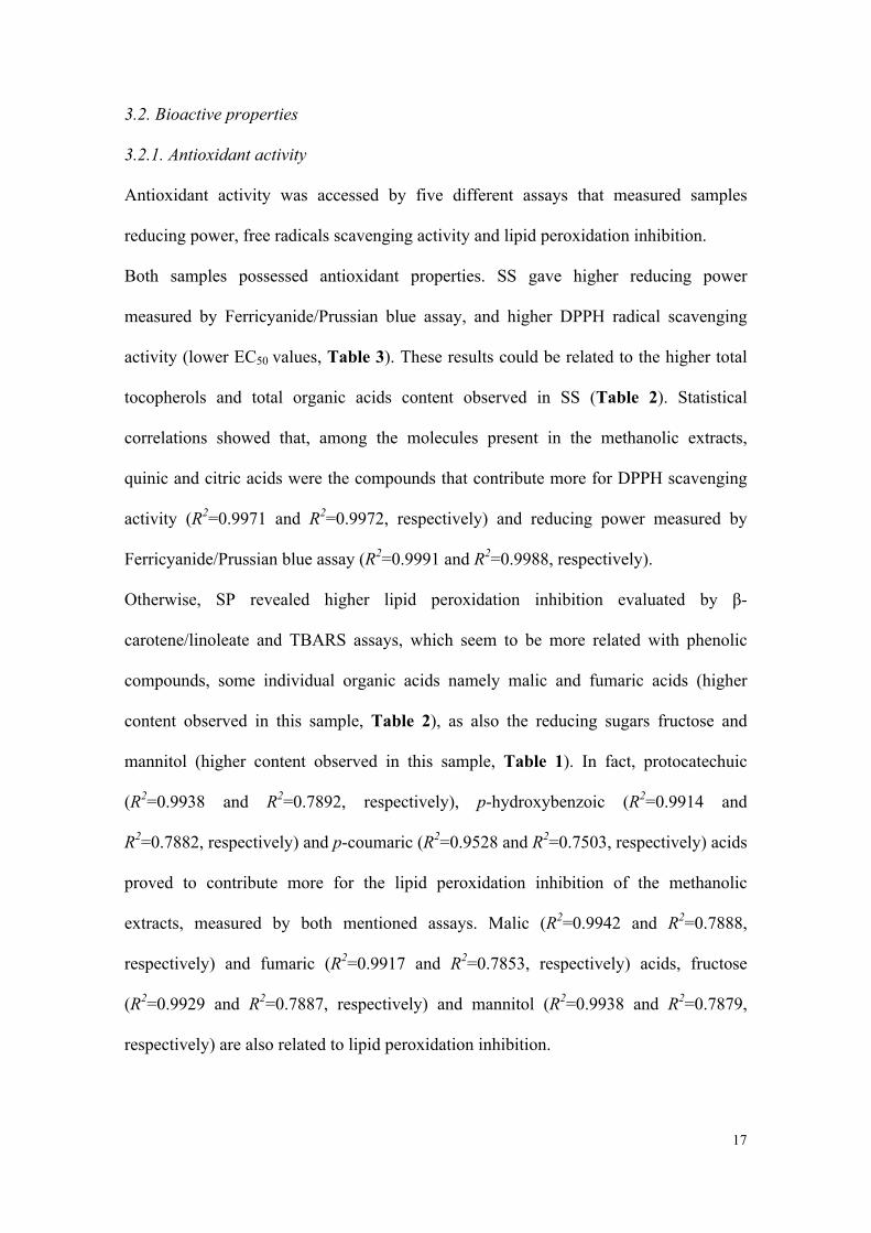
3.2. Bioactive properties
3.2.1. Antioxidant activity
Antioxidant activity was accessed by five different assays that measured samples
reducing power, free radicals scavenging activity and lipid peroxidation inhibition.
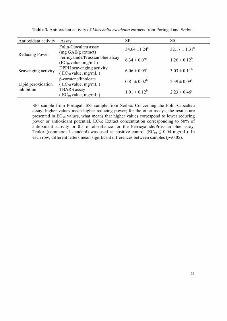
Both samples possessed antioxidant properties. SS gave higher reducing power
measured by Ferricyanide/Prussian blue assay, and higher DPPH radical scavenging
activity (lower EC50 values, Table 3). These results could be related to the higher total
tocopherols and total organic acids content observed in SS (Table 2). Statistical
correlations showed that, among the molecules present in the methanolic extracts,
quinic and citric acids were the compounds that contribute more for DPPH scavenging
activity (R2=0.9971 and R2=0.9972, respectively) and reducing power measured by
Ferricyanide/Prussian blue assay (R2=0.9991 and R2=0.9988, respectively).
Otherwise, SP revealed higher lipid peroxidation inhibition evaluated by β-
carotene/linoleate and TBARS assays, which seem to be more related with phenolic
compounds, some individual organic acids namely malic and fumaric acids (higher
content observed in this sample, Table 2), as also the reducing sugars fructose and
mannitol (higher content observed in this sample, Table 1). In fact, protocatechuic
(R2=0.9938 and R2=0.7892, respectively), p-hydroxybenzoic (R2=0.9914 and
R2=0.7882, respectively) and p-coumaric (R2=0.9528 and R2=0.7503, respectively) acids
proved to contribute more for the lipid peroxidation inhibition of the methanolic
extracts, measured by both mentioned assays. Malic (R2=0.9942 and R2=0.7888,
respectively) and fumaric (R2=0.9917 and R2=0.7853, respectively) acids, fructose
(R2=0.9929 and R2=0.7887, respectively) and mannitol (R2=0.9938 and R2=0.7879,
respectively) are also related to lipid peroxidation inhibition.
18
No significant differences were observed among reducing power measured by Folin-
Ciocalteu assay. Sample of same species from Spain gave higher DPPH scavenging
activity (Ramírez-Anguiano et al., 2007), but M. esculenta mycelium presented similar
values of EC50 (Mau et al., 2004).
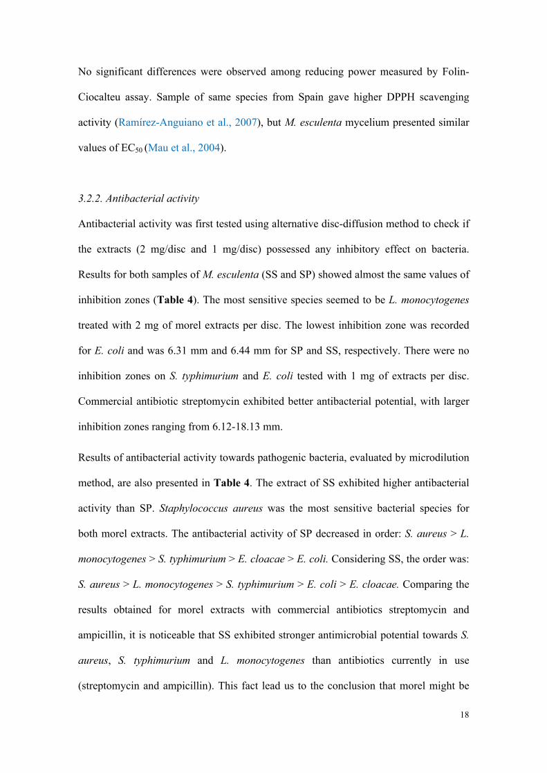
3.2.2. Antibacterial activity
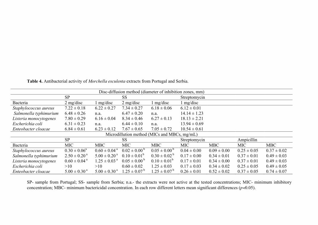
Antibacterial activity was first tested using alternative disc-diffusion method to check if
the extracts (2 mg/disc and 1 mg/disc) possessed any inhibitory effect on bacteria.
Results for both samples of M. esculenta (SS and SP) showed almost the same values of
inhibition zones (Table 4). The most sensitive species seemed to be L. monocytogenes
treated with 2 mg of morel extracts per disc. The lowest inhibition zone was recorded
for E. coli and was 6.31 mm and 6.44 mm for SP and SS, respectively. There were no
inhibition zones on S. typhimurium and E. coli tested with 1 mg of extracts per disc.
Commercial antibiotic streptomycin exhibited better antibacterial potential, with larger
inhibition zones ranging from 6.12-18.13 mm.
Results of antibacterial activity towards pathogenic bacteria, evaluated by microdilution
method, are also presented in Table 4. The extract of SS exhibited higher antibacterial
activity than SP. Staphylococcus aureus was the most sensitive bacterial species for
both morel extracts. The antibacterial activity of SP decreased in order: S. aureus > L.
monocytogenes > S. typhimurium > E. cloacae > E. coli. Considering SS, the order was:
S. aureus > L. monocytogenes > S. typhimurium > E. coli > E. cloacae. Comparing the
results obtained for morel extracts with commercial antibiotics streptomycin and
ampicillin, it is noticeable that SS exhibited stronger antimicrobial potential towards S.
aureus, S. typhimurium and L. monocytogenes than antibiotics currently in use
(streptomycin and ampicillin). This fact lead us to the conclusion that morel might be
19
alternative source of antimicrobial compounds. Nevertheless, further investigation on
structure elucidation of active compounds from morel is necessary. Considering the
differences among the results obtained by the two methods, antibacterial compounds
showed low diffusion potential around the disc used in disc-diffusion method; these
compounds (individually or in synergism) were much more active than commercial
antibiotics in microdilution method probably due to direct contact with bacterial cells
and better bioavailability. Moreover, the difference in antimicrobial activity observed
for the two morel samples might be attributed to environment factors that might
interfere in the synthesis of microbiologically active compounds. Tocopherols and
organic acids contents were higher in the sample from Serbia (Table 2) and could be
involved in its higher antimicrobial activity.
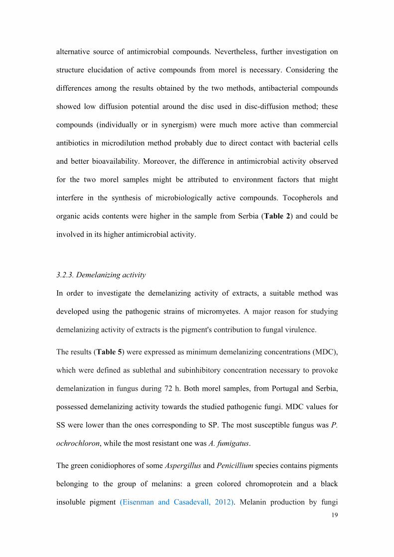
3.2.3. Demelanizing activity
In order to investigate the demelanizing activity of extracts, a suitable method was
developed using the pathogenic strains of micromyetes. A major reason for studying
demelanizing activity of extracts is the pigment's contribution to fungal virulence.
The results (Table 5) were expressed as minimum demelanizing concentrations (MDC),
which were defined as sublethal and subinhibitory concentration necessary to provoke
demelanization in fungus during 72 h. Both morel samples, from Portugal and Serbia,
possessed demelanizing activity towards the studied pathogenic fungi. MDC values for
SS were lower than the ones corresponding to SP. The most susceptible fungus was P.
ochrochloron, while the most resistant one was A. fumigatus.
The green conidiophores of some Aspergillus and Penicillium species contains pigments
belonging to the group of melanins: a green colored chromoprotein and a black
insoluble pigment (Eisenman and Casadevall, 2012). Melanin production by fungi
20
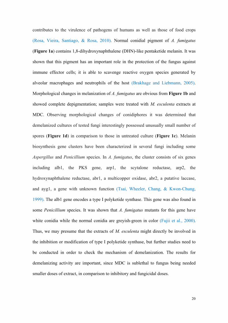
contributes to the virulence of pathogens of humans as well as those of food crops
(Rosa, Vieira, Santiago, & Rosa, 2010). Normal conidial pigment of A. fumigatus
(Figure 1a) contains 1,8-dihydroxynaphthalene (DHN)-like pentaketide melanin. It was
shown that this pigment has an important role in the protection of the fungus against
immune effector cells; it is able to scavenge reactive oxygen species generated by
alveolar macrophages and neutrophils of the host (Brakhage and Liebmann, 2005).
Morphological changes in melanization of A. fumigatus are obvious from Figure 1b and
showed complete depigmentation; samples were treated with M. esculenta extracts at
MDC. Observing morphological changes of conidiphores it was determined that
demelanized cultures of tested fungi interestingly possessed unusually small number of
spores (Figure 1d) in comparison to those in untreated culture (Figure 1c). Melanin
biosynthesis gene clusters have been characterized in several fungi including some
Aspergillus and Penicillium species. In A. fumigatus, the cluster consists of six genes
including alb1, the PKS gene, arp1, the scytalone reductase, arp2, the
hydroxynaphthalene reductase, abr1, a multicopper oxidase, abr2, a putative laccase,
and ayg1, a gene with unknown function (Tsai, Wheeler, Chang, & Kwon-Chung,
1999). The alb1 gene encodes a type I polyketide synthase. This gene was also found in
some Penicillium species. It was shown that A. fumigatus mutants for this gene have
white conidia while the normal conidia are greyish-green in color (Fujii et al., 2000).
Thus, we may presume that the extracts of M. esculenta might directly be involved in
the inhibition or modification of type I polyketide synthase, but further studies need to
be conducted in order to check the mechanism of demelanization. The results for
demelanizing activity are important, since MDC is sublethal to fungus being needed
smaller doses of extract, in comparison to inhibitory and fungicidal doses.
21
4. Conclusion
Wild samples of M. esculenta are rich sources of carbohydrates (including free sugars)
and proteins, and contain several bioactive compounds such as organic acids, phenolic
compounds and tocopherols. Polyunsaturated fatty acids also predominated over mono
and unsaturated fatty acids. SP gave higher radical scavenging activity and reducing
power, while SS showed higher lipid peroxidation inhibition. Both samples, but mainly
SS, gave antibacterial activity against five bacteria (in some cases even better than
standard antibiotics) and demelanizing activity against four micromycetes (Aspergillus
fumigatus, Aspergillus flavus, Penicillium funiculosum and Penicillium ochrochloron).
As far as we know, this is the first study reporting chemical compounds and bioactivity
of morel samples from Portugal and Serbia. Furthermore, a novel method for evaluation
of demelanizing activity was presented. Further studies are needed in order to elucidate
the mechanisms of action involved in morel bioactivity and contribution of the
identified chemical compounds.
Acknowledgements
The authors are grateful to Fundação para a Ciência e a Tecnologia (FCT, Portugal) and
COMPETE/QREN/EU (research project PTDC/AGR-ALI/110062/2009; bilateral
cooperation action Portugal/Serbia 2011; strategic projects PEst-OE/AGR/UI0690/2011
and PEst-C/QUI/UI0686/2011), and to Serbian Ministry of Education and Science
(grant number 173032) for financial support. S.A. Heleno (BD/70304/2010) and L.
Barros (BPD/4609/2008) also thank FCT, POPH-QREN and FSE.
References
22
Alves, M.J., Ferreira, I.C.F.R., Dias, J., Teixeira, V., Martins, A., & Pintado, M.A.
(2012). Review on antimicrobial activity of mushroom (Basidiomycetes) extracts
and isolated compounds. Planta Medica. Accepted.
AOAC (1995). Official methods of analysis (16th Ed.). Arlington VA, USA: Association
of Official Analytical Chemists.
Baati, T., Horcajada, P., Gref, R., Couvreur, P., & Serre, C. (2011). Quantification of
fumaric acid in liver, spleen and urine by high-performance liquid
chromatography coupled to photodiode-array detection. Journal of
Pharmaceutical and Biomedical Analysis, 56, 758-762.
Barros, L., Heleno, S.A., Carvalho, A.M., & Ferreira, I.C.F.R. (2010). Lamiaceae often
used in Portuguese folk medicine as a source of powerful antioxidants: vitamins
and phenolics. LWT- Food Science and Technology, 43, 544-550.
Bhandari, M.R., & Kawabata, J.K. (2004). Organic acid, phenolic content and
antioxidant activity of wild yam (Dioscorea spp.) tubers of Nepal. Food
Chemistry, 88, 163-168.
Brakhage, A.A., & Liebmann, B. (2005). Aspergillus fumigatus conidial pigment and
cAMP signal transduction: significance for virulence. Medical Mycology, 43, S75-
S82.
Brand-Williams, W., Cuvelier, M.E., & Berset, C. (1995). Use of a free radical method
to evaluate antioxidant activity. LWT - Food Science and Technology, 28, 25-30.
Duncan, C., Pugh, J., Pasco, G., David, N., Ross, S., & Samir, A. (2002). Isolation of a
galactomannan that enhances macrophage activation from the edible fungus
Morchella esculenta. Journal of Agricultural and Food Chemistry, 50, 5683-
5685.
23
Eismann, H.C., & Casadevall, A. (2012). Synthesis and assembly of fungal melanin.
Applied Microbiology and Biotechnology, 93, 931-940.
Elmastas, M., Turkekul, I., Ozturk, L., Gulcin, I., Isildak, O., & Aboul-Enein, H.Y.
(2006). Antioxidant activity of two wild edible mushrooms (Morchella vulgaris
and Morchella esculenta) from North Turkey. Combinatorial Chemistry & High
Throughput Screening, 9, 443-448.
Espinel-Ingroff, A. (2001). Comparation of the E-test with the NCCLS M38-P method for
antifungal susceptibility testing of common and emerging pathogenic filamentous
fungi. Journal of Clinical Microbiology, 39, 1360-1367.
Ferreira, I.C.F.R., Barros, L., & Abreu, R.M.V. (2009). Antioxidants in wild
mushrooms. Current Medicinal Chemistry, 16, 1543-156.
Ferreira, I.C.F.R., Vaz, J.A., Vasconcelos, M.H., & Martins, A. (2010). Compounds
from wild mushrooms with antitumour potential. Anti-cancer Agents in Medicinal
Chemistry, 10, 424-436.
Fujii, I., Mori, Y., Watanabe, A., Kubo, Y., Tsuji, G., & Ebizuka, Y. (2000). Enzymatic
synthesis of 1,3,5,8-tetrahydroxy-naphthalene solely from malonyl coenzyme A by
a fungal iterative type I polyketide synthase PKS1. Biochemistry, 39, 8853-8858.
Hanel, H., & Raether, W. (1988). A more sophisticated method of determining the
fungicidal effect of water-insoluble preparations with a cell harvester, using
miconazole as an example. Mycoses, 31, 148-154.
Heleno, S.A, Barros, L., Sousa, M.J., Martins, A., & Ferreira, I.C.F.R. (2009). Study
and characterization of selected nutrients in wild mushrooms from Portugal by gas
chromatography and high performance liquid chromatography. Microchemical
Journal, 93, 195-199.
24
Heleno, S.A., Barros, L., Sousa, M.J., Martins, A., & Ferreira, I.C.F.R. (2010).
Tocopherols composition of Portuguese wild mushrooms with antioxidant
capacity. Food Chemistry, 119, 1443-1450.
Heleno, S.A., Barros, L., Martins, A., Queiroz, M.J.R.P., Santos-Buelga, C., & Ferreira,
I.C.F.R. (2012). Fruiting body, spores and in vitro produced mycelium of
Ganoderma lucidum from Northeast Portugal: A comparative study of the
antioxidant potential of phenolic and polysaccharidic extracts. Food Research
International, 46, 135-140.
Kalac, P. (2009). Chemical composition and nutritional value of European species of
wild growing mushrooms: A review. Food Chemistry, 113, 9-16.
Kalyoncu, F., Oskay, M., Saglam, H., Erdogan, TF., & Tamer, AÜ. (2010).
Antimicrobial and antioxidant activities of mycelia of 10 wild mushroom species.
Journal of Medicinal Food, 13, 415-419.
Kamal-Eldin, A., & Appelqvist, L.Å. (1996). The chemistry and antioxidant properties
of tocopherols and tocotrienols. Lipids, 31, 671-701.
Kim, J.-A., Lau, E., Tay, D., & Blanco, E.J.C.D. (2011). Antioxidant and NF-kD
inhibitory constituents isolated from Morchella esculenta. Natural Products
Research: Formerly Natural Products Letters, 25, 1412-1417.
Koide, R.T., Shumway, D.L., & Stevens, C.M. (2000). Soluble carbohydrates of red
pine (Pinus resinosa) mycorrhizas and mycorrhizal fungi. Mycological Research,
104, 834-840.
Leal, A.R., Barros, L., Barreira, J.C.M., Sousa, M.J., Martins, A., Santos-Buelga, C., &
Ferreira, I.C.F.R. (2013). Portuguese wild mushrooms at the "Pharma-Nutrition"
interface: Nutritional characterization and antioxidant properties. Food Research
International, 50, 1-9.
25
Mi-Yae, S., Tae-Hun, K., & Nak-Ju, S. (2003). Antioxidants and free radical
scavenging activity of Phellinus baumii (Phellinus of Hymenochaetaceae)
extracts. Food Chemistry, 82, 593-597.
Mau, J.-L., Chang, C.-N., Huang, S.-J., & Chen, C.-C. (2004). Antioxidant properties of
methanolic extracts from Grifola frondosa, Morchella esculenta and
Termitomyces alduminosus mycelia. Food Chemistry, 87, 111-118.
Meng, F., Zhou, D., Lin, R., Jia, L., Liu, X., Deng, P., Fan, K., Wang, G., Wang, L., &
Zhang, J. (2011). Extraction optimization and in vivo antioxidant activities of
exopolysaccharide by Morchella esculenta SO-01. Bioresource Technology, 101,
4564-4569.
Nagarajkumar, M., Jayaraj, J., Muthukrishnan, S., Bhaskaran, R., & Velazhahan, R.
(2005). Detoxification of oxalic acid by Pseudomonas fluorescens strain
PfMDU2: Implications for the biological control of rice sheath blight caused by
Rhizoctonia solani. Microbiological Research, 160, 291-298.
Nagata, M., & Yamashita, I. (1992). Simple method for simultaneous determination of
chlorophyll and carotenoids in tomato fruit. Nippon Shokuhin Kogyo Gakkaish,
39, 925-928.
Ng, T.B., Liu, F., Wang, Z. (2000). Antioxidative activity of natural products from
plants. Life Sciences, 66, 709-723.
Nitha, D., Fijesh, P.V., & Janardhanan, K.K. (2011). Hepatoprotective activity of
cultured mycelium of morel mushroom, Morchella esculenta. Experimental
Toxicology and Pathology, Doi 10.1016/j.etp.2011.06.007.
Nitha, D., & Janardhanan, K.K. (2008). Aquouse-ethanolic extract of morel mushroom
mycelium Morchella esculenta, protects cisplatin and gentamicin induced
nefrotoxicity in mice. Food and Chemical Toxicology, 46, 3193-3199.
26
Nitha, D., Meera, C.R., & Janardhanan, K.K. (2007). Anti-inflammatory and antitumour
activities of cultured mycelium of morel mushroom, Morchella esculenta. Current
Science, 92, 235-239.
Ramírez-Anguiano, A.C., Santoryo, S., Reglero, G., & Soler-Rivas, C. (2007). Radical
scavenging activities, endogenous oxidative enzymes and total phenols in edible
mushrooms commonly consumed in Europe. Journal of Science and Food
Agriculture, 87, 2272-2278.
Reis, F.S., Stojković, D., Soković, M., Glamočlija, J., Cirić, A., Barros, L., & Ferreira,
I.C.F.R. (2012). Chemical characterization of Agaricus bohusii, antioxidant
potential and antifungal preserving properties when incorporated in cream cheese.
Food Research International, 48, 620-626.
Rosa, L.H., Vieira, L.M.A., Santiago, I.F., & Rosa, C.A. (2010). Endophytic fungi
community associated with the dicotyledonous plant Colobanthus quitensis
(Kunth) Bartl. (Caryophyllaceae) in Antarctica. FEMS Microbiology Ecology, 73,
178-189.
Singleton, V.L., Rossi, J.A. jr. (1965). Colorimetric of total phenolics with
phosphomolybdic-phosphotungstic acid reagents. American Journal of Enology
and Viticulture, 16, 144-158.
Sokovic, M., Glamoclija, J., Marin, P.D., Brkic, D., & van Griensven, L.J. (2010).
Antibacterial effects of the essential oils of commonly consumed medicinal herbs
using an in vitro model. Molecules, 15, 7532-7546.
Tsai, H.F., Wheeler, M.H., Chang, Y.C., & Kwon-Chung, K.J. (1999). A
developmentally regulated gene cluster involved in conidial pigment biosynthesis
in Aspergillus fumigatus. Journal of Bacteriology, 181, 6469-6477.
27
Verpoorte, R., van Beek T.A., Thomassen P.H.A.M., Aandewiel J., & Svendsen, A.B.
(1983). Screening of antimicrobial activity of some plants belonging to the
Apocynaceae and Loganiaceae. Journal of Ethnopharmacology, 8, 287-302.
Wahid, M., Sattar, A., & Khan, S. (1988). Composition of wild and cultivated
mushrooms of Pakistan. Mushroom Journal of the Tropics, 8, 47-51.
28
Legends Figure 1. (a) and (b) Cultures of Aspergillus fumigatus recorded under binocular magnifier; (a) Normal mycelium of A. fumigatus without treatment with Morchella esculenta extracts; (b) Demelanized mycelium of A. fumigatus treated with M. esculenta extracts. (c) and (d) Conidia of A. flavus recorded under light microscope; (c) Typical conidia of A. flavus with numerous spores; (d) Conidia of A. flavus with small amount of spores, treated with M. esculenta extracts.
29
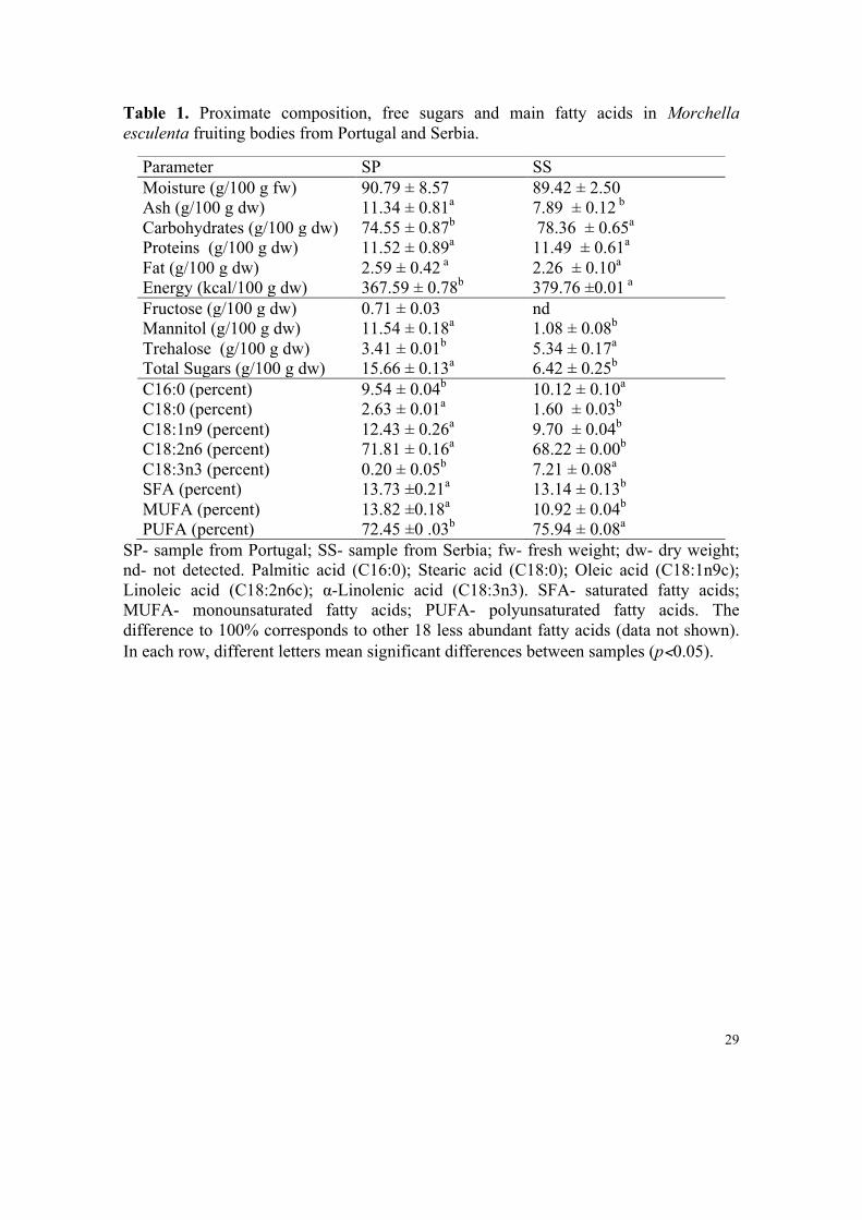
Table 1. Proximate composition, free sugars and main fatty acids in Morchella esculenta fruiting bodies from Portugal and Serbia.
SP- sample from Portugal; SS- sample from Serbia; fw- fresh weight; dw- dry weight; nd- not detected. Palmitic acid (C16:0); Stearic acid (C18:0); Oleic acid (C18:1n9c); Linoleic acid (C18:2n6c); α-Linolenic acid (C18:3n3). SFA- saturated fatty acids; MUFA- monounsaturated fatty acids; PUFA- polyunsaturated fatty acids. The difference to 100% corresponds to other 18 less abundant fatty acids (data not shown). In each row, different letters mean significant differences between samples (p<0.05).
Parameter SP SS Moisture (g/100 g fw) 90.79 ± 8.57 89.42 ± 2.50 Ash (g/100 g dw) 11.34 ± 0.81a 7.89 ± 0.12 b Carbohydrates (g/100 g dw) 74.55 ± 0.87b 78.36 ± 0.65a
Proteins (g/100 g dw) 11.52 ± 0.89a 11.49 ± 0.61a
Fat (g/100 g dw) 2.59 ± 0.42 a 2.26 ± 0.10a Energy (kcal/100 g dw) 367.59 ± 0.78b 379.76 ±0.01 a Fructose (g/100 g dw) 0.71 ± 0.03 nd Mannitol (g/100 g dw) 11.54 ± 0.18a 1.08 ± 0.08b Trehalose (g/100 g dw) 3.41 ± 0.01b 5.34 ± 0.17a Total Sugars (g/100 g dw) 15.66 ± 0.13a 6.42 ± 0.25b C16:0 (percent) 9.54 ± 0.04b 10.12 ± 0.10a C18:0 (percent) 2.63 ± 0.01a 1.60 ± 0.03b C18:1n9 (percent) 12.43 ± 0.26a 9.70 ± 0.04b C18:2n6 (percent) 71.81 ± 0.16a 68.22 ± 0.00b C18:3n3 (percent) 0.20 ± 0.05b 7.21 ± 0.08a SFA (percent) 13.73 ±0.21a 13.14 ± 0.13b MUFA (percent) 13.82 ±0.18a 10.92 ± 0.04b PUFA (percent) 72.45 ±0 .03b 75.94 ± 0.08a
30
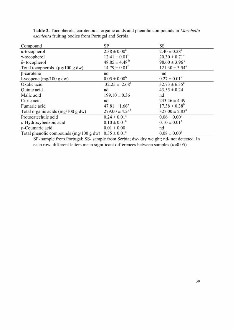
Table 2. Tocopherols, carotenoids, organic acids and phenolic compounds in Morchella esculenta fruiting bodies from Portugal and Serbia.
SP- sample from Portugal; SS- sample from Serbia; dw- dry weight; nd- not detected. In each row, different letters mean significant differences between samples (p<0.05).
Compound SP SS α-tocopherol 2.38 ± 0.00a 2.40 ± 0.28a γ-tocopherol 12.41 ± 0.01b 20.30 ± 0.71a δ- tocopherol 48.85 ± 4.48 b 98.60 ± 3.96 a Total tocopherols (µg/100 g dw) 14.79 ± 0.01b 121.30 ± 3.54a β-carotene nd nd Lycopene (mg/100 g dw) 0.05 ± 0.00b 0.27 ± 0.01a Oxalic acid 32.25 ± 2.68a 32.73 ± 6.35a Quinic acid nd 43.55 ± 0.24 Malic acid 199.10 ± 0.36 nd Citric acid nd 233.46 ± 4.49 Fumaric acid 47.81 ± 1.66a 17.38 ± 0.38b Total organic acids (mg/100 g dw) 279.00 ± 4.24b 327.00 ± 2.83a Protocatechuic acid 0.24 ± 0.01a 0.06 ± 0.00b p-Hydroxybenzoic acid 0.10 ± 0.01a 0.10 ± 0.01a p-Coumaric acid 0.01 ± 0.00 nd Total phenolic compounds (mg/100 g dw) 0.35 ± 0.01a 0.08 ± 0.00b
31
Table 3. Antioxidant activity of Morchella esculenta extracts from Portugal and Serbia.
Antioxidant activity Assay SP SS
Reducing Power
Folin-Ciocalteu assay (mg GAE/g extract) 34.64 ±1.24a 32.17 ± 1.31a
Ferricyanide/Prussian blue assay (EC50 value; mg/mL) 6.34 ± 0.07a 1.26 ± 0.12b
Scavenging activity DPPH scavenging activity ( EC50 value; mg/mL ) 6.06 ± 0.05a 3.03 ± 0.11b
Lipid peroxidation inhibition
β-carotene/linoleate ( EC50 value; mg/mL ) 0.81 ± 0.02b 2.39 ± 0.09a
TBARS assay ( EC50 value; mg/mL ) 1.01 ± 0.12b 2.23 ± 0.46a
SP- sample from Portugal; SS- sample from Serbia. Concerning the Folin-Ciocalteu assay, higher values mean higher reducing power; for the other assays, the results are presented in EC50 values, what means that higher values correspond to lower reducing power or antioxidant potential. EC50: Extract concentration corresponding to 50% of antioxidant activity or 0.5 of absorbance for the Ferricyanide/Prussian blue assay. Trolox (commercial standard) was used as positive control (EC50 ≤ 0.04 mg/mL). In each row, different letters mean significant differences between samples (p<0.05).
32
Table 4. Antibacterial activity of Morchella esculenta extracts from Portugal and Serbia.
Disc-diffusion method (diameter of inhibition zones, mm) SP SS Streptomycin Bacteria 2 mg/disc 1 mg/disc 2 mg/disc 1 mg/disc 1 mg/disc Staphylococcus aureus 7.22 ± 0.18 6.22 ± 0.27 7.34 ± 0.27 6.18 ± 0.06 6.12 ± 0.01 Salmonella typhimurium 6.48 ± 0.26 n.a. 6.47 ± 0.20 n.a. 14.14 ± 1.23 Listeria monocytogenes 7.80 ± 0.29 6.16 ± 0.04 8.34 ± 0.46 6.27 ± 0.13 18.13 ± 2.21 Escherichia coli 6.31 ± 0.23 n.a. 6.44 ± 0.10 n.a. 13.94 ± 0.69 Enteobacter cloacae 6.84 ± 0.61 6.23 ± 0.12 7.67 ± 0.65 7.05 ± 0.72 10.54 ± 0.61
Microdillution method (MICs and MBCs, mg/mL) SP SS Streptomycin Ampicillin Bacteria MIC MBC MIC MBC MIC MBC MIC MBC Staphylococcus aureus 0.30 ± 0.06a 0.60 ± 0.04 a 0.02 ± 0.00 b 0.05 ± 0.00 b 0.04 ± 0.00 0.09 ± 0.00 0.25 ± 0.05 0.37 ± 0.02 Salmonella typhimurium 2.50 ± 0.20 a 5.00 ± 0.20 a 0.10 ± 0.01b 0.30 ± 0.02 b 0.17 ± 0.00 0.34 ± 0.01 0.37 ± 0.01 0.49 ± 0.03 Listeria monocytogenes 0.60 ± 0.04 a 1.25 ± 0.03 a 0.05 ± 0.00 b 0.10 ± 0.01b 0.17 ± 0.01 0.34 ± 0.00 0.37 ± 0.01 0.49 ± 0.03 Escherichia coli >10 >10 0.60 ± 0.02 1.25 ± 0.03 0.17 ± 0.03 0.34 ± 0.02 0.25 ± 0.05 0.49 ± 0.05 Enteobacter cloacae 5.00 ± 0.30 a 5.00 ± 0.30 a 1.25 ± 0.07 b 1.25 ± 0.07 b 0.26 ± 0.01 0.52 ± 0.02 0.37 ± 0.05 0.74 ± 0.07
SP- sample from Portugal; SS- sample from Serbia; n.a.- the extracts were not active at the tested concentrations; MIC- minimum inhibitory concentration; MBC- minimum bactericidal concentration. In each row different letters mean significant differences (p<0.05).
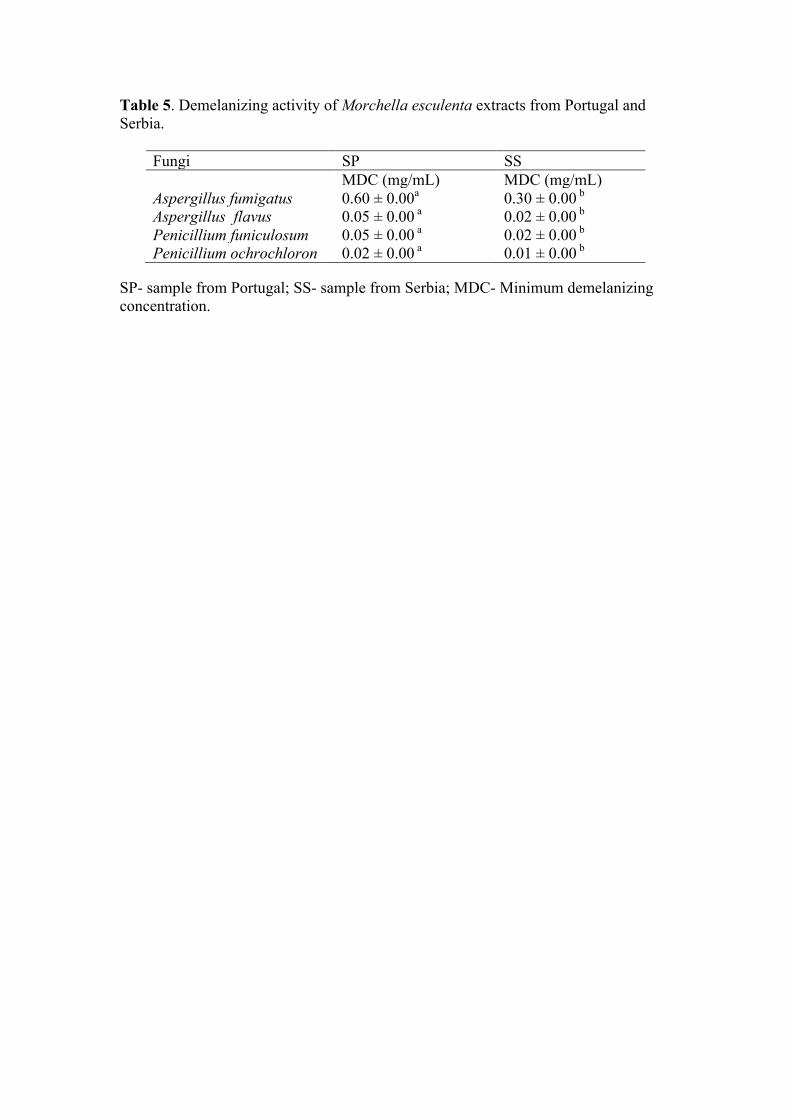
Table 5. Demelanizing activity of Morchella esculenta extracts from Portugal and Serbia.
Fungi SP SS MDC (mg/mL) MDC (mg/mL) Aspergillus fumigatus 0.60 ± 0.00a 0.30 ± 0.00 b Aspergillus flavus 0.05 ± 0.00 a 0.02 ± 0.00 b Penicillium funiculosum 0.05 ± 0.00 a 0.02 ± 0.00 b Penicillium ochrochloron 0.02 ± 0.00 a 0.01 ± 0.00 b
SP- sample from Portugal; SS- sample from Serbia; MDC- Minimum demelanizing concentration.
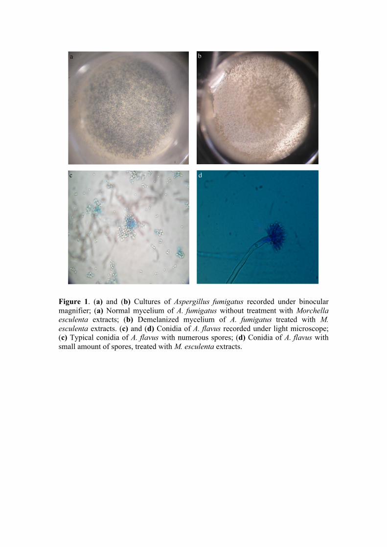
Figure 1. (a) and (b) Cultures of Aspergillus fumigatus recorded under binocular magnifier; (a) Normal mycelium of A. fumigatus without treatment with Morchella esculenta extracts; (b) Demelanized mycelium of A. fumigatus treated with M. esculenta extracts. (c) and (d) Conidia of A. flavus recorded under light microscope; (c) Typical conidia of A. flavus with numerous spores; (d) Conidia of A. flavus with small amount of spores, treated with M. esculenta extracts.