Embed Size (px)
Citation preview
ORIGINAL RESEARCH
Antioxidant, antimicrobial and cytotoxic activities of silverand gold nanoparticles synthesized using Plumbago zeylanica bark
S. Priya Velammal1 • T. Akkini Devi1 • T. Peter Amaladhas1
Received: 13 April 2016 / Accepted: 1 June 2016 / Published online: 13 June 2016
� The Author(s) 2016. This article is published with open access at Springerlink.com
Abstract Utilization of bioresources for the synthesis of
metal nanoparticles is the latest field in green chemistry.
The present work reports the utilization of the aqueous bark
extract of Plumbago zeylanica for the biosynthesis of Ag
and Au NPs. The Ag and Au NPs thus obtained were
characterized by UV–Vis, FT-IR, TEM, XRD, and EDAX
analysis. The water-soluble components of the extract were
responsible for the reduction of Ag? and Au3? ions. FT-IR
spectra revealed that the –OH and[C=O groups present in
the biomolecules were responsible for reduction and stabi-
lization of nanoparticles. TEM images showed the existence
of spherical Ag and Au NPs with average size of 28.47 and
16.89 nm, respectively, which was further substantiated by
XRD analysis. The presence of elemental Ag and Au along
with C and O from the attached biomolecules was proved by
EDAX analysis. The antimicrobial, antioxidant, and in vitro
cytotoxic activities of the synthesized nanoparticles were
studied by disc diffusion, DPPH, and MTT assay methods,
respectively. The free radical inhibition was found to be
78.17 and 87.34 % for Ag and Au nanoparticles, respec-
tively. The Ag and Au NPs showed 61.56 and 65.61 %
toxicity against DLA cell line, respectively. The DNA
binding ability of Ag and Au NPs were investigated using
CT-DNA. The hyperchromism shift inferred the groove
binding of nanoparticles with CT-DNA.
Keywords Biosynthesis � Silver nanoparticles � Goldnanoparticles � Antioxidant � Cytotoxicity � DLA cell line
Introduction
Nanoscience and nanotechnology, the two current fields
of research have strong and beautiful footprints in
ancient times. Nano mercury and its sulfide of sizes
finer than 10 nm have been used as medicines in India
since long back [1]. Silver and copper nanoparticles
have been used to produce iridescent metallic effects on
ancient ceramic objects [2]. The ancient Vedic rite of
Agnihotra has used the nanoparticles to cleanse the
environment. In Agnihotra, wood of trees like Butea
frondosa (palasa), Mangifera indica (mango), Ficus
religiosa (pipal), medicinal herbs, ghee, cloves, car-
damom, and camphor are offered to the fire god by
throwing them into the fire. This has been reported to
fill the atmosphere with nutrients in nano size and
medicinal properties, which in turn eliminate pathogenic
bacteria or germs [3]. In Ayurvedha, herbo-mineral, and
herbo-metallic drugs have been prepared by prolonged
heating which significantly reduces the particle size.
The medicinal use of colloidal gold (Swarnabhasma)
and mercury remain popular till this day and the
Swarnabhasmas (gold ash) has been characterized as
globular particles of gold with the average size between
56 and 57 nm. It has been used to treat arthiritis,
asthma, diabetes, and diseases of nervous system [4].
Electronic supplementary material The online version of thisarticle (doi:10.1007/s40097-016-0198-x) contains supplementarymaterial, which is available to authorized users.
& T. Peter Amaladhas
[email protected]; [email protected]
S. Priya Velammal
T. Akkini Devi
1 PG and Research Department of Chemistry,
V.O. Chidambaram College, Tuticorin 628008,
Tamil Nadu, India
123
J Nanostruct Chem (2016) 6:247–260
DOI 10.1007/s40097-016-0198-x
Metal and metal oxide nanoparticles have been synthe-
sized using many methods in the past, such as chemical
reduction [5], electrochemical reaction [6], microwave
method [7], reverse micelles [8], sonochemical method [9],
and biosynthesis. Biosynthesis of nanoparticles has
received great attention recently as the synthesized
nanoparticles are non-toxic and can be used for biomedical
applications. Different fungi like Fusarium oxysporum
[10], Cladosporium cladosporioides [11], Fusarium semi-
tactum [12], plants like Syzygium aromaticum [13], Med-
icago sativa [14], Azadirachta indica [15], Terminalia
cuneata [16], and Trignellafoenum graecum seeds [17]
have been employed to synthesize metal nanoparticles.
Recently, sunlight-induced rapid synthesis of silver
nanoparticles has been reported by our group [18, 19].
Biosynthesis of silver nanoparticles using bark of Cinna-
mon zeylanicum [20], Piper nigrum [21], Breynia rham-
noides [22], Avicennia marina [23], Callicarpa maingayi
[24], Cissus quadrangularis [25], Artocarpus elasticus
[26], and Saraca indica [27] has also been reported. The
use of plant barks for the synthesis of nanoparticles avoids
the problems that arise due to seasonal variation of bio-
chemicals in the leaves.
Plumbago zeylanica is a medicinal herb belonging to the
family of Plumbaginaceae. It has been used as a remedy
for skin diseases, ringworm, leprosy, sores, and ulcers. The
root or leaves are used as counter-irritant and vesicant. The
powdered bark is used to treat leprosy, spleen and liver
diseases and also for plaque [28]. It contains a variety of
important chemical compounds, such as naphthaquinones,
alkaloids, glycosides, steroids, triterpenoids, tannins, phe-
nolic compounds, flavanoids, saponins, coumarins, and
carbohydrates [29, 30]. Plumbagin, 5-hydroxy-2-methyl-
1,4-naphthoquinone is the principal active compound of P.
zeylanica [31]. In this work, biosynthesis of silver (Ag) and
gold (Au) nanoparticles (NPs) was achieved using the bark
extract of P. zeylanica. The water-soluble biocomponents
in the extract not only act as reductants to reduce Ag? and
Au3? to Ago and Auo, respectively, but also stabilize them
by attaching onto the nanoparticles. The nanoparticles
stabilized by active biomolecules may have the potential to
treat various diseases. The results of antimicrobial,
antioxidant, in vitro cytotoxicity of Ag and Au NPs and
their ability to bind DNA are reported here.
Methods
Preparation of bark extract: the reducing agent
P. zeylanica bark was purchased from the local market
from Madurai, Tamil Nadu, India. The bark was finely cut,
washed with double-distilled water to remove any
impurities and air-dried. About 2 g of bark was weighed
and boiled for 10 min with 100 mL distilled water in
Erlenmeyer flask. After cooling to room temperature, it
was filtered through Whatman no. 41 filter paper. The light
yellow colored extract (pH 6) was utilized for the synthesis
of Ag and Au NPs.
Biosynthesis of Ag and Au NPs
Analar grade silver nitrate (AgNO3) with 99.9 % purity
was purchased from Spectrochem, India and 99.999 %
pure hydrogen tetrachloroaurate(III) trihydrate (HAuCl4-3H2O) was purchased from Sigma-Aldrich, USA. To pre-
pare AgNPs, about 1 mL of 20-mM AgNO3 was diluted to
20 mL with double-distilled water and treated with 10 mL
of bark extract, and the solution was exposed to bright
sunlight. There was a visible color change from pale yel-
low to brown in 2 min, and this reaction was monitored
with time using UV–Vis spectrophotometer. In the case of
AuNPs, 5 mL of 1-mM HAuCl4 was mixed with 5 mL of
the extract, and the intensity of pink color was monitored
with time using UV–Vis spectrophotometer. In both the
cases, 1 mL of the aliquot was diluted to 10 mL with
double-distilled water before measuring the absorbance.
Characterization of Ag and Au NPs
UV–Vis spectral analysis and DNA binding studies were
performed on a JASCO, V-650 spectrophotometer. FT-IR
spectra for the dry powder of bark extract and nanoparticles
were recorded in the range 4000–400 cm-1 with Thermo
scientific, Nicolet iS5 spectrometer. The size of the
nanoparticles was assessed using PHILIPS, CM 200, TEM
microscope operated at 200 kV with a resolution of 2.4 A.
The diffraction pattern of nanoparticles was recorded using
a PANalytical X’Pert Powder X’Celerator diffractometer,
with CuKa monochromatic filter. To ascertain the ele-
mental composition of Ag and Au NPs, energy dispersive
X-ray (EDAX) analysis was performed on a JEOL, Model
JED-2300 microscope. Ultrasonic probe sonicator, Ener-
tech make, model: ENUP-500A was used to disperse CT-
DNA in Tris–HCl buffer solution.
Antimicrobial activity
Kirby–Bauer disc diffusion method was employed to assess
the antimicrobial activity of biosynthesized Ag and Au
NPs. The bacteria Escherichia coli, Pseudomonas aerugi-
nosa, Bacillus subtilis, and Staphylococcus aureus, and the
fungi Candida tropicalis and Aspergillus flaves were cho-
sen for study. For disc diffusion method, the bacterial
inoculums were standardized to a density equivalent to
barium sulfate standard of 0.5 McFarland units and
248 J Nanostruct Chem (2016) 6:247–260
123
swabbed into petridish of 4 mm depth. About 2 mm
loopful of extract, Ag and Au NPs were taken and lowered
carefully onto the paper disc. The moistened disc was then
placed on the surface of inoculated plate and incubated for
16–18 h at 35–37 �C. Netilmicin and fluconazole were
used as controls for bacteria and fungi, respectively.
Antioxidant activity
Antioxidant activity of the synthesized Ag and Au NPs was
assessed using DPPH assay. From the stock solution of the
nanoparticles (1 mg/mL), samples containing 50, 100, 150,
and 200 lg were prepared and mixed with 0.1 % DPPH.
The reaction mixture was incubated for 30 min at room
temperature. When DPPH reacts with any antioxidant, it
gets reduced, and the intensity of color decreases; and the
decrease in absorbance at 517 nm was recorded, and
butylated hydroxytoluene (BHT) was used as a standard
control. The experiment was performed in triplicate, and
the scavenging activity was calculated as % inhibition
according to the following formula, where, A is
absorbance.
% Inhibition ¼ 100
� ðA of standard control� A of sampleÞA of standard control
In vitro cytotoxicity of Ag and Au NPs
Cytotoxic effect was determined by tetrazolium dye (3-
(4,5-Dimethylthiazol-2-yl)-2,5-diphenyltetrazolium bro-
mide)-based microtitration assay (MTT assay) against
DLA (Dalton Lymphoma Ascites) cell lines. Cells were
maintained in Dulbecco’s modified Eagle’s medium
(DMEM) supplemented with 10 % fetal bovine serum
(FBS) at 37 �C in humidified atmosphere with 5 % CO2.
The cells were seeded in 96-well flat-bottom tissue culture
plates at a density of approximately 1.2 9 104 cells per
well and were allowed to attach overnight at 37 �C. Cellswere exposed to plant extract, Ag and Au NPs over a range
of concentration for 24 h. After the incubation, medium
was discarded; the MTT (10 lL of 5 mg/mL) was added to
100 lL of fresh medium and was incubated for additional
4 h. MTT assay was based on the measurement of the
mitochondrial activity of viable cells by the reduction of
the tetrazolium salt (MTT) to form a blue water-insoluble
product, formazan. After 4 h of incubation, the medium
was discarded, and 100 lL of DMSO was added to dis-
solve the formazan crystals, which were formed by the
reduction of tetrazolium salt only by metabolically active
cells [32]. The absorbance assay plates were measured at
570 nm in a micro-titer plate reader. Since the absorbance
was directly correlated with the number of viable cells, cell
survival was calculated by the following formula:
% viability ¼ At
Ac
� 100
where, At is the absorbance of sample and Ac is the
absorbance of control. Percentage cytotoxicity is calculated
by the formula: cytotoxicity % = 100 - % viability.
DNA binding studies
In every organism, deoxyribonucleic acid (DNA) is an
important genetic substance. The study of interaction of
drug and DNA has acquired great significance for design-
ing and synthesizing new drugs targeted at DNA. Pre-
suming biomolecules capped nanoparticles as drug, the
ability to bind DNA was investigated. UV–Visible spectral
studies were employed to study the binding of Ag and Au
NPs with Calf-thymus DNA (CT-DNA). DNA solutions
were prepared using the buffer 5 mM Tris–HCl/50 mM
NaCl at pH = 7.2 in water and sonicated for 30 cycles,
where each cycle consisted of 1 min with 30 s interval;
which gave ratio of absorbance at 260 and 280 nm, A260/
A280 as 1.87. This ratio indicates that the DNA was suf-
ficiently free of protein. Absorption spectra were recorded
at fixed-concentration Ag and Au NPs for varying con-
centration of the CT-DNA (2–10 lg/mL) or vice versa.
Stock solutions of DNA were stored at 4 �C and used in
4 days.
Results and discussion
Parameters influencing the formation of AgNPs
The effect of time, volume of extract, concentration of
metal precursors and pH, which influence the formation of
AgNPs were studied in detail, and the results are presented
here.
Effect of time
The mixture of the extract and silver nitrate solution was
exposed to sunlight, and the SPR band was monitored in
regular intervals using UV–Vis spectrophotometer. The
color of the solution changed from yellow to dark brown in
2 min, the intensity of the color increased with time and
finally attained a constant value in 40 min implying that the
formation of AgNPs is almost complete (Fig. 1a). The
decrease in peak width and the shift in kmax from 426 to
416 nm with time infer that the formation of monodis-
persed and small-sized nanoparticles, respectively [33].
J Nanostruct Chem (2016) 6:247–260 249
123
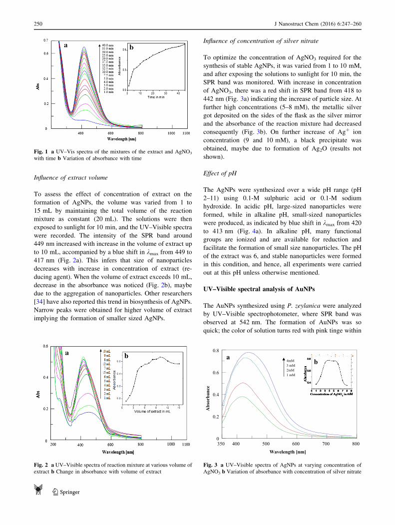
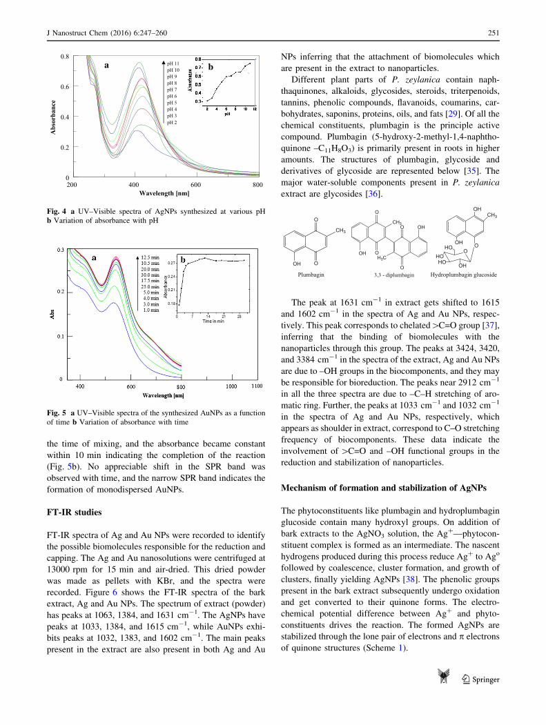
Influence of extract volume
To assess the effect of concentration of extract on the
formation of AgNPs, the volume was varied from 1 to
15 mL by maintaining the total volume of the reaction
mixture as constant (20 mL). The solutions were then
exposed to sunlight for 10 min, and the UV–Visible spectra
were recorded. The intensity of the SPR band around
449 nm increased with increase in the volume of extract up
to 10 mL, accompanied by a blue shift in kmax from 449 to
417 nm (Fig. 2a). This infers that size of nanoparticles
decreases with increase in concentration of extract (re-
ducing agent). When the volume of extract exceeds 10 mL,
decrease in the absorbance was noticed (Fig. 2b), maybe
due to the aggregation of nanoparticles. Other researchers
[34] have also reported this trend in biosynthesis of AgNPs.
Narrow peaks were obtained for higher volume of extract
implying the formation of smaller sized AgNPs.
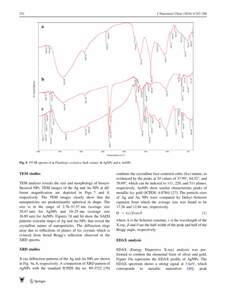
Influence of concentration of silver nitrate
To optimize the concentration of AgNO3 required for the
synthesis of stable AgNPs, it was varied from 1 to 10 mM,
and after exposing the solutions to sunlight for 10 min, the
SPR band was monitored. With increase in concentration
of AgNO3, there was a red shift in SPR band from 418 to
442 nm (Fig. 3a) indicating the increase of particle size. At
further high concentrations (5–8 mM), the metallic silver
got deposited on the sides of the flask as the silver mirror
and the absorbance of the reaction mixture had decreased
consequently (Fig. 3b). On further increase of Ag? ion
concentration (9 and 10 mM), a black precipitate was
obtained, maybe due to formation of Ag2O (results not
shown).
Effect of pH
The AgNPs were synthesized over a wide pH range (pH
2–11) using 0.1-M sulphuric acid or 0.1-M sodium
hydroxide. In acidic pH, large-sized nanoparticles were
formed, while in alkaline pH, small-sized nanoparticles
were produced, as indicated by blue shift in kmax from 420
to 413 nm (Fig. 4a). In alkaline pH, many functional
groups are ionized and are available for reduction and
facilitate the formation of small size nanoparticles. The pH
of the extract was 6, and stable nanoparticles were formed
in this condition, and hence, all experiments were carried
out at this pH unless otherwise mentioned.
UV–Visible spectral analysis of AuNPs
The AuNPs synthesized using P. zeylanica were analyzed
by UV–Visible spectrophotometer, where SPR band was
observed at 542 nm. The formation of AuNPs was so
quick; the color of solution turns red with pink tinge within
Fig. 1 a UV–Vis spectra of the mixtures of the extract and AgNO3
with time b Variation of absorbance with time
Fig. 2 a UV–Visible spectra of reaction mixture at various volume of
extract b Change in absorbance with volume of extract
0
0.8
0.2
0.4
0.6
350 800400 500 600 700
Abs
orba
nce
Wavelength [nm]
ab4mM
3 mM 2mM 1 mM
Fig. 3 a UV–Visible spectra of AgNPs at varying concentration of
AgNO3 b Variation of absorbance with concentration of silver nitrate
250 J Nanostruct Chem (2016) 6:247–260
123
the time of mixing, and the absorbance became constant
within 10 min indicating the completion of the reaction
(Fig. 5b). No appreciable shift in the SPR band was
observed with time, and the narrow SPR band indicates the
formation of monodispersed AuNPs.
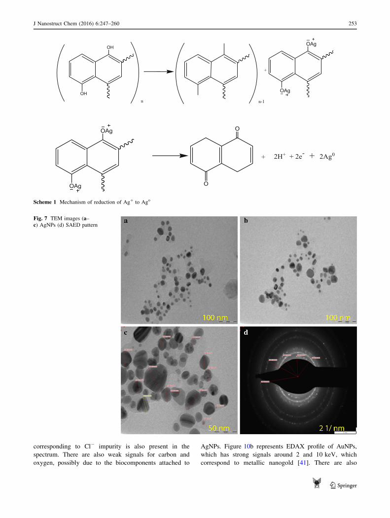
FT-IR studies
FT-IR spectra of Ag and Au NPs were recorded to identify
the possible biomolecules responsible for the reduction and
capping. The Ag and Au nanosolutions were centrifuged at
13000 rpm for 15 min and air-dried. This dried powder
was made as pellets with KBr, and the spectra were
recorded. Figure 6 shows the FT-IR spectra of the bark
extract, Ag and Au NPs. The spectrum of extract (powder)
has peaks at 1063, 1384, and 1631 cm-1. The AgNPs have
peaks at 1033, 1384, and 1615 cm-1, while AuNPs exhi-
bits peaks at 1032, 1383, and 1602 cm-1. The main peaks
present in the extract are also present in both Ag and Au
NPs inferring that the attachment of biomolecules which
are present in the extract to nanoparticles.
Different plant parts of P. zeylanica contain naph-
thaquinones, alkaloids, glycosides, steroids, triterpenoids,
tannins, phenolic compounds, flavanoids, coumarins, car-
bohydrates, saponins, proteins, oils, and fats [29]. Of all the
chemical constituents, plumbagin is the principle active
compound. Plumbagin (5-hydroxy-2-methyl-1,4-naphtho-
quinone –C11H8O3) is primarily present in roots in higher
amounts. The structures of plumbagin, glycoside and
derivatives of glycoside are represented below [35]. The
major water-soluble components present in P. zeylanica
extract are glycosides [36].
O
O
CH3
OH
Plumbagin
O
O
CH3
H3C
O
O
OH
OH
3,3 - diplumbagin
OHCH3
OOHO
HOHO
OH
Hydroplumbagin glucoside
HO
The peak at 1631 cm-1 in extract gets shifted to 1615
and 1602 cm-1 in the spectra of Ag and Au NPs, respec-
tively. This peak corresponds to chelated[C=O group [37],
inferring that the binding of biomolecules with the
nanoparticles through this group. The peaks at 3424, 3420,
and 3384 cm-1 in the spectra of the extract, Ag and Au NPs
are due to –OH groups in the biocomponents, and they may
be responsible for bioreduction. The peaks near 2912 cm-1
in all the three spectra are due to –C–H stretching of aro-
matic ring. Further, the peaks at 1033 cm-1 and 1032 cm-1
in the spectra of Ag and Au NPs, respectively, which
appears as shoulder in extract, correspond to C–O stretching
frequency of biocomponents. These data indicate the
involvement of[C=O and –OH functional groups in the
reduction and stabilization of nanoparticles.
Mechanism of formation and stabilization of AgNPs
The phytoconstituents like plumbagin and hydroplumbagin
glucoside contain many hydroxyl groups. On addition of
bark extracts to the AgNO3 solution, the Ag?—phytocon-
stituent complex is formed as an intermediate. The nascent
hydrogens produced during this process reduce Ag? to Ago
followed by coalescence, cluster formation, and growth of
clusters, finally yielding AgNPs [38]. The phenolic groups
present in the bark extract subsequently undergo oxidation
and get converted to their quinone forms. The electro-
chemical potential difference between Ag? and phyto-
constituents drives the reaction. The formed AgNPs are
stabilized through the lone pair of electrons and p electrons
of quinone structures (Scheme 1).
0
0.8
0.2
0.4
0.6
200 800400 600
Abs
orba
nce
Wavelength [nm]
pH 11 pH 10 pH 9 pH 8 pH 7 pH 6 pH 5 pH 4 pH 3 pH 2
a b
Fig. 4 a UV–Visible spectra of AgNPs synthesized at various pH
b Variation of absorbance with pH
Fig. 5 a UV–Visible spectra of the synthesized AuNPs as a function
of time b Variation of absorbance with time
J Nanostruct Chem (2016) 6:247–260 251
123
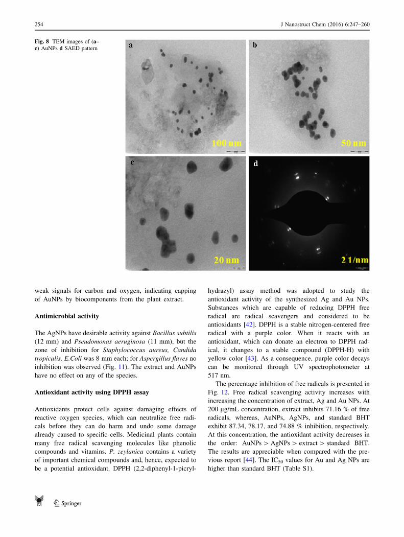
TEM studies
TEM analysis reveals the size and morphology of biosyn-
thesized NPs. TEM images of the Ag and Au NPs at dif-
ferent magnification are depicted in Figs. 7 and 8,
respectively. The TEM images clearly show that the
nanoparticles are predominantly spherical in shape. The
size is in the range of 2.76–51.57 nm (average size
28.47 nm) for AgNPs and 10–25 nm (average size
16.89 nm) for AuNPs. Figures 7d and 8d show the SAED
patterns (circular rings) of Ag and Au NPs, that reveal the
crystalline nature of nanoparticles. The diffraction rings
arise due to reflections of planes of fcc crystals which is
evinced from broad Bragg’s reflection observed in the
XRD spectra.
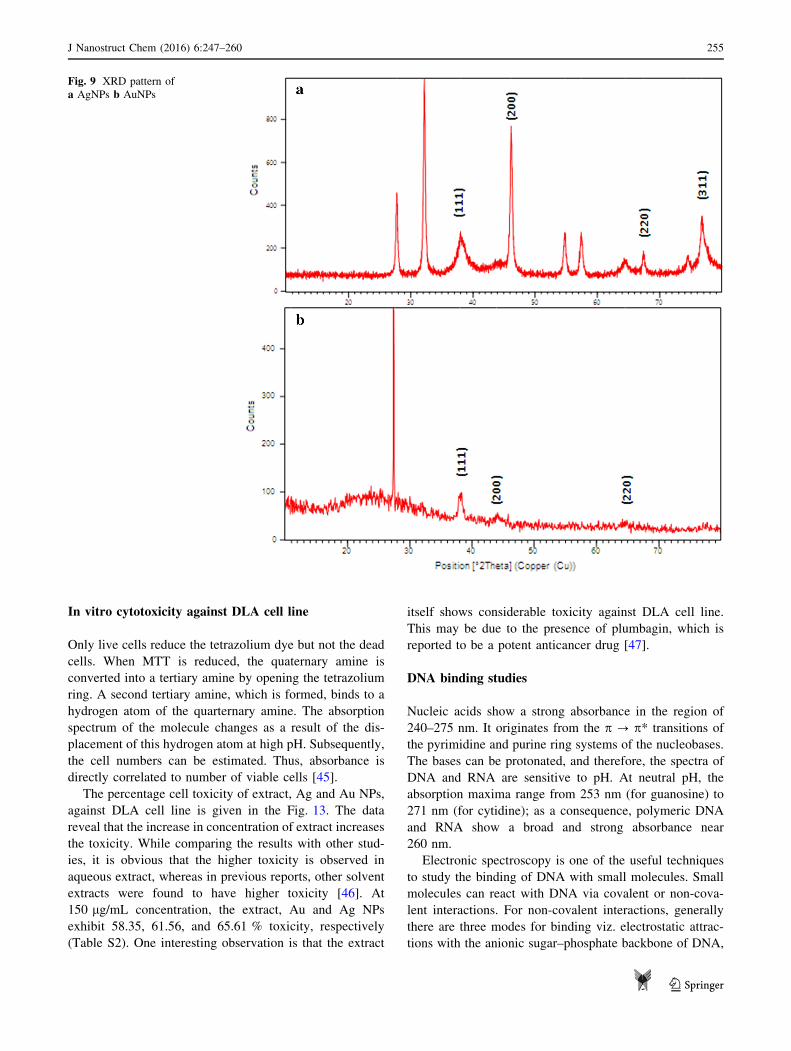
XRD studies
X-ray diffraction patterns of the Ag and Au NPs are shown
in Fig. 9a, b, respectively. A comparison of XRD pattern of
AgNPs with the standard JCPDS file no. 89-3722 [39]
confirms the crystalline face-centered cubic (fcc) nature, as
evidenced by the peaks at 2h values of 37.99�, 64.52�, and76.69�, which can be indexed to 111, 220, and 311 planes,
respectively. AuNPs show similar characteristic peaks of
metallic fcc gold (JCPDS: 4-0784) [27]. The particle sizes
of Ag and Au NPs were computed by Debye–Scherrer
equation from which the average size was found to be
17.26 and 12.84 nm, respectively.
D ¼ kk=b cos h ð1Þ
where, k is the Scherrer constant, k is the wavelength of theX-ray, b and h are the half-width of the peak and half of theBragg angle, respectively.
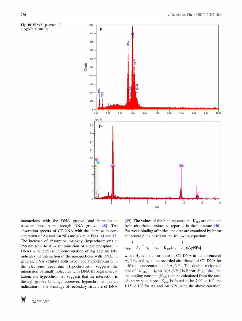
EDAX analysis
EDAX (Energy Dispersive X-ray) analysis was per-
formed to confirm the elemental form of silver and gold.
Figure 10a represents the EDAX profile of AgNPs. The
EDAX spectrum shows a strong signal at 3 keV, which
corresponds to metallic nanosilver [40]; peak
Fig. 6 FT-IR spectra of a Plumbago zeylanica bark extract, b AgNPs and c AuNPs
252 J Nanostruct Chem (2016) 6:247–260
123
corresponding to Cl- impurity is also present in the
spectrum. There are also weak signals for carbon and
oxygen, possibly due to the biocomponents attached to
AgNPs. Figure 10b represents EDAX profile of AuNPs,
which has strong signals around 2 and 10 keV, which
correspond to metallic nanogold [41]. There are also
OAg
OAg
O
O
+ 2H+ + 2e- + 2Ag0
OH
OHn n-1
+
OAg
OAg
Scheme 1 Mechanism of reduction of Ag? to Ago
Fig. 7 TEM images (a–c) AgNPs (d) SAED pattern
J Nanostruct Chem (2016) 6:247–260 253
123
weak signals for carbon and oxygen, indicating capping
of AuNPs by biocomponents from the plant extract.
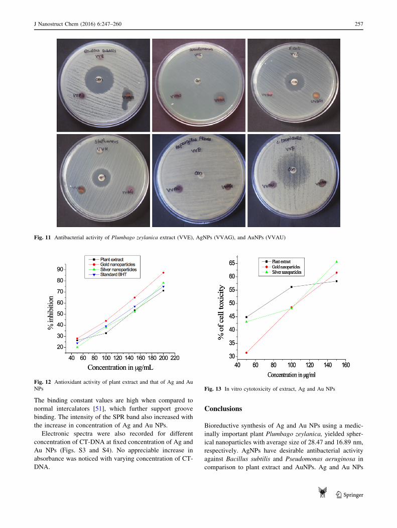
Antimicrobial activity
The AgNPs have desirable activity against Bacillus subtilis
(12 mm) and Pseudomonas aeruginosa (11 mm), but the
zone of inhibition for Staphylococcus aureus, Candida
tropicalis, E.Coli was 8 mm each; for Aspergillus flaves no
inhibition was observed (Fig. 11). The extract and AuNPs
have no effect on any of the species.
Antioxidant activity using DPPH assay
Antioxidants protect cells against damaging effects of
reactive oxygen species, which can neutralize free radi-
cals before they can do harm and undo some damage
already caused to specific cells. Medicinal plants contain
many free radical scavenging molecules like phenolic
compounds and vitamins. P. zeylanica contains a variety
of important chemical compounds and, hence, expected to
be a potential antioxidant. DPPH (2,2-diphenyl-1-picryl-
hydrazyl) assay method was adopted to study the
antioxidant activity of the synthesized Ag and Au NPs.
Substances which are capable of reducing DPPH free
radical are radical scavengers and considered to be
antioxidants [42]. DPPH is a stable nitrogen-centered free
radical with a purple color. When it reacts with an
antioxidant, which can donate an electron to DPPH rad-
ical, it changes to a stable compound (DPPH-H) with
yellow color [43]. As a consequence, purple color decays
can be monitored through UV spectrophotometer at
517 nm.
The percentage inhibition of free radicals is presented in
Fig. 12. Free radical scavenging activity increases with
increasing the concentration of extract, Ag and Au NPs. At
200 lg/mL concentration, extract inhibits 71.16 % of free
radicals, whereas, AuNPs, AgNPs, and standard BHT
exhibit 87.34, 78.17, and 74.88 % inhibition, respectively.
At this concentration, the antioxidant activity decreases in
the order: AuNPs[AgNPs[ extract[ standard BHT.
The results are appreciable when compared with the pre-
vious report [44]. The IC50 values for Au and Ag NPs are
higher than standard BHT (Table S1).
Fig. 8 TEM images of (a–c) AuNPs d SAED pattern
254 J Nanostruct Chem (2016) 6:247–260
123
In vitro cytotoxicity against DLA cell line
Only live cells reduce the tetrazolium dye but not the dead
cells. When MTT is reduced, the quaternary amine is
converted into a tertiary amine by opening the tetrazolium
ring. A second tertiary amine, which is formed, binds to a
hydrogen atom of the quarternary amine. The absorption
spectrum of the molecule changes as a result of the dis-
placement of this hydrogen atom at high pH. Subsequently,
the cell numbers can be estimated. Thus, absorbance is
directly correlated to number of viable cells [45].
The percentage cell toxicity of extract, Ag and Au NPs,
against DLA cell line is given in the Fig. 13. The data
reveal that the increase in concentration of extract increases
the toxicity. While comparing the results with other stud-
ies, it is obvious that the higher toxicity is observed in
aqueous extract, whereas in previous reports, other solvent
extracts were found to have higher toxicity [46]. At
150 lg/mL concentration, the extract, Au and Ag NPs
exhibit 58.35, 61.56, and 65.61 % toxicity, respectively
(Table S2). One interesting observation is that the extract
itself shows considerable toxicity against DLA cell line.
This may be due to the presence of plumbagin, which is
reported to be a potent anticancer drug [47].
DNA binding studies
Nucleic acids show a strong absorbance in the region of
240–275 nm. It originates from the p ? p* transitions of
the pyrimidine and purine ring systems of the nucleobases.
The bases can be protonated, and therefore, the spectra of
DNA and RNA are sensitive to pH. At neutral pH, the
absorption maxima range from 253 nm (for guanosine) to
271 nm (for cytidine); as a consequence, polymeric DNA
and RNA show a broad and strong absorbance near
260 nm.
Electronic spectroscopy is one of the useful techniques
to study the binding of DNA with small molecules. Small
molecules can react with DNA via covalent or non-cova-
lent interactions. For non-covalent interactions, generally
there are three modes for binding viz. electrostatic attrac-
tions with the anionic sugar–phosphate backbone of DNA,
Fig. 9 XRD pattern of
a AgNPs b AuNPs
J Nanostruct Chem (2016) 6:247–260 255
123
interactions with the DNA groove, and intercalation
between base pairs through DNA groove [48]. The
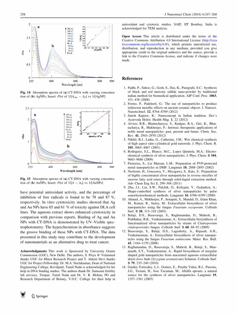
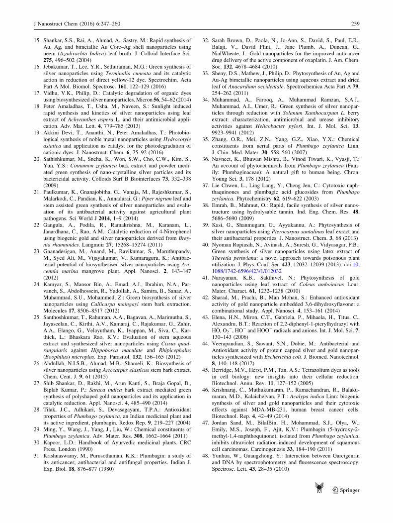
absorption spectra of CT-DNA with the increase in con-
centration of Ag and Au NPs are given in Figs. 14 and 15.
The increase of absorption intensity (hyperchromism) at
258 nm (due to p ? p* transition of sugar phosphate in
DNA) with increase in concentration of Ag and Au NPs
indicates the interaction of the nanoparticles with DNA. In
general, DNA exhibits both hyper and hypochromism in
the electronic spectrum. Hypochromism suggests the
interaction of small molecules with DNA through interca-
lation, and hyperchromism suggests that the interaction is
through groove binding; moreover, hyperchromism is an
indication of the breakage of secondary structure of DNA
[49]. The values of the binding constant, Kapp are obtained
from absorbance values as reported in the literature [50].
For weak binding affinities, the data are examined by linear
reciprocal plots based on the following equation:
1
Aobs � Ao
¼ 1
Ac � Ao
þ 1
KappðAc � A0Þ½AgNPs]
where Ao is the absorbance of CT-DNA in the absence of
AgNPs, and Ac is the recorded absorbance of CT-DNA for
different concentration of AgNPs. The double reciprocal
plot of 1/Aobs - A0 vs 1/[AgNPs] is linear (Fig. 14a), and
the binding constant (Kapp) can be calculated from the ratio
of intercept to slope. Kapp is found to be 7.03 9 107 and
1.15 9 107 for Ag and Au NPs using the above equation.
Fig. 10 EDAX spectrum of
a AgNPs b AuNPs
256 J Nanostruct Chem (2016) 6:247–260
123
The binding constant values are high when compared to
normal intercalators [51], which further support groove
binding. The intensity of the SPR band also increased with
the increase in concentration of Ag and Au NPs.
Electronic spectra were also recorded for different
concentration of CT-DNA at fixed concentration of Ag and
Au NPs (Figs. S3 and S4). No appreciable increase in
absorbance was noticed with varying concentration of CT-
DNA.
Conclusions
Bioreductive synthesis of Ag and Au NPs using a medic-
inally important plant Plumbago zeylanica, yielded spher-
ical nanoparticles with average size of 28.47 and 16.89 nm,
respectively. AgNPs have desirable antibacterial activity
against Bacillus subtilis and Pseudomonas aeruginosa in
comparison to plant extract and AuNPs. Ag and Au NPs
Fig. 11 Antibacterial activity of Plumbago zeylanica extract (VVE), AgNPs (VVAG), and AuNPs (VVAU)
Fig. 12 Antioxidant activity of plant extract and that of Ag and Au
NPs Fig. 13 In vitro cytotoxicity of extract, Ag and Au NPs
J Nanostruct Chem (2016) 6:247–260 257
123
have potential antioxidant activity, and the percentage of
inhibition of free radicals is found to be 78 and 87 %,
respectively. In vitro cytotoxicity studies showed that Ag
and Au NPs have 65 and 61 % of toxicity against DLA cell
lines. The aqueous extract shows enhanced cytotoxicity in
comparison with previous reports. Binding of Ag and Au
NPs with CT-DNA is demonstrated by UV–Visible spec-
trophotometry. The hyperchromism in absorbance suggests
the groove binding of these NPs with CT-DNA. The data
presented in this study may contribute to the development
of nanomaterials as an alternative drug to treat cancer.
Acknowledgments This work is Sponsored by University Grants
Commission (UGC), New Delhi. The authors, S. Priya @ Velammal
thanks UGC for Minor Research Project and T. Akkini Devi thanks
UGC for Project Fellowship. Dr. M.A. Neelakantan, Dean of National
Engineering College, Kovilpatti, Tamil Nadu is acknowledged for his
help in DNA binding studies. The authors thank Dr. Samasun fertility
lab services, Tirupur, Tamil Nadu and Dr. V. R. Mohan, PG and
Research Department of Botany, V.O.C. College for their help in
antioxidant and cytotoxic studies. SAIF, IIT Bombay, India is
acknowledged for TEM analysis.
Open Access This article is distributed under the terms of the
Creative Commons Attribution 4.0 International License (http://crea
tivecommons.org/licenses/by/4.0/), which permits unrestricted use,
distribution, and reproduction in any medium, provided you give
appropriate credit to the original author(s) and the source, provide a
link to the Creative Commons license, and indicate if changes were
made.
References
1. Padhi, P., Sahoo, G., Gosh, S., Das, K., Panigrahi, S.C.: Synthesis
of black and red mercury sulfide nano-powder by traditional
indian method for biomedical application. AIP Conf. Proc. 1063,431–438 (2008)
2. Fermo, P., Padeleeti, G.: The use of nanoparticles to produce
iridescent metallic effects on ancient ceramic objects. J. Nanosci.
Nanotechnol. 12, 8764–8769 (2012)
3. Satish Kapoor, K.: Nanoconcept in Indian tradition. Dav’s
Ayurveda Holist. Health Mag. 1, 22 (2012)
4. Arvizo, R.R., Bhattacharyya, S., Kudgus, R.A., Giri, K., Bhat-
tacharya, R., Mukherjee, P.: Intrinsic therapeutic applications of
noble metal nanoparticles: past, present and future. Chem. Soc.
Rev. 41, 2943–2970 (2012)
5. Nikhil, R.J., Latha, G., Catherine, J.M.: Wet chemical synthesis
of high aspect ratio cylindrical gold nanorods. J. Phys. Chem. B.
105, 4065–4067 (2001)
6. Rodriguez, S.L., Blanco, M.C., Lopez Quintela, M.A.: Electro-
chemical synthesis of silver nanoparticles. J. Phys. Chem. B 104,9683–9686 (2009)
7. Pastoriza, S., Liz Marzan, L.M.: Preparation of PVP-protected
metal nanoparticles in DMF. Langmuir 18, 2888–2895 (2002)
8. Noritomi, H., Umezawa, Y., Miyagawa, S., Kato, S.: Preparation
of highly concentrated silver nanoparticles in reverse micelles of
sucrose fatty acid esters through solid-liquid extraction method.
Adv Chem Eng Sci 1, 299–304 (2011)
9. Zhu, J.J., Liu, S.W., Palchik, O., Koltypin, Y., Gedanken, A.:
Shape-controlled synthesis of silver nanoparticles by pulse
sonoelectrochemical methods. Langmuir 16, 6396–6399 (2000)
10. Ahmad, A., Mukherjee, P., Senapati, S., Mandal, D., Islam Khan,
M., Kumar, R., Sastry, M.: Extracellular biosynthesis of silver
nanoparticles using the fungus Fusarium oxysporum. Colloids
Surf. B 28, 313–318 (2003)
11. Balaji, D.S., Basavaraja, S., Raghunandan, D., Mahesh, B.,
Prabhakar, B.K., Venkataraman, A.: Extracellular biosynthesis of
functionalized silver nanoparticles by strains of Cladosporium
cladosporioides fungus. Colloids Surf. B 68, 88–92 (2009)
12. Basavaraja, S., Balaji, D.S., Lagashetty, A., Rajasab, A.H.,
Venkataraman, A.: Extracellular biosynthesis of silver nanopar-
ticles using the fungus Fusarium semitectum. Mater. Res. Bull.
43, 1164–1170 (2008)
13. Raghunandan, D., Basavaraja, S., Mahesh, B., Balaji, S., Man-
junath, S.Y., Venkataraman, A.: Rapid biosynthesis of irregular
shaped gold nanoparticles from macerated aqueous extracellular
dried clove buds (Syzygium aromaticum) Solution. Colloids Surf.
B 79, 235–240 (2010)
14. Gardea Torresdey, L.J., Gomez, E., Peralta Videa, R.J., Parsons,
J.G., Troiani, H., Jose Yacaman, M.: Alfalfa sprouts: a natural
source for the synthesis of silver nanoparticles. Langmuir 19,1357–1361 (2003)
Fig. 14 Absorption spectra of (a) CT-DNA with varying concentra-
tion of (b) AgNPs; Insert: Plot of 1/[Aobs - A0] vs 1/[AgNP]
0
2.5
1
2
009532 400 600 800
Abs
Wavelength [nm]
200 µL 180 µL 160 µL 140 µL 120 µL 100 µL 80 µL 60 µL 40 µL 20 µL Blank
a
b
Fig. 15 Absorption spectra of (a) CT-DNA with varying concentra-
tion of (b) AuNPs; Insert: Plot of 1/[A - A0] vs 1/[AuNPs]
258 J Nanostruct Chem (2016) 6:247–260
123
15. Shankar, S.S., Rai, A., Ahmad, A., Sastry, M.: Rapid synthesis of
Au, Ag, and bimetallic Au Core–Ag shell nanoparticles using
neem (Azadirachta Indica) leaf broth. J. Colloid Interface Sci.
275, 496–502 (2004)
16. Jebakumar, T., Lee, Y.R., Sethuraman, M.G.: Green synthesis of
silver nanoparticles using Terminalia cuneata and its catalytic
action in reduction of direct yellow-12 dye. Spectrochim. Acta
Part A Mol. Biomol. Spectrosc. 161, 122–129 (2016)
17. Vidhu, V.K., Philip, D.: Catalytic degradation of organic dyes
using biosynthesized silver nanoparticles.Micron 56, 54–62 (2014)18. Peter Amaladhas, T., Usha, M., Naveen, S.: Sunlight induced
rapid synthesis and kinetics of silver nanoparticles using leaf
extract of Achyranthes aspera L. and their antimicrobial appli-
cation. Adv. Mat. Lett. 4, 779–785 (2013)
19. Akkini Devi, T., Ananthi, N., Peter Amaladhas, T.: Photobio-
logical synthesis of noble metal nanoparticles using Hydrocotyle
asiatica and application as catalyst for the photodegradation of
cationic dyes. J. Nanostruct. Chem. 6, 75–92 (2016)
20. Sathishkumar, M., Sneha, K., Won, S.W., Cho, C.W., Kim, S.,
Yun, Y.S.: Cinnamon zeylanica bark extract and powder medi-
ated green synthesis of nano-crystalline silver particles and its
bactericidal activity. Colloids Surf B Biointerfaces 73, 332–338(2009)
21. Paulkumar, K., Gnanajobitha, G., Vanaja, M., Rajeshkumar, S.,
Malarkodi, C., Pandian, K., Annadurai, G.: Piper nigrum leaf and
stem assisted green synthesis of silver nanoparticles and evalu-
ation of its antibacterial activity against agricultural plant
pathogens. Sci World J 2014, 1–9 (2014)
22. Gangula, A., Podila, R., Ramakrishna, M., Karanam, L.,
Janardhana, C., Rao, A.M.: Catalytic reduction of 4-Nitrophenol
using biogenic gold and silver nanoparticles derived from Brey-
nia rhamnoides. Langmuir 27, 15268–15274 (2011)
23. Gnanadesigan, M., Anand, M., Ravikumar, S., Maruthupandy,
M., Syed Ali, M., Vijayakumar, V., Kumaraguru, K.: Antibac-
terial potential of biosynthesised silver nanoparticles using Avi-
cennia marina mangrove plant. Appl. Nanosci. 2, 143–147
(2012)
24. Kamyar, S., Mansor Bin, A., Emad, A.J., Ibrahim, N.A., Par-
vaneh, S., Abdolhossein, R., Yadollah, A., Samira, B., Sanaz, A.,
Muhammad, S.U., Mohammed, Z.: Green biosynthesis of silver
nanoparticles using Callicarpa maingayi stem bark extraction.
Molecules 17, 8506–8517 (2012)
25. Santhoshkumar, T., Rahuman, A.A., Bagavan, A., Marimuthu, S.,
Jayaseelan, C., Kirthi, A.V., Kamaraj, C., Rajakumar, G., Zahir,
A.A., Elango, G., Velayutham, K., Iyappan, M., Siva, C., Kar-
thick, L.: Bhaskara Rao, K.V.: Evaluation of stem aqueous
extract and synthesized silver nanoparticles using Cissus quad-
rangularis against Hippobosca maculate and Rhipicephalus
(Boophilus) microplus. Exp. Parasitol. 132, 156–165 (2012)
26. Abdullah, N.I.S.B., Ahmad, M.B., Shameli, K.: Biosynthesis of
silver nanoparticles using Artocarpus elasticus stem bark extract.
Chem. Cent. J. 9, 61 (2015)
27. Shib Shankar, D., Rakhi, M., Arun Kanti, S., Braja Gopal, B.,
Biplab Kumar, P.: Saraca indica bark extract mediated green
synthesis of polyshaped gold nanoparticles and its application in
catalytic reduction. Appl. Nanosci. 4, 485–490 (2014)
28. Tilak, J.C., Adhikari, S., Devasagayam, T.P.A.: Antioxidant
properties of Plumbago zeylanica, an Indian medicinal plant and
its active ingredient, plumbagin. Redox Rep. 9, 219–227 (2004)
29. Ming, Y., Wang, J., Yang, J., Liu, W.: Chemical constituents of
Plumbago zeylanica. Adv. Mater. Res. 308, 1662–1664 (2011)
30. Kapoor, L.D.: Handbook of Ayurvedic medicinal plants. CRC
Press, London (1990)
31. Krishnaswamy, M., Purusothaman, K.K.: Plumbagin: a study of
its anticancer, antibacterial and antifungal properties. Indian J.
Exp. Biol. 18, 876–877 (1980)
32. Sarah Brown, D., Paola, N., Jo-Ann, S., David, S., Paul, E.R.,
Balaji, V., David Flint, J., Jane Plumb, A., Duncan, G.,
NialWheate, J.: Gold nanoparticles for the improved anticancer
drug delivery of the active component of oxaplatin. J. Am. Chem.
Soc. 132, 4678–4684 (2010)
33. Sheny, D.S., Mathew, J., Philip, D.: Phytosynthesis of Au, Ag and
Au-Ag bimetallic nanoparticles using aqueous extract and dried
leaf of Anacardium occidentale. Spectrochemica Acta Part A 79,254–262 (2011)
34. Muhammad, A., Farooq, A., Muhammad Ramzan, S.A.J.,
Muhammad, A.I., Umer, R.: Green synthesis of silver nanopar-
ticles through reduction with Solanum Xanthocarpum L. berry
extract: characterization, antimicrobial and urease inhibitory
activities against Helicobacter pylori. Int. J. Mol. Sci. 13,9923–9941 (2012)
35. Zhang, O.R., Mei, Z.N., Yang, G.Z., Xiao, Y.X.: Chemical
constituents from aerial parts of Plumbago zeylanica Linn.
J. Chin. Med. Mater. 30, 558–560 (2007)
36. Navneet, K., Bhuwan Mishra, B., Vinod Tiwari, K., Vyasji, T.:
An account of phytochemicals from Plumbago zeylanica (Fam-
ily: Plumbaginaceae): A natural gift to human being. Chron.
Young Sci. 3, 178 (2012)
37. Lie Chwen, L., Ling Lang, Y., Cheng Jen, C.: Cytotoxic naph-
thaquinones and plumbagic acid glucosides from Plumbago
zeylanica. Phytochemistry 62, 619–622 (2003)
38. Emrah, B., Mahmut, O.: Rapid, facile synthesis of silver nanos-
tructure using hydrolysable tannin. Ind. Eng. Chem. Res. 48,5686–5690 (2009)
39. Kasi, G., Shanmugam, G., Ayyakannu, A.: Phytosynthesis of
silver nanoparticles using Pterocarpus santalinus leaf exract and
their antibacterial properties. J. Nanostruct. Chem. 3, 68 (2013)
40. Nyoman Rupiasih, N., Avinash, A., Suresh, G., Vidyasagar, P.B.:
Green synthesis of silver nanoparticles using latex extract of
Thevetia peruviana: a novel approach towards poisonous plant
utilization. J. Phys. Conf. Ser. 423, 12032–12039 (2013). doi:10.
1088/1742-6596/423/1/012032
41. Narayanan, K.B., Sakthivel, N.: Phytosynthesis of gold
nanoparticles using leaf extract of Coleus amboinicus Lour.
Mater. Charact. 61, 1232–1238 (2010)
42. Sharad, M., Prachi, B., Man Mohan, S.: Enhanced antioxidant
activity of gold nanoparticle embedded 3,6-dihydroxyflavone: a
combinational study. Appl. Nanosci. 4, 153–161 (2014)
43. Elena, H.N., Miron, C.T., Gabriela, P., Mihaela, H., Titus, C.,
Alexandru, B.T.: Reaction of 2,2-diphenyl-1-picrylhydrazyl with
HO, O2-, HO- and HOO- radicals and anions. Int. J. Mol. Sci. 7,
130–143 (2006)
44. Veerapandian, S., Sawant, S.N., Dobie, M.: Antibacterial and
Antioxidant activity of protein capped silver and gold nanopar-
ticles synthesized with Escherichia coli. J. Biomed. Nanotechnol.
8, 140–148 (2012)
45. Berridge, M.V., Herst, P.M., Tan, A.S.: Tetrazolium dyes as tools
in cell biology: new insights into their cellular reduction.
Biotechnol. Annu. Rev. 11, 127–152 (2005)
46. Krishnaraj, C., Muthukumaran, P., Ramachandran, R., Balaku-
maran, M.D., Kalaichelvan, P.T.: Acalypa indica Linn: biogenic
synthesis of silver and gold nanoparticles and their cytotoxic
effects against MDA-MB-231, human breast cancer cells.
Biotechnol. Rep. 4, 42–49 (2014)
47. Jordan Sand, M., BilalBin, H., Mohammad, S.J., Olya, W.,
Emily, M.S., Joseph, F., Ajit, K.V.: Plumbagin (5-hydroxy-2-
methyl-1,4-naphthoquinone), isolated from Plumbago zeylanica,
inhibits ultraviolet radiation-induced development of squamous
cell carcinomas. Carcinogenesis 33, 184–190 (2011)
48. Yunhua, W., Guangzhong, Y.: Interaction between Garcigenrin
and DNA by spectrophotometry and fluorescence spectroscopy.
Spectrosc. Lett. 43, 28–35 (2010)
J Nanostruct Chem (2016) 6:247–260 259
123
49. Nahid, S., Saba, H.: Spectroscopic studies on the interaction of
calf thymus DNA with the drug levetiracetam. Spectrochim. Acta
Part A Mol. Biomol. Spectrosc. 96, 278–283 (2012)
50. Swarup, R., Ratan, S., Utpal, G., Tapan, K.D.: Interaction studies
between biosynthesized silver nanoparticle with calf thymus
DNA and cytotoxicity of silver nanoparticles. Spectrochim. Acta
Part A Mol. Biomol. Spectrosc. 141, 176–184 (2015)
51. Pin-xian, X., Zhi-hong, X., Feng-juan, C., Zheng-zhi, Z., Xiao-
wen, Z.: Study on synthesis, structure and DNA binding of Ni, Zn
complexes with 2-phenylquinoline-4 carboylhydrazide. J. Inorg.
Biochem. 103, 210–218 (2009)
260 J Nanostruct Chem (2016) 6:247–260
123