Embed Size (px)
Citation preview
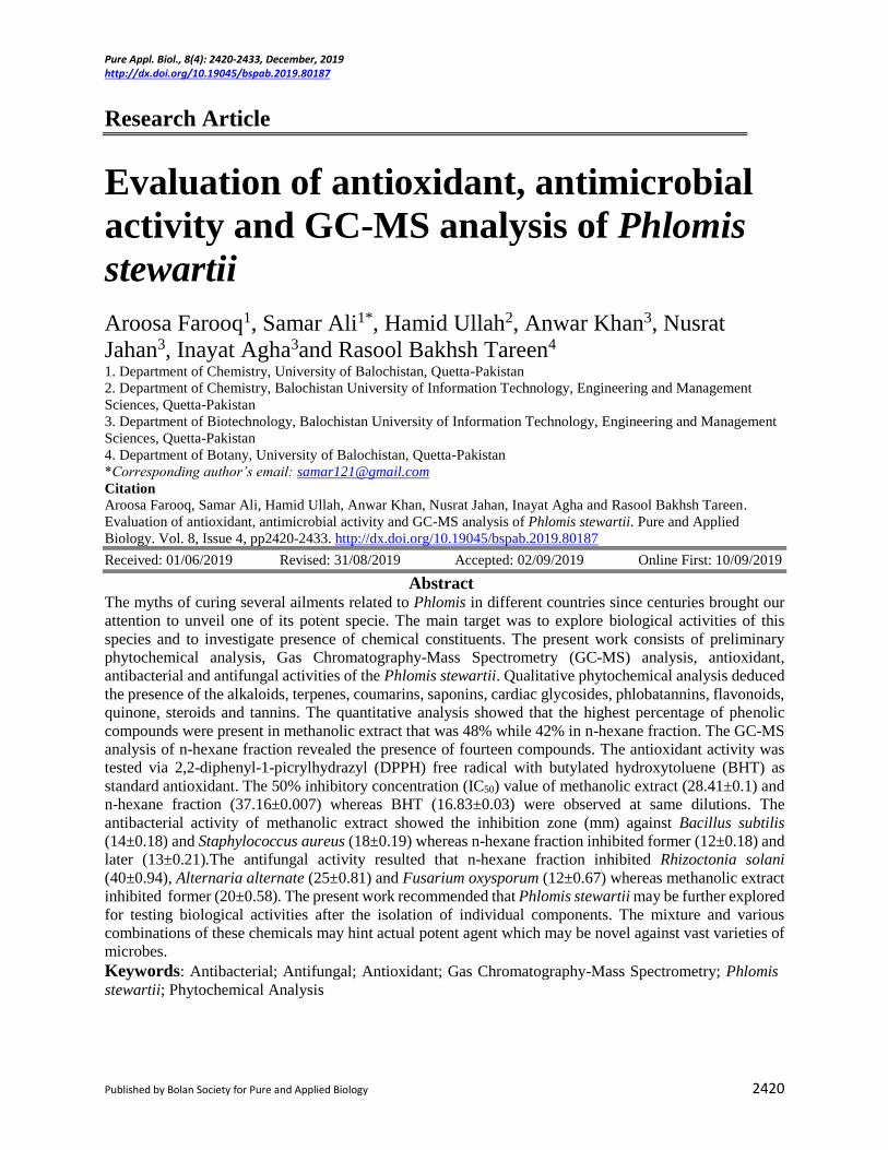
Pure Appl. Biol., 8(4): 2420-2433, December, 2019 http://dx.doi.org/10.19045/bspab.2019.80187
Published by Bolan Society for Pure and Applied Biology 2420
Research Article
Evaluation of antioxidant, antimicrobial
activity and GC-MS analysis of Phlomis
stewartii
Aroosa Farooq1, Samar Ali1*, Hamid Ullah2, Anwar Khan3, Nusrat
Jahan3, Inayat Agha3and Rasool Bakhsh Tareen4 1. Department of Chemistry, University of Balochistan, Quetta-Pakistan
2. Department of Chemistry, Balochistan University of Information Technology, Engineering and Management
Sciences, Quetta-Pakistan
3. Department of Biotechnology, Balochistan University of Information Technology, Engineering and Management
Sciences, Quetta-Pakistan
4. Department of Botany, University of Balochistan, Quetta-Pakistan
*Corresponding author’s email: [email protected]
Citation Aroosa Farooq, Samar Ali, Hamid Ullah, Anwar Khan, Nusrat Jahan, Inayat Agha and Rasool Bakhsh Tareen.
Evaluation of antioxidant, antimicrobial activity and GC-MS analysis of Phlomis stewartii. Pure and Applied
Biology. Vol. 8, Issue 4, pp2420-2433. http://dx.doi.org/10.19045/bspab.2019.80187
Received: 01/06/2019 Revised: 31/08/2019 Accepted: 02/09/2019 Online First: 10/09/2019
Abstract The myths of curing several ailments related to Phlomis in different countries since centuries brought our
attention to unveil one of its potent specie. The main target was to explore biological activities of this
species and to investigate presence of chemical constituents. The present work consists of preliminary
phytochemical analysis, Gas Chromatography-Mass Spectrometry (GC-MS) analysis, antioxidant,
antibacterial and antifungal activities of the Phlomis stewartii. Qualitative phytochemical analysis deduced
the presence of the alkaloids, terpenes, coumarins, saponins, cardiac glycosides, phlobatannins, flavonoids,
quinone, steroids and tannins. The quantitative analysis showed that the highest percentage of phenolic
compounds were present in methanolic extract that was 48% while 42% in n-hexane fraction. The GC-MS
analysis of n-hexane fraction revealed the presence of fourteen compounds. The antioxidant activity was
tested via 2,2-diphenyl-1-picrylhydrazyl (DPPH) free radical with butylated hydroxytoluene (BHT) as
standard antioxidant. The 50% inhibitory concentration (IC50) value of methanolic extract (28.41±0.1) and
n-hexane fraction (37.16±0.007) whereas BHT (16.83±0.03) were observed at same dilutions. The
antibacterial activity of methanolic extract showed the inhibition zone (mm) against Bacillus subtilis
(14±0.18) and Staphylococcus aureus (18±0.19) whereas n-hexane fraction inhibited former (12±0.18) and
later (13±0.21).The antifungal activity resulted that n-hexane fraction inhibited Rhizoctonia solani
(40±0.94), Alternaria alternate (25±0.81) and Fusarium oxysporum (12±0.67) whereas methanolic extract
inhibited former (20±0.58). The present work recommended that Phlomis stewartii may be further explored
for testing biological activities after the isolation of individual components. The mixture and various
combinations of these chemicals may hint actual potent agent which may be novel against vast varieties of
microbes.
Keywords: Antibacterial; Antifungal; Antioxidant; Gas Chromatography-Mass Spectrometry; Phlomis
stewartii; Phytochemical Analysis
Farooq et al.
2421
Introduction
Natural products either obtained from
plants, animals or microbes are considered
as important tools in medicinal field. A
large number of natural products are used
as starting material for drug development.
Additionally, they are allowed to be
marketed after several modifications to
overcome side effects [1]. Natural
products are categorized as primary and
secondary metabolites, the later one are
actually responsible for making them
unique with respect to several
characteristic benefits provided to
different life forms [2, 3].
Medicinal plants are potent with respect to
ailments cure and famous since ancient
history. The plants are used for curing
several ailments from simple fever to
major diseases caused by pathogens in
different parts of the world [4].The hidden
chemistry for such curing potential lies in
their phytochemicals which brings some
uniqueness in plant and these substances
are actually the center of attraction for
several activities. These reported
substances are up to 4500 and
350substances are completely
characterized by using different
techniques. These phytochemicals make
the plant attractive by imparting color,
flavor, and taste as well as protect them
against foreign attack of living as well as
non-living like exposure of ultraviolet
rays, polluting agents etc. [5]. These
substances when isolated and applied on
other living organisms then they impart
mixture of two behaviors that is
detrimental as well as benign [6]. The
most potent phytochemicals found in most
of the plants are flavonoids, carotenoids,
polyphenols, lignins, anthocyanins,
indole-3-carbinol and glycosides [7].
Phlomis stewartii belongs to Lamiaceae
family. It consists of rosy pink flowers;
located in different areas of Pakistan
especially in Suleiman and salt range. It is
perennial plant with erect stems having 30-45
cm height. It consists of dense hairs and much
branching at lower side.The leaves are thick-
textured, aromatic, elliptic or narrow oblong,
crenulate, acute at apex,length of calyx is
12mm. The corolla are rose to dusky pink
with length of 2 cm, tube exerted beyond
calyx, calyx is surrounded by thin bracts
nearly 18 in number [8, 9].
In present work, Phlomis stewartii was
studied for the GC-MS analysis of n-hexane
fraction and preliminary phytochemical
analysis of the plant along with evaluation of
antibacterial, antifungal and antioxidant
activities.
Materials and methods
Plant material
The plant sample was collected from Ziarat,
Balochistan in the month of April, 2017 and
identified by Dr. Rasool Bakhsh Tareen,
Professor Department of Botany, University
of Balochistan, and Quetta. The plant (7 kg)
was extracted thrice with methanol in order
to obtain maximum chemical constituents in
the extract [10].The obtained methanolic
extract was fractionated into n-hexane and
ethyl acetate fractions.
GC-MS analysis
Gas Chromatography (GC) was used to
determine the retention time and Mass
spectrometry (MS) was used to determine the
molecular ion peak and fragment ion peaks of
compounds. The column
(30m×250µm×0.25µm) was packed with 5%
phenyl methyl siloxane as stationary phase.
The helium gas used as mobile phase which
allowed the sample to move across the
column. The oven temperature was initially
set at 50 0C for at least 3 minutes and then it
was increased from 10-180 0C /15 minutes. In
the second step, temperature was raised up to
a maximum of 300 0C, the maximum
temperature and pressure of oven recorded as
360 0C and 9.05psi respectively. The
complete run up time was 68 minutes [11]

Pure Appl. Biol., 8(4): 2420-2433, December, 2019 http://dx.doi.org/10.19045/bspab.2019.80187
2422
and the mass spectra thus obtained were
matched with already available data in Replib
and Mainlib [12].The electron impact (EI)
used as a source for ionization at 250 0C.
Qualitative analysis
The methanolic extract and n-hexane fraction
were evaluated for the presence of alkaloids,
terpenes, coumarins, saponins, cardiac
glycosides, phlobatannins, flavonoids,
quinone, steroids and tannins by adopting
methods described in previous reported
literature [12]. The test methodsare included
in (Table 1).
Table 1. Qualitative analysis of Methanolic extract and n-hexane fraction
Compounds type Methanolic extract n- Hexane fraction
Alkaloids Dark red color appeared Dark red color appeared
Cardiac Glycosides
Two rings formed, lower one
reddish brown while upper one
was bluish green
Two rings formed, lower one
reddish brown while upper one was
bluish green
Coumarin Light Yellow colored Light Yellow colored
Flavonoids
Yellow color appeared which was
changed later due to introduction
of HCl
Yellow color appeared which was
later disappeared as soon as dil.
HCl was introduced
Phlobatannins No change No change
Quinone Intense red color appeared Medium red color appeared
Saponins Foam appeared Foams appeared
Steroids Red layer at lower side of test
tube appeared
Red layer at lower side of test tube
appeared
Tannins Light greenish ppt. appeared Light greenish ppt. appeared
Terpenoids Reddish ring appeared Medium reddish rings appeared
Quantitative analysis
The quantitative analysis of n-hexane and
methanolic extract were determined for total
alkaloid contents, total phenolic contents,
total saponins and total flavonoids contents.
The methodology thus adopted was already
reported [13, 14].
Antioxidant activity
The n-hexane fraction and methanolic extract
were evaluated for antioxidant activity. The
2,2-diphenyl-1-picrylhydrazyl(DPPH) was
used as free radical while BHT as a standard
antioxidant. The plant sample and DPPH
were dissolved in methanol, so methanol was
used as reference. The DPPH (0.004 g) was
dissolved in 100 mL of methanol. The
dilutions (250, 125, 50, 10) mg/L were
prepared from the 5mg/L stock solution for
plant sample and BHT standard. All the
prepared samples were kept in dark for a
period of 20-25 minutes in dark for providing
them the time to complete the reaction. The
UV Spectrophotometer (Shimadzu, Japan)
was used and all the prepared dilutions
absorbance measured at 517 nm. The
decrease in absorbance confirmed the
scavenging activity of sample [10,15]. The
following formula was used to assayed
antioxidant activity of plant sample.
%age scavenging of DPPH
= Abs control- Abs sample/Abs control
The effectiveness of sample was further
checked by IC50 value obtained from slope by
using formula as;
IC50 = 50-b/a (a and b are obtained from
slope)
Antifungal activity
The antifungal activity of sample was tested
by using disc diffusion method and agar well
diffusion method. The Potato Dextrose Agar
(PDA) was used as nutrient and fungi were
Farooq et al.
2423
Alternaria alternate, Rhizoctonia solani and
Fusarium oxysporum.
Disc diffusion method
The fungi were placed at center of the discs
and samples applied at peripheral region. The
diameter of disc was 6mm and allowed for
incubation for 3 days at 37 0C in dark. This
method was used to assess the samples on the
basis of qualitative analysis [16].
Agar well diffusion method
This method adopted for the quantitative
analysis of the plant samples against fungi.
The different concentrations of sample were
prepared by dissolving in Dimethyl sulfoxide
(DSO). The concentrations ranging from 20-
100µL were prepared and then added each
into the well. The fungi inoculum was
allowed to spread on dish and well bored to a
depth of 8mm. The samples kept for 3-4
hours in inverted position and after that
placed them in incubator for 48-72 hours at
37 0C. The mycotic inhibition was
determined by following formula;
%age mycelial Inhibition = [(dc-dt)/dc] ×100
dc = Diameter of fungal growth in control
dt = Diameter of fungal growth under test
The procedure repeated in triplicate in order
to confirm the authenticity and liability of
work.
Antibacterial activity
The methanolic extract and n-hexane fraction
of the plant were subjected for antibacterial
activity. The two gram positive bacteria,
Escherichia coli and Shigella dysenteriae
and two gram negative bacteria Bacillus
subtilis and Staphylococcus aureus were
selected to analyze the antibacterial activity.
Macchonkey agar (MA) and Eosin methylene
blue (EMB) were used as selective media to
confirm the presence of gram negative
bacteria whereas Mannitol Salt Agar (MSA)
used as selective media to test the presence of
gram positive bacteria. After the initial
screening, nutrient agar, Muellar Hinton
Agar (MHA) was prepared for all the four
bacteria, and samples incubated for 24 hours
at 24-28 0C.
Disc diffusion method
A fresh colony of each bacterium was taken
and dissolved in saline water to retain the cell
wall of bacteria. They were compared with
McFarland standard 0.05 and the colonies
transparency of bacteria should be match able
with it [17]. The stock solution of sample was
diluted as 0.1, 0.12, 0.14, 0.16 and 0.18g/µL
and used to determine minimum inhibitory
concentration (MIC). The antibiotic
Novobiocin acted as positive control while
DMSO used as negative control. The
conclusion was drawn by comparing the
samples with positive control.
Results and discussion
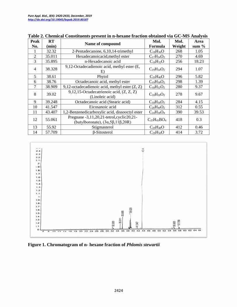
GC-MS analysis
The n-hexane fraction of Phlomis stewartii
was investigated via GC-MS technique. The
list of compounds, their molecular formula
(MF), molecular weight (MW), and retention
time (RT), area sum % peaks are incorporated
in (Table 2). The GC chromatogram showed
relative concentration of each compound
verses retention time. The chromatograms
included as (Figure 1 & 2).The mass spectra
of most abundant compounds are added as
(Figure 3-6).
The data obtained from the present work
showed the presence of 14 compounds
including phytol, steroids, eicosanoic acid,
Linoleic acid, pregnane, stigmasterol, β-
Sitosterol, 9,12-octadecadioic acid,
hexadecadioic acid etc. Among them,
stigmasterol, β-sitosterol and phytol have
antioxidant potential. The ester based
compounds have the potential of scavenging
free radicals [18]. The long chain fatty acids
including eicosanoic acid, linoleic acid etc.
are potent agent for antibacterial activities.
The presence of above compounds in n-
hexane fraction revealed its medicinal
importance [19-21].
Pure Appl. Biol., 8(4): 2420-2433, December, 2019 http://dx.doi.org/10.19045/bspab.2019.80187
2424
Table 2. Chemical Constituents present in n-hexane fraction obtained via GC-MS Analysis
Peak
No.
RT
(min) Name of compound
Mol.
Formula
Mol.
Weight
Area
sum %
1 32.32 2-Pentadecanone, 6,10,14-trimethyl C18H36O 268 1.05
2 35.011 Hexadecanoicacid,methyl ester C17H34O2 270 4.69
3 35.895 n-Hexadecanoic acid C16H32O 256 18.23
4 38.328 9,12-Octadecadienoic acid, methyl ester (E,
E) C19H34O2 294 1.07
5 38.61 Phytol C20H40O 296 5.82
6 38.76 Octadecanoic acid, methyl ester C19H38O2 298 1.39
7 38.909 9,12-octadecadienoic acid, methyl ester (Z, Z) C18H32O2 280 9.37
8 39.02 9,12,15-Octadecatrienoic acid, (Z, Z, Z)
(Linoleic acid) C18H30O2 278 9.67
9 39.248 Octadecanoic acid (Stearic acid) C18H36O2 284 4.15
10 41.547 Eicosanoic acid C20H40O2 312 0.55
11 43.407 1,2-Benzenedicarboxylic acid, dissooctyl ester C24H38O4 390 39.53
12 55.061 Pregnane -3,11,20,21-tetrol,cyclic20,21-
(butylboronate), (3α,5β,11β,20R) C25H43BO4 418 0.3
13 55.92 Stigmasterol C29H48O 412 0.46
14 57.709 β-Sitosterol C29H50O 414 3.72
Figure 1. Chromatogram of n- hexane fraction of Phlomis stewartii

Farooq et al.
2425
Figure 2. Chromatogram obtained from n-hexane fraction
Figure 3. Mass Spectrum of β-Sitosterol

Pure Appl. Biol., 8(4): 2420-2433, December, 2019 http://dx.doi.org/10.19045/bspab.2019.80187
2426
Figure 4. Mass Spectrum of 1,2-Benzenedicarboxylic acid, disoctyl ester
Figure 5. Mass Spectrum of Pregnane-3,11,20,21-tetrol, cyclic 20,21-(butyl boronate), (3α,
5β, 11β, 20R)

Farooq et al.
2427
Figure 6. Mass Spectrum of Linoleic acid
Qualitative analysis
The methanolic extract and n-hexane fraction
were assessed for the qualitative analysis and
it was observed that n-hexane fraction
contained the largest amount of alkaloids
while terpenoids, cardiac glycosides,
steroids, flavonoids were in moderate
amount. The methanolic extract indicated the
presence of alkaloids, cardiac glycosides,
coumarins, flavonoids, quinones, saponins,
steroids, tannins, terpenoids. These results
revealed the presence of such important
phytochemicals which were found to be
helpful in imparting antimicrobial,
antioxidant, anti-analgesic, anti-
inflammatory actions [22-24]. The said
chemical constituents are reported as active
agents for antioxidant and antimicrobial
activities [16, 25, 26]. The results are
included in (Table 3).
Quantitative analysis
The methanolic extract and n-hexane fraction
were evaluated for quantitative analysis. It
was revealed that highest %age of phenolic
compounds were present in methanolic
extract that was 48% while 42% in n-hexane
fraction. The results are incorporated in
(Table 4).
The compounds present in methanolic extract
and n-hexane fraction are reportedly
biologically important such as flavonoids are
antioxidant and antiviral agents [27].
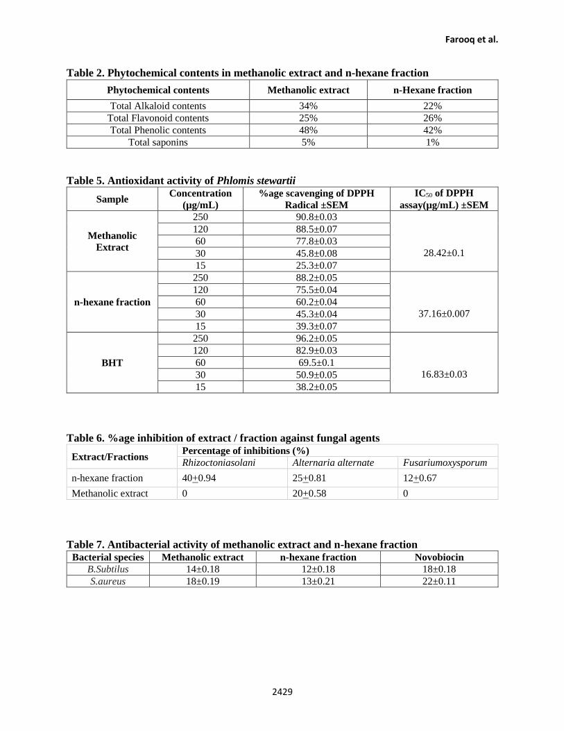
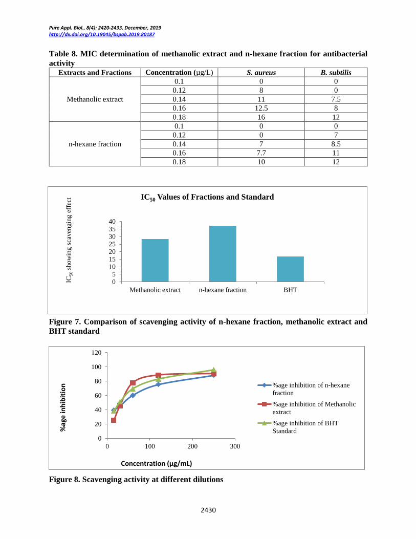
Antioxidant activity
The antioxidant activity of Phlomis stewartii
was tested and IC50 obtained from different
dilutions showed the potency of sample as an
antioxidant. The decoloration of DPPH from
purple color indicated the scavenging of it by
sample and BHT. The results showed that
methanolic fraction (28.42±0.1) is more
potent antioxidant than n-hexane fraction
(37.16±0.007). The results are shown in
(Table 5).
The DPPH scavenging effect was calculated
by absorbance of control and sample
difference, the samples were tested in
triplicate and then IC50 was obtained from the
DPPH scavenging activity graphs at different
dilutions. The graphical representation is
included in (Figure 7 & 8). The scavenging
activity indicating the presence of some
antioxidant in plant sample such as

Pure Appl. Biol., 8(4): 2420-2433, December, 2019 http://dx.doi.org/10.19045/bspab.2019.80187
2428
polyphenols and quinines can be reason for
antioxidant activity.
The antioxidant activity was found to be
dependent upon hydrophilic and hydrophobic
nature of components, larger electron donor
groups like -OH in phenylpropanoids favours
more activity [10]. BHT has such phenolic
group which resembled with phenolic
skeleton of antioxidants present in plants.
The stable free radical (DPPH) was chosen
and absorbance was measured at 517nm and
reduction in absorbance was observed due to
engulfing of free radicals. It was due to
provision of hydrogen by antioxidant [15].
Flavonoids and several glycosides which
contain hydroxyl groups are much effective
agents of such activities [26].
Antifungal activity
The n-hexane and methanolic extract were
assessed for antifungal activity and
Alternaria alternate, Rhizoctonia solani and
Fusarium oxysporum were used as fungal
agents. The potency of n-hexane fraction and
methanolic extract was tested by means of
qualitative as well as quantitative analysis.
The results revealed that n-hexane fraction
inhibited the R. solani (40+0.94), A. alternate
(25+0.81) and F. oxysporum (12+0.67)
whereas methanolic extract inhibited only A.
alternate (20+0.58). The results are shown in
(Table 6 & Figure 9).
The use of medicinal plants as antifungal
agents is in practice since ancient times and
their use in development of drugs against
fungal pathogens has minimized the chances
of adverse side effects on human health. The
demand for appropriate anti-fungal agents
also get importance after finding of adverse
action of many fungi, especially in destroying
the quality, quantity, mortality and shelf life
of useful crops. The discovery of new drugs
for inhibiting fungi is actually a new aspect
of human pathology [28, 29].
Antibacterial activity In present study, methanolic extract and n-
hexane fraction were also assayed for
antibacterial activity against gram negative
bacteria (E. coli, B. Subtilus) and gram
positive bacteria (S. aureus & S. dysenteriae).
It was revealed n-hexane fraction showed
MIC against S. aureus (13±0.21) and B.
subtilus(12±0.18) while methanolic extract
was more active against B. subtilus
(14±0.18)and S. aureus (18±0.19). The
results are shown in (Table 7, 8 & Figure 10).
The previous findings related to this activity
supported present results, antibacterial
activity depends upon synergism of
components in a mixture rather than a single
isolated compounds, while for some bacteria
one compound can be effective than extract
or fraction [16].
Table 1. Qualitative analysis for checking strength of components in n-hexane fraction and
methanolic extract
Compounds type Methanolic extract n- Hexane fraction
Alkaloids +++ +++
Cardiac Glycosides ++ ++
Coumarin + +
Flavonoids ++ ++
Phlobatannins - -
Quinone +++ ++
Saponins ++ +
Steroids ++ ++
Tannins + +
Terpenoids +++ ++ +++ indicated the more abundance of respective phytochemicals, while ++ revealed moderate level of phytochemicals and +
showed the lowest concentration of that phytochemical in particular sample. – indicated the absence of that substance in sample
Farooq et al.
2429
Table 2. Phytochemical contents in methanolic extract and n-hexane fraction
Table 5. Antioxidant activity of Phlomis stewartii
Sample Concentration
(µg/mL)
%age scavenging of DPPH
Radical ±SEM
IC50 of DPPH
assay(µg/mL) ±SEM
Methanolic
Extract
250 90.8±0.03
28.42±0.1
120 88.5±0.07
60 77.8±0.03
30 45.8±0.08
15 25.3±0.07
n-hexane fraction
250 88.2±0.05
37.16±0.007
120 75.5±0.04
60 60.2±0.04
30 45.3±0.04
15 39.3±0.07
BHT
250 96.2±0.05
16.83±0.03
120 82.9±0.03
60 69.5±0.1
30 50.9±0.05
15 38.2±0.05
Table 6. %age inhibition of extract / fraction against fungal agents
Extract/Fractions Percentage of inhibitions (%)
Rhizoctoniasolani Alternaria alternate Fusariumoxysporum
n-hexane fraction 40+0.94 25+0.81 12+0.67
Methanolic extract 0 20+0.58 0
Table 7. Antibacterial activity of methanolic extract and n-hexane fraction
Bacterial species Methanolic extract n-hexane fraction Novobiocin
B.Subtilus 14±0.18 12±0.18 18±0.18
S.aureus 18±0.19 13±0.21 22±0.11
Phytochemical contents Methanolic extract n-Hexane fraction
Total Alkaloid contents 34% 22%
Total Flavonoid contents 25% 26%
Total Phenolic contents 48% 42%
Total saponins 5% 1%
Pure Appl. Biol., 8(4): 2420-2433, December, 2019 http://dx.doi.org/10.19045/bspab.2019.80187
2430
Table 8. MIC determination of methanolic extract and n-hexane fraction for antibacterial
activity
Extracts and Fractions Concentration (µg/L) S. aureus B. subtilis
Methanolic extract
0.1 0 0
0.12 8 0
0.14 11 7.5
0.16 12.5 8
0.18 16 12
n-hexane fraction
0.1 0 0
0.12 0 7
0.14 7 8.5
0.16 7.7 11
0.18 10 12
Figure 7. Comparison of scavenging activity of n-hexane fraction, methanolic extract and
BHT standard
Figure 8. Scavenging activity at different dilutions
05
10152025303540
Methanolic extract n-hexane fraction BHT
IC50 Values of Fractions and Standard
IC50
sho
win
g s
caven
gin
g e
ffec
t
0
20
40
60
80
100
120
0 100 200 300
%age inhibition of n-hexane
fraction
%age inhibition of Methanolic
extract
%age inhibition of BHT
Standard
Concentration (µg/mL)
%ag
ein
hib
itio
n

Farooq et al.
2431
Figure 9. Petri plates showing inhibition zones of fractions against fungal strains
Figure 10. Petri plates showing inhibition zones of fractions against antibacterial strains
against
Conclusion
The present work concluded that Phlomis
stewartii is one of the important medicinal
plants. The GC-MS analysis of n-hexane
fraction revealed the presence of long chain
acids, esters, stigmasterol, polyphenols and

Pure Appl. Biol., 8(4): 2420-2433, December, 2019 http://dx.doi.org/10.19045/bspab.2019.80187
2432
other sterols. The qualitative and quantitative
screening confirmed the presence of
flavonoids, saponins and total phenolic
contents in methanolic extract and n-hexane
fraction. The presence of these
phytochemicals can be reason behind the
biological activities. The methanolic extract
showed the more antioxidant potential, it may
be due to combined effect of many
compounds such sterols, particularly esters
have the potential of scavenging free radicals.
n-hexane fraction showed significant
antibacterial activity. The presence of long
chain fatty acids including eicosanoic acid
and linoleic acid etc. are potent agent for
antibacterial activity. n-hexane fraction also
exhibited the higher antifungal activity. The
Phlomis stewartii is a potential plant which
may further be explored and studied for the
bioassay guided isolation of pure
compounds.
Author’s contributions
Conceived and designed the experiments: S
Ali, Performed the experiments: A Farooq,
Analyzed the data: S Ali & H Ullah,
Contributed reagents/materials/ analysis
tools: A Khan, N Jahan, I Agha & RB Tareen,
Wrote the paper: A Farooq & S Ali.
Acknowledgement
The authors warmly acknowledge
Department of Chemistry, University of
Balochistan, Quetta and Faculty of Life
Sciences and Informatics, BUITEMS, Quetta
for providing the necessary equipments and
labs in order to conduct lab work smoothly.
References
1. Cooper R & Nicolo G (2014). Natural
Products Chemistry: Sources,
Separations and Structures. 1st Ed. CRC
Press, Taylor & Francis group, pp206.
2. Abozenadah, Bishop H, Bittner & Flatt
(2018). Allied Health Chemistry. 1st Ed.
Western Oregon University, Oregon
State (America), pp 208.
3. Dhawan D & Gupta J (2017).
Comparison of different solvents for
Phytochemical extraction potential from
Datura metel plant leaves. Intl J Biol
Chem 11 (1): 17-22.
4. Koche D, Shirsat R & Kawale M (2016).
An overview of major classes of
Phytochemicals: their types and role in
disease prevention. Hislopia J 9(1/2): 1-
11.
5. Donmez AA (2003). Iridoid and
Phenylpropanoid Glycosides from
Phlomis samia, P. monocephala and P.
carica. Turk J Chem 27: 295-305.
6. Paiva, SA, and RM Russell (1999). Beta-
Carotene and Other Carotenoids as
Antioxidants. J Am Coll Nutr 18(5):
426–433.
7. Cheij I (1984). Studies on bark of Morus
species used to relieve tooth ache and
other medicinal purpose. The Macdonald
Encyclopedia of Medicinal Plants.
Macdonald & Co (Publishers) Ltd, pp
365.
8. Jabeen B, Riaz N, Saleem M, Naveed
MA, Ashraf M, Alam U, Rafiq HM,
Tareen RB & Jabbar A (2013). Isolation
of Natural Compounds from Phlomis
stewartii showing α-hlucosidase
inhibitory activity, Phytochem 96: 443-
448.
9. Sarker S & Nahar L (2007). Chemistry
for Pharmacy Students: General Organic
and Natural Product Chemistry. John
Wiley & Sons, pp 283.
10. Mensor LL, Menezes FS, Leitao GG,
Reis AS, Santos TCS, Coube CS &
Leitao SG (2001).Screening of Brazilian
plant extracts for antioxidant activity by
using DPPH free radical method.
Phytother Res 15: 127-130.
11. Hussain SZ & Maqbool K (2014). GC-
MS principle, technique and its
application in food science. Intl J Curr
Sci 13: 116-126.
12. Stein SE (2008). NIST Standard
Reference Database 1A. Gaithersburg 2:
975-980.
Farooq et al.
2433
13. Mir MA, Sawhney SS & Jassal MMS
(2012). Qualitative and quantitative
analysis of phytochemicals of
Taraxacum officinale. J Pharm
Pharmacol 2(1): 1-5.
14. Saxena M, Saxena J, Nema R, Singh D
& Gupta A (2013). Phytochemistry of
medicinal plants. J Pharmacogn
Phytochem 1(6): 168-182.
15. Koleva II, Beek TAV, Linssen JPH,
Groot AD & Evstatieva LN (2002).
Screening of Plant Extracts for
Antioxidant Activity: A Comparative
Study on three testing methods.
Phytochem Anal 13: 8-17.
16. Semnani KM, Saeedi M, Mahdavi MR &
Rahimi F (2006). Antimicrobial studies
on extracts of three species of Phlomis.
Pharm Biol 44(6): 426-429.
17. Smania EDFA, DelleMonache F, Yunes,
RA, Paulert R, & Smania Junior A
(2007). Antimicrobial activity of methyl
australate from Ganoderma australe.
Rev Bras Farmacogn 17(1): 14-16.
18. Ozkan G, Sagdic O, Baydar NG &
Baydar H (2004). Note: Antioxidant and
antibacterial activities of Rosa
damascena flower extracts. Food Sci
Tech Int 10(4): 277-281.
19. Achuthan CR, Babu BH & Padikkhala J
(2003). Antioxidant and
hepatoprotective effects of Rosa
damascena. Pharm Biol 41(5): 357-361.
20. Harput US, Saracoglu I, Calis I, Donmez
AA & Nagatsu A (2004). Secondary
metabolites from Phlomis kotschyana.
Turk J Chem 28: 767-774.
21. Shang X, Wang J, Li M, Miao X, Pan H,
Yang Y & Wang Y (2011).
Antinociceptive and anti-inflammatory
activities of Phlomis umbrosa Turcz
extract. Fitoterapia 82: 716-721.
22. Tsitsimi E, Loukis A &Verykokidou E
(2011). Composition of the Essential Oil
of the Flowers of Phlomis fruticosa L.
from Greece. J Essent Oil Res 12(3):
355-356.
23. Zhang Y & Wang ZZ (2008).
Comparative analysis of essential oil
components of three Phlomis species in
Qinling Mountains of China. J Pharm
Biomed Anal 47: 213-217.
24. Shayan SZ, Moradkhani S & Dastan D
(2016). Analysis of fatty acid
composition of two selected Phlomis
species. J HerbMed Pharmacol 5(4):
153-156.
25. Lofty RA & Abd El-Moaty HI (2016).
Antibacterial activities of some active
constituents isolated from Phlomis
Floccosa D. Don. Egypt J Desert Res 66
(1): 69-78.
26. Nazemiyeh H, Rahman MM, Gibbons S,
Nahar L, Delazar A, Ghahramani MA,
Talebpour AH, & Sarker SD (2008).
Assessment of antibacterial activity of
phenylethanoid glycosides from Phlomis
lanceolata against multidrug resistant
strains of Staphylococcus aureus. J Nat
Med 62: 91-95.
27. Shtayen MSA & Ghdeib SIA (1999).
Antifungal activity of plant extract
against dermatophytes. Mycoses,
42:665-672.
28. Hammer KA, Carson CF & Riley TV
(1999). Antimicrobial activity of
essential oils and other plant extracts. J
Appl Microbiol 86: 985-990.
29. Dellavalle PD, Cabrera A, Alem D,
Larranaga P, Ferriera F & Rizza MD
(2011). Antifungal activity of medicinal
plant extracts against Phytopathogenic
fungus Alternaria spp. Chil J Agr Res
71(2): 231-239.