Embed Size (px)
Citation preview
SquintFree Papers
70th AIOC Proceedings, Cochin 2012
620
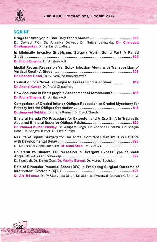
SQUINTDrugs for Amblyopia: Can They Stand Alone? .............................................803Dr. Dwivedi P.C., Dr. Anamika Dwivedi, Dr. Sujata Lakhtakia, Dr. Charudatt Chalisgaonkar, Dr. Pankaj Choudhary
Is Minimally Invasive Strabismus Surgery Worth Going For? A Paired Study ..................................................................................................................805Dr. Richa Sharma, Dr. Amitava A.K.
Medial Rectus Recession Vs. Botox Injection Along with Transposition of Vertical Recti - A Study ..................................................................................809 Dr. Roshani Desai, Dr. K. Namitha Bhuvaneswari
Evaluation of a Novel Technique to Assess Fundus Torsion .....................812Dr. Anand Kumar, Dr. Praful Chaudhary
How Accurate is Photographic Assessment of Strabismus? .....................815Dr. Richa Sharma, Dr. Amitava A.K.
Comparison of Graded Inferior Oblique Recession to Graded Myectomy for Primary Inferior Oblique Overaction ..............................................................818Dr. Jaspreet Sukhija, Dr. Neha Kumari, Dr. Parul Chawla
Bilateral Harada ITO Procedure for Extorsion and V Eso Shift in Traumatic Acquired Bilateral Superior Oblique Palsies .................................................820Dr. Pramod Kumar Pandey, Dr. Anupam Singh, Dr. Abhishek Sharma, Dr. Shagun Sood, Dr. Sanjeev kumar, Dr. Ekta Kumari
Results of Squint Surgery for Horizontal Comitant Strabismus in Patients with Developmental Delay ...............................................................................823Dr. Meenakshi Gopalakrishnan, Dr. Sanil Shah, Dr. Aarthy G. ......................................
Unilateral Vs Bilateral LR Recession in Divergent Excess Type of Small Angle IDS - 4 Year Follow-up ...........................................................................827Dr. Kamlesh, Dr. Shilpa Goel, Dr. Yuvika Bansal, Dr. Manav Sachdev
Role of Binocular Potential Score (BPS) in Predicting Surgical Outcome of Intermittent Exotropia (X[T]) ...........................................................................831Dr. Arti Elhence, Dr. (MRS.) Vinita Singh, Dr. Siddharth Agrawal, Dr. Arun K. Sharma

Squint correction with spectacles
Orthophoric child
Squint Free Papers
803
Drugs for Amblyopia: Can They Stand Alone?Dr. Dwivedi P.C., Dr. Anamika Dwivedi, Dr. Sujata Lakhtakia, Dr. Charudatt Chalisgaonkar, Dr. Pankaj Choudhary
Amblyopia is an acquired maldevelopment of central visual pathways resulting in reduced vision. Various treatment modalities are constantly
being proposed for amblyopia including medical treatment, a dream of strabismologists for a long time. Past efforts to treat amblyopia medically were not successful in terms of applicability and effectivity. More recently there have been attempts based on catecholamines. Levodopa is a precursor for the catecholamine neurotransmitters, dopamine and noradrenaline. Levodopa has demonstrated improvement in visual acuity and contrast sensitivity in amblyopic eyes by influencing visual system at the retina1 and cortical levels.2 Another agent, Citicoline, has been reported to have an effect similar to levodopa.3 Fluoxetine, a drug used for treatment of depression, shown to restore neuronal plasticity in the visual system. Fluoxetine has potential for clinical application in treatment of amblyopia They are well tolerated with minimal adverse effects but overall improvement in vision is small. Thus, whether pharmacological treatment will become a useful mode of therapy for amblyopia is unclear.
We conducted a retrospective study to assess efficacy and safety of pharmacological agents in comparison with occlusion therapy in amblyopia management.
MATERIALS AND METHODSRetrospective analysis of records of 200 pts of amblyopia was done who attended squint and amblyopia clinic of G M hospital, SSMC Rewa. The record of comprehensive ophthalmic examination of patients were reviewed, selecting the patient between 2-17 years of age with strabismic, anisometropic and mixed amblyiopia. Patients, visual acuity and history of any previous treatment noted. All patient underwent refraction under cycloplegia and best possible optical correction was prescribed before starting specific therapy for amblyopia.
Various treatment modalities received by the patients were grouped as: (1) Part Time Occlusion (PTO); (2) Levodopa/Carbidopa; (3) Citicholine; (4) Fluoxetine
SqUINTChairman: Dr. Santhan Gopal K.S.; Co-Chairman: Dr. (MRS.) Vinita Singh
Convenor: Dr. Elizabeth Joseph Moderator: Dr. Vidyavati Mundada
70th AIOC Proceedings, Cochin 2012
804
with PTO. Out of 200 patients selected 141 patients were treated with part time occlusion of 6hrs/day. 14 patients received medical therapy, combination of Levodopa (250mg) and Carbidopa (25 mg), dose titrated according to age, for a period of 3 months. 25 patients received CDP choline 500-1000mg (7-28mg/kg/day) daily orally for a period of 14 days. 20 patients were given 20mg Fluoxetine for 4 months in addition to part time occlusion of 6 hrs/day. All patients were followed up monthly for 6 months.
Patients with any degree of visual improvement considered “Responders”. Number of patient responding and degree of response of therapy compared among groups.
RESULTS Out of 200 patients’ records studied for this study 141 received part time occlusion with good compliance. Of these 141 patients 99 patients showed visual improvement with the therapy with average 4 line of improvement in LogMAR scale. This constitute 70% of patients in part time occlusion group to be responders.
Of the 14 cases in this study who underwent medical therapy with Levodopa+ Carbidopa only 2 cases showed improvement in their visual acuity while 11 cases showed no improvement in visual acuity even after 3 months of initiation of therapy. After which medical therapy was terminated and patients were advised other treatment modality or cosmetic surgical correction for strabismus. Of these 2 (14%) responders average improvement in visual acuity was 2 LogMAR lines. 10 patients out of 25 receiving CDP Choline showed visual improvement. Improvement started at two weeks with maximum patients improving by 4 weeks. These 40% responders showed 1.4 LogMAR lines of visual improvement. In 20 patients, where Fluoxetine was added to part time occlusion, 16 patients showed improvement in visual acuity with 4.53 LogMAR lines of visual improvement. None of the patients recieveing medical therapy (Levodopa or CDP choline) reported any adverse effect of the drug.
DISCUSSION Our retrospective analysis of data in patients treated with medical therapy alone met with very marginal success in terms of both, number of patients responding to therapy and number of lines of visual improvement. Responders in various treatment groups noted were 70%, 14%, 40% and 80% respectively. In responders the mean LogMAR line of improvement noted was 4 lines in PTO group, 1.4 lines in Citicholine group, 2 lines in Levodopa group and 4.53 in Fluoxetine with PTO group. This clearly showed better results in occlusion group and in patients where occlusion was supplemented with medical
Squint Free Papers
805
therapy. Medical therapy for amblyopia was well tolerated with no significant adverse effects reported.
In conclusion though medical therapy for amblyopia was well tolerated, it was met with very marginal success without occlusion. Occlusion therapy remains the gold standard for the treatment of amblyopia, medical therapy can be used as adjuvant to PTO or in patients with poor compliance for occlusion.
REFERENCES 1. Gottlob I, Weghaupt H, Vass C. Effect of levodopa on the human luminance
electroretinogram. Invest Ophthalmol Vis Sci 1990;31:1252-8. 2. Leguire LE, Walson PD, Rogers GL, et al. Levodopa/carbidopa treatment for
amblyopia in older children. J Pediatr Ophthalmol Strabismus 1995;32:143-51. 3. Methods Find Exp Clin Pharmacol. Citicholine: pharmacological and clinical
review, 2006 update. 2006 Sep;28 Suppl B:1-56.4. José Fernando Maya Vetencourt, et al The Antidepressant Fluoxetine Restores
Plasticity in the Adult Visual Cortex. Science 2008;320, 385.
Is Minimally Invasive Strabismus Surgery Worth Going For? A Paired StudyDr. Richa Sharma, Dr. Amitava A.K.
Minimal access surgery are common in all the fields of medicine.1
Ophthalmology too has witnessed an effort to reduce the size of incision For example: phacoemulsification, miniature implant drainage surgery, sutureless vitrectomy, endoscopic lacrimal surgery etc.
Apart from providing functional benefit such as fusion and stereopsis, strabismus surgery serves to improve cosmesis. In an immediate post op period, that would mean an eye with less redness, congestion, chemosis and watering. This can be achieved by giving smaller size of the incision, less tissue trauma and ability to dispense off the sutures at least on conjunctiva.
Most of the strabismus surgeons prefer to give limbal incision in the quadrant of interest introduced by Harms and later popularized by Von noorden.2,3 Some surgeons also prefer to chose Fornix incision approach popularized by Parks.4 Several other conjunctival approaches have been introduced by these authors Velez, Santiago, Swan And Talbott.5,6,7
Recently, in 2007 a novel technique for muscle exposure has been introduced by Daniel S Mojon in which 2 incisions parallel to muscle strap were given and further recessions and plication done through the tunnel made after
70th AIOC Proceedings, Cochin 2012
806
separating muscle from the surrounding tissue. This study was done in 39 patients, 20 of which underwent Minimally invasive strabismus surgery (MISS) and it was compared with 19 patients who underwent limbal incision surgery retrospectively. There outcomes were the alignment in the two groups, binocular single vision, variation in vision , patient’s s discomfort and number and type of complications.8
While the above mentioned study was a group randomization, we designed a parallel study in which one eye was randomized to MISS and other to Standard paralimbal surgery (SPS) to compare post operative outcome in terms of cosmesis and discomfort in the two techniques. Oue primary outcomes being redness, congestion, chemosis, discomfort and foreign body sensation. Final alignment was not our primary but secondary outcome. We also evaluated total time taken, visible scarring, and any complications.
MATERIALS AND METHODS20 eyes of ten patients were included in the study. After proper consent, eyes were randomized to each group.
Both the eyes were anaesthetized by giving peribulbar block using xylocaine (2%), sensoricaine (0.5%) with hyaluronidase. After separating the lids with universal eye speculum, a 5-0 silk traction suture (Johnson & Johnson Ltd Aurangabad NW 5079) was passed through the superficial sclera near the limbus in the quadrant of the muscle to be operated upon. Care was taken that the 5-0 silk suture does not contact/ abrade the cornea. Linear conjunctival incisions, parallel to the edges of the muscle of interest were given, their anterior limits being adjacent to the insertion of the muscle. For recessions Their posterior limits were about 1 mm short of the planned recession. From the access available through the two linear parallel cuts, after hooking the muscle, the episcleral tissue was cleared, and careful dissection to expose the muscle margins (including intermuscular septum) and surface was undertaken with Westcott scissors, till about 7 mm behind the insertion. Vicryl 6-0 (Johnson & Johnson Ltd. Aurangabad NW 2670) bites were taken from the muscle margins from near the insertion, and the muscle disinserted using spring scissors. Hemostasis was be undertaken at this stage in case a need is felt. After measuring the distance for the desired recession, Vicryl 6-0 scleral bites (of the previously passed suture through the muscle margins) was taken so as to provide a new anchor to the EOM. Care was exercised to ensure that the cut edge of the muscle is stretched, so as to prevent the central sagging of the muscle tendon. If the parallel conjunctival cut edges appeared to be in
Squint Free Papers
807
good apposition no sutures were applied to close the cuts. Good apposition was considered where in the edges came in contact or are not more than 2 mm apart at a point of maximum separation, despite gently smoothening the conjunctival surface. If the edges of the cut appear to be wider than 2 mm apart at any point, then a single Vicryl 8-0 (Johnson and Johnson Ltd.Aurangabad NW 2348) suture was applied in the centre. In case of any unplanned tear or a planned extension of the cuts two sutures may be given.
After a similar conjunctival approach (their length being 1 mm short of the planned resection), as in recessions, and clearing of the EOM, two Vicryl 6-0 sutures were applied at the edges of the EOM, at the point of desired resection. These sutures were passed through the insertion of the EOM. The muscles were cut between both the suture-bites, taking care not to cut the sutures. Hemostasis was carried out at this stage if a need was felt. Tying the sutures brought the point of resection to the insertion, effecting a resection. Decision to suture the conjunctival cuts was based on criteria as mentioned in recessions.
We evaluated the patient at first post operative day, 2-4 weeks and at 6 weeeks. Our parameters being redness, congestion, chemosis, foreign body senasation and drop intolerance, they were graded on a scale of 0 to 3. Redness and congestion were graded using standard photographs. Other parameters were graded on a scale of severity. Time taken from conjunctival incision to muscle insertion was measured in both eyes using a stop watch and rounded off. Visible scarring was defined as if an obvious scarring was present at 6 weeks, visible in ambient light at a distance of one meter.
Final alignment was also evaluated and termed successful if it was within 10 pd of targeted surgery at 6 weeks. Any complications encountered preoperatively or post operatively were noted. LogMAR Vision was checked at 1st post-op day. Indirect ophthalmolscopy and Slit lamp examination were also done.
RESULTSThe mean duration of reinsertion of muscle to sclera (for recessions) or muscle stump (in case of resections) in SPS eye was 29.6 (±SD) minutes as compared to 40.4 (SD) minutes in MISS eye with mean difference being 10.8 minutes (95% CI 2.67 and 18.92 minutes). The 2 tailed p value on Mann Whitney u test was 0.004.
Redness, congestion, chemosis and drop intolerance were similar in both eyes at first post-op day. But foreign body sensation was significantly more in SPS eyes. Redness, congestion, Foreign body sensation and total inflammatory scores were less in MISS eyes at 2-4 weeks. At 6 weeks, only redness showed the significant difference while other parameters were more or less equal. P
70th AIOC Proceedings, Cochin 2012
808
values at different period are given in the table below.
Redness congestion chemosis Foreign Drop Total body intolerance inflammatory senastion score P value (day 1) 0.655 0.655 1.00 0.01 0.083 0.04
2-4 weeks 0.033 0.033 1.00 0.02 0.317 0.04
6 weeks 0.046 0.059 1.00 0.317 1.00 0.05
Scarring was seen in all the eyes which underwent SPS but present only in 6 eyes. (p value 0.0867) which was insignificant in our study.
None of the MISS eyes required conversion to SPS. No complications for example corneal dellen, tenon prolapse were encounterd in any of the groups.
6 out of ten patients attained successful outcome while rest three were within 15 pd.
In conclusion the immediate post-op period, MISS causes less patient’s discomfort as the sutures if required to be placed are away from the limbus and less drop intolerance.
It does not have any effect on redness in immediate post-op period but definitely cause less redness, congestion, discomfort at intermediate period of 2-4 wks providing better cosmesis. Over all outcome is also better. Expertise is required as there is less exposure. MISS is more time taking surgery but we believe with experience this difference can be decreased.
REFERENCES1. Darzi A, Mackay S. Recent advances in minimal access surgery. BMJ2002;324:31–4.2. Harms H. U¨ber Muskelvorlagerung. Klin Monatsbl Augenheilk 1949;115:319–24.3. Von Noorden GK. The limbal approach to surgery of the rectus muscles. Arch
Ophthalmol 1968;80:94–7.4. Parks MP. Fornix incision for horizontal rectus muscle surgery. Am J Ophthalmol.
1968;65:907–15.5. Swan KC, Talbott T. Recession under Tenon’s capsule. Arch Ophthalmol 1954;51:32–
41.6. Velez G. Radial incision for surgery of the horizontal rectus muscles. J Pediatr
Ophthalmol Strabismus 1980;17:106–7.7. Santiago AP, Isenberg SJ, Neumann D, et al. The paralimbal approach with
deferred conjunctival closure for adjustable strabismus surgery. Ophthalmic Surg Lasers 1998;29:151–6.
8. Comparision of new minimally invasive strabismus surgery technique with the usual limbal approach for rectus muscle recession and plication. D S Mojon. Br J Ophthalmol 2007;91:76–82.
Squint Free Papers
809
Medial Rectus Recession Vs. Botox Injection Along with Transposition of Vertical Recti - A Study Dr. Roshani Desai, Dr. K. Namitha Bhuvaneswari
Acquired sixth nerve palsy has been shown to be the most common type of cerebral nerve palsy in some studies.1 It contributes to 45 percent
of all cases of cerebral nerve palsy.2 The abducens nerve is susceptible to damage from various types of intracranial pathologies. This is due to its long intracranial pathway and its anglulation at the petrous tip of the temporal bone. It is non-flexible at the path between the brainstem and the meningeal entrance site. The nerve is commonly affected by meningeal inflammation, cerebral edema and any displacement of the brain stem. The sixth cranial nerve, similar to other cranial nerves, is also sensitive to toxic substances, demyelinative processes and viral diseases. The acquired variant improves spontaneously over 3 to 6 months. Based on current information, 78% of patients recover in one year and 40% of the remaining patients have a severe underlying condition such as intracranial aneurysms or vascular disorders (brainstem stroke), carotid-cavernous fistula or cerebral tumors which need neurosurgical intervention.4
In patients with slow improvement, contracture of the medial rectus muscle may result in concomitant esotropia with positive forced duction test (FDT). After identifying the underlying cause, several techniques such as patching, corrective prisms or Botulinum toxin injections are used to overcome diplopia and abnormal head posture during the first 6 months following sixth nerve palsy. When esotropia is not improving and becomes stable for at least 6 months, surgical intervention is warranted. A variety of procedures may be performed such as recession and resection, bilateral medial rectus recession, different muscle transposition procedures (Hummelschiem and Jensen) with or without medial rectus (MR) recession, and bilateral lateral rectus (LR) resection along with medial rectus myotomy. In this study, we present the surgical outcomes of a consecutive series of 20 patients with sixth nerve palsy over 16 months at Regional Institute of Ophthalmology and Government Ophthalmic Hospital, Egmore Chennai -08 Tamil Nadu.
MATERIALS AND METHODSThis interventional prospective study was performed on 20 consecutive patients with sixth nerve palsy who were referred to Regional Institute of Ophthalmology and Government Ophthalmic Hospital during the period May 2009 to December 2010. Relevant data included age, gender, reasons for referral,
70th AIOC Proceedings, Cochin 2012
810
laterality of the palsy, etiology of sixth nerve dysfunction (traumatic, ischemic, infectious, idiopathic), angle of deviation at presentation, head posture, residual deviation at the end of 6 months (primary and secondary deviation as measured by prism bar cover test), positive FDT and type of treatment (vertical transposition with medial rectus recession v/s Botulinum toxin). Patients were followed one day, one week, six and twelve weeks following surgery. Visual acuity was measured using Snellen’s optotypes. Patients were divided into 2 different groups; group A underwent surgical intervention with transposition with medial rectus recession, group B underwent muscle transposition with one dose (10-20 units) of Botulinum toxin injected into the MR muscle. Data were compared pre and postoperatively using paired t-test with significance level set at P<0.05.
RESULTSThis study included 20 patients consisting of 12 (60%) male and eight (40%) female subjects with mean age of 44.8 years (range, 19 years to 72 years). Mean follow-up was 4 months after surgery. The right eye was involved in eight (40%) patients, the left eye in nine (45%) patients and both eyes in the remaining three (15%) individuals. Chief complaints included deviation in 16 (80%) patients, diplopia in three (15%) patients and abnormal head posture in one (5%) patient. The sixth nerve dysfunction was traumatic in 4 (20%) patients, post surgical in 1 (5 %), idiopathic in 5 (25%) and ischemic in 10 (50%) patients. Regarding treatment, 10 patients (50%) were operated with muscle transposition and MR recession (group A) and 10 patients (50%) underwent muscle Transposition with Botulinum toxin injections (group B). Preoperatively, the mean deviation was 36 PD (range, 10 - 90) in group A, which was reduced to 5 PD (range, 15 PD esotropia to 5 PD exotropia) after the first operation, and 17 PD (range, 5-30 PD) in group B which reduced to 3 PD (range, 0-10 PD esotropia). Botulinum toxin injections were repeated in 3 patients from group B following surgery whereas one patient required MR recession after 3 months of the previous surgery in this group. 2 patients in Group A were diagnosed with ocular ischemic syndrome after 3 muscle surgery. In both groups, greater the primary deviation and limitation of motility, the larger the improvement. Furthermore, the higher the primary deviation, the better the response to primary strabismus surgery.
DISCUSSIONSixth nerve palsy has been reported to be the most common type of extraocular nerve paralysis based on some studies2,5 but ranking second following fourth nerve palsy according to other reports.6-8 This condition has been reported to
Squint Free Papers
811
be more prevalent in male patients7-8 which may be due to the higher exposure of men to trauma such as accidents and work related injuries. Men are also more susceptible to ischemic damage.
Among acquired cases of sixth nerve palsy in our series, ischemia was the most common cause followed by idiopathic and trauma. The most prevalent chief complaint was squint rather than diplopia. This may be explained by the patient population with higher potential for suppression. The success rate gained after primary intervention in both groups is comparable, if appropriate selection of patients is made. Treatment should be based on the severity of paralysis, its onset and the amount of motility limitation to get optimal response. The outcome of surgery for strabismus depends on the severity and surgical technique. Despite good cosmetic outcome, eyes in group A had the least amount of improvement in motility. The significant improvement in the Botulinum group reflects the possibility of utilizing minimally invasive methods such as Botulinum toxin. In a study on 33 patients with a clinical diagnosis of traumatic sixth nerve paresis, Holmes et al8 showed that 86% of unilateral and 38% of bilateral cases improved spontaneously after 3 months and recommended that Botulinum toxin can alleviate diplopia.
REFERENCES1. Nelson LB. Strabismus disorders. In: Nelson LB, Calhoun JH, Harley RD, eds.
Pediatric Ophthalmology. 3rd ed. Philadelphia: WB Saunders; 1991:149-68.2. Rush JA, Younge BR. Paralysis of cranial nerves III, IV, and VI: cause and prognosis
in 1,000 cases. Arch Ophthalmol 1981;99:76-9.3. Shrader EC, Schlezinger NS: Neuro-ophthalmologic evaluation of abducens nerve
paralysis. Arch Ophthalmol 1960;63:84-91.4. King AJ, Stacey E, Stephenson G, Trimble RB. Spontaneous recovery rates for
unilateral sixth nerve palsies. Eye 1995;9:476-8.5. Bagheri A, Khodabakhshi M, Anisian A, Mirdehghan A. Epidemiology and
Etiologic Characteristics of Patients with Paralytic Strabismus. Bina J Ophthalmol 2004;9:323-32.
6. Von Noorden GK, Campos EC. Paralytic strabismus. In: Von Noorden GK, Campos EC (eds). Binocular Treatment of Sixth Nerve Palsy; Bagheri et al JOURNAL OF OPHTHALMIC AND VISION RESEARCH 2010; Vol. 5, No. 1 37 vision and ocular motility. 6th ed. New York: CV Mosby; 2002:414-57.
7. Sharpe JA. Neural control of ocular motor systems. In: Miller NR, Newman NJ (eds). Walsh and Hoyt’s clinical neuro-ophthalmology. 5th ed. Baltimore: Williams and Wilkins; 1998:1101-68.
8. Holmes JM, Droste PJ, Beck RW. The natural history of acute traumatic sixth nerve palsy or paresis. J AAPOS 1998;2:265-8.
70th AIOC Proceedings, Cochin 2012
812
Evaluation of a Novel Technique to Assess Fundus Torsion Dr. Anand Kumar, Dr. Praful Chaudhary
Assessment of ocular torsion is crucial for the diagnosis and management of various ocular motility disorders. Both subjective and objective
methods have been described for measurement of this important parameter. The most common objective method used clinically to assess torsion is based upon the relationship between the position of the fovea and the optic disc.1 It is now generally believed that in normal individuals, a horizontal line passing through the fovea would cross the optic disc within its lower one-third diameter, and this is equivalent to a torsional range of about 9 degrees.2,3 A grading system proposed by Guyton, based upon the position of the fovea with respect to the optic disc is widely followed clinically to estimate abnormal torsion.2
Recently, Parsa et al have proposed incorporating the assessment of the orientation and axis of major retinal vessels as an accessory tool in the estimation of ocular torsion (unpublished data). The purpose of this paper is to further characterize the cues offered by the retinal vasculature and to evaluate this novel technique using these cues against the widely followed disc-macula relationship for assessment of ocular torsion.
MATERIALS AND METHODSIn a pilot study, 10 fundus photographs were evaluated for the correlation between the disc-foveal angle (DFA) and the angles subtended on the vertical axis by imaginary lines joining two corresponding points on the retinal blood vessels in the superior and inferior halves of the retina at varying distances from the centre of the optic disc (e.g., one disc diameter, two disc diameters, etc.). It was found that the angle made by the line joining the blood vessels at one disc diameter distance from the centre of the optic disc in the supero-temporal and infero-temporal quadrant had the highest correlation with the DFA (r=0.78, p=0.008, Pearson correlation co-efficient, 2- tailed). This imaginary line was then used as one of the cues to assess torsion using the retinal vasculature in the subsequent fundus pictures.
Thirty eight fundus pictures, taken by a method described elsewhere4 from patients having varying ocular motility disorders were assessed by two independent masked examiners. Examiner I (an ophthalmologist) assessed the fundus torsion using the conventional disc fovea relationship, and subsequently graded the torsion based on Guyton’s classification.2 Examiner II (a pediatric ophthalmologist) was provided with the same set of fundus pictures but with
Squint Free Papers
813
the macular area digitally covered by an opaque circle. He then assessed the torsion using the following vascular cues: (i) angle made by an imaginary line joining the superior and inferior branches of the central retinal artery as they emerge out from the centre of the optic disc, (ii) the orientation of the “axis of symmetry” passing through the centre of disc as the vertex, if the temporal vascular arcade is considered as a parabola (iii) the angle subtended by an imaginary line joining the arteries in the supero-temporal and infero-temporal quadrant at a distance of one disc diameter from the centre of the optic disc. For each fundus picture, three responses were noted based on each of the above 3 cues as “no torsion, intorsion or extorsion.” The final response for a particular fundus picture was the response that was repeated atleast twice. The final response was noted as “cannot be commented” if each of the 3 responses were different.
The readings of the two examiners were then compared and analyzed to test the sensitivity and specificity of the new method.
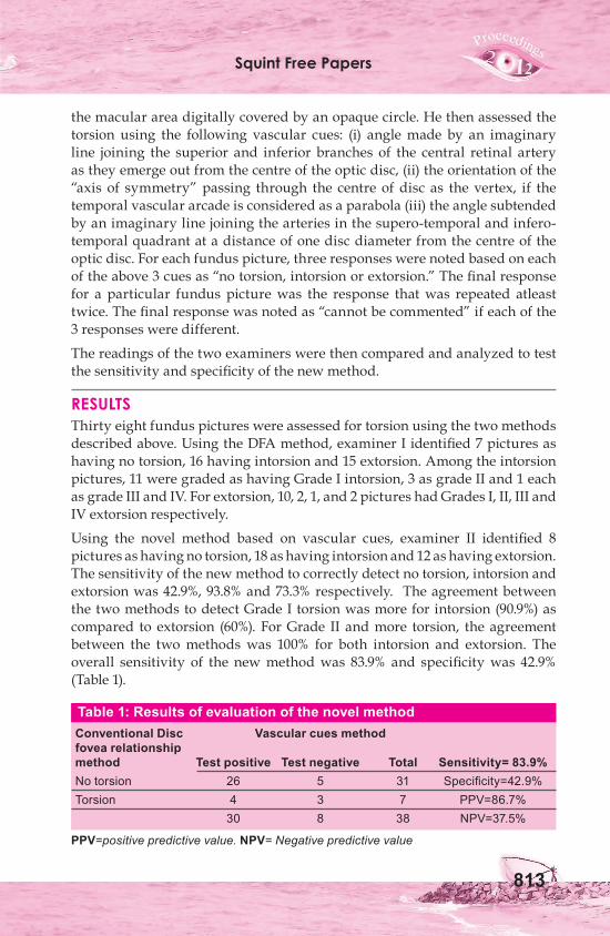
RESULTSThirty eight fundus pictures were assessed for torsion using the two methods described above. Using the DFA method, examiner I identified 7 pictures as having no torsion, 16 having intorsion and 15 extorsion. Among the intorsion pictures, 11 were graded as having Grade I intorsion, 3 as grade II and 1 each as grade III and IV. For extorsion, 10, 2, 1, and 2 pictures had Grades I, II, III and IV extorsion respectively.
Using the novel method based on vascular cues, examiner II identified 8 pictures as having no torsion, 18 as having intorsion and 12 as having extorsion. The sensitivity of the new method to correctly detect no torsion, intorsion and extorsion was 42.9%, 93.8% and 73.3% respectively. The agreement between the two methods to detect Grade I torsion was more for intorsion (90.9%) as compared to extorsion (60%). For Grade II and more torsion, the agreement between the two methods was 100% for both intorsion and extorsion. The overall sensitivity of the new method was 83.9% and specificity was 42.9% (Table 1).
Table 1: Results of evaluation of the novel methodConventional Disc Vascular cues methodfovea relationshipmethod Test positive Test negative Total Sensitivity= 83.9%No torsion 26 5 31 Specificity=42.9%Torsion 4 3 7 PPV=86.7% 30 8 38 NPV=37.5%
PPV=positive predictive value. NPV= Negative predictive value
70th AIOC Proceedings, Cochin 2012
814
DISCUSSIONData from our study indicate that assessment of fundus torsion using cues from the retinal vasculature has good agreement with that assessed using the conventional disc fovea relationship method, especially in Grade II or more torsion. The novel method was more sensitive for detecting intorsion as compared to extorsion when the fundus picture had Grade I torsion. This method can thus find application in assessing fundus torsion clinically in situations where using the disc fovea method may be unreliable or invalid. These conditions include, but are not restricted to, optic disc anomalies such as optic disc hypoplasia, disc coloboma, megalopapillae; or macular abnormalities such as geographic atrophy, macular scars or macular hemorrhage. In conditions such as significant anisometropia, albinism and high ametropia the disc foveal angle may be confounded by differing disc sizes and the disc macula distances. The novel method would be more reliable in such conditions as it is not based upon these anatomic landmarks.The true angle of cyclodisparity is best determined by the the torsional alignment of retinas between the two eyes together5, rather than any relative anatomic relationship between disc and fovea in each eye individually. The location of the major retinal blood vessels can serve as a good indicator about the distribution of the retinal ganglion cells, with axon densities well coupled to the location of the superior and inferior temporal retinal vascular arcades.6 It is also well accepted that in the retina, guidance molecules for axons direct and regulate the developing vasculature as well, thereby lending basis for the positional relationship between the two structures.The vascular cues used in this report to assess torsion using the novel method too are based on the intimate correlation of the axon densities with the major retinal blood vessels. The blood vessels entering the eye are considered to be roughly perpendicular to an imaginary horizontal line passing through the centre of the disc. Thus any significant change in the orientation of the imaginary line joining these blood vessels would be indicative of abnormal ocular torsion. Furthermore, in the absence of abnormal ocular torsion, an imaginary line bisecting the superior and inferior temporal vascular arcades which corresponds to the midline raphe would be almost horizontal. Any rotational bias in the orientation of such a projected line would indicate abnormal torsion. It has been demonstrated that retinal nerve fibre layer thickness profiles, as measured using scanning laser polarimetry correlate with the location of the main temporal superior and inferior blood vessels.6
So these blood vessels could serve as alternate landmarks for assessment of fundus torsion.
Limitations of using a method based on the location of retinal blood vessels as indicator of fundal torsion include its subjective nature, inability to grade
Squint Free Papers
815
torsion and unsuitability in vascular anomalies. Although the method showed a good sensitivity to detect higher grades of torsion, it tended to over-diagnose torsion. The low sensitivity of the test to correctly detect ‘no torsion’ could be explained by an absence of a ‘normal range’ of ocular torsion based on this new method. So cases which are ‘borderline normal’ on the DFA method would be designated as having torsion by this method. Of the 7 fundus pictures having no torsion by the conventional disc fovea relationship method, three were incorrectly labelled as having intorsion and one as extorsion by the new method. Further refinements of the cues using larger number of fundus pictures in subsequent studies may aid in improving the specificity of this technique.
In summary, this novel method of assessing fundus torsion using the cues from retinal vasulature can serve as a fairly accurate and accessory clinical tool, especially in cases of optic disc or foveal anomalies.
REFERENCES 1. Phillips PH, Hunter DG. Evaluation of ocular torsion and principles of management.
In: Rosenbaum AL, Santiago AP, eds. Clinical strabismus management. Philadelphia: Saunders; 1999:52–72.
2. Guyton D. Clinical assessment of ocular torsion. Am. Orthopt. J. 1983;33:7-15.3. von Noorden GK. Clinical observations in cyclodeviations. Ophthalmology.
1979;86:1451-61.4. Kushner BJ, Hariharan L. Observations about Objective and Subjective Ocular
Torsion. Ophthalmology. 2009;116:2001-10.5. Burian HM. Fusional movements in permanent strabismus: a study of the role of
the central and peripheral retinal regions in the act of binocular vision in squint. Arch. Ophthalmol. 1941;26:626.
6. Resch H, Brela B, Resch-Wolfslehner C, Vass C. Position of retinal blood vessels correlates with retinal nerve fibre layer thickness profiles as measured with GDx VCC and ECC. Br. J. Ophthalmol. 2011;95:680-4.
How Accurate is Photographic Assessment of Strabismus?Dr. Richa Sharma, Dr. Amitava A.K.
We can often notice strabismus in photographs, but can we measure it? And if so how accurately? It may then benefit patients who stay far away and
provide them an option to send their photographs to allow the surgeon to get a reasonable estimate of the amount of strabismus and subsequent tentative treatment options can then be discussed telephonically.
70th AIOC Proceedings, Cochin 2012
816
Although cameras have been used in the past to estimate strabismus, it has often involved other costly equipments like PlusOptix and Medical technology and Innovation (MTI) photoscreeners.1 In any case they have been used to detect strabismus, not quantify it. Such photo screeners have been widely used to screen for amblyopia, refractive errors and strabismus. Others have utilized cameras to capture Purkinje images to quantify ocular misalignment.2,3,4 But this involves a complicated set of equipment with little clinical applicability.
From the photographs and measurement data available in our strabismus clinic records, we designed a study to estimate the agreement of the quantity of strabismus by using the Bland and Altman graphic method.
MATERIALS AND METHODSFrom the strabismus clinic records we identified data of 40 patients with largely horizontal strabismus in whom photographs, taken in our photography section, were also available. We excluded patients with vertical strabismus > 5 prism diopter (PD). In our clinic the flash photograph is taken with an SLR camera (Nikon Asahi) from a distance of 50 cm, with the patient directed to look into the camera lens. Only the photographs were provided to the assessor and the following method was used to quantify the horizontal strabismus: The normal corneal diameter was assumed to be 12 mm, from limbus to limbus; while 1 mm of rotation was assumed to involve an angular shift of 7.5 degrees, for an average sized globe of 23 mm axial length, and the eye was assumed to be a perfect sphere. Horizontal corneal diameter in photograph was measured using a Vernier caliper which could measure 1/10th of a millimeter.
This permitted us to calculate a magnification (or minification) factor if any: by assuming that the measure of corneal diameter on the photograph (say, A mm) was equal to 12 mm: thus : A mm (on the photograph) ≈ 12 mm (actual), and therefore each 1 mm (in the photograph) = A/12 mm (actual). Next we measured the horizontal distance from the corneal reflex to the nearest limbus in the strabismic eye, and a symmetrical measurement was made in the non-strabismic eye: the difference providing us a measure of deviation.
This was then converted to actual by multiplying by A/12, to get actual difference in mm. Thereafter, the value so obtained was converted to degrees by multiplying by 7.5 and then to prism diopters (PD) by the standard mathematical formula: PD = 100×Tan (degrees). Once strabismus had been estimated from photographs, the clinic record was accessed to find out the actual strabismus recorded by using prism bar cover test (PBCT) or prism bar reflex test (PBRT). Agreement between the photographic measures and the clinic measures were then statistically evaluated using graphic Bland and Altman plots.
Squint Free Papers
817
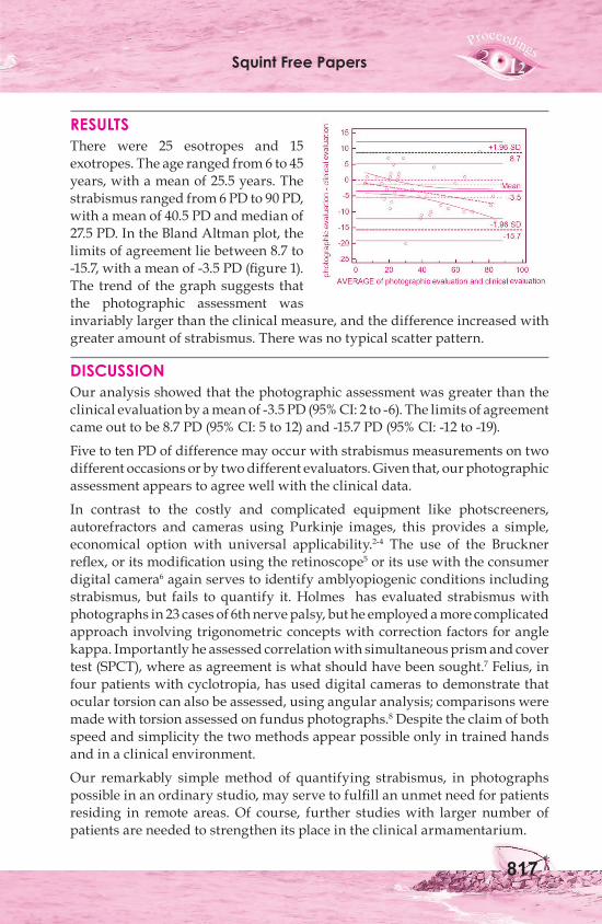
RESULTSThere were 25 esotropes and 15 exotropes. The age ranged from 6 to 45 years, with a mean of 25.5 years. The strabismus ranged from 6 PD to 90 PD, with a mean of 40.5 PD and median of 27.5 PD. In the Bland Altman plot, the limits of agreement lie between 8.7 to -15.7, with a mean of -3.5 PD (figure 1). The trend of the graph suggests that the photographic assessment was invariably larger than the clinical measure, and the difference increased with greater amount of strabismus. There was no typical scatter pattern.
DISCUSSION Our analysis showed that the photographic assessment was greater than the clinical evaluation by a mean of -3.5 PD (95% CI: 2 to -6). The limits of agreement came out to be 8.7 PD (95% CI: 5 to 12) and -15.7 PD (95% CI: -12 to -19).
Five to ten PD of difference may occur with strabismus measurements on two different occasions or by two different evaluators. Given that, our photographic assessment appears to agree well with the clinical data.
In contrast to the costly and complicated equipment like photscreeners, autorefractors and cameras using Purkinje images, this provides a simple, economical option with universal applicability.2-4 The use of the Bruckner reflex, or its modification using the retinoscope5 or its use with the consumer digital camera6 again serves to identify amblyopiogenic conditions including strabismus, but fails to quantify it. Holmes has evaluated strabismus with photographs in 23 cases of 6th nerve palsy, but he employed a more complicated approach involving trigonometric concepts with correction factors for angle kappa. Importantly he assessed correlation with simultaneous prism and cover test (SPCT), where as agreement is what should have been sought.7 Felius, in four patients with cyclotropia, has used digital cameras to demonstrate that ocular torsion can also be assessed, using angular analysis; comparisons were made with torsion assessed on fundus photographs.8 Despite the claim of both speed and simplicity the two methods appear possible only in trained hands and in a clinical environment.
Our remarkably simple method of quantifying strabismus, in photographs possible in an ordinary studio, may serve to fulfill an unmet need for patients residing in remote areas. Of course, further studies with larger number of patients are needed to strengthen its place in the clinical armamentarium.
70th AIOC Proceedings, Cochin 2012
818
REFERENCES1. Matta NS, Arnold RW, Singman EL, Silbert DI. Comparison between the plusoptiX
and MTI Photoscreeners. Arch Ophthalmol.2009;127:1591-5.2. Barry JC, Effert R, Koupp A, Burrhof A. Measurement of ocular alignment with
photographic purkinje I and IV reflection pattern evaluation. Invest Ophthalmol Vis Sci.1994;35:4219-35.
3. Barry JC, Effert R, Kaupp A. Objective measurement of small angles of strabismus in infants and children with photographic reflection pattern evaluation. Ophthalmology.1992;99:320-8.
4. Effert R, Barry JC, Colberg R, Kaupp A, Scherer G. Self-assessment of angles of strabismus with photographic Purkinje I and IV reflection pattern evaluation. Graefes Arch Clin Exp Ophthalmol. 1995;233:494-506.
5. Amitava AK, Kewlani D, Khan Z, Razzaq A. Assessment of a modification of Brückner’s test as a screening modality for anisometropia and strabismus. Oman J. Ophthalmol.2010;3:131-5.
6. Arnold RW. Vision Screening in Alaska: experience with Enhanced Bruckner test. Alaska Med.1993;35:212-5.
7. Jonathom M.Holmes, George G Hohberger, David A. Leske. Photographic and clinical techniques for outcome assessment in sixth nerve palsy. Ophthalmology.2001;108:1300-07.
8. Joost Felius, Kirsten G. Locke RN, Mohamed A. Hussain, David R.Stager Jr, David R. Stager Sr.Photographic assessment of changes in torsional strabismus. JAAPOS.2009;13:593-95.
Comparison of Graded Inferior Oblique Recession to Graded Myectomy for Primary Inferior Oblique OveractionDr. Jaspreet Sukhija, Dr. Neha Kumari, Dr. Parul Chawla
Primary Inferior Oblique (IO) Overaction may cause a socially noticeable vertical hypertropia of the affected eye in primary position and contralateral
gaze. Surgical management is often required to improve alignment. Treatment of this entity is varied. Among the various weakening techniques of inferior oblique muscle overaction, the most commonly used techniques include myectomy and recession. Previous reports have suggested that unilateral inferior oblique weakening may lead to secondary hyperactivity of the opposite inferior oblique muscle postoperatively.1-4 In this study, inferior oblique recessions and myectomy were compared to evaluate the surgical results in primary inferior oblique overaction in bilateral and unilateral cases.
Squint Free Papers
819
MATERIALS AND METHODSFifty eyes of 32 patients were evaluated. Group 1 included 28 eyes of 17 cases with IO recession and group 2 had 22 eyes of 15 patients where myectomy was performed. Bilateral surgery was done in 11 cases in group-1 and 7 in group-2. Unilateral weakening of IO was performed in eyes with unilateral IO overaction. Oblique muscle dysfunction was graded in approximately 45° adducted eye as decribed by Min et al.5 Amount of recession done was based on grade of overaction (Finks for grade 2, Parks for grade 3, Modified Eliott nankin for grade 4). Similarly a 5, 6 and 7 mm myectomy was done in grade 2,3 and 4 IO overaction. Standard of success was based on grade 1 or less inferior oblique overaction at three months operative.
RESULTS4/28 eyes which underwent inferior oblique recessions had zero inferior oblique overaction post surgery(success rate 85.7%). In eyes where Finks recession was done the success rate was 100%. Successful outcome was achieved in 14/16 eyes with parks recession and 5/7 eyes with modified Eliott Nankin recession. 1/6 cases where unilateral recession was done developed IO overaction in the other eye. The overall success rate in myectomy group was 72.7%. 2/6 eyes where 5 mm myectomy was done developed hypotropia. Residual IO overaction was present in 3/10 eyes and 1/6 eyes were a 6mm and 7 mm myectomy was done. 3/8 eyes in unilateral group developed contralateral IO overaction.
DISCUSSIONBoth recession and myectomy reduced the function of the overactive inferior oblique muscle, but eyes with recession tended to be more effective. In eyes where a myectomy was performed showed underaction of the inferior oblique with hypotropia, which is probably due to non-attachment of the inferior oblique muscle to the globe. However this persisted even at 3 months which shows that performing myectomy in grade 2 IO overaction can sometime produce unpredictable results. In the myectomy group where a 6 mm and 7 mm procedure was done residual overaction was observed more often as compared to the recession group. However this finding did not reach statistically significant levels. Elliott and Nankin6 compared the anterior transposition with the inferior oblique muscle recession, and Del Monte and Parks7 compared the denervation-extirpation on one eye with the “14 mm” recession of the inferior oblique muscle on the other eye in 16 patients with bilateral symmetrical +4 primary inferior oblique muscle overaction. Their results at the final assessment showed 100% normal inferior oblique muscle action for the denervationextirpation eyes, while 88% of the eyes receiving the recession had a residual +1 to +4 overactive inferior oblique muscle. In our series the success rate with recession was 85.7% and with myectomy 72.7%.
70th AIOC Proceedings, Cochin 2012
820
Unilateral inferior oblique recession has been recommended in patients with strictly unilateral muscle overaction. Singh et al have shown that all patients with asymmetry of 3 grades and some patients with asymmetry of 2 grades can be safely treated by unilateral surgery.8
In conclusion this small series shows that inferior oblique muscle recessions and myectomies are almost equally effective in the vast majority of patients with primary inferior oblique overaction. Recessions however give more controlled effect in unilateral cases.
REFERENCES1. Von Noorden GK, Campos EC. A and V patterns. In: von Noorden GK, Campos EC,
eds. Binocular Vision and Ocular Motility: Theory and Management of Strabismus, St. Louis: Mosby; 2001;6:396-410.
2. Von Noorden GK, Campos EC. Principles of surgical treatment. In: von Noorden GK, Campos EC, eds. Binocular Vision and Ocular Motility: Theory and Management of Strabismus, St. Louis: Mosby; 2001;6:566-624.
3. Stein LA, Ellis FJ. Apparent contralateral inferior oblique overaction after unilateral inferior oblique muscle weakening procedures. J AAPOS. 1997;1:2-7.
4. Oguz V, Devranoglu K, Arslan O, Tolun H, Celikkol L. Secondary hyperactivity of the contralateral oblique muscle after unilateral weakening of the inferior oblique muscle [article in French]. J Fr Ophtalmol. 1996;19:327-9.
5. Min BM, Park JH, Kim SY, Lee SB. Comparison of inferior oblique muscle weakening by anterior transposition or myectomy: a prospective study of 20 cases. Br J Ophthalmol. 1999;83:206-8.
6. Elliott RL, Nankin S. Anterior transposition of the inferioroblique. J Pediatr Ophthalmol Strabismus 1981;18:35–8.
7. Del Monte MA, Parks MM. Denervation and extirpation of the inferior oblique: an improved weakening procedure for marked overaction. Ophthalmology 1983;90:1178–85.
8. Singh V, Agrawal S, Agrawal S. Outcome of Unilateral Inferior Oblique Recession. J Pediatr Ophthalmol Strabismus 2009;46:350-7.
Bilateral Harada ITO Procedure for Extorsion and V Eso Shift in Traumatic Acquired Bilateral Superior Oblique PalsiesDr. Pramod Kumar Pandey, Dr. Anupam Singh, Dr. Abhishek Sharma, Dr. Shagun Sood, Dr. Sanjeev kumar, Dr. Ekta Kumari
Superior oblique palsy (SOP) is the commonest cause of paralytic strabismus and of vertical strabismus in large strabismus series.1 Congenital and
traumatic etiologies are most frequent and 20 to 80% of traumatic cases can
Squint Free Papers
821
be bilateral. Over and above masked bilateral SOPs may account for upto 8 to 28% of cases of unilateral SOPs.2 There are thus no paradigmatic presentations of bilaterality, as presentations are diverse and may often be abstruse. Markers of bilaterality include V eso of >20 PD, Extorsion of >12, chin down position and history of trauma. Torsional and horizontal diplopia in down gaze are most trouble some symptoms in patients with acquired BL SOPs. Extorsion of >100 requires a procedure specifically to address it and Harada Ito3 procedure has been done conventionally to ameliorate it. V esotropia in down gaze gives rise to horizontal diplopia and often is the presenting complaint of the patient even surpassing torsional diplopia and is reason for adopting chin down head position in most cases. Little attention though seems to have been paid in literature to ameliorate esotropia in down gaze in these patients, who are troubled most by it. Inferior rectus recessions with nasal transposition, weakening procedures on inferior oblique may work but have attendant unwanted baggage of under corrections or untoward effects, inferior transposition of horizontal recti may work for eso shift but it worsens extorsion. The surgical modality that will treat extorsion and eso shift in down gaze simultaneously, still evades us. We evaluated Fell’s4 modification of Harada Ito procedure to simultaneously treat extorsion and V eso in 5 patients with bilateral traumatic SOPs.
MATERIALS AND METHODS consecutive 5 patients diagnosed as BL traumatic SOPs seen over last 2 years at a tertiary care institution were included in the study. Period of 6 months was allowed for recovery of paresis. Patients’ complaints and their evolution/ regression were recorded. Visual acuity, abnormal head posture, primary position deviation, deviation fixing with either eye in 9 cardinal gazes were recorded. V eso was assessed by recording deviation in 250 up and down gazes at 6 meters using an accommodative target. Extorsion was evaluated subjectively by double Maddox rods and objectively by fundus photography. Neuro-imaging was done in all. Modified Harada Ito procedure was executed in all bilaterally under local anesthesia. Anterior 1/3 to ½ of superior oblique tendon was identified by superior temporal approach and split for about 10 mm, it was attached along superior border of lateral rectus muscle 6 to 8 mm behind it’s insertion. The more eso shift was to be treated more posterior was the placement. Conjunctive was closed by 8/0 Vicryl sutures. Resolution of patients’ complaints, primary position alignment, extorsion and V eso in down gaze were evaluated at 6 weeks and results analyzed
RESULTSAge ranged from 24 to 42 years. All were males and involved in road
70th AIOC Proceedings, Cochin 2012
822
traffic accidents with 3 having suffered concussions. Neuro-imaging was unremarkable in all. Best corrected visual acuity was 6/6 in all OU.All complained of troublesome diplopia in down gaze with horizontal and torsional component and reading difficulties. A chin down head position was adopted in all. Two complained of vertical diplopia in side gazes. Subjective extorsion by DMR ranged from 12 to 22 0. Esotropia in down gaze ranged from 12 to 20 PD with PP horizontal deviation of 6 to 12 PD. One had hypertropia of 5 PD in PP. four had positive Bielschowsky’s head tilt test and 1 was masked BL SOP.
Postoperatively all were relieved of trouble some horizontal and torsional diplopia. Subjective extorsion of 4 to 6 0 in 2 patients by DMR was noted. V esotropia of 5 and 8 PD was noted in 2 patients. No untoward outcomes were encountered.
DISCUSSIONBilateral acquired SOPs may engender diagnostic and therapeutic dilemmas as presentation may be confused with acute acquired comitant esotropia, decompensated monofixational esotropia, accommodative esotropia or laterally alternating skew to name a few. Therapeutic challenges include exuberant amount of subjective extorsion as well as eso shift in down gaze. If extorsion exceeds 100 some form of torsional surgery is indicated. Recession of inferior recti with nasal transposition may be helpful if residual extorsion is still present but it may worsen esotropia in down gaze. For V pattern medial recti may be transposed inferiorly but that worsens extorsion and may be counterproductive. Inferior oblique weakening procedures do not address esotropia and extorsion in down gaze effectively. Fell’s modification of Harada Ito procedure helps ameliorate both, extorsion upto 200 as well as esotropia of upto 30 PD in down gaze with no untoward effects and thus may be most suited to tackle symptoms emanating from excyclotorsion and esotropia in down gaze in patients with acquired bilateral superior oblique palsies. A paradigmatic surgical approach for the management of Bilateral traumatic SOPs still evades us.
REFERENCES1. Von Noorden GK, Murry E, Wong SY; Superior Oblique Palsies, A Review of 270
Cases; Arch. Ophthalmol. 1988;104:1771-1776.2. Kushner B J; The diagnosis and treatment of bilateral masked superior oblique
palsy; Amer. J. Ophthalmol. 1988;105:186-1943. Harada M, Ito Y; Surgical Correction of Cyclotropia; Jpn. J. Ophthalmol.; 8; 88 19644. Fells P; Surgical Management of Cyclotropia; Int. Ophthalmol. Clin.; 16; 161; 1976.
Squint Free Papers
823
Results of Squint Surgery for Horizontal Comitant Strabismus in Patients with Developmental DelayDr. Meenakshi Gopalakrishnan, Dr. Sanil Shah, Dr. Aarthy G.
It is known that children with developmental delay have higher rate of ophthalmic abnormalities. Strabismus has been found to be one of the most
frequent ophthalmic anomaly in these children.1,2,3,4,5,6,7 Little has been published about surgical outcomes after strabismus surgery in this group.4,8,9,10,11,12 Most Studies found that strabismus surgery for neurologically impaired children is less predictable, they have a poorer prognosis, and the common thought is that treating these patients by the standard surgical schedules will attain a high rate of overcorrection.9,10 Reduction in surgical dose may attain a higher rate of surgical success. Therefore, the goal of our study was to determine results of more conservative amount of surgery for both esodeviaton and exodeviation among developmentally delayed children.
MATERIALS AND METHODSRecords of all the children with developmental delay who underwent squint surgery for horizontal comitant deviation during a 6 year period from 2005 to 2011 were reviewed.
Exclusion criteria were, vertical deviation, limited ocular movements, incomitant deviation, and strabismus secondary to other eye pathology, previous eye/muscle surgery, or postoperative follow-up of less than 6 weeks. Forty-four children were found to meet our inclusion and exclusion criteria, 29 children with esotropia with a mean age of 5.08±3.27 years (range, 1-12 years) at surgery and 15 children with exotropia with a mean age of 7.73±4.34 years (range, 3-18 years) at surgery.
All the children underwent comprehensive eye examination preoperatively and postoperative follow-up. Visual acuity was assessed by CSM method, Snellen’s chart or Leas symbols depending on age and ability. Cooperation for binocular function test was poor in many patients. Strabismus angle measurement was obtained by the use of the alternate cover test; the modified Krimsky method was used when cooperation was poor. Refractive errors were measured by cyclopentolate 1% or homatropine 2% (in patients with CNS or seizure disorders). Glasses were prescribed for children for both amblyopic and accommodative concerns. All refractive errors and amblyopia were treated before surgery.
The preoperative and postoperative angle of deviation was calculated for each subject as the mean of distant and near angles if measured by cover test or the modified Krimsky measurement at near. Out of 29 patients having esotropia
70th AIOC Proceedings, Cochin 2012
824
25 patients were operated by bilateral MR recession (BMRc), 3 patient under went BMRc + LR resection and one patient under went unilateral recession-resection. Out of 15 patients with exotropia bilateral LR recession (BLRc) was done in 7 patients, unilateral recession-resection in 5 patients and BLRc + MR resection was done in 3 patients. Average preoperative angle of deviation was (A) 43.64±13.61 PD (ET 46.21±14.67 PD and XT 38.67±9.9 PD). Amount of surgery performed on horizontal muscles in each patient was correlated with standard table used by individual surgeon for their routine squint surgeries (in normal children). This comparison with the standard table gave us angle of deviation (B) which would have been corrected in a normal child with the same amount of surgery.
No complications were observed during or after the surgery. Ocular examination including angle of deviation, ocular movements and cycloplegic refraction, were performed at 6 week in all patients. Successful outcome was defined as within 10 PD of orthophoria.
RESULTSOut of total 44 (30 male, 14 female) children, esotropia was found in 29 (65.90%)(21 male, 8 female) and exotropia was present in 15 (34.09%)children (9 male, 6 female). Mean spherical equivalent pre-operatively was +1.05 ± 2.30 in patients with esotropia and +0.42 ±1.35 in patients with exotropia. Nystagmus was present in 9 (5 esotropic and 4 exotropic) children.
Average preoperative angle of deviation was (A) 43.64±13.61 PD (ET 46.21±14.67 PD and XT 38.67±9.9 PD). Amount of surgery performed on horizontal muscles in each patient was correlated with standard table used for normal children to get the angle of deviation (B) which would have been corrected in a normal child with the same amount of surgery. This operated angle of deviation (B) was expressed as percentage of pre operative angle of deviation (A). On an average 75.67%±14.71 (ET-73.9% ±15.62 and XT–79.08%±12.53) of angle of deviation was operated. For example one patient having 25 PD esotropia was operated for both eyes MR recession 3.5 mm by surgeon 1. When compared with the standard surgical table used by surgeon 1, this amount of surgery is supposed to correct 20 PD of esotropia. So amount of surgery performed was 80% (20PD) of the pre-operative angle (25PD).
Surgical success was determined at 6 week post operative follow up. Longer follow-up was not selected because long term stability of angle is affected by many factors other than surgical factors. Out of 44 patients successful outcome was noted in 27 (61.36%) patients at 6 weeks post operative. Among patients with esotropia successful outcome was noted in 17 out of 29 (58.62%) and among patients with exotropia 10 out of 15 (66.66%) patients had successful outcome. Among patients with successful outcome average
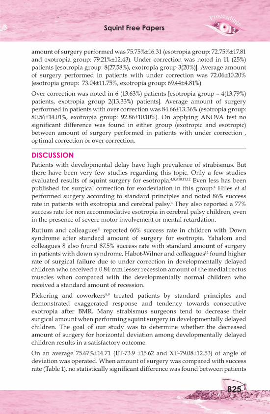
Squint Free Papers
825
amount of surgery performed was 75.75%±16.31 (esotropia group: 72.75%±17.81 and exotropia group: 79.21%±12.43). Under correction was noted in 11 (25%) patients [esotropia group: 8(27.58%), exotropia group 3(20%)]. Average amount of surgery performed in patients with under correction was 72.06±10.20% (esotropia group: 73.04±11.75%, exotropia group: 69.44±4.81%)
Over correction was noted in 6 (13.63%) patients [esotropia group – 4(13.79%) patients, exotropia group 2(13.33%) patients]. Average amount of surgery performed in patients with over correction was 84.66±13.36% (esotropia group: 80.56±14.01%, exotropia group: 92.86±10.10%). On applying ANOVA test no significant difference was found in either group (exotropic and esotropic) between amount of surgery performed in patients with under correction , optimal correction or over correction.
DISCUSSIONPatients with developmental delay have high prevalence of strabismus. But there have been very few studies regarding this topic. Only a few studies evaluated results of squint surgery for esotropia.4,8,9,10,11,12 Even less has been published for surgical correction for exodeviation in this group.4 Hiles et al performed surgery according to standard principles and noted 86% success rate in patients with exotropia and cerebral palsy.4 They also reported a 77% success rate for non accommodative esotropia in cerebral palsy children, even in the presence of severe motor involvement or mental retardation.
Ruttum and colleagues11 reported 66% success rate in children with Down syndrome after standard amount of surgery for esotropia. Yahalom and colleagues 8 also found 87.5% success rate with standard amount of surgery in patients with down syndrome. Habot-Wilner and colleagues12 found higher rate of surgical failure due to under correction in developmentally delayed children who received a 0.84 mm lesser recession amount of the medial rectus muscles when compared with the developmentally normal children who received a standard amount of recession.
Pickering and coworkers8,9 treated patients by standard principles and demonstrated exaggerated response and tendency towards consecutive exotropia after BMR. Many strabismus surgeons tend to decrease their surgical amount when performing squint surgery in developmentally delayed children. The goal of our study was to determine whether the decreased amount of surgery for horizontal deviation among developmentally delayed children results in a satisfactory outcome.
On an average 75.67%±14.71 (ET-73.9 ±15.62 and XT–79.08±12.53) of angle of deviation was operated. When amount of surgery was compared with success rate (Table 1), no statistically significant difference was found between patients
70th AIOC Proceedings, Cochin 2012
826
with 80-100% surgery compared to less than 80% surgery. But it was seen that rate of over correction increased with more than 80% surgery and that of under correction increased with lesser amount of surgery.
Table 1amount of surgery performed under correction Optimal correction Over correction ET group(12) 7(58.33%) 2(16.66%) 3(25%)80-100% XT group(6) 0 4(66.66%) 2(33.33%) ET group(17) 6(35.29%) 10(58.82%) 1(5.88%)50 - 80% XT group(9) 3(33.33%) 6(66.66%) 0
When amount of surgery was compared (Table 2) with outcome no significant difference was found. Standard deviation was very high among all groups which suggest unpredictability of strabismus surgery in this group of patients. Mean amount of surgery performed in patients with optimal outcome was 75.75%±16.31 (esotropia group: 72.75%±17.81 and exotropia group: 79.21%±12.43).
Table 2Groups No. of patients (%) Amount of surgery (ET group -total 29 patients) performed (esotropia) (XT group– total 15 patients) Under ET group 8(27.58%) 73.04±11.75%,correction XT group 3(20%) 69.44±4.81% Combined 11(25%) 72.06±10.20%Optimal ET group 17(58.62%) 72.75%±17.81correction XT group 10(66.66%) 79.21%±12.43 Combined 27(61.36%) 75.75%±16.31Over ET group 4(13.79%) 80.56±14.01%,correction XT group 2(13.33%) 92.86±10.10% Combined 6(13.63%) 84.66±13.36%
In conclusion, surgical outcome in patients with developmental delay is very unpredictable. Though there was no statistically significant difference, operating for 75.75%±16.31 of angle of deviation is more likely to be successful.
REFERENCES1. Bankes JLK. Eye defects of mentally handicapped children. Br J Ophthalmol
1974;2:533-5.2. Seaber JH, Chandler AC. A five-year study of patients with cerebral palsy and
strabismus. In: Moore S, Mein J, Stockbridge L, editors. Orthoptics: past, present, future. New York: Grune and Stratton; 1976. p. 271-7.
Squint Free Papers
827
3. Losseff S. Ocular findings in cerebral palsy. Am J Ophthalmol 1962;54:1114-8.4. Hiles DA, Wallar PH, McFarlane F. Current concepts in the management of
strabismus in children with cerebral palsy. Ann Ophthalmol 1975;7:789-98.5. Buckley E, Seaber JH. Dyskinetic strabismus as a sign of cerebral palsy. Am J
Ophtalmol 1981;91:652-7.6. Buckley E, Seaber JH. Unique ocular findings in cerebral palsy patients with
strabismus. Am Orthop J. 1981;31:53-9.7. Bankes JLK, Thornhill DM, Corr PE, et al. The management and binocular
achievement of mentally handicapped children with squint. In: Moore S, Mein J, Stockbridge L, editors. Orthoptics: past, present, future. New York: Grune and Stratton; 1976. p. 293-8.
8. Yahalom C, Mechoulam H, Cohen E, Anteby I. Strabismus surgery outcome among children and young adults with Down syndrome. J AAPOS 2010;14:117-9
9. Pickering JD, Simon JW, Lininger LL, et al. Exaggerated effect of bilateral medial rectus recession in developmentally delayed children. J Pediatr Ophthalmol Strabismus 1994;31:374-7.
10. Pickering JD, Simon JW, Ratliff CD, et al. Alignment Success following medial rectus recessions in normal and delayed children. J Pediatr Ophthalmol Strabismus 1995;32:225-7.
11. Ruttum MS, Kivlin JD, Hong P. Outcome of surgery for esotropia in children with Down Syndrome. Am Orthoptic J 2004;54:98-101.
12. Zohar Habot-Wilner, Abraham Spierer, Joseph Glovinsky, and Tamara Wygnanski-Jaffe. Bilateral Medial Rectus Muscle Recession: Results in Children With Developmental Delay Compared With Normally Developed Children MD J AAPOS 2006;10:150-4.
Unilateral Vs Bilateral LR Recession in Divergent Excess Type of Small Angle IDS - 4 Year Follow-upDr. Kamlesh, Dr. Shilpa Goel, Dr. Yuvika Bansal, Dr. Manav Sachdev
The traditional treatment for intermittent exortropia has been a bilateral rectus recession or a unilateral lateral rectus recession combined with
a medial rectus resection. The use of single lateral rectus muscle recession has been controversial. Many Surgeons avoid using this technique on lateral rectus muscle because of concern that it may produce lateral incommittance and abduction deficits.
Although some authors report unilateral recessions as ineffective or producing inconsistent results, the approach has been found to be successful for deviations of 20 PD or less. We have found this procedure to be useful in deviation less than 25 PD. The advantage of unilateral lateral rectus recession for this
70th AIOC Proceedings, Cochin 2012
828
disorder includes limiting the risk of surgery to one eye, lower incidence of overcorrection, little change in their alignment from the early post-operative period and shorter duration of anesthesia.
The purpose of this study was to compare the surgical and functional outcome of unilateral and bilateral rectus recession in the treatment of divergence excess type of small angle exotropia.
MATERIALS AND METHODS100 patients with intermittent exotropia less than 25 PD were enrolled for this study. Criteria for enrollment were: Divergence excess type of intermittent exotropia (a) with devialtion less than 25 PD (b) best corrected VA>= 6/9. (c) no A or V Pattern (d) No history of previous strabismus surgery. (e) full pre-operative duction and version.
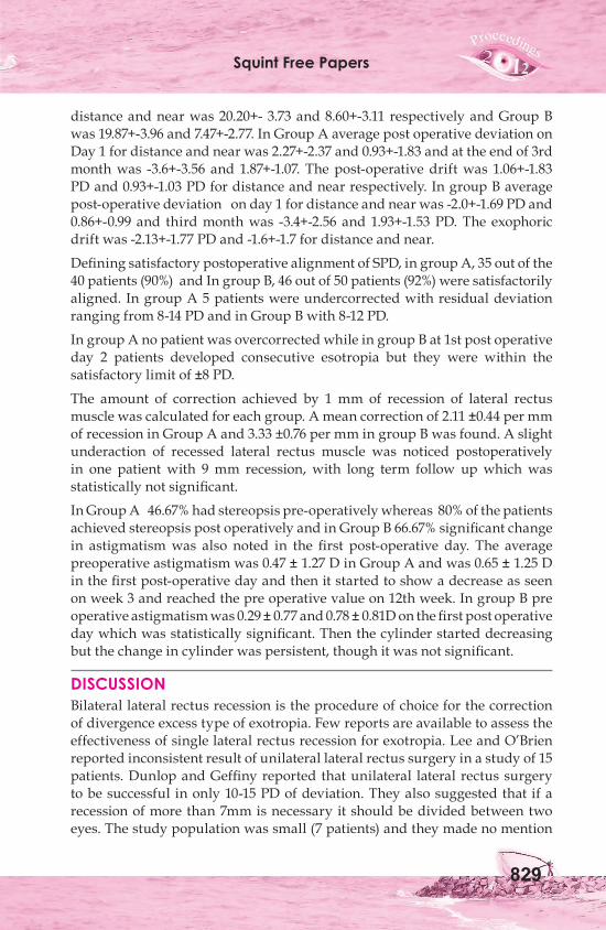
The patients were randomly divided into two groups of 50 each. Group A underwent unilateral rectus recession and Group B underwent bilateral lateral rectus recession. Measurment were taken at 6m for distance and 30 cm for near deviation after patching one eye for 24 hrs in primary position and in 30 degree side gaze for lateral incomittance. Side gaze measurement was made by rotating the face an estimated 30 degree and having the patient to look forward. Biocular status of the patient was assessed by worth 4 dot test and synaptophore. Corneal topography was done both preoperatively and postoperatively to compare any change in refractive states. All surgeries were performed by conventional hangback technique by one surgeon. The decision on the amount of lateral rectus recession was taken on an individual basis keeping in mind the preoperative factors influencing the outcome of surgery, in each case following the guidelines described by strabismologist.
Table 1: Guidelines for Lateral Rectus Recession Surgery PD Group A Group B15 7 mm 4 mm20 8 mm 5 mm25 9 mm 6 mm
A satisfactory result was considered when the alignment is within 8 PD of orthophoria. An incommitant postoperative result was considered present, when alignment changed for more than 20% greater from primary to lateral side.
RESULTSThe mean age of the patients at the time of surgery was 9.60+-4.79 in Group A and 9.93+-4.71 yrs in Group B. The preoperative mean angle in Group A for
Squint Free Papers
829
distance and near was 20.20+- 3.73 and 8.60+-3.11 respectively and Group B was 19.87+-3.96 and 7.47+-2.77. In Group A average post operative deviation on Day 1 for distance and near was 2.27+-2.37 and 0.93+-1.83 and at the end of 3rd month was -3.6+-3.56 and 1.87+-1.07. The post-operative drift was 1.06+-1.83 PD and 0.93+-1.03 PD for distance and near respectively. In group B average post-operative deviation on day 1 for distance and near was -2.0+-1.69 PD and 0.86+-0.99 and third month was -3.4+-2.56 and 1.93+-1.53 PD. The exophoric drift was -2.13+-1.77 PD and -1.6+-1.7 for distance and near.
Defining satisfactory postoperative alignment of SPD, in group A, 35 out of the 40 patients (90%) and In group B, 46 out of 50 patients (92%) were satisfactorily aligned. In group A 5 patients were undercorrected with residual deviation ranging from 8-14 PD and in Group B with 8-12 PD.
In group A no patient was overcorrected while in group B at 1st post operative day 2 patients developed consecutive esotropia but they were within the satisfactory limit of ±8 PD.
The amount of correction achieved by 1 mm of recession of lateral rectus muscle was calculated for each group. A mean correction of 2.11 ±0.44 per mm of recession in Group A and 3.33 ±0.76 per mm in group B was found. A slight underaction of recessed lateral rectus muscle was noticed postoperatively in one patient with 9 mm recession, with long term follow up which was statistically not significant.
In Group A 46.67% had stereopsis pre-operatively whereas 80% of the patients achieved stereopsis post operatively and in Group B 66.67% significant change in astigmatism was also noted in the first post-operative day. The average preoperative astigmatism was 0.47 ± 1.27 D in Group A and was 0.65 ± 1.25 D in the first post-operative day and then it started to show a decrease as seen on week 3 and reached the pre operative value on 12th week. In group B pre operative astigmatism was 0.29 ± 0.77 and 0.78 ± 0.81D on the first post operative day which was statistically significant. Then the cylinder started decreasing but the change in cylinder was persistent, though it was not significant.
DISCUSSION Bilateral lateral rectus recession is the procedure of choice for the correction of divergence excess type of exotropia. Few reports are available to assess the effectiveness of single lateral rectus recession for exotropia. Lee and O’Brien reported inconsistent result of unilateral lateral rectus surgery in a study of 15 patients. Dunlop and Geffiny reported that unilateral lateral rectus surgery to be successful in only 10-15 PD of deviation. They also suggested that if a recession of more than 7mm is necessary it should be divided between two eyes. The study population was small (7 patients) and they made no mention
70th AIOC Proceedings, Cochin 2012
830
about pre-operative deviation and the amount of recession performed. Nelso et al reported 90% success rate by performing 7-8 mm unilateral recession of 15-20 PD of exotropia with no over correction.
In our study, success rate between the two groups are comparable with 86.67% in group A and 93.33% in group B. none of the previous study had compared the results of Unilateral and Bilateral surgeries. We have compared the results between two symmetrical groups with respect to the amount of deviation and only one operating surgeon thus eliminating the inter-surgeon variability. Study by Abraham had compared the results between unilateral and bilateral surgeries but the two groups were not identical in terms of their amount of deviation.Raab and Parks showed that 80% of the patients undergoing bilateral lateral rectus recession changes their alignment in the post operative period with 71% becoming divergent, 81% of the patients with an initial exodeviation of 11-20 PD and 60% of patients with esodeviation upto 10 PD eventually became satisfactorily aligned. According to our study post-operative drift in group A was 1.06±1.83 and in group B was 2.13±1.77, the difference of which was not statistically significant. The results showed little change in alignment from the immediate post-operative period. Unlike bilateral surgery, initial orthophoria or small exodeviation carries good prognosis for satisfactory end results. It has been well documented, that immediate over correction of exotropia following bilateral lateral rectus recession is desirable for better long term alignment. However, prolonged consecutive esotropia in very young children can lead to amblyopia and suppression. Recession of single lateral rectus recession can be safely done in children less than 3 yrs of age with minimum risk of overcorrection.Surgeon in favor of bilateral surgery emphasizes the benefits of bilateral symmetrical surgery and risk of incommitance in unilateral surgery . Mild lateral gaze incommitance was noted in one patient which was less than 20% from the lateral gaze in the first post-operative day. Fretis reported a moderate restriction of abduction associated with some lateral incommitance during the first post-operative week but it gradually reduced to minimal. This could be because they have done large recession of 11.5-12mm. In our case also 9 mm recession was associated with mild restriction.Change in refractive status after strabismus surgery is well documented. In our study the change in astigmatism in immediate post-op period was significant in group B although the long term results were same. The difference in both these groups could possibly be explained by difference in the amount of recession as has been proposed by various authors. The further away the muscle in insertion from the limbus, the lesser is the risk of traction and thus the deformation of cornea.
Squint Free Papers
831
In conclusion it can be concluded from the current study that unilateral lateral rectus recession is comparable to bilateral lateral rectus recession for divergent excess type of intermittent exotropia with less than 25PD of deviation. In unilateral surgery only one muscle is being operated upon, the surgery is quicker with less anesthesia time, lesser complication with the added advantage of only one eye undergoing significant refractive changes. Thus we feel that unilateral muscle surgery is an easy and effective alternative to conventional bilateral lateral rectus recession and it should be strongly considered by the strabismus surgeon as a procedure of choice for small angle exotropia.
Role of Binocular Potential Score (BPS) in Predicting Surgical Outcome of Intermittent Exotropia (X[T])Dr. Arti Elhence, Dr. (MRS.) Vinita Singh, Dr. Siddharth Agrawal, Dr. Arun K. Sharma
Intermittent exotropia X[T] represents a stage in the natural history of exodeviations which begin as an exophoria [X] and progress to X [T] and
constant deviation [XT] as progressive deterioration of binocularity occurs. X[T] can be managed conservatively as well as with surgery but there are multiple variables that affect the outcome. The preoperative variables that have been studied in different permutations and combinations include, age at presentation, age at surgery, refractive error and preoperative binocular functions. An important variable is timing of surgery especially while dealing with a progressive condition like X[T]. Many authors have attempted to study the same and propose that surgical intervention at a younger age has better post operative outcome1-3 and delay can lead to suppression and loss of binocularity even after surgical correction. However, early surgery has the risk of consecutive esotropia.4 Apart from the age at surgery it is also essential to identify the appropriate timing with respect to the stage of disease which is influenced by the fusional control of the patient. The pre operative variables which determine the outcome have been quantitatively evaluated using the Binocular Potential Score (BPS) which has been developed by us for use in horizontal concomitant squints [APPENDIX-1].5 The seven variables included in the score are age of onset, duration of squint, intermittency, variability, visual acuity and responses on synoptophore and WFDT. Each is assigned a score from 1 to 5. The sum total (Range = 7- 35) of the individual BPS scores is then graded as follows: Very good(I): 28-35, Good(II): 21-27, Fair(III): 14-20, Poor (IV): 7-13. We studied the correlation between pre-operative BPS score and
70th AIOC Proceedings, Cochin 2012
832
surgical outcome (motor as well as sensory) to evaluate its predictive value. This correlation may aid in decision making regarding surgery for X[T].
MATERIALS AND METHODSThe study was a retrospective and prospective interventional study on 28 patients operated for X[T]. Patients were selected for surgery based on standard protocol6 (those who complained of deviation >50% of waking hours, had deteriorating binocular functions or were concerned cosmetically). Written and informed consent of patients or parents was obtained. Patients unwilling for inclusion in study or who had significant vertical deviation >10PD in primary gaze, those with gaze incomitance7 and retrospective patients whose pre-operative and post-operative records were incomplete were excluded. Patients underwent a complete orthoptic and ophthalmic workup including calculation of pre-operative BPS. Binocular functions were tested on the synoptophore (Appasamy ASP902) using simultaneous macular perception and binocular fusion slides. Binocular single vision (BSV) was tested on Worth 4 dot test using red-green glasses for distance and near and the better response used for analysis. Near stereoacuity was recorded using Randot ® stereotest (Stereo Optical Co, Chicago, IL). Selected patients underwent conventional strabismus surgery. Basic type deviations underwent unilateral recession-resection. Divergence excess type underwent bilateral lateral rectus recession.
Patients were reviewed at 1 week and 4 weeks after surgery. The motor outcome was defined as good (≤10PD), fair (11-19PD), poor (≥20PD). Sensory outcome was defined by the binocular function score (BF score-variable 6 and 7 of BPS i.e. responses on synoptophore and WFDT on a scale of 2 to 10). The readings at the end of 4 weeks were taken for calculations. STATISTICAL ANALYSIS: the data was analysed using statistical software package, STATA 11.2 The comparison of means for the normally distributed data between two groups was done using two-sample t-test. For data which was not distributed normally Mann-Whitney test was applied. The comparison among three groups was done using one way analysis of variance (ANOVA). Kruskal Wallis test was applied if data was not normally distributed or the variance was not homogeneous across groups. Difference was considered to be significant if the two tailed ‘p’ value was found to be ≤0.05.
RESULTS28 patients were recruited in this study with 11 retrospective and 17 prospective patients. Mean age of patients was 18.4±8.9 years range: 6-35 years, mean age at surgery was 18.4 ±8.7 years. 68% patients had good motor outcome, 12% fair and 20% poor. The distribution of good, fair and poor motor outcomes was not significantly different between BPS grades (p=0.506) [Table 1].
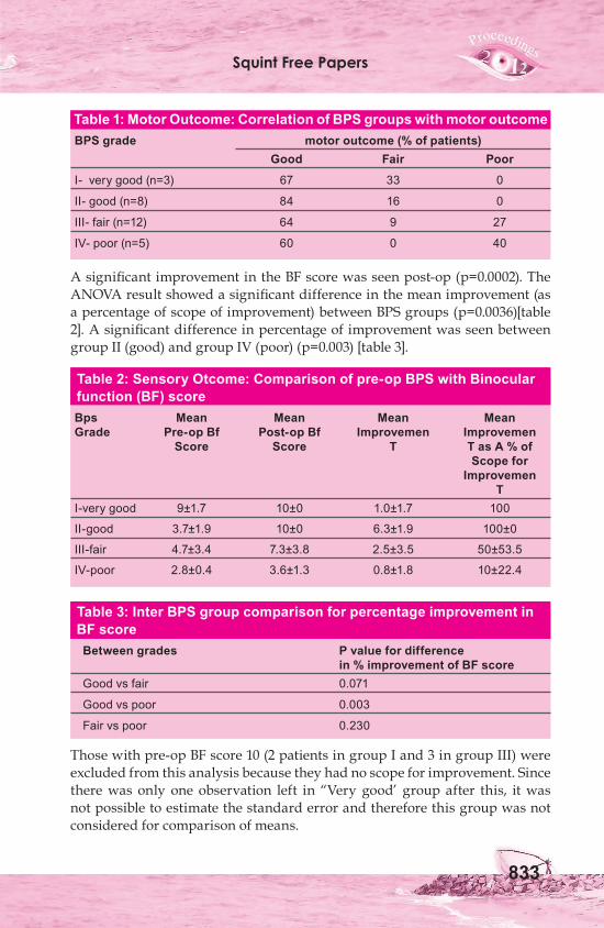
Squint Free Papers
833
Table 1: Motor Outcome: Correlation of BPS groups with motor outcome BPS grade motor outcome (% of patients) Good Fair Poor
I- very good (n=3) 67 33 0
II- good (n=8) 84 16 0
III- fair (n=12) 64 9 27
IV- poor (n=5) 60 0 40
A significant improvement in the BF score was seen post-op (p=0.0002). The ANOVA result showed a significant difference in the mean improvement (as a percentage of scope of improvement) between BPS groups (p=0.0036)[table 2]. A significant difference in percentage of improvement was seen between group II (good) and group IV (poor) (p=0.003) [table 3].
Table 2: Sensory Otcome: Comparison of pre-op BPS with Binocular function (BF) scoreBps Mean Mean Mean MeanGrade Pre-op Bf Post-op Bf Improvemen Improvemen Score Score T T as A % of Scope for Improvemen TI-very good 9±1.7 10±0 1.0±1.7 100
II-good 3.7±1.9 10±0 6.3±1.9 100±0
III-fair 4.7±3.4 7.3±3.8 2.5±3.5 50±53.5
IV-poor 2.8±0.4 3.6±1.3 0.8±1.8 10±22.4
Table 3: Inter BPS group comparison for percentage improvement in BF scoreBetween grades P value for difference in % improvement of BF scoreGood vs fair 0.071
Good vs poor 0.003
Fair vs poor 0.230
Those with pre-op BF score 10 (2 patients in group I and 3 in group III) were excluded from this analysis because they had no scope for improvement. Since there was only one observation left in “Very good’ group after this, it was not possible to estimate the standard error and therefore this group was not considered for comparison of means.
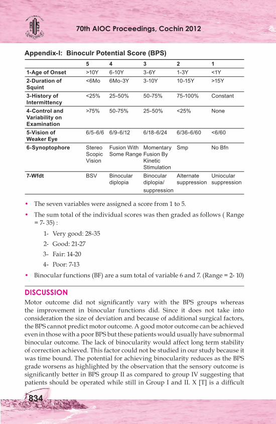
70th AIOC Proceedings, Cochin 2012
834
DISCUSSIONMotor outcome did not significantly vary with the BPS groups whereas the improvement in binocular functions did. Since it does not take into consideration the size of deviation and because of additional surgical factors, the BPS cannot predict motor outcome. A good motor outcome can be achieved even in those with a poor BPS but these patients would usually have subnormal binocular outcome. The lack of binocularity would affect long term stability of correction achieved. This factor could not be studied in our study because it was time bound. The potential for achieving binocularity reduces as the BPS grade worsens as highlighted by the observation that the sensory outcome is significantly better in BPS group II as compared to group IV suggesting that patients should be operated while still in Group I and II. X [T] is a difficult
Appendix-I: Binoculr Potential Score (BPS) 5 4 3 2 1 1-Age of Onset >10Y 6-10Y 3-6Y 1-3Y <1Y 2-Duration of <6Mo 6Mo-3Y 3-10Y 10-15Y >15Y Squint 3-History of <25% 25-50% 50-75% 75-100% Constant Intermittency 4-Control and >75% 50-75% 25-50% <25% None Variability on Examination 5-Vision of 6/5-6/6 6/9-6/12 6/18-6/24 6/36-6/60 <6/60 Weaker Eye 6-Synoptophore Stereo Fusion With Momentary Smp No Bfn Scopic Some Range Fusion By Vision Kinetic Stimulation 7-Wfdt BSV Binocular Binocular Alternate Uniocular diplopia diplopia/ suppression suppression suppression
• The seven variables were assigned a score from 1 to 5.
• The sum total of the individual scores was then graded as follows ( Range = 7- 35) :
1- Very good: 28-35 2- Good: 21-27 3- Fair: 14-20 4- Poor: 7-13
• Binocular functions (BF) are a sum total of variable 6 and 7. (Range = 2- 10)
Squint Free Papers
835
disorder to treat, particularly because of reluctance on the part of the patient to undergo surgery for a condition that is only occasionally present and also on the part of the surgeon who has to weigh the pros and cons of surgery and its appropriate timing. The issue of binocularity and its effect on the daily working of the individual is perhaps the most important consideration while making such decisions. An attempt has been made with the help of this study to elucidate the fact that BPS can help simplify decision making both for the surgeon as well as the patient. The BPS combines multiple variables hence is an effective predictive tool that could be used to explain prognosis to patients and can also serve as a guideline for planning surgery at an appropriate stage. The patients can be counselled that though the disease is intermittent waiting will lead to deterioration of the BPS grade and consequently poorer sensory results. Thus the primary utility of BPS is for follow up and for deciding appropriate timing for surgery.
REFERENCES1. Yang CQ, Shen Y, Gu YS, Han W. Clinical investigation of surgery for intermittent
exotropia. J ZhejiangUniv Sci B 2008;9:470-3.2. Wu QZ, Wu X, Lu W, Wang JH, Clinical investigation on binocular vision in
intermittent exotropia before and after surgery. Zhonghua Yan Ke Za Zhi. 2007; 43:968-71.
3. Saunders RA, Trivedi RH. Sensory results after lateral rectus recession for intermittent exotropia operated before two years of age. J AAPOS 2008;12:132-5.
4. Sharma P. Evaluation of distance and near stereoacuity and fusional vergence in intermittent exotropia. Indian J OPhthalmol. 2008;56:121-5.
5. Singh V, Panday M, Agrawal S. Binocular potential score: A novel concept. J Pediatr Ophthalmol Strabismus 2008;45:104-8.
6. Von Noorden GK. Binocular Vision and Ocular Motility. St Louis: Mosby Year Book; 2002;6:367.
7. Pineles SL, Ela-Dalman N, Zvansky AG, Yu F, Rosenbaum AL. Long-term results of the surgical management of intermittent exotropia. JAAPOS. 2010;14:298-304.