Embed Size (px)
DESCRIPTION
Manual of Squint
Citation preview
Manual ofSQUINT

Manual ofSQUINT
Leela AhujaEx-Professor of Strabismology
Ex-Director, Institute of OphthalmologyAligarh Muslim University
Aligarh, UPIndia
JAYPEE BROTHERS MEDICAL PUBLISHERS (P) LTDNew Delhi • Ahmedabad • Bengaluru • Chennai • Hyderabad
Kochi • Kolkata • Lucknow • Mumbai • Nagpur
®
Published by
Jitendar P VijJaypee Brothers Medical Publishers (P) LtdCorporate Office4838/24, Ansari Road, Daryaganj, New Delhi 110 002, IndiaPhone: +91-11-43574357
Registered OfficeB-3, EMCA House, 23/23B Ansari Road, Daryaganj, New Delhi 110 002, IndiaPhones: +91-11-23272143, +91-11-23272703, +91-11-23282021, +91-11-23245672Rel: +91-11-32558559 Fax: +91-11-23276490, +91-11-23245683e-mail: [email protected] Visit our website: www.jaypeebrothers.com
Branches• 2/B, Akruti Society, Jodhpur Gam Road Satellite
Ahmedabad 380 015 Phones: +91-79-26926233, Rel: +91-79-32988717Fax: +91-79-26927094 e-mail: [email protected]
• 202 Batavia Chambers, 8 Kumara Krupa Road, Kumara Park EastBengaluru 560 001 Phones: +91-80-22285971, +91-80-22382956, +91-80-22372664Rel: +91-80-32714073 Fax: +91-80-22281761 e-mail: [email protected]
• 282 IIIrd Floor, Khaleel Shirazi Estate, Fountain Plaza, Pantheon RoadChennai 600 008 Phones: +91-44-28193265, +91-44-28194897, Rel: +91-44-32972089Fax: +91-44-28193231 e-mail: [email protected]
• 4-2-1067/1-3, 1st Floor, Balaji Building, Ramkote Cross RoadHyderabad 500 095 Phones: +91-40-66610020, +91-40-24758498 Rel:+91-40-32940929Fax:+91-40-24758499, e-mail: [email protected]
• No. 41/3098, B & B1, Kuruvi Building, St. Vincent RoadKochi 682 018, Kerala Phones: +91-484-4036109, +91-484-2395739, +91-484-2395740e-mail: [email protected]
• 1-A Indian Mirror Street, Wellington SquareKolkata 700 013 Phones: +91-33-22651926, +91-33-22276404, +91-33-22276415Rel: +91-33-32901926 Fax: +91-33-22656075, e-mail: [email protected]
• Lekhraj Market III, B-2, Sector-4, Faizabad Road, Indira NagarLucknow 226 016 Phones: +91-522-3040553, +91-522-3040554e-mail: [email protected]
• 106 Amit Industrial Estate, 61 Dr SS Rao Road, Near MGM Hospital, ParelMumbai 400012 Phones: +91-22-24124863, +91-22-24104532, Rel: +91-22-32926896Fax: +91-22-24160828, e-mail: [email protected]
• “KAMALPUSHPA” 38, Reshimbag, Opp. Mohota Science College, Umred RoadNagpur 440 009 (MS) Phone: Rel: +91-712-3245220, Fax: +91-712-2704275e-mail: [email protected]
USA Office1745, Pheasant Run Drive, Maryland Heights (Missouri), MO 63043, USAPh: 001-636-6279734 e-mail: [email protected], [email protected]
Manual of Squint© 2008, Jaypee Brothers Medical Publishers
All rights reserved. No part of this publication should be reproduced, stored in a retrieval system, ortransmitted in any form or by any means: electronic, mechanical, photocopying, recording, orotherwise, without the prior written permission of the author and the publisher.
This book has been published in good faith that the material provided by author is original. Everyeffort is made to ensure accuracy of material, but the publisher, printer and author will not beheld responsible for any inadvertent error(s). In case of any dispute, all legal matters are to besettled under Delhi jurisdiction only.
First Edition: 2008ISBN 978-81-8448-382-6
Typeset at JPBMP typesetting unitPrinted at Ajanta

To— Shridivale Sai Baba
— My Husband Prof. (Dr) OP AhujaEx-Director of Institute of Ophthalmology andDirector, Founder of Ahuja Eye Centre, Aligarh
— My grandchildren– Ashir, Arjun, Shishir, Aanchal,Rhea and Savani — the future of India

Preface
A lot of literary works have been done on squint but still there is adearth of standard books on strabismus for postgraduate students. Nodoubt, surgery of squint is done by many ophthalmologists, but mostly,it is on cosmetic grounds and that too without the help of proper orthopticdepartment. It is also a fact that general public is reluctant to havetreatment, particularly surgical treatment of squint, as this malady isconsidered to be due to displeasure of some Goddess. The importanceis not to cure deviation, but to improve binocular function. Blindnesshas existed since time immemorial as illustrated in the story of ShravanKumar.
I realize that some of the topics are very much comprehensive so Ihave tried to simplify them by providing their description in simple andeasily understood language. Most controversial aspects of certainconditions have been deliberately left out for the sake of easyunderstanding. This book includes material from Duke-Elder, Kyth Lyle,von Noordan, Kanski, Muller and Paymann. The first three chapters onapplied anatomy of paralytic squint are venture of my husband Prof OPAhuja, Ex-Director, Institute of Ophthalmology, and Founder andDirector of Ahuja Eye Centre, Aligarh, UP.
I owe so much to Prof GP Gupta, Ex-Director of Institute ofOphthalmology, Aligarh, Prof BS Goel, Ex-Director, Institute ofOphthalmology, Aligarh and my son Dr Anupam Ahuja, Consultant,Ahuja Eye Centre, Aligarh for help and providing me photographs.
I am immensely thankful to Late (Prof) LP Agarwal, Ex-DirectorAIIMS, Delhi, Prof (Dr) Manoj Shukla, Ex-Director, Institute ofOphthalmology, Aligarh, Prof (Dr) SS Soodan, Principal and Director ofAscon College of Medical Science, Jammu, Prof S Mittal, Head,Department of Ophthalmology, Meerut Medical College, Prof BD Sharma,Head, Department of Ophthalmology, Agra Medical College, Prof RCNagpal, Head, Dept. of Ophthalmology, Jolly Grant Medical College,Dehradun and Dr Bhavna Chawla, Assistant Professor, Department ofOphthalmology, AIIMS, New Delhi for their support.
Moreover, I would like to thank Dr Namrata Bhardwaj, Dr AwadeshBhardwaj, Dr Gyatri Ahuja, Dr Indira Mehrotra, Dr Naintara Vasudeva,Dr Sheela Sachdeva, Dr Usha Chawla, Dr Shashi Ahuja, Mrs VimalNarula, Mrs Manju Ahuja and Mrs Aruna Ahuja for their encouragementto me.
My sincere thanks to Mr Zaheer Ahmad (Limra Computers), RumanaNaz (Artist) and Mr Kanaihya (Typist) for help.
I am extremely grateful to Aligarh Muslim University for giving meopportunity to serve in the Department of Ophthalmology for 33 years.
My special thanks to my publisher Shri JP Vij, CEO, editorial boardand the other staff of M/s Jaypee Brothers Medical Publishers (P) Ltd.,New Delhi for giving me this opportunity to author this book.
Last, but not the least, the strength and energy given by God alonecould have made me complete this book.
Leela Ahuja
viii Manual of Squint
Contents
1. Introduction........................................................................................... 1
2. Anatomy of Extraocular Muscles ...................................................... 2
3. Neurological Control of Ocular Movements .................................. 6
4. Binocular Vision ................................................................................. 16
5. Visual Acuity ...................................................................................... 20
6. Abnormalities of Binocular Vision ................................................ 27
7. Accommodative Convergence/Accommodation Ratio ................ 32
8. Heterophoria ....................................................................................... 38
9. Pseudostrabismus ............................................................................... 57
10. Manifest and Concomitant Squints ............................................... 59
11. Paralytic Squints .............................................................................. 114
12. Vertical Strabismus .......................................................................... 139
13. A-V and X Syndromes ..................................................................... 144
14. Musculofascial Anomalies ............................................................. 156
15. Abnormal Retinal Correspondence .............................................. 164
16. Amblyopia ......................................................................................... 176
17. Aniseikonia ....................................................................................... 196
18. Nystagmus ......................................................................................... 204
Index ..................................................................................................... 207
The strabismus, a condition of lack of coordination between the twoeyes is known and recognized since the earliest time. In the primitivefolklore and mythology, it was considered to be an effect of evil eye.The word strabismus was derived from the name of Greek Geographernamed, ‘STRABO’ who had a horrible and unbecoming squint. Thereorganistic and documentation of the condition of the squint in theliterature dated back to 2600 BC. It was stated that Egyptian GoddessMaya Squinted and also Egyptian King D Joser (2600 BC) for whom thefirst pyramid was built, has gross internal squint, Guillemean describedstrabismus as a wrestling or within which drawth the sight unequally ora convulsion and pulling of muscles which move the eye or so samemuscles of the eye are loosened and shortened, so the eyes as drawndownward, upward, to the right side or to the left side.
Hippocrates first noted the cross eye in children of cross eyes parentsuse of a mask with two holes in front of the eyes to straighten them wasdescribed by Paulus, Worth in 1903 classified the binocular vision inthree grades and devised the four dot test. Maddox emphasized thetreatment of abnormal retinal correspondence and Mary Maddox wasfirst to organize the orthoptic clinic in London.
The prevalence of squint in Indian population to be 3-4% andprevalence of amblyopia 1%.
1 Introduction
The eyeball is moved by a set of six extraocular muscles, consisting offour recti and two oblique muscles. These arise from the wall of theorbit, and are inserted into the sclera.
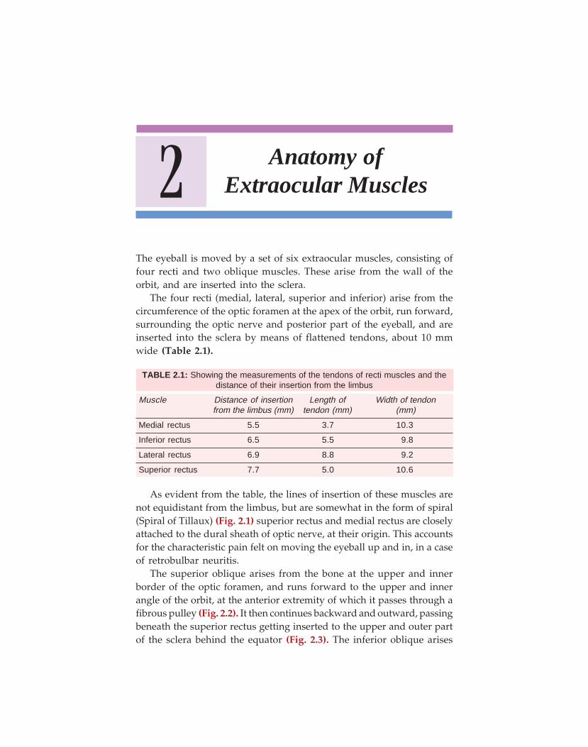
The four recti (medial, lateral, superior and inferior) arise from thecircumference of the optic foramen at the apex of the orbit, run forward,surrounding the optic nerve and posterior part of the eyeball, and areinserted into the sclera by means of flattened tendons, about 10 mmwide (Table 2.1).
TABLE 2.1: Showing the measurements of the tendons of recti muscles and thedistance of their insertion from the limbus
Muscle Distance of insertion Length of Width of tendonfrom the limbus (mm) tendon (mm) (mm)
Medial rectus 5.5 3.7 10.3
Inferior rectus 6.5 5.5 9.8
Lateral rectus 6.9 8.8 9.2
Superior rectus 7.7 5.0 10.6
As evident from the table, the lines of insertion of these muscles arenot equidistant from the limbus, but are somewhat in the form of spiral(Spiral of Tillaux) (Fig. 2.1) superior rectus and medial rectus are closelyattached to the dural sheath of optic nerve, at their origin. This accountsfor the characteristic pain felt on moving the eyeball up and in, in a caseof retrobulbar neuritis.
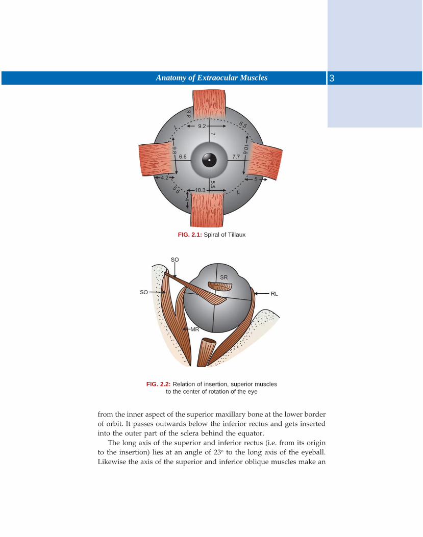
The superior oblique arises from the bone at the upper and innerborder of the optic foramen, and runs forward to the upper and innerangle of the orbit, at the anterior extremity of which it passes through afibrous pulley (Fig. 2.2). It then continues backward and outward, passingbeneath the superior rectus getting inserted to the upper and outer partof the sclera behind the equator (Fig. 2.3). The inferior oblique arises
2 Anatomy ofExtraocular Muscles
3Anatomy of Extraocular Muscles
FIG. 2.1: Spiral of Tillaux
FIG. 2.2: Relation of insertion, superior musclesto the center of rotation of the eye
from the inner aspect of the superior maxillary bone at the lower borderof orbit. It passes outwards below the inferior rectus and gets insertedinto the outer part of the sclera behind the equator.
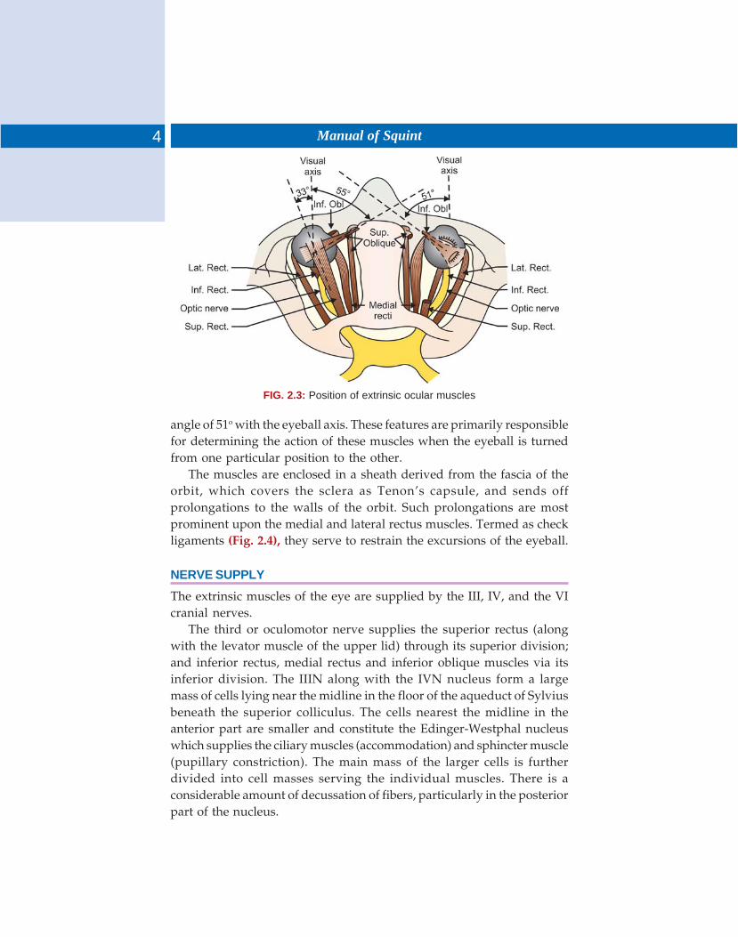
The long axis of the superior and inferior rectus (i.e. from its originto the insertion) lies at an angle of 23o to the long axis of the eyeball.Likewise the axis of the superior and inferior oblique muscles make an
4 Manual of Squint
FIG. 2.3: Position of extrinsic ocular muscles
angle of 51o with the eyeball axis. These features are primarily responsiblefor determining the action of these muscles when the eyeball is turnedfrom one particular position to the other.
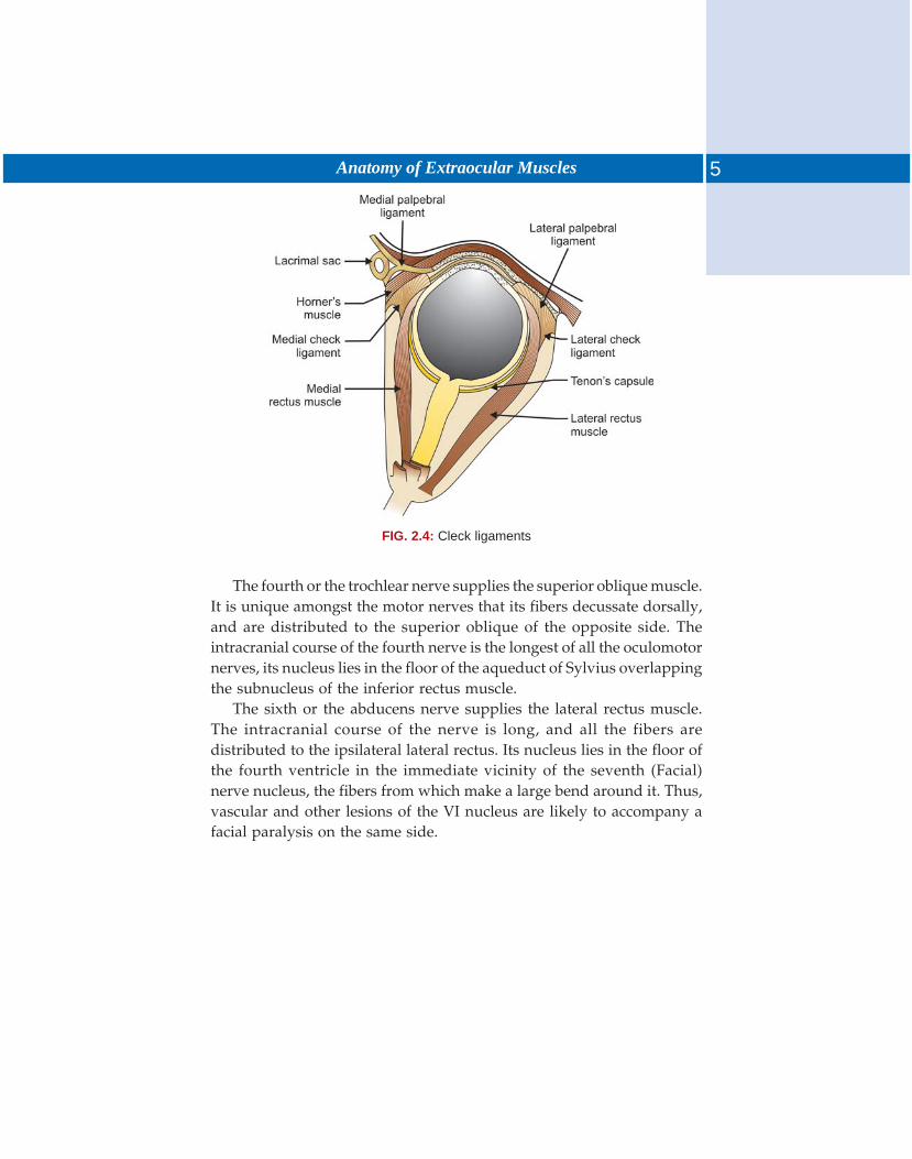
The muscles are enclosed in a sheath derived from the fascia of theorbit, which covers the sclera as Tenon’s capsule, and sends offprolongations to the walls of the orbit. Such prolongations are mostprominent upon the medial and lateral rectus muscles. Termed as checkligaments (Fig. 2.4), they serve to restrain the excursions of the eyeball.
NERVE SUPPLY
The extrinsic muscles of the eye are supplied by the III, IV, and the VIcranial nerves.
The third or oculomotor nerve supplies the superior rectus (alongwith the levator muscle of the upper lid) through its superior division;and inferior rectus, medial rectus and inferior oblique muscles via itsinferior division. The IIIN along with the IVN nucleus form a largemass of cells lying near the midline in the floor of the aqueduct of Sylviusbeneath the superior colliculus. The cells nearest the midline in theanterior part are smaller and constitute the Edinger-Westphal nucleuswhich supplies the ciliary muscles (accommodation) and sphincter muscle(pupillary constriction). The main mass of the larger cells is furtherdivided into cell masses serving the individual muscles. There is aconsiderable amount of decussation of fibers, particularly in the posteriorpart of the nucleus.
5Anatomy of Extraocular Muscles
The fourth or the trochlear nerve supplies the superior oblique muscle.It is unique amongst the motor nerves that its fibers decussate dorsally,and are distributed to the superior oblique of the opposite side. Theintracranial course of the fourth nerve is the longest of all the oculomotornerves, its nucleus lies in the floor of the aqueduct of Sylvius overlappingthe subnucleus of the inferior rectus muscle.
The sixth or the abducens nerve supplies the lateral rectus muscle.The intracranial course of the nerve is long, and all the fibers aredistributed to the ipsilateral lateral rectus. Its nucleus lies in the floor ofthe fourth ventricle in the immediate vicinity of the seventh (Facial)nerve nucleus, the fibers from which make a large bend around it. Thus,vascular and other lesions of the VI nucleus are likely to accompany afacial paralysis on the same side.
FIG. 2.4: Cleck ligaments
The action of III, IV and VI nerve is controlled and coordinated by acomplex intermediary complex and ‘centers’ lying in the region ofmidbrain. The nuclei are interconnected to a considerable extent by fibersparticipating the posterior longitudinal bundle. These fibers playan important role in the coordination of ocular movements andequilibration. One of the most important of such connections is the linkbetween the VI nerve nucleus of one side with the III nerve nucleus ofthe other. In this region there are also ‘centers’ that control the conjugatemovements.
This elaborate mechanism in the midbrain is, in turn, controlled fromthree sources, one voluntary and three reflex.
Voluntary ocular movements. These are initiated in the motor areaof frontal lobe of both sides. The fibers travel along the internal capsule,leaving it in the midbrain first the fibers for vertical movements andmovements of the upper lid and then those for lateral movements. Thesefibers control the conjugate movement of both eyes, but movements ofindividual muscles are not represented. Stimulation of cortex or the tracttherefore produces a conjugate deviation of eyes in the opposite direction,while a destruction would lead to a paralysis of conjugate movementsaway from affected side.
Psychoptic reflexes like fixation, fusional movements and convergence,etc. are centered in the visual cortex of occipital lobe. The afferentpathway is through the visual pathways, and the efferent run down theoptic radiations to the posterior longitudinal bundle and then theoculomotor nerves.
Statokinetic reflex controls the position of eyes when the head isrotated in space. The afferent fibers run from the semicircular canals ofthe inner ears to the midbrain centers.
Static reflexes coordinate movements of eyes in respect of movementof the head on the body. These are initiated by the proprioceptive
3 Neurological Controlof Ocular Movements
7Neurological Control of Ocular Movements
impulses arising from the neck muscles which are linked to the oculomotornerves through the posterior longitudinal bundle.
THE PHYSIOLOGY OF OCULAR MOVEMENTS
Ocular movements in various directions are referred to be the onesinitiating from the primary position.1. Primary position: The eyes are looking straight ahead, the visual axes
are parallel, the vertical meridians of corneas are vertical and parallel,and the head is vertical.
2. Secondary position: These are the positions of the eyes assumed whenthe eyes are moved around the transverse, vertical or anteroposterioraxis.
3. Tertiary position: These positions are assumed when the eyes aremoved along an oblique axis. Two laws govern the movements ofthe eyes into the tertiary position. These are:i. Dander’s law: “For any determinate position of the line of fixation
with respect to the head, there corresponds a definite and invariableangle of torsion, independent of the volition of the observer, andindependent of the manner in which the line of fixation has beenbrought into the position in question”. More simply stated, it isthat for every rotation of the eye to a tertiary position there is adefinite and measurable amount of torsion.
ii. Listing’s law: When the line of fixation passes from its primary toany other position, the angle of torsion of the eye in this secondposition is the same as if the eye had arrived at this position byturning about a fixed axis perpendicular to the initial and finalpositions of the line of fixation. In other words, in rotation to atertiary position the eye will turn about that oblique axis which isperpendicular to the initial and final positions of the line of fixation.
Ocular Movements
The ocular movements may be described as monocular (ductions) orbinocular (versions and vergences). Ductions include the followingmovements:1. Adduction: An inward movement of the eye towards the nose, a medial
rotation along the vertical axis.2. Abduction: An outward movement, a lateral rotation along the vertical
axis.
8 Manual of Squint
3. Supraduction (Sursumduction): An upward movement or elevationalong the horizontal axis.
4. Infraduction: When the eye moves down (depression) along thehorizontal axis.
5. Incycloduction (intorsion): When the eye makes a rotatory movementalong the anteroposterior axis such that the superior pole (12 O’clockpoint) rotates towards the nose.
6. Excycloduction (extorsion): When the eye rotates in a manner that the12 O’clock point rotates away from the nose.
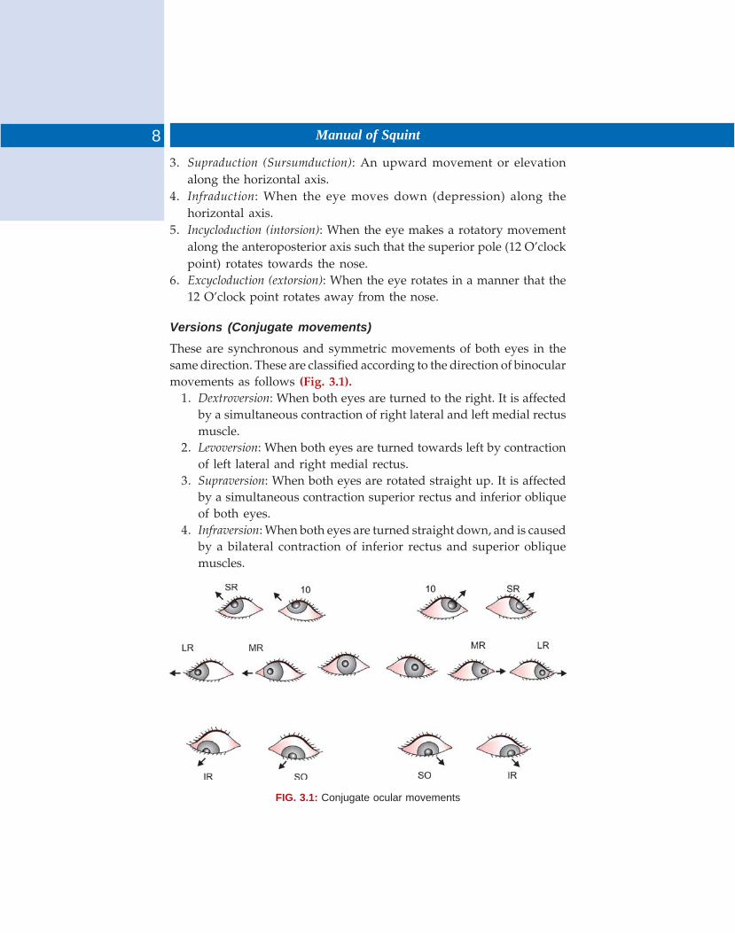
Versions (Conjugate movements)
These are synchronous and symmetric movements of both eyes in thesame direction. These are classified according to the direction of binocularmovements as follows (Fig. 3.1).
1. Dextroversion: When both eyes are turned to the right. It is affectedby a simultaneous contraction of right lateral and left medial rectusmuscle.
2. Levoversion: When both eyes are turned towards left by contractionof left lateral and right medial rectus.
3. Supraversion: When both eyes are rotated straight up. It is affectedby a simultaneous contraction superior rectus and inferior obliqueof both eyes.
4. Infraversion: When both eyes are turned straight down, and is causedby a bilateral contraction of inferior rectus and superior obliquemuscles.
FIG. 3.1: Conjugate ocular movements
9Neurological Control of Ocular Movements
5. Dextrodepression: When both eyes are turned down and to the right.It is caused by a simultaneous contraction of right inferior rectusand left superior oblique.
6. Dextroelevation: When both eyes are turned up and to the right. It iscaused by a simultaneous contraction of right superior rectus andleft inferior oblique.
7. Levoelevation: When both eyes are turned up and to the left, aposition achieved by a simultaneous contraction of left superiorrectus and right inferior oblique.
8. Levodepression: When both eyes are turned down and to the left.This position is brought about by a simultaneous contraction of leftinferior rectus and right superior oblique.
9. Dextrocyclovesion: When the eyes rotate along the anteroposterioraxis so that the superior pole (12 O’clock point) rotates to the rightside. This movement is brought about a simultaneous contractionof inferior rectus and inferior oblique muscle of the right eye, andsuperior rectus and the superior oblique of left eye.
10. Levocycloversion: A movement just opposite of dextrocycloversion.
Vergences
Vergences are disjugate, synchronous and symmetric movements of botheyes in the opposite direction. Depending upon the direction ofmovement vergences may be described as follows:1. Convergence: It is a synchronous inward movement of both eyes
brought about by a simultaneous contraction of both medial recti.2. Divergence: It is a simultaneous and synchronous outward movement
of both eyes brought about by a simultaneous contraction of bothlateral recti. All ocular movements take place around a hypotheticalpoint-center of rotation which lies 13.5 mm behind the apex of cornea.Though located slightly posterior, for practical purposes, it may beconsidered to coincide with the geometrical center of the eyeball. Allrotations of the eyeball take place along three axes—Tick’s axes whichare perpendicular to each other and intersect at the center of rotation.These axes are:X Horizontal axis: It lies horizontally when the head is in upright
position. Rotation along this axis results in elevation or depression.Y Anteroposterior axis: It lies anteroposteriorly and at right angle to
the horizontal axis. The axes in the two eyes are parallel. Rotationalong this axis results in torsional movements (extorsion andintorsion).
10 Manual of Squint
Z Vertical axis: It lies vertically when the head is in upright position,and is at right angle to the X and Y axis. Rotation along this axiscauses adduction or abduction.
The ocular movements may be of two types — voluntary andinvoluntary. The latter are either fusional or due to vestibule ocularreflexes.
Voluntary
1. Dextroversion and levoversion: When both eyes are turned to the rightor left respectively.
2. Supraversion and infraversion: When both eyes are turned up or downrespectively.
3. Oblique parallel movements: When both eyes are turned up and right(Dextroelevation), up and left (levoelevation), down and right(Dextrodepression), down and left (levodepression).
4. Convergence: When both eyes are turned in during the process ofconverging on the point of fixation. This is essentially an involuntaryphenomenon, but can also be achieved by a conscious effort.
Involuntary
1. Psychoptic reflexes, such as fixation, fusional movements, convergence,etc.
2. Statokinetic reflexes coordinate the position of the eyes when thehead is rotated in space.
3. Static reflexes coordinate the movements of the eyes in respect of theposition of the head upon the body.
ACTIONS OF EXTRAOCULAR MUSCLES
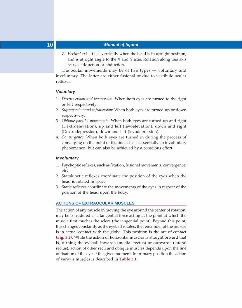
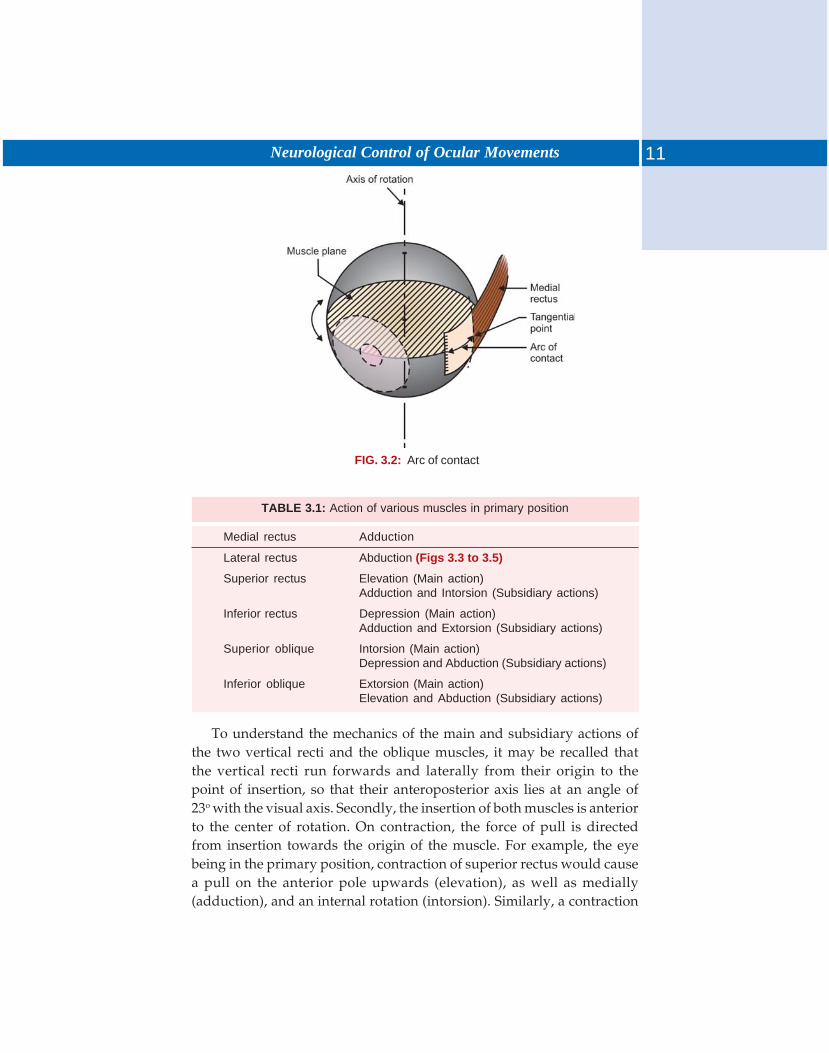
The action of any muscle in moving the eye around the center of rotation,may be considered as a tangential force acting at the point at which themuscle first touches the sclera (the tangential point). Beyond this point,this changes constantly as the eyeball rotates, the remainder of the muscleis in actual contact with the globe. This position is the arc of contact(Fig. 3.2). While the action of horizontal muscles is straightforward thatis, turning the eyeball inwards (medial rectus) or outwards (lateralrectus), action of other recti and oblique muscles depends upon the lineof fixation of the eye at the given moment. In primary position the actionof various muscles is described in Table 3.1.
11Neurological Control of Ocular Movements
FIG. 3.2: Arc of contact
TABLE 3.1: Action of various muscles in primary position
Medial rectus Adduction
Lateral rectus Abduction (Figs 3.3 to 3.5)
Superior rectus Elevation (Main action)Adduction and Intorsion (Subsidiary actions)
Inferior rectus Depression (Main action)Adduction and Extorsion (Subsidiary actions)
Superior oblique Intorsion (Main action)Depression and Abduction (Subsidiary actions)
Inferior oblique Extorsion (Main action)Elevation and Abduction (Subsidiary actions)
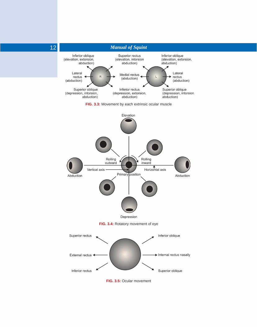
To understand the mechanics of the main and subsidiary actions ofthe two vertical recti and the oblique muscles, it may be recalled thatthe vertical recti run forwards and laterally from their origin to thepoint of insertion, so that their anteroposterior axis lies at an angle of23o with the visual axis. Secondly, the insertion of both muscles is anteriorto the center of rotation. On contraction, the force of pull is directedfrom insertion towards the origin of the muscle. For example, the eyebeing in the primary position, contraction of superior rectus would causea pull on the anterior pole upwards (elevation), as well as medially(adduction), and an internal rotation (intorsion). Similarly, a contraction
12 Manual of Squint
FIG. 3.5: Ocular movement
FIG. 3.3: Movement by each extrinsic ocular muscle
FIG. 3.4: Rotatory movement of eye
13Neurological Control of Ocular Movements
of inferior rectus muscle will affect a depression and adduction. But,being inserted on the inferior aspect of the globe it will cause rotation ofthe inferior pole inwards (thus causing an outward rotation of thesuperior pole-extorsion).
On the other hand if the eyeball is turned 23° outwards, the axes ofthe two recti shall coincide with the visual axis and the muscularcontraction would cause maximal elevation or depression with a minimalamount of any subsidiary movement of adduction and torsion. If theglobe could be turned in, at an angle of 67°, the plane of action of thetwo muscles would be perpendicular to the anteroposterior axis, theaction of the muscles will be entirely torsion).
The actions of oblique muscles can be explained on a similar basis.Contrary to the recti the general direction of the oblique is from frontbackwards, the effective origin of the superior oblique being from thefibrous pulley at the upper and inner angle of the orbit. Secondly, bothmuscles are inserted behind the equator in the outer part of sclera. Thuscontraction of superior oblique will pull the posterior pole up, causing adownward movement of the anterior pole (depression). Similarly theposterior pole will be pulled medially causing a movement of the anteriorpole laterally (abduction). Its insertion being in the outer part of sclera,the pull of the muscle will tend to pull the globe inwards along theanteroposterior axis (intorsion). Likewise, contraction of inferior obliquewill pull the posterior pole down (towards its origin) and hence theanterior pole up (elevation). The contraction will also pull the posteriorpole medially and hence the anterior pole laterally (abduction). A rotationof the outer sclera (site of insertion) along the anteroposterior axis, shallbe towards the floor of the orbit (extorsion).
The action of muscles described above are in the situation when theeyeball is in primary position. However if the globe is turned inwardsmaking an angle of 51° with the visual axis, the plane of the obliqueswill coincide with the anteroposterior axis and the muscle will act purelyas elevator or depressor with negligible subsidiary actions.
Thus, as far as elevation and depression are concerned, the obliquesact when the eyeball is adducted while superior and inferior recti actwhen the ball is abducted. In the primary position, the recti are responsiblefor 63.3% of vertical motion while the obliques are responsible for 36.7%.An understanding of these actions is important in functional testing ofvertical plane muscles.
14 Manual of Squint
In accordance with the action of an individual muscle uniocularly orin relation to the action of other muscles in the same eye or thecontralateral eye the muscles can be classified as follows:1. Agonist: It refers to a particular muscle causing a specific ocular
movement. For example, in rotation of the eyeball to the left, lateralrectus of the left eye is agonist.
2. Synergists: The set of muscles which move the same eye in oneparticular direction are called synergists. For example, superior rectusand inferior oblique of the same eye are synergists in the movementof elevation of that eye.
3. Antagonists: These are the muscles having opposite action in the sameeye, such as medial and lateral rectus.
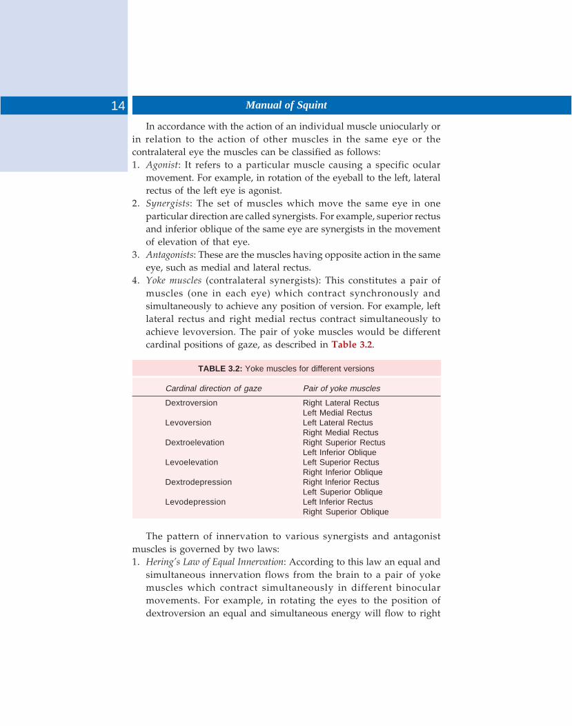
4. Yoke muscles (contralateral synergists): This constitutes a pair ofmuscles (one in each eye) which contract synchronously andsimultaneously to achieve any position of version. For example, leftlateral rectus and right medial rectus contract simultaneously toachieve levoversion. The pair of yoke muscles would be differentcardinal positions of gaze, as described in Table 3.2.
TABLE 3.2: Yoke muscles for different versions
Cardinal direction of gaze Pair of yoke muscles
Dextroversion Right Lateral RectusLeft Medial Rectus
Levoversion Left Lateral RectusRight Medial Rectus
Dextroelevation Right Superior RectusLeft Inferior Oblique
Levoelevation Left Superior RectusRight Inferior Oblique
Dextrodepression Right Inferior RectusLeft Superior Oblique
Levodepression Left Inferior RectusRight Superior Oblique
The pattern of innervation to various synergists and antagonistmuscles is governed by two laws:1. Hering’s Law of Equal Innervation: According to this law an equal and
simultaneous innervation flows from the brain to a pair of yokemuscles which contract simultaneously in different binocularmovements. For example, in rotating the eyes to the position ofdextroversion an equal and simultaneous energy will flow to right
15Neurological Control of Ocular Movements
lateral rectus and left medial rectus. Similarly, if the eyes are turnedthe position of dextroelevation an equal and simultaneous amount ofenergy (innervation) will flow to right superior rectus and left inferioroblique.
2. Sherrington’s Law of Reciprocal Innervation: This law states that duringan ocular movement an increased amount of innervation flow to theagonist muscle is accompanied by a decreased amount of innervationto the relaxing antagonist muscle. Thus, on moving the eyes to theright (dextroversion) an increased amount of innervation to the rightlateral rectus and left medial rectus will be accompanied by adecreased amount of innervation to the right medial rectus and leftlateral rectus.The resultant clinical picture following an extraocular muscle palsy is
influenced by this set of laws and will be discussed subsequently underthe head—Paralytic Squint.
The two eyes being located some distance away from each other, imageof any object formed in each eye cannot be identical, as each eye regardsa slightly different aspect of the object observed. But the two slightlydissimilar images are mentally fused into a single image. In addition,such a fusion provides the perception of a third dimension to the image-stereopsis one of the greatest advantages of binocular vision. There aremany factors involved in the successful development of binocular vision,which consist of complex and closely related sensory, motor and centralmechanisms.
MECHANISMS
Sensory Mechanisms
Retinal Sensitivity
The two eyes should have a reasonably good and equal visual acuity.The refractive status of the two eyes may not be very different so thatthe images formed do not differ greatly.
Retinal Correspondence
Normally, any point of retinal receptors in one eye corresponds to anotherpoint in the other eye. For example, a point located 10° on the nasal sideof one retina corresponds to another point located 10° placed temporarilyin the other. Foveas in the two eyes provide the best example ofcorresponding points. Such points do not refer to individual retinalreceptors but a group of receptors in a small area—Pannum area. Eacheye contains many such areas and the sum of points in space the imageswill fall upon corresponding retinal areas is called horopter. In otherwords horopter can be considered as a sum total of points in the physicalspace that stimulate corresponding elements of two eyes. Conversely,an object which does not lie on the horopter forms image on
4 Binocular Vision
17Binocular Vision
noncorresponding points of the retina of two eyes, and if attention isdirected to this object it would look double-Diplopia, which may behomonymous or crossed.
Visual Pathways
The development of binocular vision is dependent on a hemidecussationof the afferent optic nerve fibers at the optic chiasma because this enablesthe nerve fibers from corresponding retinal areas of the two eyes tobecome associated with one and other in the visual cortex. The retinamay be divided, from the functional point of view, to be divided verticallythrough the midpoint of fovea. All retinal fibers from the temporal halfof the retina including the temporal half of fovea pass through the chiasmawithout decussation, traveling along the ipsilateral optic tract. On theother hand, all retinal fibers from the nasal of the retina including thenasal half of fovea decussate at the chiasma and travel along thecontralateral optic tract. It follows therefore, that fibers from thecorresponding retinal areas (temporal retina of one eye and nasal retinaof the other eye) travel in the same optic tract, terminate in the samelateral geniculate body, getting relayed to the same side of opticradiations to reach the striate area of the same visual cortex.
Motor Mechanisms
These are responsible for maintaining the eyes in the correct position atall times, i.e. inrest and during all movements, and may be consideredin three groups:
Anatomical Factors
These are concerned with the structure of the bony orbits and theircontents as well as the structure of the two eyeballs so that the eyes maylie within orbits in a manner that the visual axes be parallel to each otherin all states of rest and various movements.
Physiological (or dynamic) Factors
These are the postural reflexes (static, statokinetic) which determine theposition of eyes and are independent of visual stimuli. In addition, certainpsychoptic reflexes make a significant contribution to the achievementof binocular vision, such as:
i. Fixation reflex: This relates to the ability of each eye to independentlyfix at the same object. It is dependent mainly on adequatelyfunctioning fovea and to some extent, on an adequate field of vision.
18 Manual of Squint
ii. Refixation reflex: This is an elaboration of fixation reflex, and consistsof the ability of the two eyes to change fixation from one object tothe other object (active refixation), or the ability of eyes to retainfixation of a moving object (passive refixation).
iii. Disjunctive or vergence fixation reflex: This the application of fixationreflex in which the eyes retain fixation during the course of adisjunctive movement such as convergence or divergence.
Central Mechanisms
These concern the development of fusion, which, though partly a sensoryphenomenon, also partly concerns the cortical control of ocular move-ments which is a motor function. Perception of a single mental impressionof two slightly different images as seen by the two eyes, is an essentialpart of the functions of visual cortex. The motor component of the pheno-menon concerns the centers in the frontal and occipital parts of the centralhemispheres which control the intermediary centers and the cranial nucleiconcerned in the final impulses controlling the movements of extraocularmuscles.
GRADES OF BINOCULAR VISION
The phenomenon of binocular vision has three different components:
Simultaneous Perception
This is the first grade of binocular vision. It refers to the simultaneousperception of the impulses, received from the two eyes, by the cerebralcortex. It is the faculty to see two dissimilar objects simultaneously. Itdoes not necessarily mean that the image of two different objectsconcerned can be superimposed. This grade of binocular vision can bedemonstrated on a major amblyoscope by using slides of two differentpictures like a lion and a cage presented to the eyes individually.Simultaneous binocular perception can be:
i. Simultaneous paramacular perceptionii. Simultaneous macular perception
iii. Simultaneous foveal perception.Under certain conditions human being have the faculty to suppress
the image of one eye, though both eyes are open, such as looking througha monocular microscope, or shooting with a gun.
19Binocular Vision
Fusion
This second grade of binocular vision. This is the faculty of producing acomposite picture of two similar objects, each of which is incomplete ina different manner. When picture of two rabbits (one with a bunch offlowers in hand but without the tail, and the other with the tail butwithout flowers) is seen on a major amblyoscope, a single picture of therabbit is seen in a complete form with a tail as well as a bunch of flowersin hand.
Fusion can be of two types:i. Central
ii. Peripheral fusion.
Stereopsis
It is the highest form of binocular cooperation that adds a new qualityof vision. It refers to the ability to obtain an impression of depth by thesuperimposition of two pictures of the same object taken from a slightlydifferent angle. It is not just the depth perception which concerns theperceptions of distance between the objects, which can be judged evenon a monocular vision. But stereopsis refers to the visual appreciation ofthree dimensions during binocular vision. Various tests to judge thequality of this faculty are described in subsequent chapters.
Visual acuity is defined as the power to differentiate object from eachother and to appreciate their details. It is highly complex functionconsisting of:
i. The ability to detect an object in the field of vision.ii. The ability to name a symbol or specify the position of a critical
element in it.Optically, the visual acuity is expressed as the minimum visual angle
substended at the anterior focal plane when accommodation is entirelyrelaxed. Binocular visual acuity is always better than the monocular acuity.
Basically, the visual process can be considered as the reception ofinformation by the retina, and the transmission of that coded informationalong the optic nerves and radiations to the cerebral cortex. The eyesees nothing as it is simply the input mechanisms of computer. Perceptionis the read-out mechanisms of that computer. It is of course the cortexalone which sees. Vision is a continuous process of receiving, sampling,analysing and coding information until the final decoding and read-outmechanism occurs. The pupillary reflex is present at birth demonstratingthat neonate is sensitive to differences in intensity of the visual stimulicortical cells in immature kitten leave a normal receptive field arrangementbefore their eyes are opened, demonstrating that patterned light stimuliare not necessary for the development of the functional architecture ofthe cerebral cortex.
Infants as young as 15 days can discriminate colors. By 1 month ofage an infant sees complex forms and can see the difference between agray patch and square composed of 3mn stripes. By the age of 6 monthsa baby’s coordination has reached a stage where he will repeat responseswhich produce interesting results, such as swinging a toy, and clearly todo this, his vision must have developed accordingly. So that fixationand following movement occur as well as the recognition of familiarand interesting objects. By a month baby will knock down pillow to
5 Visual Acuity
21Visual Acuity
find a toy and he is able visually to differentiate objects easily. As ageincreases, through trial and error experiment (11-18 months) and laterthinking about the effects of various responses (18-24 months) the childbuilds up his memory store so that at 12-18 months he will look for anobject hidden under a second pillow and at 18-24 months he will lookfor the object even when it has been removed.
Thus, with increasing age the percepts breaks up and instead of seeingthings as a whole, he is able to differentiates the stimuli in hissurroundings, the percept can begin to be seen as its components parts.Discrimination of symbol and letters develops gradually so that by theage of 1 years a child can distinguish simple symbols and by 5 or 6 yearshe can distinguish letters.
At birth foveal sensitivity and the cortical control behind it is notwell-developed. It is by continued use and by the reception of repeatedand useful information, that the cortex is able to program itself andbuild up a satisfactory memory alone, so that it is able to compare datasamples presented to it and increase its ability. At first a lot of data, isrequired to produce a simple response.
The infant will respond simply to complex colored, patterns andshapes. As age increase, and with, repeated stimulation the cortical cellsincrease the selectivity of their response in infancy. Visual sensitivity isrecognized by means of pupillary reflexes demonstrating integrity ofthe nervous pathway to the lateral geniculate body, and later, by theresponse to complex forms, demonstrating integrity of the prewiredmechanism of the cortex.
This is followed by recognition of complex forms, demonstratingintegrity of an elementary memory store for perception. Presentation ofsymbols containing the same amount of information within decreasingareas gives us our test of visual acuity. By the time a child is 3 years hecan distinguish and demonstrate his acuity by recognition of suchsymbols.
It should be realized that 6/9 using a simple symbol does notnecessarily mean that the vision will be 6/9 after further developmentof the visual mechanism using more complex tests. If the vision is 6/9 at3 years of age using symbols than one expects it to be 6/9 with Snellen’stest at 6 year. But this assumes a normal development of the cortex andretina. The excellence of Snellen’s test of vision is due to these factors,large amount of information is packed into a confined area and the areacontaining this information can be varied easily. It is not until a childcan read line of 6/6 Snellen letter at a resonable speed that we can be
22 Manual of Squint
certain that the visual mechanism is normal. 6/6 visions determines theability of the sensitivity curve of the fovea. The speed at which the linecan be read determines the effectiveness of the position control systemof that sensitivity curve and the ability of whole decoding mechanismof the visual cortex.
ANGULAR AND CORTICAL VISUAL ACUITY
The response to a single optotype has been termed angular vision, whilethe response to a row of letter is known as cortical vision, the reading ofa row of letter involves interpretation by the cortex, whereas angularvision, or recognizing simple optotypes, depends simply on the angularmagnification of the letter. It is obvious that all visual processes mustinvolve cortical activity, the eye is only the axons by which visualsensations are transmitted to the cortex. We do not see through one eyeor through both eyes, but through the brain as through Cyclopean eye.
RECORDING OF VISUAL ACUITY
Snellen visual acuity represents the patient’s resolving capability on lettertargets. Vernier visual acuity is a test of resolving minimal separationsof a grid pattern. The essence of both these methods of testing visualacuity is that an object subtends different angles on the retina whenpresented at different distances from the eye.
The angle subtended by the object at the nodal point of the eye iscalled the visual angle. Visual resolution is measured by the angle atwhich the components of an object can be appreciated. They are commonlymeasured in minutes of arc and decimal fractions of minutes. The Snellennotation 6/6 means that the subject can read letters composed of blacklines on a white background 6 meter away when the width of each linesubtends 1 minutes of arc on the retina.
The notation 6/12 correspondents to 2 minutes of arc the Snellennotation, therefore, can be expressed by the formula.
Visual angle (minutes) = 1 / Snellen notation
Occasionally, the Snellen notation is expressed as a decimal fraction,thus 6/6 is 1.0, 6/12 is 0.5 and so on. The smallest detectable visualangle has been found to be 0.5 second of arc against a uniformlyilluminated background such a line producer a geometric retinal imageapproximately 0.033 mm, which is the diameter of single foveal cone.“Snellen’s chart” should be accepted as international chart to determine
23Visual Acuity
the subjective visual acuity. Each component of top letter of Snellen’schart subtends an angle of vision at 60 meters. Whole the letter in theline indicating normal visual acuity (6/6) subtend the same angle at adistance of 6 meters. Six meters is accepted from practical point of viewbecause most rays from a distance of 6 meter and more are as good asparallel rays. Depending on the number of lines the patient can read,distance vision is recorded as 6/60 to 6/6 with Snellen’s chart illuminatedeither externally or internally with uniform illumination. The intensityof the light over the chart should be between 20 to 30 foot candles in adiffuse manner and at the same time there should be no brilliant light inthe visual field of the patient. The chart is placed over a white wall, or ifit is necessary, it can be mounted on top of white paper. The chart isplaced in such a manner that the eyes of the patients one level with the20/20 line. The patients can be standing or sitting. The chart can beelevated or lowered according to the different heights of the patients.A line is made at 20 feet from the chart, and if the person to be tested isstanding his head should be at the level of the line. Some chart evenhave letter for recording visual acuity up to 6/5 to 6/4. If a personmisses, or incorrectly reads some letters of a line, the record is qualifiedas ‘partial’. Farther more vision should be recorded for each eyeseparately as well as binocularly. It is to be noted that the binocularvision (both eyes open) is always one line more than the uniocular visionprovided both eyes have equal visual acuity.
The macular part of the retina is most sensitive part and most visualacuity is derived from this area. Retinal sensitivity gradually diminishesfrom the center to the periphery, so much so that the peripheral retinahas only 10% of the central sensation. It is an every day experience thata person with a gross localized foveal lesions with whole of the remainingretina normal will not have visual acuity more than 6/60 or 6/36 partial.On the other hand with gross pathological lesions in peripheral retinabut an unaffected macular area, patient may have 6/6 vision, althoughthis will be tubular in character because of the loss of peripheral field. Inthe grades of vision take 6/60, 6/36…………..6/9, 6/6 the constantnumber 6 in the numerator indicates the distance from which patient isreading and the denominator indicates the distance in meters from whichthe patient should be able to read that line. Countries not following themetric system denote it is feet as 20/200 to 20/20.
If the vision with both eyes open is 3/60 or less (with correction ifnecessary), it is total blindness because a person with that poor visualacuity cannot independently move about except in very familiar
24 Manual of Squint
surroundings. If vision, again with both eyes open, and with correction,if necessary, is more that 3/60 but 6/60 or less, it is considered economicblindness, because such a person by virtue of his visual cannot earn hisliving independently, vision better that 6/60 but 6/18 or less again withboth eyes open and with correction is considered a visual handicapbecause such a person is visually handicapped and may be unfit forservice or jobs needing good visual acuity.
Three types of charts are being used for illiterate pupil. The Landolt’s‘C’ charts are accepted as standard for testing visual acuity for variousprogressive in preference to others. The ‘E’ charts are also identical andcan be used under the same guidelines as ‘C’ charts. The dot chartsshowing different number of dots of different sizes are also covenant.Multicolored balls can be used from different distances for the toddlers.
It is rarely possible to obtain any significant subjective responses forvisual acuity determination of children under the age of 3 years andhence recourse has to be made entirely to the objective methodsassessment. Quantitative upto kinetic test can be carried out with mostsmall children. Visual four test pattern equal width of 1/8,1/16. 1/32and 1/64 inches mounted on the C, K, N drum. At the test distance of 12inches they represent 36, 18,9 and 4.5 minutes of visual angle. The levelof illumination was set at 100 foot candles. Minimum separable acuitythreshold were established by observing prompt and properly directedrhythmic optokinetic responses in both direction of the rotation of thecylinder in eight out of ten trials with each test pattern.
Forced choice preferential looking test by employing patterns andacuity grating is useful in infants and young children. This test allowsthe child to look at screens while observing the behavior of the eye andhead.
Normal adult acuity can be attained by 4-5 months. This can be elicitedby visual evoked responses (VEH) to square move gratings of variousspatial frequencies.
VISION IN VARIOUS REFRACTIVE ERRORS
Hypermetropia
The uncorrected visual acuity in hypermetropes varies with the degreeof optical error and the portion which cannot be overcome byaccommodation.
The corrected visual acuity frequently does not come upto standard,particularly in higher degrees of the defect, usually when the refractive
25Visual Acuity
error was not corrected in early childhood (Ametropic amblyopia) butthe acuity improves to same extent after wearing correcting spectaclesfor some months. Hypermetropes who do not wear correcting spectaclesor wear them intemittently. See better without them. A variable incidenceof amblyopia has been reported. The commonest cause of such acondition is hypermetropic refractive error and amblyopia could beprevented by early use of glasses.
Myopia
Visual acuity beyond the far point is seriously affected in incorrectedmyopia, being reduced by about the same ratio as in hypermetropia. Thecorrected visual acuity in the absence of degenerative changes is usuallygood and even better with contact lenses. Individual who use spectacleshabitually see less well without them than those who do wear themintermittently or not at all incidence of amblyopia in myopia is much lessalmost unknown for the reason that myopia at least sees the near objectsmore clearly than in hypermetropia where all accommodation reserve isup for distance and he neither sees distance nor see near objects clearly.Therefore, near vision stimulus is not derived in myopia.
Astigmatism
The vision in astigmatism is characteristic. In higher degree of astigmatismeye cannot form a sharply defined image on the retina in anycircumstance, therefore, vision may be diminished very considerably.The dimension of visual acuity is about equal for corresponding degreeof simple hypermetrope and myopia astigmatism can usually be broughtupto normal standard. But in higher degrees this is by no means alwaysthe case particularly if the optical correction is not made early life andalso if the astigmatism is oblique. This deficiency is essentially perceptualand there may be a tendency for poor differentiation in the meridian ofgreatest astigmatism. Astigmatic amblyopia or meridional amblyopia ispresent then. Amblyopia ex-anopsia affecting all meridia is more commonin higher degrees of astigmatism and there is a tendency to developstrabismus particularly in the presence of hypermetropic errors.
Anisometropia
Binocular vision is the rule in smaller degree of the defect with highergrades of error, fusion is usually impossible and alternating and unocularvision may occur. Alternating vision may result in which case each of
26 Manual of Squint
the two eyes is used one at a time and is specially so if both eyes havegood visual acuity and when one is hemitropic or moderatelyhypermetropic and other is myopic. The patient uses the former fordistant vision and latter for near vision. He may therefore remain verycomfortable and at times be unaware of the defect. If the defect in oneeye is high and especially if the visual acuity is not good it may bealtogether excluded from vision and the better eye is relied upon inunocular vision.
OBSTACLES TO VISION AT VARIOUSAGES FROM BIRTH TO INFANCY
The fixation reflex is innate being present at birth but is only feeblydeveloped, responding momentarily to strong stimulus such as brightlight, in general. The movements of the eyes are independent irregularand unconjugated. Obstacles to vision at birth lead to failure indevelopment of fixation and congenital nystamus results. By the age of5 or 6 weeks the conjugate fixation reflex becomes established but it isnot until almost 6 months that conjugate deviations become completelyaccurate. Owing to the inter position of some obstacles in the reflexpath, fusion may be embarrassed and maintained with difficulty, resultingin heterophoria later: squint or not attained at all resulting intoconcomitant squint. Again some structural obstacles (neuromuscular)may prohibit the development of adequate conjugate movements frombirth, so that a congenital nondominant squint develops. Desjugatefixation reflexes are developed after 6 months. Failure of the desjugatefixation reflexes are firmly established towards the end of the first yearand if obstacles become insuperable diplopia results.
If there would be obstacle to any of the reflexes developing at variousages, various types of neuromuscular anomalies would develop. Apartfrom, this, the visual acuity may be permanently impaired if there is anyobstacle whether refractive error, strabismus, congenital cataract andptosis. The amount and density of amblyopia would thus depend on thevisual acuity that has developed by that age.
The binocular reflexes may be greatly modified by the presence ofobstacle in the reflex path. Although these obstacles are more hunderingwhen reflexes are immature, they can even interference with the fullydeveloped reflexes. The presence of these abnormal obstacles results inthe development of perverted reflexes, any of structural anomalies, whichreplace the normal. The younger the patient, the more likely is a slightobstacle to produce a permanent effect.
There obstacle may be divided into sensory, motor and central. Thepenalty suffered by an adult through such a simple sensory obstacle asincorrect glasses may not exceed headache and various irritability, but achild in such a circumstances may have pay with his sight. A carefulconsideration of motor obstacles isolate large group of paralytic squintfrom what has ordinary concomitant squint. The chief factor inincomplicated accommodational squint is a congenital and hereditarydeformity, excessive hypermetropia, and the factor next in importanceis weakness of the neuromuscular mechanism of accommodation. Theresulting insufficiency of accommodation axial on one hand and dynamicon other hand, instead of being overcome by the occipital accommodationreflex alone, elicits an attempt at correction by a frontal effort whichensues as accommodation and convergence in abnormal association,excessive convergence resulting in a, attempt to over-accommodation.
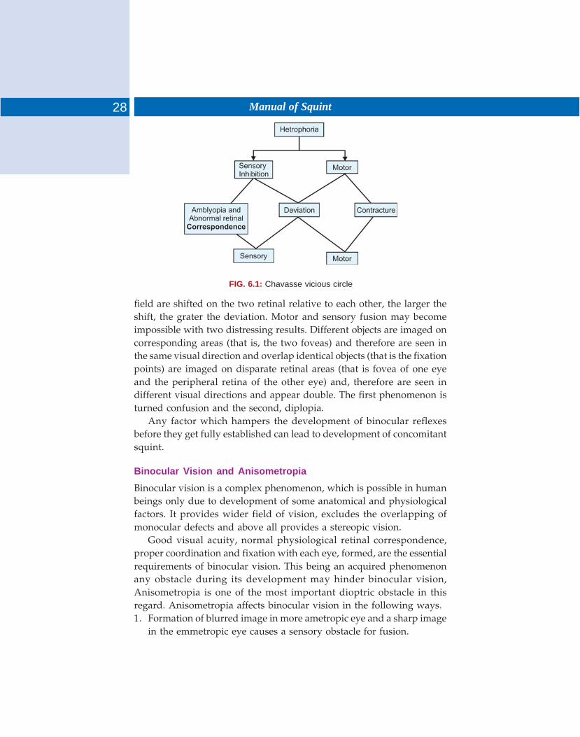
According to chavasse, in any case of dissociation whether this isdue to a sensory or a motor obstacle, two vicious circles, linked togetheras a figure of eight, are in action, whether the type of deviation isconcomitant, paralytic or mixes. Whatever the cause of dissociationchanges rapidly develop as shown in Figure 6.1.
MECHANISM
According to von Noorden, whenever there is a manifest deviation ofthe visual axes of the two eyes, the images of all objects in the binocular
6 Abnormalities ofBinocular Vision
28 Manual of Squint
FIG. 6.1: Chavasse vicious circle
field are shifted on the two retinal relative to each other, the larger theshift, the grater the deviation. Motor and sensory fusion may becomeimpossible with two distressing results. Different objects are imaged oncorresponding areas (that is, the two foveas) and therefore are seen inthe same visual direction and overlap identical objects (that is the fixationpoints) are imaged on disparate retinal areas (that is fovea of one eyeand the peripheral retina of the other eye) and, therefore are seen indifferent visual directions and appear double. The first phenomenon isturned confusion and the second, diplopia.
Any factor which hampers the development of binocular reflexesbefore they get fully established can lead to development of concomitantsquint.
Binocular Vision and Anisometropia
Binocular vision is a complex phenomenon, which is possible in humanbeings only due to development of some anatomical and physiologicalfactors. It provides wider field of vision, excludes the overlapping ofmonocular defects and above all provides a stereopic vision.
Good visual acuity, normal physiological retinal correspondence,proper coordination and fixation with each eye, formed, are the essentialrequirements of binocular vision. This being an acquired phenomenonany obstacle during its development may hinder binocular vision,Anisometropia is one of the most important dioptric obstacle in thisregard. Anisometropia affects binocular vision in the following ways.1. Formation of blurred image in more ametropic eye and a sharp image
in the emmetropic eye causes a sensory obstacle for fusion.
29Abnormalities of Binocular Vision
2. Unequal size of the retinal images (Aniseikonia) causes difficulty infusion.
3. Prismatic effect due to unequal power of the correcting spectaclescauses unequal peripheral fusion.
4. Difficulty in binocular—spatial judgment because of aniseikonia.A blurred image and aniseikonia may lead to the development of
foveal suppression, amblyopia, abnormal retinal correspondence andstrabismus. It has been observed that if a patient of anisometropia ishaving binocular vision and if given treatment for amblyopia he improvesby better visual status and longer maintenance than those cases wholack binocular function. In few cases, if aniseikonia and prismatic effectare overcome by using contact lenses, there patients maintain goodbinocular vision.
There is no rigid relationship between anisometropia and aniseikonia.It has generally accepted that 25 diopter difference of refraction causes0.5% differences in image size.
Vision in Anisometropia
The vision in significant anisometropia may be binocular, alternating orexclusively uniocular.a. Binocular vision: Binocular vision is noticed in smaller degree of
anisometropia.Each 0.25D difference between the refraction of the two eyes
causes 0.5> difference in the size between the two retinal images.Probably the difference of 5D is the limit which can usually betolerated with case. Moreover since the incorrected image of oneeye is always blurred binocular vision is rarely perfect, and attemptsof fusion frequently, although not always, bring on symptoms ofaccommodative asthenopia. The symptomatology of this group thusresembles that of small refractive errors.
b. Alternating vision: This occur in higher degrees of anisometropia, hereeach of the two eyes is used one at a time. This is apt to occur whenthe visual acuity of both the eyes are good and one is emmetropic ormoderately hypermetropic and other myopic. Here the patient fallsinto the easy and legitimate habit of using the eye which is emmetropicor hypermetropic for the distant vision and the other eye which ismyopic for near work, and he may remain very comfortable andindeed quiet unaware of his defect and if the anisometropia is mixed,require no optical correction for any distance at any time of life.
30 Manual of Squint
c. Uniocular vision (Suppression): If the refractive error in one is very highand if its visual acuity is poor, it may be altogether excluded from thevision and the other eye alone being relied upon in uniocular vision.In this event the defective eye may become not a uncommonly deviated.
Relationship between Anisometropia and Amblyopia
Visual acuity in the anisometropic eye is lower under binocular conditionsthen when tested monocularly. This is because of the fact that inanisometropic patients, the purpose of active inhibition of fovea is toeliminate sensory interference caused by super imposition of a focusedand a defocused image originating from the fixation point (abnormalbinocular interaction). Apart from this the foveal form vision-deprivationdue to uncorrected refractive error plays a role in producing amblyopia.After optical correction of anisometropia, the resulting aniseikonia maybe another causal factor of amblyopia.
Intensity of amblyopia rended to very directly with the amount ofanisometropia. Amblyopia is more common and a higher degree inpatients with anisohypermetropia than in those with anisomyopia. Retinaof the more ametropic of a pair of hypermetropia eyes never receivesclearly defined image, since with details clearly focused on the fovea ofthe better eye no stimulus is provided for the further accommodativeeffort required to produce a clear image in the fovea of the morehypermetropic eye when myopia is unequal, the more myopic eye canbe used for near work and the less myopic eye for distance. Therefore,unless the myopia is of high degree both retinal receive adequate stimuleand amblyopia does not develop. Apart from this, myopia is rarelypresent in early childhood, Amblyopia frequently occurs when thedegree of anisometropia is higher than 2.0.
In anisometrop amblyopia the central suppression scotoma is normallysmall so that the optic phenomenon of Haidinger’s brushes may be obtainable, a capacity which indicates that the prognosis after treatment isrelatively good.
Relationship with Squint
In anisometropia the influence which accommodation convergencerelationship may exert on development of squint depends largely onwhether one is used constantly for fixation irrespective of distance ofgaze or whether one eye is used for fixation for near objects and theother eye for fixation for those situated at a distance. When one eye isdominant and has only a moderate degree of hypermetropia the othereye tends to remain straight irrespective of wheather it is more
31Abnormalities of Binocular Vision
hypermetropic or less hypermetropic than the dominant eye or evenwhen it is myopic, a clear illustration of the fact that in early infancy theeyes are associated with one another by the more primitive posturalreflexes without any regard to the presence of a high refractive error inone eye. When one eye is dominant and has a fairely marked degree ofhypermetropia, the other eye may remain straight or may tend to divergewhen either eye is dominant so that one eye is used for distance and theother eye for near, divergence may occur because there is not reward tobe obtained from the exercise of accommodation convergence reflex.Anisometropia also constitutes a central obstacle of a sensory type. Thereis also evidence that errors of refraction even fully corrected by spectaclelenses, may favor the development of squint in certain cases when thereis moderate degree of difference between the refraction of the two eyes(anisometropia) leading to a sufficient size difference of the retinal images(aniseikonia) which prevent the normal fulfilment of fusion mechanismdespite the clarity of each separate image in the visual cortex. In suchcases a positive attempt to prevent fusion (a state termed horror fusion)may lead to the development of purposive strabismus.
It seems, likely therefore that a primary failure in the developmentof the fusion faculty plays significant part in the production of certainsquint although it must be realized that in most of the cases the defect offusion faculty is largely secondary to some motor or sensory obstacle sothat the duration of the visual axes in the direct cause of the lack ofreinforcement of the fusion reflex.
In unilateral myopia of moderate degree the myopia eye can diverge.In anisometropia of moderate degree in which one eye is myopic andother hypermetropic or relatively so, the myopic eye is usually used fornear fixation and the hypermetropic eye for distance fixation in whichcase an alternating divergent strabismus develop.
Anisometropia and Eccentric Fixation
There are several hypothesis regarding the cause of eccentric fixation.According to “Scotoma hypothesis”, central inhibitional scotoma or lossof macular function is the cause of eccentric fixation which developssimilar to anomalous correspondence on the basis of constant deviationof the visual axis. Eccentric fixation and anomalous retinalcorrespondence (ARC) are only different stages of same pathophysiologicevent occurring as an adoptation to faulty “binocular position”. Accordingto “motor-hypothesis”, fixation is significantly influenced by motorfactors.
Whenever a person exerts a certain amount of accommodation adetermined amount of convergence is called into play, calledaccommodative convergence. The convergence response of an individualto a unit stimulus of accommodation may be expressed in a numbertermed his accommodative convergence accommodation ratio. It isreasonable to assume that the basic convergence requirement is fulfilledthrough accommodative convergence. Tonic and fusional convergencehave their own functions and proximal convergence is a supplementaryone. Therefore a normal emmetropic person should be expected to exectIMA of convergence for each diopter of accommodation, but this is notthe case. Each individual responds to a unit stimulus of accommodationwith a specific amount of convergence that may be greater or smaller thanis called for by the convergence requirement. The convergence responseof an individual to a unit stimulus of accommodation may be expressedin a number termed accommodative convergence/accommodation ratio(AC/A ratio). This ratio which has the dimensions (D/D) is a measure ofthe responsiveness of person’s convergence function to a unit of stimulationof accommodation. Quantitative studies on persons with normalsensorimotor system have shown that in the vast majority of people, theAC/A ratio does not fulfil the convergence requirement. The normalrange of the AC/A ratio is between three and five. Values above five areconsidered to denote excessive accommodative convergence and valuesunder three as in sufficiency.
The association between accommodation and convergence developsearly in life as a result of constantly repeated simultaneous use of relateddegrees of the two functions, that is a learned association has beenaccepted and elaborated on by many workers. An acquired associationimplies a certain degree of independence in the relationship of twofunctions. This elastic relationship is expressed as “relative accommo-dation” and “relative convergence”. Any change in the stimulus to
7AccommodativeConvergence/
Accommodation Ratio
33Accommodative Convergence/Accommodation Ratio
accommodation that can be shown to lead to a change in convergence orthat accommodation can be changed by forced convergence would favoran innate and stable relationship between the two types of convergence.Furthermore if the association is learned, one would not expect it toexist in patients who have had strabismus throughout most or all theirlives. There is an increase in AC/A ratio in early presbyopia which isattributed to an increase in impulse to accommodation, somewhat similarto that required with cycloplegia. It is observed that AC/A is a factor inthe inheritance of esotopia.
METHODS FOR DETERMINATION OF RATIO
Various methods are devised for measuring AC/A ratioa. Heterophoric methodb. Gradient methodc. Fixation-desparity methodd. Haloscopic methode. Graphic method.
Changes in AC/A ratio with glasses, drugs operation and exercise,both accommodation and convergence have a central and peripheralmechanism. There is a gradual decrease of esotropia. At near fixationwithout changes of the angle at distance in children wearing bifocal. Itwears that spectacle lenses have changed AC/A ratio. It is demonstratedthat AC/A ratio is reduced by using parasympathomimetic drug suchas echothiophate iodide. This drug is cholinesterase inhibitor and itenhances the effect of acetylcholine on the ciliary muscle. There is areduction in AC/A ratio by gradient method when the eyes were underthe influence of di-iso-propyl fluorophosphates (DFP) and phospholineiodide (PI). This is because parasympathomimetic drugs affect the pupil.The greater depth of focus of an eye with a narrow pupil would reducethe need to accommodate and hence, reduce the accommodation effort.Weakening the action of the medial rectus muscle effect the AC/A ratio.This can be explained by a change in the relationship between muscularconstructions and the resulting rotation of the eyes. Operations on themedial recti muscle reduces the mechanical effectiveness and the changeis long lasting. Ethanol not only increases tonic convergence but alsoreduces AC/A ratio.
Generally, orthoptic exercise do not change AC/A ratio but sometimesin patients with exophoris orthoptic exercises induce a small increase inAC/A ratio.
34 Manual of Squint
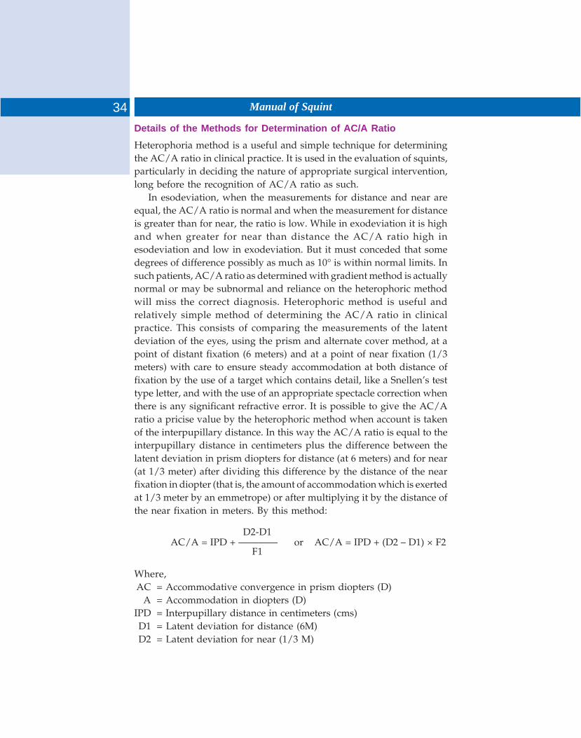
Details of the Methods for Determination of AC/A Ratio
Heterophoria method is a useful and simple technique for determiningthe AC/A ratio in clinical practice. It is used in the evaluation of squints,particularly in deciding the nature of appropriate surgical intervention,long before the recognition of AC/A ratio as such.
In esodeviation, when the measurements for distance and near areequal, the AC/A ratio is normal and when the measurement for distanceis greater than for near, the ratio is low. While in exodeviation it is highand when greater for near than distance the AC/A ratio high inesodeviation and low in exodeviation. But it must conceded that somedegrees of difference possibly as much as 10° is within normal limits. Insuch patients, AC/A ratio as determined with gradient method is actuallynormal or may be subnormal and reliance on the heterophoric methodwill miss the correct diagnosis. Heterophoric method is useful andrelatively simple method of determining the AC/A ratio in clinicalpractice. This consists of comparing the measurements of the latentdeviation of the eyes, using the prism and alternate cover method, at apoint of distant fixation (6 meters) and at a point of near fixation (1/3meters) with care to ensure steady accommodation at both distance offixation by the use of a target which contains detail, like a Snellen’s testtype letter, and with the use of an appropriate spectacle correction whenthere is any significant refractive error. It is possible to give the AC/Aratio a pricise value by the heterophoric method when account is takenof the interpupillary distance. In this way the AC/A ratio is equal to theinterpupillary distance in centimeters plus the difference between thelatent deviation in prism diopters for distance (at 6 meters) and for near(at 1/3 meter) after dividing this difference by the distance of the nearfixation in diopter (that is, the amount of accommodation which is exertedat 1/3 meter by an emmetrope) or after multiplying it by the distance ofthe near fixation in meters. By this method:
D2-D1AC/A = IPD + ———— or AC/A = IPD + (D2 – D1) × F2
F1
Where,AC = Accommodative convergence in prism diopters (D)
A = Accommodation in diopters (D)IPD = Interpupillary distance in centimeters (cms)D1 = Latent deviation for distance (6M)D2 = Latent deviation for near (1/3 M)
35Accommodative Convergence/Accommodation Ratio
F1 = Distance of near fixation in dioptersF2 = Distance of near fixation in meters
Example:If IPD = 6 cmD1 = 4 DexoD2 = 10 DexoF1 = 3 DAC1A = 6 = (–10 – (–4)6 + (–10 + 4)
—————— 3
= 6 + (–2)= 4Or if IPD = 6 cmD1 = 4 DexoD2 = 10 DexoF2 = 1/3 MAC/A = 6 + (–10 (–4) × 1/3= 6 + (–10 + 4) × 1/3= 6 – (–2)= 4
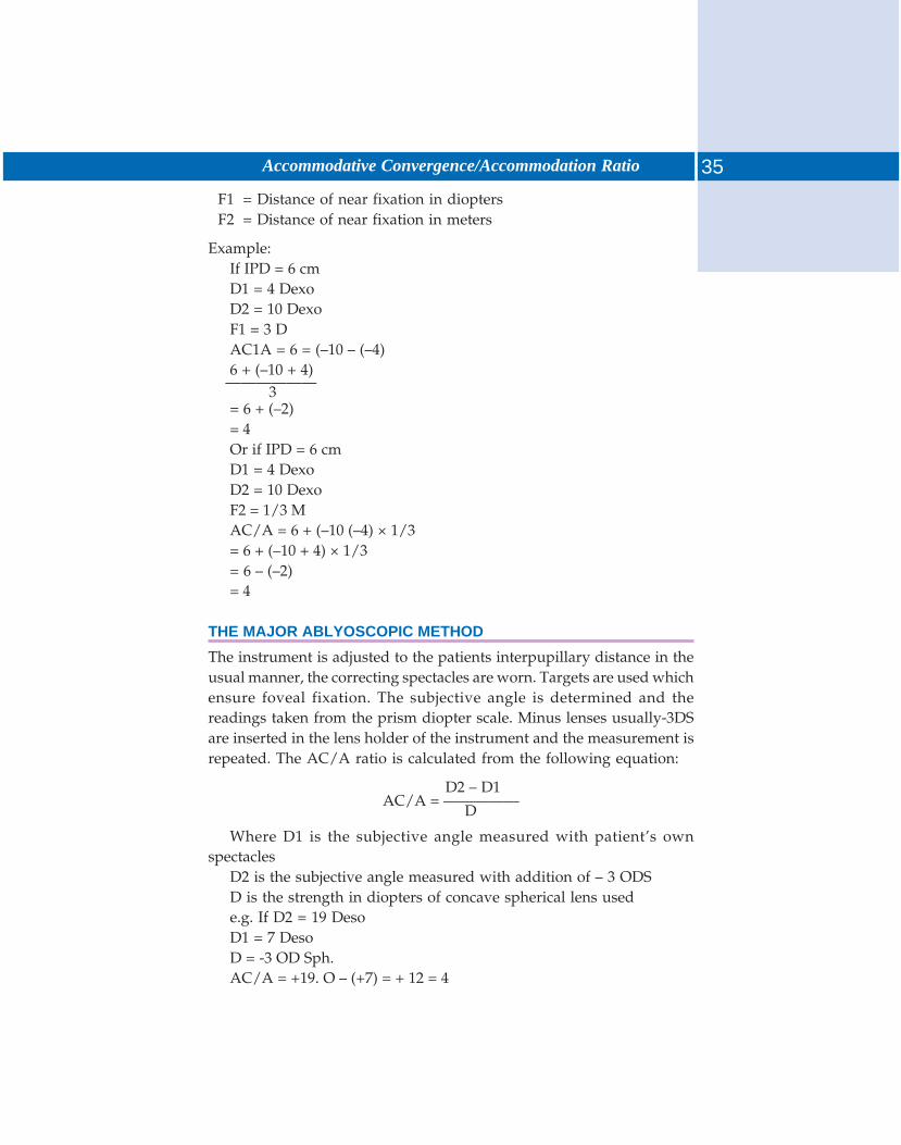
THE MAJOR ABLYOSCOPIC METHOD
The instrument is adjusted to the patients interpupillary distance in theusual manner, the correcting spectacles are worn. Targets are used whichensure foveal fixation. The subjective angle is determined and thereadings taken from the prism diopter scale. Minus lenses usually-3DSare inserted in the lens holder of the instrument and the measurement isrepeated. The AC/A ratio is calculated from the following equation:
D2 – D1AC/A = —————
D
Where D1 is the subjective angle measured with patient’s ownspectacles
D2 is the subjective angle measured with addition of – 3 ODSD is the strength in diopters of concave spherical lens usede.g. If D2 = 19 DesoD1 = 7 DesoD = -3 OD Sph.AC/A = +19. O – (+7) = + 12 = 4
36 Manual of Squint
This method is comparable to the gradient method when usingSnellen’s test types. The advantage of using this method is that smalldeviations can be more accurately measured than may be possible bymeans of the prism and cover test.
Graphic Method
By this method we measure the ratio and determine its character byusing the major amblyoscope along with the graph. The aim of the testis to determine whether the accommodative convergence response isslow or rapid. Each measurement so obtained must be compared withnormal convergence which accompanies each diopter of accommodationin the maintenance of binocular single vision, so that there is a directcomparison between this and the patient’s subjective angle as recordedon the prism-diopter scale.
Method of Fixation Disparity
It is apparent that the magnitude of the fixation disparity givesinformation about a heterophoria. Which is not strickly comparable tothat revealed by most other methods because it had the advantage ofnot creating dissociation of the eyes. It is possible also to change thestate of the heterophoria by altering the vergence of the eyes by the useof prisms and of the accommodation by the use of spherical lenses. Inthis way the value of the muscular imbalance may be related to theaccommodative convergence relationship so that is provides onassessment of the AC/A ratio.
There are several advantages in exploring AC/A ratio by the methodof fixation disparity as compared with the others. Both eyes receive thesame stimuli for accommodation both are subjected to the same type ofestimation and fusion of the two eyes is maintained during the periodof the test so that there is no element of dissociation of the eyes. But thisis complicated and time consuming procedure and not suitable for routineclinical determinations particularly in young children.
Holoscopic Method
When the subject reads a line of fine print to maintain his/her accuracyof focusing, the deviation of the eyes and the degree of accommodationare measured simultaneously at different lavels. It is found that thedeviation increases as the eye accommodates and is usually measuredby the phoria for distant vision and also at the near point with the
37Accommodative Convergence/Accommodation Ratio
appropriate spectacle correction in place, the result is calculated bydividing the change of phoria from the one for the near distance by thediopteric change occurring between the two distances. Modern majoramblyoscope is widely used for calculating this ratio.
Gradient Method
In determining the AC/A ratio by this method the change in the stimulusto accommodation is produced by means of ophthalmic lenses. For agiven fixation distance minus lenses placed before the eyes increase therequirement for accommodation and plus lenses relax accommodation.It is assured that – 1D lenses produce an equivalent of 1D of accommo-dation whereas + lenses relax accommodation by 1D and that theaccommodative response to the lenses is linear within a certain range.In the gradient method the AC/A ratio is measured by an estimation ofthe difference between the deviations of the eyes for a given distanceusing a Maddox rod in front of one eye and correcting prisms in front ofother eyes go that there is change in their accommodation and thereforein their convergence. Convex lenses by decreasing the amount ofaccommodation necessary for the given distance decreases the amountof convergence and concave lenses by increasing the amount ofaccommodation increase the amount of convergence. The importance indetermining there deviation of the eyes is to ensure that the patientexerts the full amount of accommodation required for the particularfixation distance. This is achieved best by the use of an object whichcontain much fine detail in conjunction with the alternate prism andcover test, in preference to the use simply of a fixation light as in theusual Maddox rod test. Difference of the deviation are measured bysubtracting the first deviation from the second deviation, due regard tosign, plus measurements when esodeviation and minus when anexodeviation. The final figure of the ratio is obtained by dividing thedifference in the deviations by the power of the lenses used, to reduce itto a simple unit of accommodation for the care of comparison. As ageneral rule the values for the AC/A ratio by this method are slightlylower than those obtained by the heterophoric method because the fixdistances which is adopted throughout the gradient method precludessome of the influence of the factor of proximal convergence. This methodhas the advantage of inducing convergence which is mainly due to thepatient’s subjective accommodative error.
Heterophoria/latent deviation is a condition of imperfect balance of theextrinsic ocular muscles in which there is a tendency if the eyes to deviatefrom their norm a relative position. This tendency, however, is kept inchecked by the desire for binocular vision and by the reserve neuro-muscular power of the eye.
Since the position of rest is usually of a slight divergence, only a fewpeople are really orthophoric, hence some degree of heterophoria isuniversal.
CLASSIFICATION OF HETEROPHORIA
1. Exophoria2. Esophoria3. Hyperphoria4. Hypophoria5. Cyclophoria
i. Incyclophoriaii. Excyclophoria
Exophoria is again divided intoi. Divergence excesses
Exphoria is greater for distanceii. Convergence weakness
Exophoria is greater for neariii. Mixedor tonic
Esophoria is further divided intoi. Convergence excess type
ii. Divergence insufficiency typeiii. Mixed type
8 Heterophoria
39Heterophoria
ETIOLOGY OF HETEROPHORIA
Heterophoria can be classified into the following types.
Exophoria
Persistent use of accommodation by the hypermetropic favors thedevelopment of esophoria. There are two groups of causes for constantexophorias: (1) static causes and (2) anomalies of sensorimotor system.Innervational factors for causation of exophoria. Congenital abnormalitiesof orbit, e.g. in extreme forms of hypertelorism, a wide interpupillarydistance is produced leading to exophoria. Exophoria may also occur inexophthalmos in which there is some displacement of the eyeballoutwards. They also laid the emphasis of certain occupations, e.g.watchmaker or microscopist which entail prolonged uniocular activitytend to produce exophoria in later life which is accompanied by ocularneglect or suppression.
In the production of exophoria, AC/A ratio plays an important role.A high ratio with exophoria is sometimes seen in myopes due to therelative weakness of the response of the ciliary muscles compared withthat of the medial recti. It is also sometimes seen in presbyopes in whomaccommodation diminishes. In contrast, in exophoria (convergenceweakness type) the AC/A ratio is usually low but may be normal inwhich an uncorrected refractive error may be an important influence inproducing the exodeviation.
Esophoria
Persistent use of accommodation by the hypermetrope in excess of hisconvergence in order to attain clear vision favors the development ofesophoria. On the other hand, in congenital or infantile myopia there isincreased convergence leading to esodeviation. Due to central overactivity through convergence impulses, esophoria is typically seen inenergetic or unrestrained, in the young, strong, asthenic or neurotic incontrast with exophoria.
Esophoria could be produce if the orbits are set close together with anarrow interpupillary distance. The displacement of the eyeball inwardsin cases of enophthalmos can lead to esophoria. They also regardedphysiological defects (e.g. lack of coordination of reflexes associatedwith convergence or divergence) as cause of heterophoria and thusexplained the basis of esophoria as an underlying cause of excessiveapplication to close work. The most common factor etiologically to
40 Manual of Squint
produce esophoria is an increased convergence innervation associatedwith increased accommodation determined either by a hypermetropicrefractive error or arising from optical cause associated withaccommodative strain.
Hyperphoria
Hyperphoria is of three different types with three different reasons.
Static Hyperphoria
It is due to the anatomical factors which determine the position of rest.
Paretic Hyperphoria
It is due to the paresis of an elevator or a depressor muscle.
Spastic Hyperphoria
It is due to an over action of one or both inferior oblique muscles.
ROLE OF REFRACTIVE ERRORS
Influence of refraction on heterophoria is as follows:
Esophoria may result from a demand for:1. Increased accommodation, as in:
a. Bilateral superable hypermetropia orb. Superable hypermetropia of the eye which sees better at all
distances, whatever the refraction of the other eye.2. Increased convergence, as in bilateral congenital myopia.
Exophoria may result form a demand for:a. Decreased accommodation, as in bilateral acquired myopia.b. Decreased convergence, as in recession of the near point in
presbyopia.Decreased accommodation of one eye and decreased convergence,
as in myopic hypermetropic anisometropia, in which the dominant eyeis myopic or subnormally hypermetropic.
SYMPTOMS OF HETEROPHORIA
Heterophoria can be described as fully compensated or uncompensated.In the fully compensated type of heterophoria ocular symptoms do notdevelop due to: (i) strong reserve neuromuscular power available tomaintain the eyes in the physiological position and also (ii) strong strength
41Heterophoria
of desire for a binocular vision. If however, either one or both of thesefactors are weak, the muscle imbalance tends to become uncompensated/decompensated and symptoms occur.
Factors predisposing towards decompensation of heterophoria are:Bodily ill health : Symptoms may arise during illness.Ocular fatigue : Symptoms may arise during periods of
overwork.Mental ill health : Symptoms may arise during periods of anxiety
and worry.Certain occupations : Jobs which entail prolonged ocular activity
whether it be for close work as in clerks, typistsor for distance as in night drivers.
Advancing age : At the less easily adaptable age of middle life,symptom may begin to arise.
Classified the symptoms of heterophoria into four main types:
1. Symptoms due to muscular fatigue (caused by the continuous use ofthe reserve neuromuscular power). These are:
• Headaches (especially occurring during or following prolonged useof eyes as in reading, watching TV/film, etc.)
• Difficulty in changing the focus for near objects after looking at adistance and vice versa.Photophobia (sometimes) occurring in bright light, not relieved by
wearing dark glasses but getting relieved by closing one eye.
2. Symptoms due to failure to maintain constant binocular vision:a. Blurring of print/running together of words while reading.b. Intermittent diplopia — occur under conditions of fatigue or general
debility. Horizontal diplopia particularly when viewing distant objectssuggest esophoria, when viewing near objects suggest exophoria.Vertical diplopia suggests hyperphoria. Sometimes intermittent squintwithout diplopia is usually noticed by patient’s friends. It is seen insome cases of exophoria associated with intermittent divergent squint.Intermittent convergent squint occurs in some cases of esophoria.
3. Symptoms due to defective postural sensation: Transmitted from theocular muscles as a result of alteration of muscle tonus: like difficultyin judging the position of moving objects, difficulty in judgment incarrying out precision tool work and difficulty in estimating distancesfrom the ground.
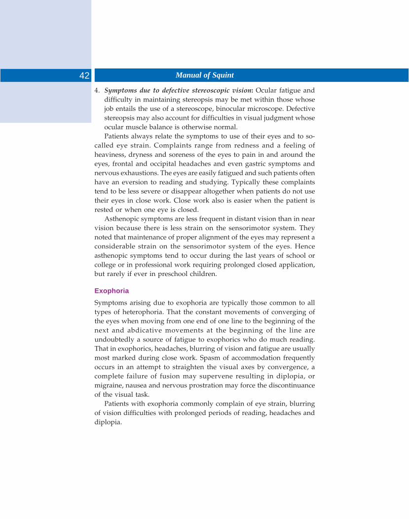
42 Manual of Squint
4. Symptoms due to defective stereoscopic vision: Ocular fatigue anddifficulty in maintaining stereopsis may be met within those whosejob entails the use of a stereoscope, binocular microscope. Defectivestereopsis may also account for difficulties in visual judgment whoseocular muscle balance is otherwise normal.Patients always relate the symptoms to use of their eyes and to so-
called eye strain. Complaints range from redness and a feeling ofheaviness, dryness and soreness of the eyes to pain in and around theeyes, frontal and occipital headaches and even gastric symptoms andnervous exhaustions. The eyes are easily fatigued and such patients oftenhave an eversion to reading and studying. Typically these complaintstend to be less severe or disappear altogether when patients do not usetheir eyes in close work. Close work also is easier when the patient isrested or when one eye is closed.
Asthenopic symptoms are less frequent in distant vision than in nearvision because there is less strain on the sensorimotor system. Theynoted that maintenance of proper alignment of the eyes may represent aconsiderable strain on the sensorimotor system of the eyes. Henceasthenopic symptoms tend to occur during the last years of school orcollege or in professional work requiring prolonged closed application,but rarely if ever in preschool children.
Exophoria
Symptoms arising due to exophoria are typically those common to alltypes of heterophoria. That the constant movements of converging ofthe eyes when moving from one end of one line to the beginning of thenext and abdicative movements at the beginning of the line areundoubtedly a source of fatigue to exophorics who do much reading.That in exophorics, headaches, blurring of vision and fatigue are usuallymost marked during close work. Spasm of accommodation frequentlyoccurs in an attempt to straighten the visual axes by convergence, acomplete failure of fusion may supervene resulting in diplopia, ormigraine, nausea and nervous prostration may force the discontinuanceof the visual task.
Patients with exophoria commonly complain of eye strain, blurringof vision difficulties with prolonged periods of reading, headaches anddiplopia.
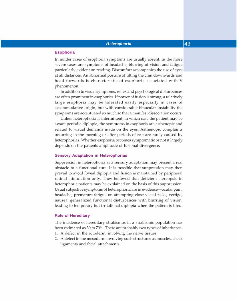
43Heterophoria
Esophoria
In milder cases of esophoria symptoms are usually absent. In the moresevere cases are symptoms of headache, blurring of vision and fatigueparticularly evident on reading. Discomfort accompanies the use of eyesat all distances. An abnormal posture of tilting the chin downwards andhead forwards is characteristic of esophoria associated with Vphenomenon.
In addition to visual symptoms, reflex and psychological disturbancesare often prominent in esophorics. If power of fusion is strong, a relativelylarge esophoria may be tolerated easily especially in cases ofaccommodative origin, but with considerable binocular instability thesymptoms are accentuated so much so that a manifest dissociation occurs.
Unless heterophoria is intermittent, in which case the patient may beaware periodic diplopia, the symptoms in esophoria are asthenopic andrelated to visual demands made on the eyes. Asthenopic complaintsoccurring in the morning or after periods of rest are rarely caused byheterophorias. Whether esophoria becomes symptomatic or not it largelydepends on the patients amplitude of fusional divergence.
Sensory Adaptation in Heterophorias
Suppression in heterophoria as a sensory adaptation may present a realobstacle to a functional cure. It is possible that suppression may thenprevail to avoid foveal diplopia and fusion is maintained by peripheralretinal stimulation only. They believed that deficient stereopsis inheterophoric patients may be explained on the basis of this suppression.Usual subjective symptoms of heterophoria are in evidence—ocular pain,headache, premature fatigue on attempting close visual tasks, vertigo,nausea, generalized functional disturbances with blurring of vision,leading to temporary but irritational diplopia when the patient is tired.
Role of Hereditary
The incidence of hereditary strabismus in a strabismic population hasbeen estimated as 30 to 70%. There are probably two types of inheritance.1. A defect in the ectoderm, involving the nerve tissues.2. A defect in the mesoderm involving such structures as muscles, check
ligaments and facial attachments.
44 Manual of Squint
INVESTIGATIONS
History
a. Visual symptoms: Difficulty in doing near/far work blurring of vision,running of letters, intermittent diplopia/deviation, difficulty inchanging focus, difficulty in judging distances from the ground,difficulty in judging position of moving objects, and difficulty inmaintaining stereopsis.
b. Ocular symptoms: Headache, eyeache, lacrimation, tiredness of eyes,heaviness of eyes and photophobia.
c. General symptoms: Headache, giddiness, nausea, vomiting, mental illhealth and bodily ill health.Past history regarding any ocular trouble, wearing of glasses,
previous refractive status, or general illness, etc. was elicited.
Ophthalmic Examination
a. Visual acuity It was tested both for near and distance, with and withoutglasses.
b. Ocular examination was done by torch light.c. Ocular movements: Uniocular and binocular movements were recorded
in all the cardinal nine gazes.d. Orthoptic investigations: The cases were fully investigated to find out
the condition of muscle balance as indicated below:
Interpupillary Distance
Cover test: The presence of heterophoria may be detected by noting thatone eye deviates when it is covered, and that it makes a movement toregain binocular fixation when the cover is removed.
The cover test was carried out both for near and distance and ifthere was a relevant refractive error, then the test was performed bothwith and without the spectacle corrections. The fixation object used wasa small light placed at about 1/3 meter distance and at 6 meters distance.
The findings of the cover test were recorded as follows:The test was also repeated several times, in order to detect even a
small degree of latent deviation.
Maddox rod and Maddox wing test: Heterophoria for distance was measuredby Maddox rod (Fig. 8.1). Heterophoria for near was measured byMaddox wing (Fig. 8.2). Both these tests cause dissociation of the twoeyes so that a true reading can only be obtained when the subject hasgot binocular vision.
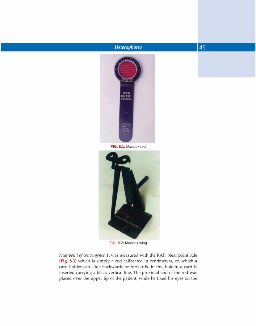
45Heterophoria
FIG. 8.1: Maddox rod
FIG. 8.2: Maddox wing
Near point of convergence: It was measured with the RAF. Near point rule(Fig. 8.3) which is simply a rod calibrated in centimeters, on which acard holder can slide backwards or forwards. In this holder, a card isinserted carrying a black vertical line. The proximal end of the rod wasplaced over the upper lip of the patient, while he fixed his eyes on the
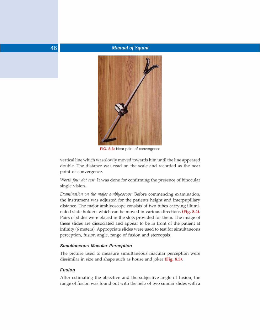
46 Manual of Squint
FIG. 8.3: Near point of convergence
vertical line which was slowly moved towards him until the line appeareddouble. The distance was read on the scale and recorded as the nearpoint of convergence.
Worth four dot test: It was done for confirming the presence of binocularsingle vision.
Examination on the major amblyoscope: Before commencing examination,the instrument was adjusted for the patients height and interpupillarydistance. The major amblyoscope consists of two tubes carrying illumi-nated slide holders which can be moved in various directions (Fig. 8.4).Pairs of slides were placed in the slots provided for them. The image ofthese slides are dissociated and appear to be in front of the patient atinfinity (6 meters). Appropriate slides were used to test for simultaneousperception, fusion angle, range of fusion and stereopsis.
Simultaneous Macular Perception
The picture used to measure simultaneous macular perception weredissimilar in size and shape such as house and joker (Fig. 8.5).
Fusion
After estimating the objective and the subjective angle of fusion, therange of fusion was found out with the help of two similar slides with a
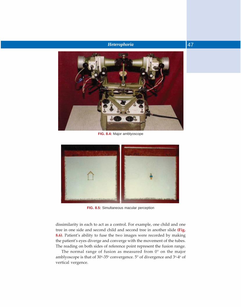
47Heterophoria
FIG. 8.4: Major amblyoscope
FIG. 8.5: Simultaneous macular perception
dissimilarity in each to act as a control. For example, one child and onetree in one side and second child and second tree in another slide (Fig.8.6). Patient’s ability to fuse the two images were recorded by makingthe patient’s eyes diverge and converge with the movement of the tubes.The reading on both sides of reference point represent the fusion range.
The normal range of fusion as measured from 0° on the majoramblyoscope is that of 30o-35o convergence. 5° of divergence and 3o-4o ofvertical vergence.
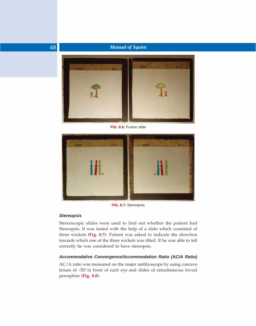
48 Manual of Squint
FIG. 8.6: Fusion slide
FIG. 8.7: Stereopsis
Stereopsis
Stereoscopic slides were used to find out whether the patient hadStereopsis. It was tested with the help of a slide which consisted ofthree wickets (Fig. 8.7). Patient was asked to indicate the directiontowards which one of the three wickets was tilted. If he was able to tellcorrectly he was considered to have stereopsis.
Accommodative Convergence/Accommodation Ratio (AC/A Ratio)
AC/A ratio was measured on the major amblyoscope by using concavelenses of -3D in front of each eye and slides of simultaneous fovealperception (Fig. 8.8).
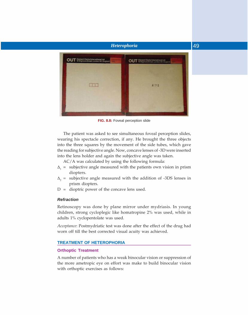
49Heterophoria
FIG. 8.8: Foveal perception slide
The patient was asked to see simultaneous foveal perception slides,wearing his spectacle correction, if any. He brought the three objectsinto the three squares by the movement of the side tubes, which gavethe reading for subjective angle. Now, concave lenses of -3D were insertedinto the lens holder and again the subjective angle was taken.
AC/A was calculated by using the following formula:Δ1 = subjective angle measured with the patients own vision in prism
diopters.Δ2 = subjective angle measured with the addition of -3DS lenses in
prism diopters.D = dioptric power of the concave lens used.
Refraction
Retinoscopy was done by plane mirror under mydriasis. In youngchildren, strong cycloplegic like homatropine 2% was used, while inadults 1% cyclopentolate was used.
Acceptance: Postmydriatic test was done after the effect of the drug hadworn off till the best corrected visual acuity was achieved.
TREATMENT OF HETEROPHORIA
Orthoptic Treatment
A number of patients who has a weak binocular vision or suppression ofthe more ametropic eye on effort was make to build binocular visionwith orthoptic exercises as follows:
50 Manual of Squint
1. Antisuppression exercises: On cherioscope, chasing and flashing exerciseson major amblyoscope were with the use of simultaneous macularperception slides. It was given in those cases who has complete orpartial suppression of more ametropic eye with a view to providestimulus to the suppressed eye. The exercises were given 10 to 15times daily.
2. Fusion exercises: A fusion exercises on major amblyoscope: Fusionexercises were given on major amblyoscope with the fusion slides.Fusion range could be increased by gradually converging both thetubes of major amblyoscope till the fusion breaks. Exercises weregiven daily or on alternate days for 10 to 15 minutes depending onthe rolerance and convenience of the patients.
When difficulty is experienced accommodation may be inducedby inserting –3D sph. In the lens carriers. Patients should be taughtto relax accommodation while adducting, i.e. keeping the picturesclear to 20o to 25o. Abduction should be performed without any sineof spasm. As a final exercises, when adduction to 50oC is achievedwith fusion picture and voluntary adduction with simultaneousperception pictures should be attained. It should on no account begiven when adduction is unsteady with fusion pictures, as it is difficultexercise to perform smoothly.
i. Home exercises: Home exercises comprising of convergence to nearpoint (Pencil to nose exercise) and reorganization of physiologicaldiplopia for near and distance was explained to the patient.Patients were instructed to do exercises almost two to three timesdaily for 10 to 15 minutes.
ii. Fusion exercises on diploscope: It is based on physiological diplopiaand require simultaneous use of the eyes and providesconvergence to the eyes.
iii. Physiological diplopia with pencil and distant light.3. Ex. diploscope exercise4. Exercise on Remy separator5. Exercise with the help of stereogram cards6. Occlusion to induce use of eye with marked suppression.
Great care must be taken if this is undertaken and the occluder isbest worn for reading, cinema, etc. not worn walking about. Treatmentof all types of heterophoria is basically the same.
Prisms: Prisms to correct esophoria on exophoria are not advised. Patientswho are unable to attend for treatment, who are unfit or too old mayget relief from symptoms with prisms. Prism to correct a vertical deviationare often necessary.
51Heterophoria
Operation
Operation is necessary:a. If the deviation is becoming manifest.b. If the deviation is large and the patient is unable to maintain
comfortable ocular vision.Patient, should be totally occluded for a short period before the
operation into manifest the full deviation.
Hyperphoria
Small degrees of hyperphoria give rise to symptoms. Large degrees areusually suppressed and do not give rise to symptoms.
Patients with hyperphoria lose their fusion range which may be thecause of symptoms. So lateral muscles range need attention.
It is rarely possible to reduce or compensate for a hyperphoria withorthoptic treatment.
Prisms should be used to compensate the vertical deviation.Large hyperphoria are usually paretic in origin and often require
surgery to compensate for the deviation.
Basic Orthoptic Treatment
a. Clip-on vertical prisms where necessary.b. Make sure that the fusion range and muscle control is within normal
limits.
Cyclophoria
Never seen unless associated with a paralysis of an elevator or depressormuscle, (External rectus palsy slight cyclo on extreme).1. In traumatic cases, if treated early, it will disappear as the range of
fusion increases and the patient obtains binocular single vision (BSV)with the help of prisms.
2. In cases of diplopia of long standing, cyclophoria cannot be overcomeexcept by surgical treatment.
3. Small degrees often appear with an aphakia who has diplopia whenwearing a contact lens. If the contact lens is given reasonably soonafter operation, the cyclophoria can be overcome.
Convergence Insufficiency
It can be defined as a condition in which the parallel movements of theeyes are normal but the associated movement of simultaneous contraction
52 Manual of Squint
of medial rectus muscles is reduced in power, normal near point ofconvergence is between 6–10 cm. Even it may be a normal limit butthere may be inability to maintain convergence without undue effortwhich constitutes some degree of convergence insufficiency.
Convergence insufficiency may exist as a separate entity or may existin association with exophoria or esophoria, etc.
There are seven types of convergence insufficiency:1. Primary idiopathic2. Secondary to primary divergent strabismus (divergence excess type)3. Secondary to a vertical muscle defect4. Convergence insufficiency due to refractive error5. Systemic convergence insufficiency (poor general health)6. Convergence insufficiency associated with presbyopia7. Surgically induced convergence insufficiency.
General Physical Causes
Intoxications and diseases of endocrine glands (Moebius sign orconvergence insufficiency in exophthalmos).
Psychologic causes are anxiety, neurosis.The symptoms are those of visual fatigue in general. When
convergence insufficiency alone is involved, the symptoms appear innear work after some time, and disappear quickly with rest.
Examination shows, orthophoria for distance and exophoria for near.During the effort of convergence, the pupil may remain in relativemydriasis.
In pure convergence insufficiency, which is rather rare, there will beorthophoria to 30 cm, and only from this point can the insufficiency bedemonstrated.
Treatment
To treat convergence insufficiency, additional fusional convergenceshould be developed with the appropriate exercises.
Fusional convergence can be developed by teaching the patient toconverge on objects progressively closer to his eyes while maintainingbinocular vision.
The patient is taught to constantly check that he is using both eyes inany fusional convergence training. To check on the use of both eyes, thepatient must have some clue. For example, if the patient tries to bringthe tip of the pencil closer and closer to his nose, a different color pencil
53Heterophoria
should be held further away. The image of the pencil held further awaywill fall on noncorresponding retinal points, and the patient will seetwo images of the distant pencil.
The patient attempts to bring one pencil closer to his nose whileseeing two images of the pencil held farther away. He sees only oneimage of the nearer pencil if he is aligning his foveas because the imagesof the near pencil strike corresponding foveal areas on the retina.
The two pencil fusional convergence exercise is easy to teach to mostpatients. The patient is instructed to bring the pencil progressively closerto him and try to see the point singly while seeing two of the pencilsheld farther away. The single point may be blurred because the limit ofaccommodative convergence has been exceeded and only fusionalconvergence is being used. Practice in the above would be expected toincrease the patient’s fusional convergence.
a. Convergence paralysis: In this condition the patient gets diplopia onplacing even the smallest power of prism before the eye. On the contrarythe patient with convergence insufficiency does tolerate prisms to theextent permitted by the amount of convergence present.
Secondly, the patient will demonstrate constriction of pupil onattempted convergence. In the case of convergence insufficiency thepupillary constriction will accompany convergence movement but itwill dilate as soon as the limits of convergence is crossed and the eyesdiverge.
b. Accommodative effort syndrome: The patients of convergenceinsufficiency are usually associated with an exophoria for near, whilein case of accommodative effort syndrome no heterophorias areassociated.
When a lens of -3D is placed before the eye in a case of convergenceinsufficiency, there is an enhancement of convergence, while under similarcircumstances the case of accommodative effort syndrome willdemonstrate a tropia.
Placing of plus lenses before the eye reduces convergence by onaccount of relaxation of the accommodative convergence, while theaccommodation is helped in cases of accommodative effort syndrome.
Treatment
It is indicated in children with poor fusional reserve and a child startshaving intermittent exotropia. In adults the treatment is indicated whenthe symptoms are present. It consists of:
54 Manual of Squint
Optical Treatment
Any refractive error present is corrected after a meticulous refraction.While a full correction is prescribed for myopes, a slight under correctionis made in hypermetropes. This strategy helps to improve the accommo-dational convergence.
Orthoptic Treatment
Orthoptic treatment is primarily aimed at improving the amplitude ofconvergence. Same set of exercises are done as in the case of treatingexophoria, and consist of:
A. Improving near point of convergence, which include:i. Advancement exercises: In this exercise the patient is asked to hold an
object (preferably with some minute details) some distance awayfrom his nose, and then gradually bring it closer to the nose untilhe sees it double. At this point he is told to withdraw the objectslightly away till it becomes single again. This position is to bemaintained for few moments following which the exercise processis repeated several times. Over a period of time the patient shouldbe able to bring and keep the object almost to the tip of his nose,maintaining a binocular single vision (i.e. single object is being seen)all the time.
ii. Jump convergence exercise: This, in fact is an extension of advancementexercise, and should be undertaken after a successful completionof the latter. It trains the patient to maintain binocular single visionunder the circumstances when a rapid change in the amount ofconvergence is required. Two objects are used for this exercise,one being placed at a distance of 6 meters, and the other at 33 cmaway front of the patient. The patient is then asked to look at thetwo objects alternately. Gradually, the distance of the near objectis brought closer or about 5 cm away from the nose, whilemaintaining a binocular single vision all the timer though the nearobject may look blurred.
This exercise can also be done with the help of prisms by askingthe patient to fix at a near object and then placing a 10D prism withbase out in front of one eye. The patient is then encouraged tomaintain single vision for which he has to converge. Gradually, thedemand for more and more convergences brought about by agranual increase in the power of the prism until the patient canconverge to maintain single vision with prism of 40D.
55Heterophoria
B. Improving amplitude of fusional convergence: The following exercises maybe undertaken to improve the amplitude of fusional convergence:
i. Exercises with prisms: Prisms of increasing power, with base out, areplaced before the patient’s eye while he is fixing at a near object.He is encouraged to maintain single vision when the prism’s isbeing increased. Use of a prism bar for this purpose is moreappropriate.
ii. Exercises on synoptophore: The patient is asked to fuse the twostereoscopic slides and then the tubes are slowly converged untilhe fusion is broken as evidenced by the loss of stereopsis. Theprocedure is repeated again and again for about five minutes onweekly basis. In the intervening period home exercises arecontinued.
iii. Physiological diplopia exercise: This is performed with help of a card.Before starting the procedure the patient is first made to appreciatephysiological diplopia. The stereogram is held at arm’s length infront of the patient and he is asked to fix at the picture. At thispoint a pencil is placed midway between the card and the patient.
iv. Exercise on diploscope: Exercises for voluntary convergence—This isa very useful exercise that needs the cooperation of an intelligentpatient who is asked to fix at a distant object, preferably a smallsource of light. At this stage another object say a pencil or a fingeris interposed and placed in front of the patient at about an arm’slength. The patient is now asked to fix his gaze at the pencil and isencouraged to appreciate doubling of the distant fixation object,which results as the pencil is being fixed (physiological diplopia).The pencil is then removed from the field of vision and the patientis asked to keep on with seeing double images of the distant object.This procedure may be repeated several times. In due course oftime the patient is trained to see double images of the distant object,even without the introduction of the pencil.
Prism Treatment or Prismotherapy
Prism treatment or prismotherapy is reserved for cases not respondingfavorably to the orthoptic treatment. Base in prisms are corporated inthe correcting glasses. In general, prescription of prisms is avoided inchildren.
56 Manual of Squint
Surgical Treatment
Operative interference should be made as the 1st alternative when allother forms of treatment fail to relieve the symptoms. A unilateral orbilateral resection of the medial rectus muscle may be undertaken.
Convergence Paralysis
As already described it is the result of some intracranial lesions involvingthe midbrain and the III N nucleus. The diagnostic features are:• Sudden onset• Exotropia and crossed diplopia on attempted convergence.
Normal Adduction
• Usually normal accommodation.• Preservation of miosis and accommodation on attempted
convergence.• Evidence of intracranial lesion.
Diplopia caused by the weak base out prism (while a case orconvergence deficiency tolerates base out prisms to a certain extent).
Treatment: Appropriate prisms are prescribed for near vision. If binocularsingle vision cannot be achieved, occlusion of one eye be done whiledoing near work. Surgical interference is not indicated.
Convergence Spasm
It is rare anomaly of convergence which is mostly of functional nature.Rarely it may be caused by some intracranial disease. It is characterizedby:• Intermittent attacks of extreme convergence resembling a bilateral
palsy of VI N• Intermittent homonymous diplopia• Blurring of vision caused by associated spasm of accommodation• Miotic pupils, as a part of the near reflex• Myopia upto 6D, induced by the spasm of accommodation.
Treatment
Most of the cases need psychiatric treatment, after the possibility of anintracranial has been excluded. The palliative measures may be adoptedin the form of prolonged atropinization or occlusion of one eye as analternative.
PSEUDOESOTROPIA
Epicanthus—It is a bilateral condition, which may be associated withptosis. A more or less vertical fold of skin runs from the root of the noseto the inner end of the lower eyelid, covering the medical canthus andthe caruncle.
If such folds are prominent they produce or apparent convergentstrabismus.
PSEUDODIVERGENT STRABISMUS
An apparent divergent strabismus may be produced by one or more ofthe following factors:1. A large positive angle alpha2. A wide interpupillary distance3. Exophthalmos4. A wide palpebral fissure.
If binocular single vision is present, the parents should reassuredthat there is no actual deviation.
i. If the orbits are set wide apart producing a wide interpupillarydistance, exophoria is common. If orbits are set close together,resulting in narrow interpupillary distance, esophoria is common.
ii. If there is exophthalmos (as seen in hyperthyroidism), there willbe some displacement of the eyeball out made as well as forwards,exophoria usually occurs. The presence of an undue narrowing ofthe lateral canth causes an apparent divergent squint because ofthe reduced amount of the eyeball which is visible on the lateralside of each canthus.
iii. The presence of abnormally large angle alpha.The presence of a large positive (or nasal) angle alpha may produce
an apparent divergent strabismus.
9 Pseudostrabismus
58 Manual of Squint
In the average normal eye the visual axis does not coincide with theoptic axis (or midpupillary line) but cuts the corneal to the nasal side ofthe latter. (This is because the fovea is situated slightly downwards andto the temporal side of the point at which the optic axis cuts the ratina)this means that there is a small angle between the optic axis and visualaxis. This angle, which rarely exceeds 5° to 7° is known as angle alpha.When visual axis cuts the cornea on the nasal side of the optic axis, it issaid to be positive angle alpha and when visual axis cuts the cornea onthe temporal side, it is side to be negative angle alpha. For purpose ofmeasurement of angle alpha on major amblyoscope, we use a specialslide consisting of row of numbers and letters at intervals of one degree.This slide is placed in front of the eye under observation. The patientshould be told to look at the ‘0’. If the corneal reflection in observed tobe to the nasal side of the pupil the angle alpha is positive, if it is to thetemporal side the angle alpha is negative. Then we ask the patient tolook at each of the numbers or letters in turn, until the reflection on thecornea is observed to be central. This procedure is then repeated usingthe slide before the other eye and in this way we can record positiveand negative angle alpha.
PSEUDOHYPERTROPIA
If one orbit is slightly higher than the other due to a symmetry of theskull, then are appearance of vertical strabismus may result. Facialasymmetrics or orbital tumors, the mass can displace the globe verticallyand may stimulate vertical ocular deviation.
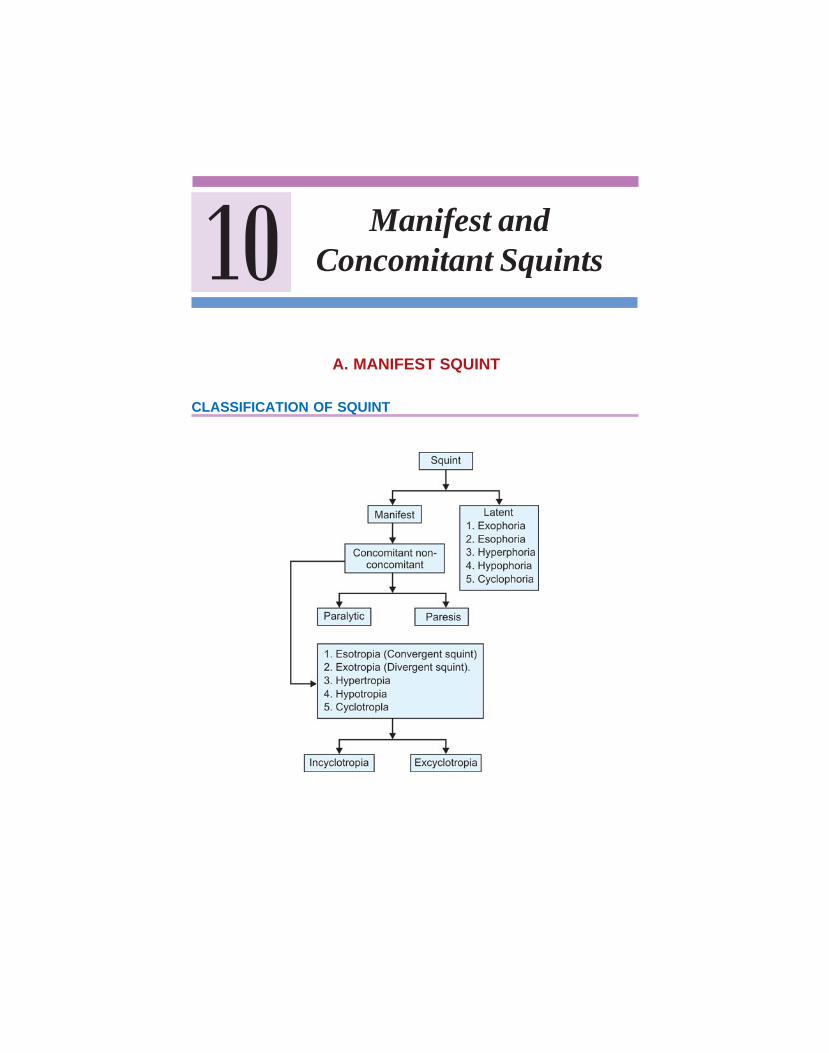
CLASSIFICATION OF SQUINT
10 Manifest andConcomitant Squints
A. MANIFEST SQUINT
60 Manual of Squint
Normally, both eyes are maintained at the point of fixation under variouscentral and peripheral influences. In ordinary activity both eyes aredirected at the point of fixation together. If the position of fixation isnot maintained so that one eye gets deviated, the condition ofheterotropia or manifest squint arises.
The manifest squint may be either concomitant or incomitant. Theword concomitant is derived from Latin ‘concomitor’ meaning ‘I attend,I accompany’. In fact, in a concomitant squint the deviation remains thesame, or approximately so, in all positions of the gazes. The eyes thusmove together in coordination retaining the abnormal relationshipbetween them with all ocular movements.
According to Duke Elder, concomitant squint is of two types —primary and secondary. Primary concomitant squint develops due toobstacle in sensory or afferent pathway of binocular vision so that eyesare dissociated but coordinated by postural reflexes which retains themotor axis unimpaired. It is a bilateral condition, the deviation beingshared equally between two eyes. When one eye fixes the deviation ofboth becomes manifest in the other eye. In secondary concomitant squint,there is a peripheral muscular basis; it follows secondarily upon theincomitant squint.
Classification
The concomitant squint is clinically classified based on differentparameters.1. Depending upon the direction of deviation:
Esotropia—the deviating eye turns inwardsExotropia—the deviating eye turns outwardsHypertropia—the deviating eye turns upwardsHypotropia—the deviating eye turns downwardsCyclotropia—torsional defect where the deviation takes the form ofa rotation round the fixation axis.
2. Depending upon the constant or intermittent presence of thedeviation: Constant and intermittent types.
3. Depending upon the fixation preference:Uniocular squint—affects one eye so that the other eye is preferredfor fixation.
B. CONCOMITANT SQUINT
61Manifest and Concomitant Squints
Alternating squint—alternate involvement of the eyes and no obviouspreference for fixing with either eye.
4. Depending upon the time of onset:Congenital squint—deviation noticed in early months of life.Infantile squint—deviation noticed before 1 year of age.Acquired squint—deviation noticed after 2 years of age. In primaryacquired squint no definite cause can be found and it is acute in onset.In secondary type, the deviation arises from known cause such asdisease of the eyes or trauma or operation. Consecutive cases are theresult of change in the nature of squint—either spontaneous oroccurring after attempted squint surgery.
5. Depending upon the fusional and accommodative vergence:Convergence weakness or excess, divergence weakness or excess,basic types; typical accommodative, partially accommodative, hypo-accommodative, nonaccommodative types.Periodic squint is a special type as reported by Duke-Elder, which
differs in degree depending on far or near fixation. If the squint isgreater for near, it is called directly periodic; if greater for distance,inversely periodic.
Cyclic squint—Cyclic squint is another special type in which the squintappears and disappears in a rhythmic manner, most frequently at 48hours intervals.
Convergent concomitant squint or esotropia may be primary,secondary due to loss of vision or consecutive following overcorrectionof exotropia by surgery.
According to Duke-Elder there are three main forms of primaryesotropia.1. Esotropia of the convergence excess type in which the deviation is
significantly greater for near.a. Accommodative esotropia
i. Typical accommodative esotropia in which the deviation iscured by correction of the underlying hypermetropia.
ii. Atypical accommodative esotropia with no significantrefractive error. It may be either hypoaccommodative orhyper-accommodative with high AC/A ratio.
iii. Partially accommodative esotropia—the commonestaccommodative squint where uncorrected hypermetropia ispartly responsible for the deviation.
62 Manual of Squint
b. Tonic esotropiac. Esotropia with congenital myopia.
2. Esotropia of the divergence weakness type in which the deviation isgreater for distance.
3. Esotropia of the basic type in which the deviation is almost same indistance and near.A few other forms of esotropia are:
4. Essential (infantile) esotropia—characterized by early onset, largedeviation, no significant refractive error and frequent association ofvertical component.
5. Acute concomitant esotropia.6. Cyclic esotropia.7. Microtropia—characterized by small deviation, amblyopia, eccentric
fixation with central scotoma and harmonious retinal correspondence.Among the causes of primary concomitant convergent squint few
important ones are excess use of accommodation in hypermetropia,uncompensated esophoria, congenital myopia and anatomical conditionslike asymmetry of orbits, etc. There is a special form of convergent squintwhich may be associated with the following:1. Congenital myopia: Near object seen clearly, but distant objects cannot
be seen and so all distant objects appear blurred to him so there is noinitiative to keep the eyes straight and they converge.
2. Other causes:i. Corneal opacity
ii. Lenticular opacityiii. Chorioretinal atrophyiv. Optic atrophyv. Pseudoglioma
vi. Retinoblastoma3. Consecutive convergent squint: Over correction of divergent squint can
lead to consecutive convergent squint.4. Ocular palsy: Primary vertical concomitant squint is rare. Most of the
vertical components are associated with primarily horizontalconcomitant squint cases, usually esotropias. The etiology of trulyconcomitant vertical deviations of magnitude rarely exceding fewprism diopters is not clear. There can be A-V pattern, overaction ofinferior obliques and dissociated vertical deviations associated withprimarily horizontal concomitant squint cases.
63Manifest and Concomitant Squints
Etiological Causes (Duke Elder)
1. Optical obstacles: Preventing the formation of suitable retinal images.These are of 2 types:a. Extraneous factors: Seen in young children due to prolonged period
of disease of one eye despite absence of any pathological lesionslike:— Congenital ptosis— Effect of wrong spectacles— Seen in watch makers— Prolonged occlusion of an eye.
b. Ocular factors: Like— High refractive errors— Anisometropia— Aniseikonia— Opacities in the ocular media— Damage to fovea/Parafovea as seen in congenital toxoplasmosis
and congenital rubella.2. Sensory obstacles: Preventing the association of corresponding
retinocerebral points like— Disease of retina and optic disk— Lesions of the visual pathways.
3. Motor obstacles: Preventing adequate coordination of the 2 eyes. Thesecan be of following types:a. Static obstacles:
— Anomalies of symmetry and inclination of orbits and shapeof skull as in craniofacial dysostosis where divergence incommon
— Abnormalities in the shape of globe as in high myopia— Abnormality of position of globe — as in proptosis— Space occupying lesions of orbits— Congenital abnormalities of muscles— Paresis of the muscles— Abnormalities of insertion of the muscles.
4. Central obstacles: Preventing the emergency of unitary binocularperception like:a. Faculty development of faculty of fusion
64 Manual of Squint
b. After the establishment of the binocular reflexes, fall in theefficiency at highest level may lead to their breakdown. Thedampening of the cerebral activity is seen in:— Severe illness— Fatigue— Alcohol poisoning— Coma.
c. The factors which leads to disturbance of neural equilibrium, whichin turn leading to a latent and compensated ocular imbalance tobecome manifest squint like in:— Hyperexcitability state as been in teething— Emotional states.
General Features
1. The deviation of the eyeball is same in all the directions of the gaze.2. The primary deviation (deviation of the squinting eye under cover
when sound eye is fixating) is equal to secondary deviation (deviationof sound eye under cover when squinting eye is fixating).
3. There is no limitation of movements of the eyeball.4. In case of uniocular concomitant squint, the vision in the squinting
eye is usually defective.5. There is no compensatory head posture.6. There is no abnormal projection/orientation.
Symptoms
1. Patient may complain of manifest deviation of the eye. It may beeither intermittent or constant.
2. Patient may complain of diminition of vision in one or both eyes.i. If it is constant, there is development of suppression, which leads
to amblyopia. The patient gets relief of symptoms like diplopiaand headache with development of suppression.
ii. Most of times it is associated with horizontal squint. It may beprimary vertical squint with secondary horizontal or vice versa.The most common secondary horizontal or vice versa. The mostcommon secondary vertical elements are elevation in adductionor abduction due to imbalance of vertical recti (superior and
65Manifest and Concomitant Squints
inferior rectus) and oblique muscle (superior and inferior obliquemuscles).
Cyclotropia
There are torsional defects and are extremely rare. These are usuallyparetic in nature and due to congenital defects.
Sequalae of Events in a Case
In a case of concomitant squint, apart from loss of binocular vision, thepatient may be asymptomatic. Initially, there might be confusion, i.e.simultaneous formation of two dissimilar images of two objects, orDiplopia, i.e. simultaneous formation of two images of same object on 2-corresponding points of retina. But these symptoms disappear with thedevelopment of suppression in the squinting eye in favor of the othereye in order to avoid confusion or diplopia. The suppression may becentral, i.e. inhibition of the foveal image of the squinting in order toavoid confusion or it may be peripheral, i.e. inhibition of the imagefrom the periphery of retina or extrafoveal point in order to avoiddiplopia. The suppression may be facultative in initial stages, i.e. inhibitionof image when eye is deviating or obligatory, i.e. inhibition of the imageirrespective of whether eye is deviated or not.
The suppression in all cases is aided by the peripheral situation ofthe image in the squinting eye, but the essential seat of suppression is inthe brain. As the image of any object falling on noncorresponding pointscauses diplopia, which brain finds difficult to fuse, so it actively suppressesthe image.
The prolonged suppression of image leads to permanent lowering ofvision in the squinting eye, leading to a condition called amblyopia. Inthe long-standing cases, the squinting eye may or may not show anymovement to take up fixation on cover test. This position of the deviationwhich is less than the actual deviation as called as eccentric fixation, i.e.fixation in the squinting eye is being assumed by extrafoveal point. Themaximum visual acuity on the extra-foveal point is made available tothe squinting eye. This is a uniocular phenomenon. While the conditionwhere fovea of one eye corresponds with the extrafoveal point in othereye and it is called as anomalous retinal correspondence. It is a binocularphenomenon.
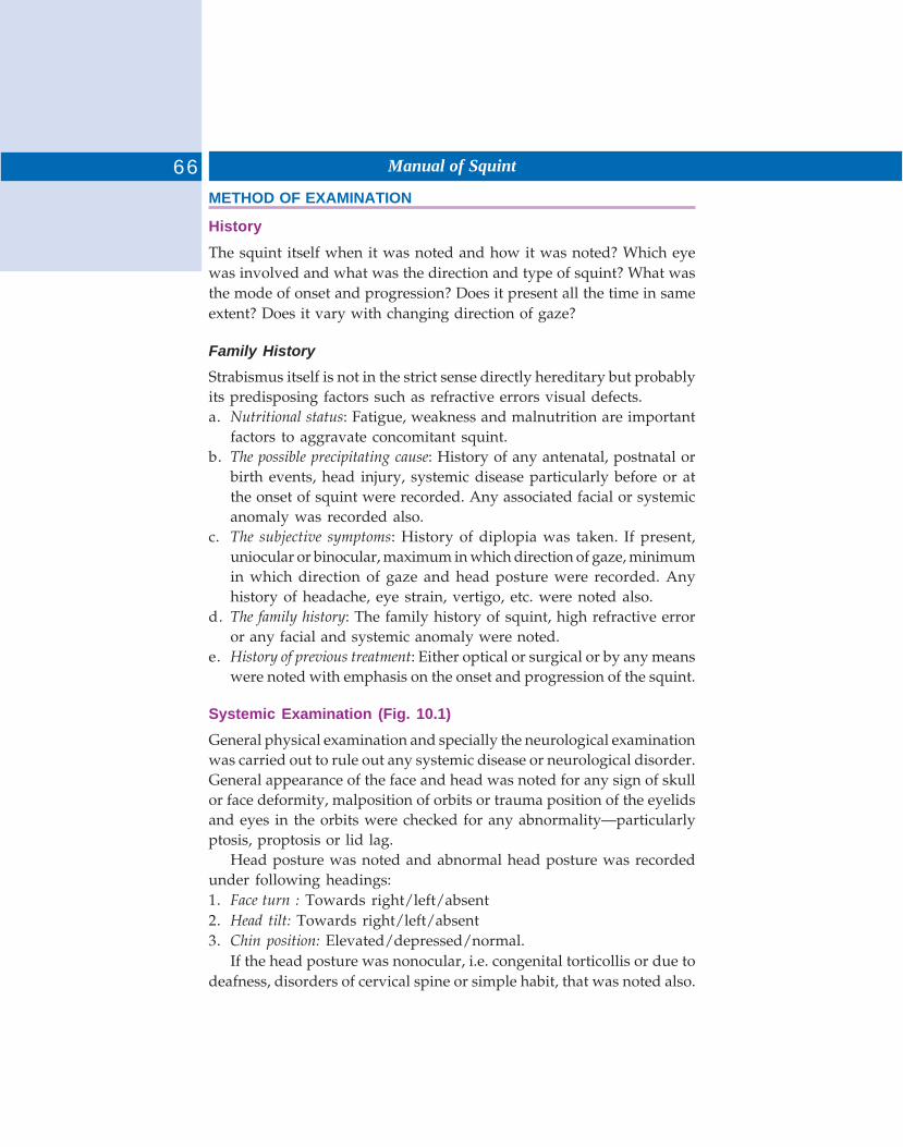
66 Manual of Squint
METHOD OF EXAMINATION
History
The squint itself when it was noted and how it was noted? Which eyewas involved and what was the direction and type of squint? What wasthe mode of onset and progression? Does it present all the time in sameextent? Does it vary with changing direction of gaze?
Family History
Strabismus itself is not in the strict sense directly hereditary but probablyits predisposing factors such as refractive errors visual defects.a. Nutritional status: Fatigue, weakness and malnutrition are important
factors to aggravate concomitant squint.b. The possible precipitating cause: History of any antenatal, postnatal or
birth events, head injury, systemic disease particularly before or atthe onset of squint were recorded. Any associated facial or systemicanomaly was recorded also.
c. The subjective symptoms: History of diplopia was taken. If present,uniocular or binocular, maximum in which direction of gaze, minimumin which direction of gaze and head posture were recorded. Anyhistory of headache, eye strain, vertigo, etc. were noted also.
d. The family history: The family history of squint, high refractive erroror any facial and systemic anomaly were noted.
e. History of previous treatment: Either optical or surgical or by any meanswere noted with emphasis on the onset and progression of the squint.
Systemic Examination (Fig. 10.1)
General physical examination and specially the neurological examinationwas carried out to rule out any systemic disease or neurological disorder.General appearance of the face and head was noted for any sign of skullor face deformity, malposition of orbits or trauma position of the eyelidsand eyes in the orbits were checked for any abnormality—particularlyptosis, proptosis or lid lag.
Head posture was noted and abnormal head posture was recordedunder following headings:1. Face turn : Towards right/left/absent2. Head tilt: Towards right/left/absent3. Chin position: Elevated/depressed/normal.
If the head posture was nonocular, i.e. congenital torticollis or due todeafness, disorders of cervical spine or simple habit, that was noted also.
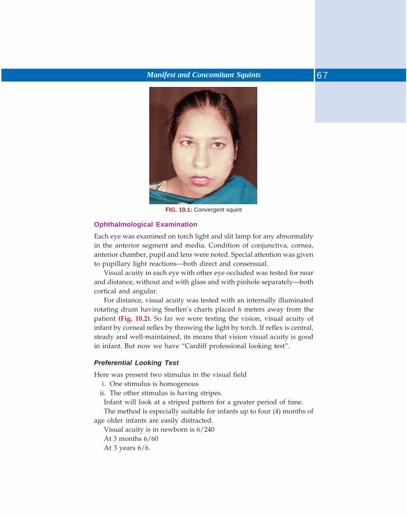
67Manifest and Concomitant Squints
Ophthalmological Examination
Each eye was examined on torch light and slit lamp for any abnormalityin the anterior segment and media. Condition of conjunctiva, cornea,anterior chamber, pupil and lens were noted. Special attention was givento pupillary light reactions—both direct and consensual.
Visual acuity in each eye with other eye occluded was tested for nearand distance, without and with glass and with pinhole separately—bothcortical and angular.
For distance, visual acuity was tested with an internally illuminatedrotating drum having Snellen’s charts placed 6 meters away from thepatient (Fig. 10.2). So far we were testing the vision, visual acuity ofinfant by corneal reflex by throwing the light by torch. If reflex is central,steady and well-maintained, its means that vision visual acuity is goodin infant. But now we have “Cardiff professional looking test”.
Preferential Looking Test
Here was present two stimulus in the visual fieldi. One stimulus is homogenous
ii. The other stimulus is having stripes.Infant will look at a striped pattern for a greater period of time.The method is especially suitable for infants up to four (4) months of
age older infants are easily distracted.Visual acuity is in newborn is 6/240At 3 months 6/60At 3 years 6/6.
FIG. 10.1: Convergent squint
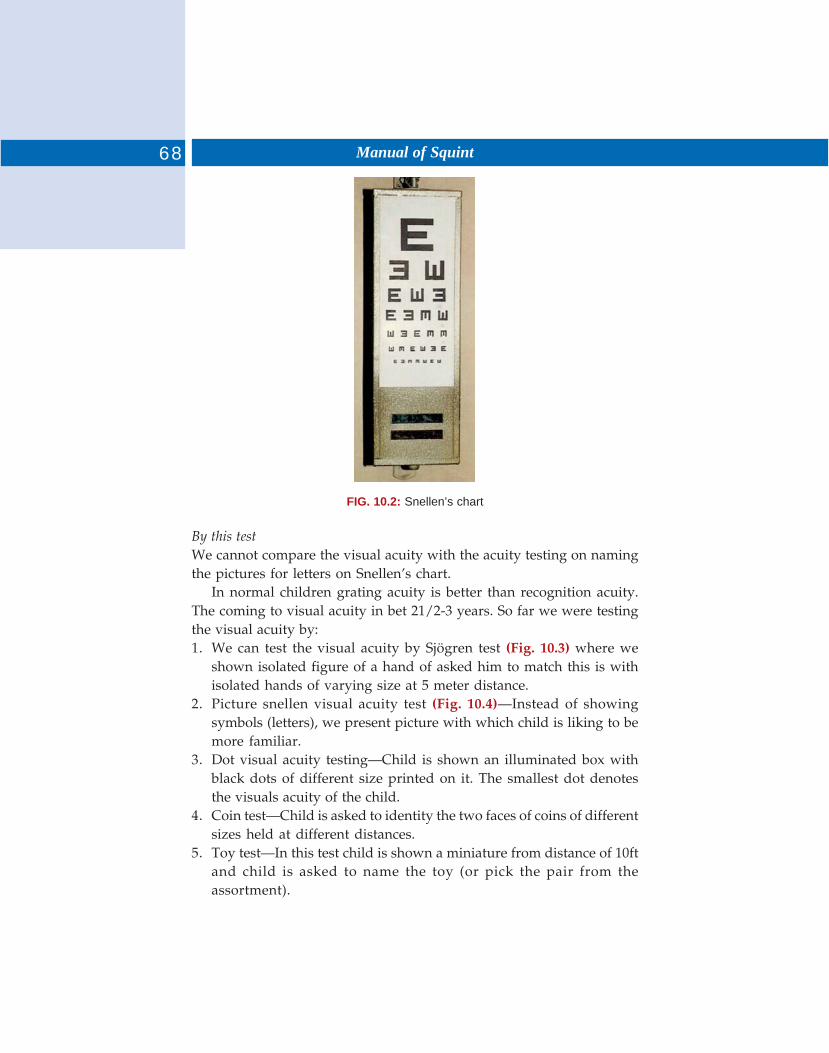
68 Manual of Squint
FIG. 10.2: Snellen’s chart
By this testWe cannot compare the visual acuity with the acuity testing on namingthe pictures for letters on Snellen’s chart.
In normal children grating acuity is better than recognition acuity.The coming to visual acuity in bet 21/2-3 years. So far we were testingthe visual acuity by:1. We can test the visual acuity by Sjögren test (Fig. 10.3) where we
shown isolated figure of a hand of asked him to match this is withisolated hands of varying size at 5 meter distance.
2. Picture snellen visual acuity test (Fig. 10.4)—Instead of showingsymbols (letters), we present picture with which child is liking to bemore familiar.
3. Dot visual acuity testing—Child is shown an illuminated box withblack dots of different size printed on it. The smallest dot denotesthe visuals acuity of the child.
4. Coin test—Child is asked to identity the two faces of coins of differentsizes held at different distances.
5. Toy test—In this test child is shown a miniature from distance of 10ftand child is asked to name the toy (or pick the pair from theassortment).
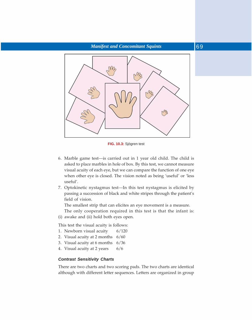
69Manifest and Concomitant Squints
FIG. 10.3: Sjögren test
6. Marble game test—is carried out in 1 year old child. The child isasked to place marbles in hole of box. By this test, we cannot measurevisual acuity of each eye, but we can compare the function of one eyewhen other eye is closed. The vision noted as being ‘useful’ or ‘lessuseful’.
7. Optokinetic nystagmus test—In this test nystagmus is elicited bypassing a succession of black and white stripes through the patient’sfield of vision.The smallest strip that can elicites an eye movement is a measure.The only cooperation required in this test is that the infant is:
(i) awake and (ii) hold both eyes open.
This test the visual acuity is follows:1. Newborn visual acuity 6/1202. Visual acuity at 2 months 6/603. Visual acuity at 6 months 6/364. Visual acuity at 2 years 6/6
Contrast Sensitivity Charts
There are two charts and two scoring pads. The two charts are identicalalthough with different letter sequences. Letters are organized in group
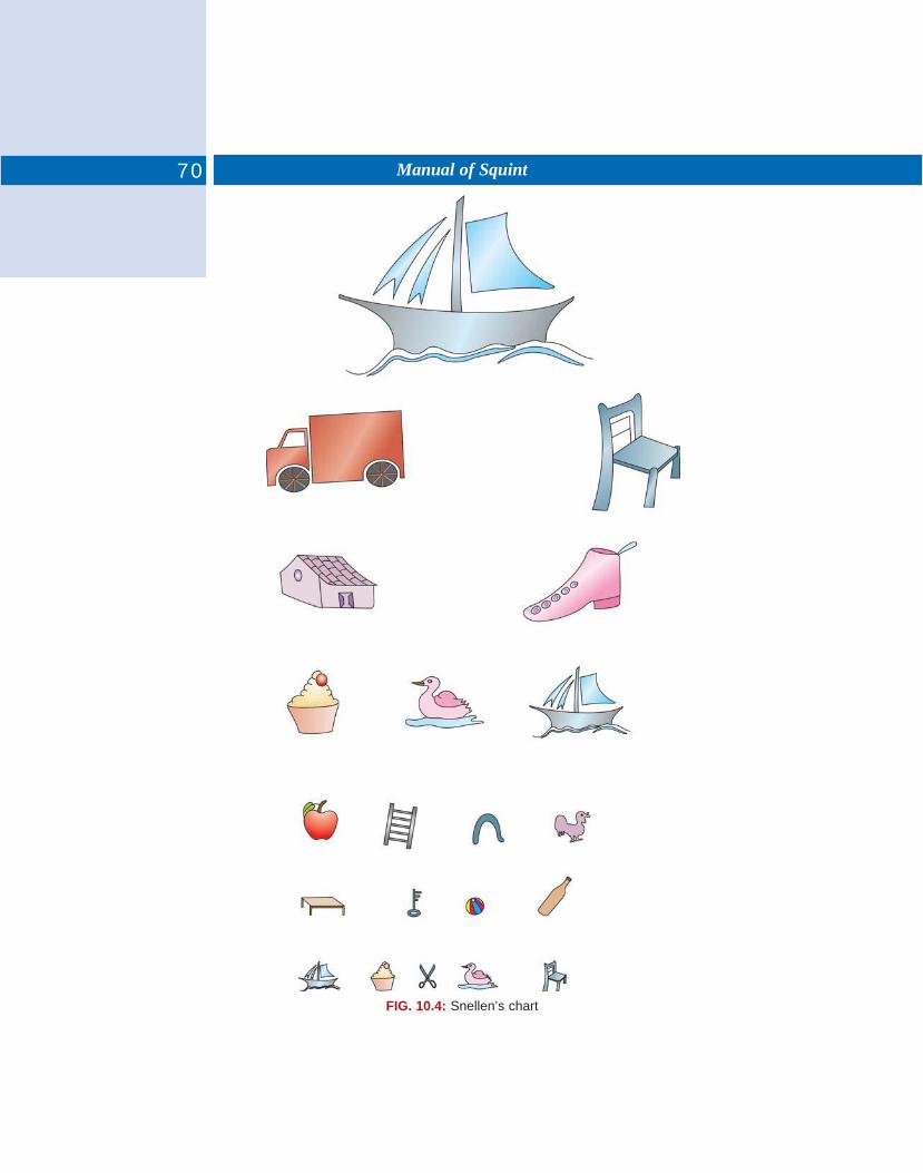
70 Manual of Squint
FIG. 10.4: Snellen’s chart
71Manifest and Concomitant Squints
of three. With each triplet all letters have the same contrast. The contrastdecreases from one triplet to the next. The division into triplets isindicated on the scoring pad. In Snellen chart the difficulty in readingline increases from line to line but in Pelli-Robson chart the difficultyincreases in the middle of each line as well. The center of chart shouldbe approximately at the level of the patient eye. The chart should beilluminated as uniformly as possible so that luminance of the white areasis about 85ed/m2.
We test the patient before dilating the people or applying any otherdrug to their eyes.
The patient should sit or stand directly in front of the chart so thatthe distance from the eyes to the chart is about 1 meter or 40 inches(patient should sit at 1 meter distance away from the chart and the levelof the eyes should be at the center of the chart). The patient shouldwear their best distance correction and if necessary an addition of 0.75D.Patient should read letter from upper left hand corner and he has toread each letter on the chart. On the scoring pad underline or circle eachletter correctly and strike any letter read incorrectly. Patients should bemade to guess even when they believe that the letters are invisible. Donot let the patient give up too soon. You should allow several secondsfor the finest letters to appear, but do not let the patient give up until heor she has guessed incorrectly 2 of 3 letters in a triplet. The reliability ofthe result depends on this.
Scoring pads — The patient’s sensitivity is indicated by the faintesttriplet for which 2 of the 3 letters are named correctly.
The patient should be tested three times. Test each eye separatelyand both eyes together. When you test one eye, the other eye is covered.The three measurements should take no more than 8 minutes in all.
Binocular Log contrast sensitivity is normally 0.15 higher thanmonocular.
The chart’s plastic substrate and special ink were chosen for theirgreat stability and contrast clarity.
The chart should not be touched by fingertips. If necessary, wipe thechart gently with soft cloth using a highly diluted solution of mild soapor detergent (e.g. lvory liquid) in water then rinse with clean water.Avoid exposure to direct sunlight or any UV light source. To prolongthe life of the chart it is suggested that the chart is tuned to face the wallwhen not in use.
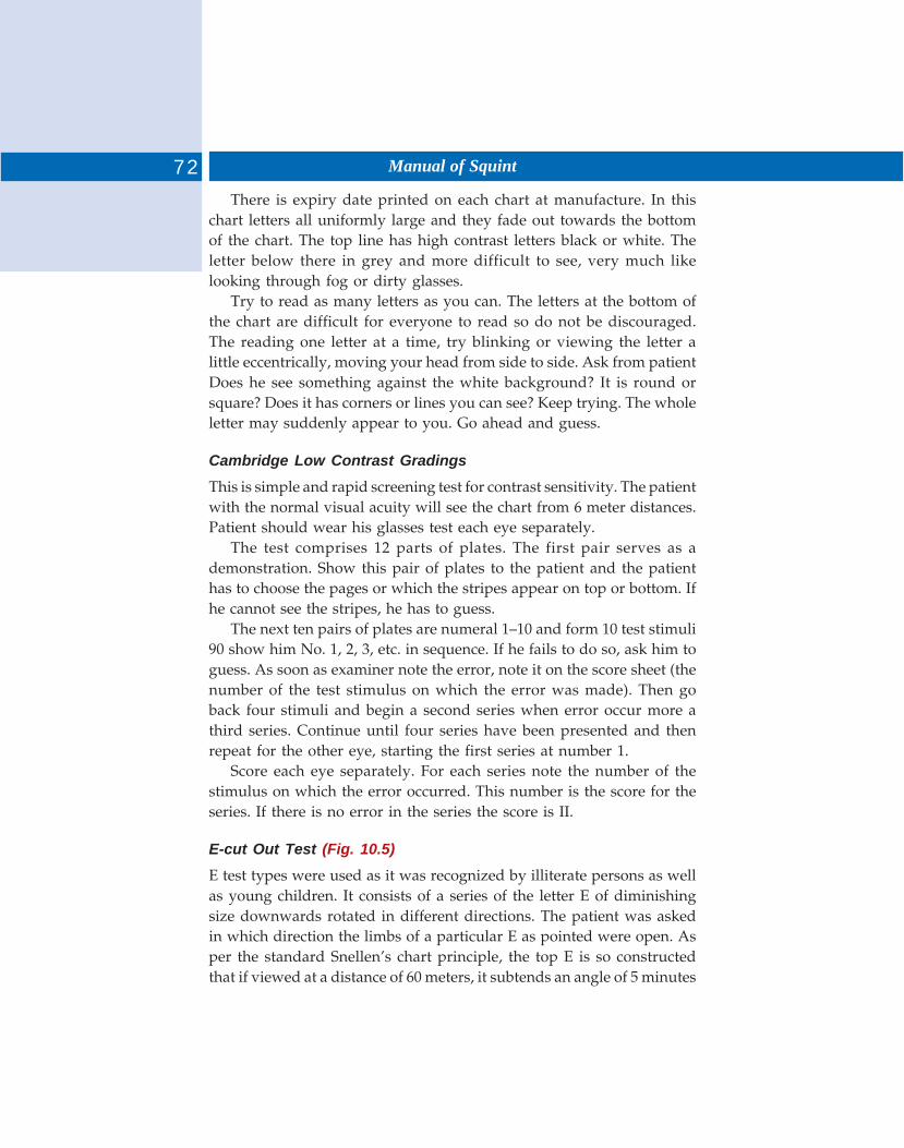
72 Manual of Squint
There is expiry date printed on each chart at manufacture. In thischart letters all uniformly large and they fade out towards the bottomof the chart. The top line has high contrast letters black or white. Theletter below there in grey and more difficult to see, very much likelooking through fog or dirty glasses.
Try to read as many letters as you can. The letters at the bottom ofthe chart are difficult for everyone to read so do not be discouraged.The reading one letter at a time, try blinking or viewing the letter alittle eccentrically, moving your head from side to side. Ask from patientDoes he see something against the white background? It is round orsquare? Does it has corners or lines you can see? Keep trying. The wholeletter may suddenly appear to you. Go ahead and guess.
Cambridge Low Contrast Gradings
This is simple and rapid screening test for contrast sensitivity. The patientwith the normal visual acuity will see the chart from 6 meter distances.Patient should wear his glasses test each eye separately.
The test comprises 12 parts of plates. The first pair serves as ademonstration. Show this pair of plates to the patient and the patienthas to choose the pages or which the stripes appear on top or bottom. Ifhe cannot see the stripes, he has to guess.
The next ten pairs of plates are numeral 1–10 and form 10 test stimuli90 show him No. 1, 2, 3, etc. in sequence. If he fails to do so, ask him toguess. As soon as examiner note the error, note it on the score sheet (thenumber of the test stimulus on which the error was made). Then goback four stimuli and begin a second series when error occur more athird series. Continue until four series have been presented and thenrepeat for the other eye, starting the first series at number 1.
Score each eye separately. For each series note the number of thestimulus on which the error occurred. This number is the score for theseries. If there is no error in the series the score is II.
E-cut Out Test (Fig. 10.5)
E test types were used as it was recognized by illiterate persons as wellas young children. It consists of a series of the letter E of diminishingsize downwards rotated in different directions. The patient was askedin which direction the limbs of a particular E as pointed were open. Asper the standard Snellen’s chart principle, the top E is so constructedthat if viewed at a distance of 60 meters, it subtends an angle of 5 minutes
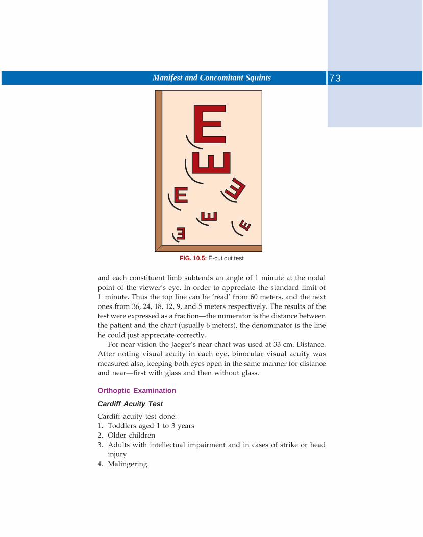
73Manifest and Concomitant Squints
FIG. 10.5: E-cut out test
and each constituent limb subtends an angle of 1 minute at the nodalpoint of the viewer’s eye. In order to appreciate the standard limit of1 minute. Thus the top line can be ‘read’ from 60 meters, and the nextones from 36, 24, 18, 12, 9, and 5 meters respectively. The results of thetest were expressed as a fraction—the numerator is the distance betweenthe patient and the chart (usually 6 meters), the denominator is the linehe could just appreciate correctly.
For near vision the Jaeger’s near chart was used at 33 cm. Distance.After noting visual acuity in each eye, binocular visual acuity wasmeasured also, keeping both eyes open in the same manner for distanceand near—first with glass and then without glass.
Orthoptic Examination
Cardiff Acuity Test
Cardiff acuity test done:1. Toddlers aged 1 to 3 years2. Older children3. Adults with intellectual impairment and in cases of strike or head
injury4. Malingering.
74 Manual of Squint
The principle of the target design is that of the vanishing optotype.The targets are drawn with a white band bordered by two bands, eachone is half the thickness of the white band. All on a neutral background.If the target lies beyond the visual acuity of the child, it merges with thegrey background and becomes simply invisible. Thus resolution,detection and recognition acuity threshold are all brought together. Thetargets used are picture, but decreasing in width of white and blackbands. The narrowest white band for which the target is visible. Theprinciple of the test is that of preferential looking are infant will chooseto look towards a target, rather than towards a plain stimulus. In Cardifftest, each target is positioned either in the top half or in the bottom.Half of the card.
If the target is visible, the child will look toward sit and the examinerwatch the child’s eye movements.
An important feature of the preferential looking technique is that theexaminer should not know in advance the position of target.
For any given target width, if examiner estimates the positioncorrectly on two consecutive occasions, the target is assumed to be visibleto the child. If the examiner is unable to make a judgment from thechild’s responses, then target is assumed to be beyond the child’s acuitylimit.
In Cardiff test, we present two cards to the child.The target is positioned up and down so that the eye movements are
easier to discriminate in cases of congenital nystagmus. The child is seatedat one meter distance from the target at this distance the examiner canappreciate eye movement of the child.
This test can done at 50 cm. At this distance visual acuity is 6/120 to6/12.
Procedure—for each visual acuity level shuffle the three cards and beginwith widest target (lowest acuity) present the first card at the child’seye level. In order to draw, child attention talk about the picture, orencourage the child to point to the picture.
We can establish the visual acuity by child eye movement byestimating the position top/bottom of the target. Once you make yourdecision, present the second card to confirm your decision. If two correctestimates are made, proceed to the next level. If incorrect estimate ismade return to the next larger target and repeat the test.
Shuffle the card between each presentation. The end point can thenbe taken at the highest level at which at least two out of the three cards
75Manifest and Concomitant Squints
are scored correctly. The calibration for the cards it given in table, whichpresents acuity levels for two distances, in both Log MAR and equivalentsmaller acuity.
Cardiff preferential looking tests tend to give a higher acuitythan a visual acuity on Snellen chart because the child is target and notto identify it.
Cardiff test is carried out at a near distance. Test the visual acuitywith glasses, if there is any reflective error. In malingering the patient isnot aware of their eye movements and will look consistently at the targeteven while visiting that they cannot see it.
Ductions
The test was performed at near. Each eye was covered in turn while theother eye fixated spot light held at 33 cm. and was moved in all cardinaldirections of gaze. Any overaction or underaction in any direction wasnoted. Presence of nystagmoid movements were checked if any,particularly in full duction.
Versions: Carried out in similar manner keeping both eye uncovered todetect any underaction of one muscle with or without overaction of itsantagonist and contralateral synergist, up shoot or down shoot of anyeye in horizontal versions, retraction of the globe and narrowing ofpalpebral fissure in any direction of gaze.
Diplopia in any position of binocular gaze was noted if stated by thepatient on enquiry.
Cover Test (Figs 10.6A and B)
For near the test was performed with and without glasses with a sharpobject of fixation such as a spot sized small light situated at a distance of33 cm. from the patient. The patient was asked to fixate the object andone eye was covered. Any movement in the uncovered eye was noted.An inward movement meant exotropia, outward movement esotropia,upward movement hypotropia and downward movement hypertropiaof the uncovered eye. At times combination of horizontal and verticalmovements were also detected indicating the presence of both horizontaland vertical squint. The test was repeated covering the other eye andlooking for movement of the now uncovered eye for any manifest squintof this eye. In unilateral squint the sound eye took fixation whenever itwas uncovered. In alternating squint the fixation was retained by theuncovered eye irrespective of which eye was covered.
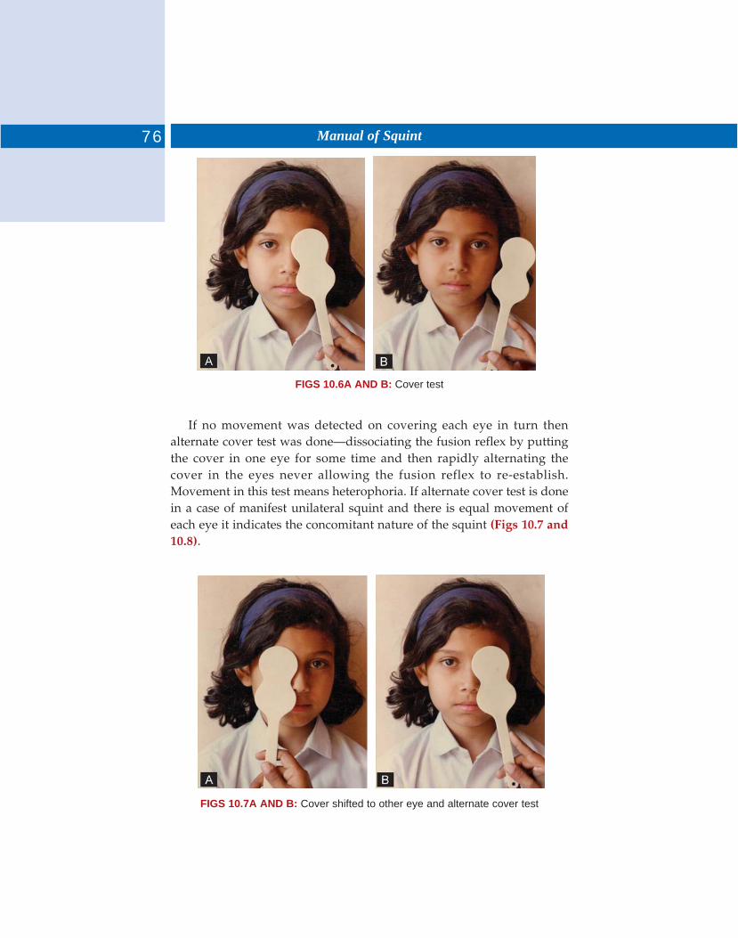
76 Manual of Squint
FIGS 10.6A AND B: Cover test
If no movement was detected on covering each eye in turn thenalternate cover test was done—dissociating the fusion reflex by puttingthe cover in one eye for some time and then rapidly alternating thecover in the eyes never allowing the fusion reflex to re-establish.Movement in this test means heterophoria. If alternate cover test is donein a case of manifest unilateral squint and there is equal movement ofeach eye it indicates the concomitant nature of the squint (Figs 10.7 and10.8).
FIGS 10.7A AND B: Cover shifted to other eye and alternate cover test
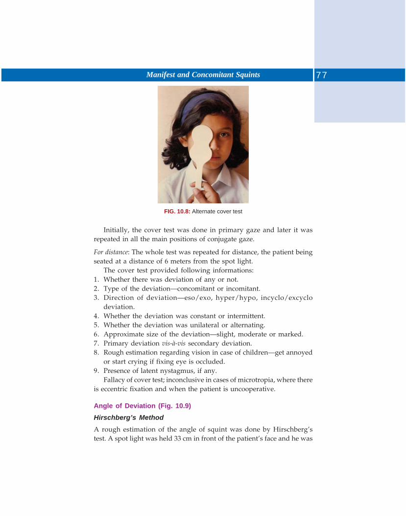
77Manifest and Concomitant Squints
FIG. 10.8: Alternate cover test
Initially, the cover test was done in primary gaze and later it wasrepeated in all the main positions of conjugate gaze.
For distance: The whole test was repeated for distance, the patient beingseated at a distance of 6 meters from the spot light.
The cover test provided following informations:1. Whether there was deviation of any or not.2. Type of the deviation—concomitant or incomitant.3. Direction of deviation—eso/exo, hyper/hypo, incyclo/excyclo
deviation.4. Whether the deviation was constant or intermittent.5. Whether the deviation was unilateral or alternating.6. Approximate size of the deviation—slight, moderate or marked.7. Primary deviation vis-à-vis secondary deviation.8. Rough estimation regarding vision in case of children—get annoyed
or start crying if fixing eye is occluded.9. Presence of latent nystagmus, if any.
Fallacy of cover test; inconclusive in cases of microtropia, where thereis eccentric fixation and when the patient is uncooperative.
Angle of Deviation (Fig. 10.9)
Hirschberg’s Method
A rough estimation of the angle of squint was done by Hirschberg’stest. A spot light was held 33 cm in front of the patient’s face and he was
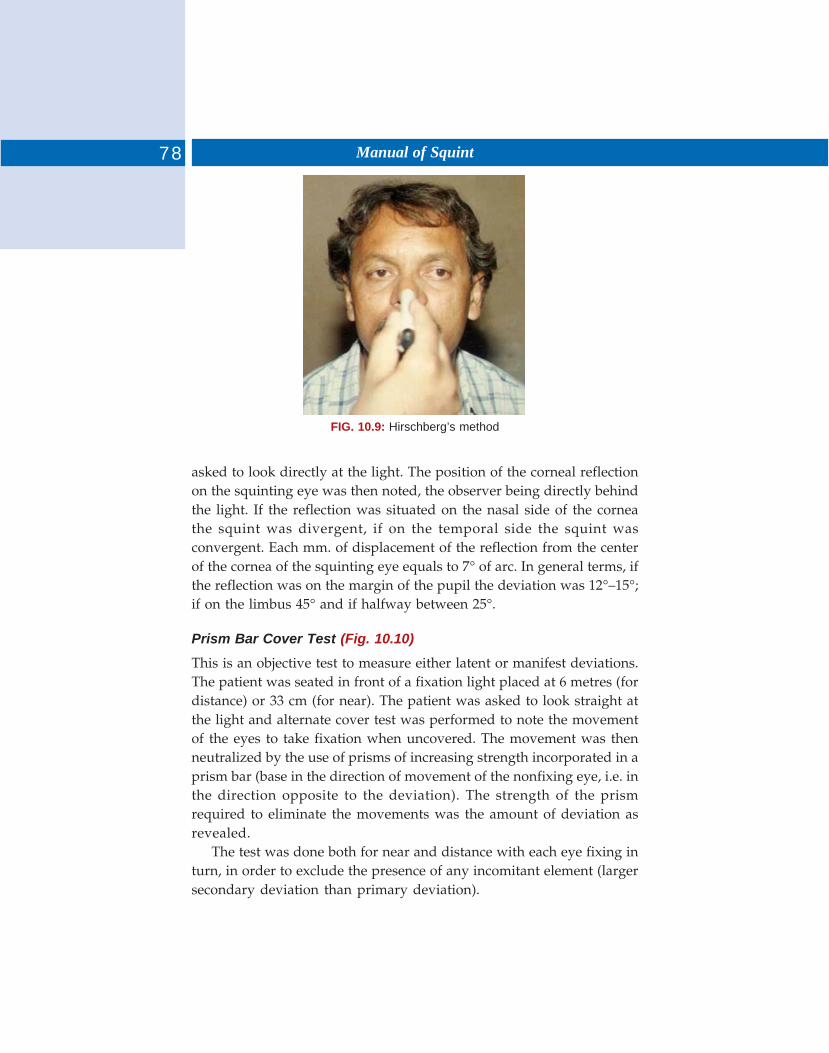
78 Manual of Squint
FIG. 10.9: Hirschberg’s method
asked to look directly at the light. The position of the corneal reflectionon the squinting eye was then noted, the observer being directly behindthe light. If the reflection was situated on the nasal side of the corneathe squint was divergent, if on the temporal side the squint wasconvergent. Each mm. of displacement of the reflection from the centerof the cornea of the squinting eye equals to 7° of arc. In general terms, ifthe reflection was on the margin of the pupil the deviation was 12°–15°;if on the limbus 45° and if halfway between 25°.
Prism Bar Cover Test (Fig. 10.10)
This is an objective test to measure either latent or manifest deviations.The patient was seated in front of a fixation light placed at 6 metres (fordistance) or 33 cm (for near). The patient was asked to look straight atthe light and alternate cover test was performed to note the movementof the eyes to take fixation when uncovered. The movement was thenneutralized by the use of prisms of increasing strength incorporated in aprism bar (base in the direction of movement of the nonfixing eye, i.e. inthe direction opposite to the deviation). The strength of the prismrequired to eliminate the movements was the amount of deviation asrevealed.
The test was done both for near and distance with each eye fixing inturn, in order to exclude the presence of any incomitant element (largersecondary deviation than primary deviation).
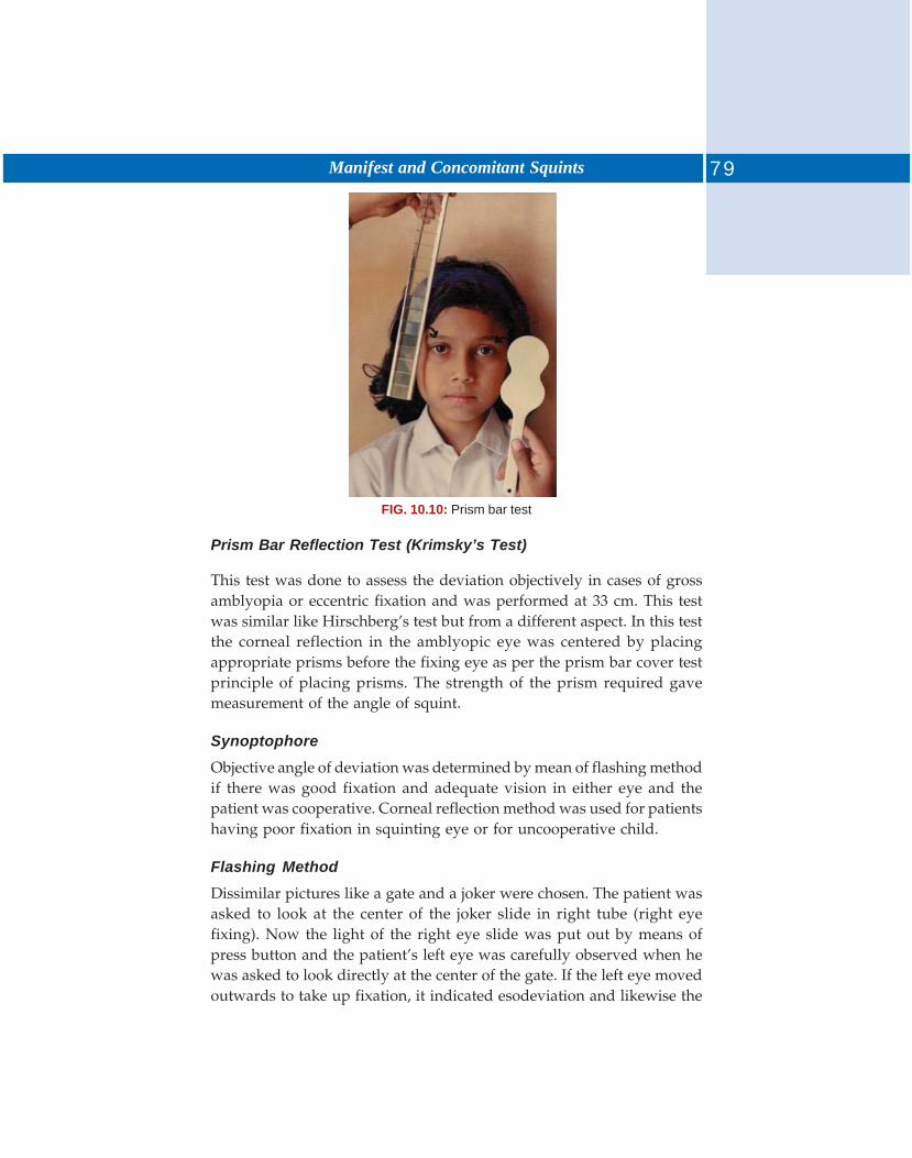
79Manifest and Concomitant Squints
FIG. 10.10: Prism bar test
Prism Bar Reflection Test (Krimsky’s Test)
This test was done to assess the deviation objectively in cases of grossamblyopia or eccentric fixation and was performed at 33 cm. This testwas similar like Hirschberg’s test but from a different aspect. In this testthe corneal reflection in the amblyopic eye was centered by placingappropriate prisms before the fixing eye as per the prism bar cover testprinciple of placing prisms. The strength of the prism required gavemeasurement of the angle of squint.
Synoptophore
Objective angle of deviation was determined by mean of flashing methodif there was good fixation and adequate vision in either eye and thepatient was cooperative. Corneal reflection method was used for patientshaving poor fixation in squinting eye or for uncooperative child.
Flashing Method
Dissimilar pictures like a gate and a joker were chosen. The patient wasasked to look at the center of the joker slide in right tube (right eyefixing). Now the light of the right eye slide was put out by means ofpress button and the patient’s left eye was carefully observed when hewas asked to look directly at the center of the gate. If the left eye movedoutwards to take up fixation, it indicated esodeviation and likewise the
80 Manual of Squint
left tube position was then adjusted or the slide in that tube was raisedor lowered (in cases of vertical movements) to eliminate movement inleft eye. The reading as shown on the scale that time was the objectiveangle of deviation fixing right eye. Placing the joker in left tube and thegate in right the test was repeated to measure the objective angle ofdeviation fixing left eye.
Corneal
Reflection Method: Dissimilar pictures like a cot and a joker were chosen.The tube before the fixing eye was placed at zero except in large angleof deviation where the deviation was divided between two arms of theinstrument. The patient was encouraged to look directly at the picturebefore fixing eye. Tube before the deviating eye was then adjustedhorizontally and vertically as required so that the corneal reflection inthat eye coincide as accurately as possible with the same in fixing eye.Reading from the scale at this point was the objective angle of deviationmeasured by corneal reflection method.
Subjective Angle of Deviation
Dissimilar pictures like a gate and a joker were chosen. First the jokerwas kept in the right tube and the patient was asked to look directly atthis. Left arm of the instrument was then moved horizontally andvertically as required so that the joker was completely into the gate. Thescale reading as noted was the subjective angle of deviation fixing righteye. Placing the joker in left tube and moving the right arm of theinstrument the test was repeated to measure the subjective angle ofdeviation fixing left eye.
By comparing subjective and objective angles of deviation, anomalousretinal correspondence was noted for its presence or absence.
The angle of deviation was measured subjectively or objectively witheach eye fixing in turn in all the cardinal directions of gaze and wasrecorded in a table form.
Assessment of Binocular Functions (On Synoptophore)
Simultaneous macular perception (SMP) was already ascertained whilemeasuring the subjective angle of deviation with dissimilar simultaneousperception slides.
Fusion was checked with similar slides with different controls like atree with a boy and a tree with a girl.
81Manifest and Concomitant Squints
After appropriate adjustment of the tubes the two pictures gotsuperimposed and a single picture with both the controls were seen bythe patient. The angle at which the fusion occurred was noted.Thereafter, fusion range was seen in abduction and adduction when thetubes were moved accordingly in coordinated manner while the patienttried to maintain the fusion of the images.
Stereopsis was noted after putting the stereoscopic slides such asthose of a different color wickets and a ball while the patient was askedto tell the position of middle wicket—whether straight or inclined toany side.
The Maddox rod consists of several rods or grooves, colored redand mounted in a disk so that it refracts light rays in one direction andconverts a point of light source into a red line of light when placed infront of an eye.
Method: The Maddox rod with its grooves horizontal was placed in frontof one eye, while keeping the fixation of spot light (at 6 meters distance)by other eye. In exodeviation the images crossed and in esodeviationthe images did not cross. The amount of deviation was measured by aprism bar till the spot light image was on the vertical line.
Vertical deviations can be ascertained and measured also in similarmanner keeping the Maddox rod grooves vertical and placing base downprisms for hyperdeviation and base up prisms for hypodeviations. Thewhole test was repeated keeping Maddox rod in front of the other eyeand the readings were recorded separately.
Maddox Wing Test
The patient was asked to look through two horizontal apertures madein the instrument holding the instrument in such a manner as requiredin day-to-day near working conditions. After some time he was askedto tell the number under which the two arrows, red and white, came torest. The number under which the white arrow came to rest indicatedthe amount of horizontal deviation in prism diopters while that of thered arrow indicated the vertical deviation in prism diopters.
This test was applicable to small angle deviations with no suppression.
Near Point of Convergence
This was measured with the help of a RAF near point rule, the face pieceof the instrument was placed on the cheek bones and the patient wasinstructed to look at the line marked on the card which was moved near
82 Manual of Squint
to the eyes till the line became double or blurred. This gave the subjectivemeasurement of the near point of convergence. While performing thetest the eyes of the patient were under constant observation till eithereye or both stopped converging or diverged. This gave the reading forobjective near point of convergence.
Near Point of Accommodation
This was also measures with the RAF near point rule in the same waywhen the patient was asked to look at the letters printed on the cardinstead of a line. When he first noticed the letters becoming blurred wasthe near point of accommodation.
The near point of accommodation was measured uniocularly as wellas binocularly.
Worth’s Four Dot Test
The apparatus consists of a box containing four apertures of coloredglasses illuminated internally — the two lateral apertures are green, theupper one red and the lower one white.
6/6 method: The patient was seated at 6 meters distance from the boxwearing red green goggles, red glass being in front of right eye. If thepatient had binocular single vision he would see four dots. If the patienthad any manifest or latent deviation he would see five dots. The patientwould see two red dots in cases of suppression of left eye and threegreen dots in suppression of right eye. If a patient saw four dots inpresence of manifest deviation it indicated anomalous retinalcorrespondence. In presence of binocular single vision the color of thelower spot as seen, indicated which eye was dominant.
Bagolini’s Striated Glass Test
This test was carried out by asking the patient to fixate binocularly on aspot light, after being provided with plano lenses with narrow finestriations accross one meridian. The lenses were placed with the striationsperpendicular to each other. When the cross seen by the patient bisectedthe fixation light (at 6 meters or 33 cm) it indicated harmonious anomalousretinal correspondence in presence of a manifest squint. If only one linewas seen passing through the light there was total suppression of othereye. If any line was discontinuous at the light it meant fixation pointscotoma. If the light was seen double with one line passing through
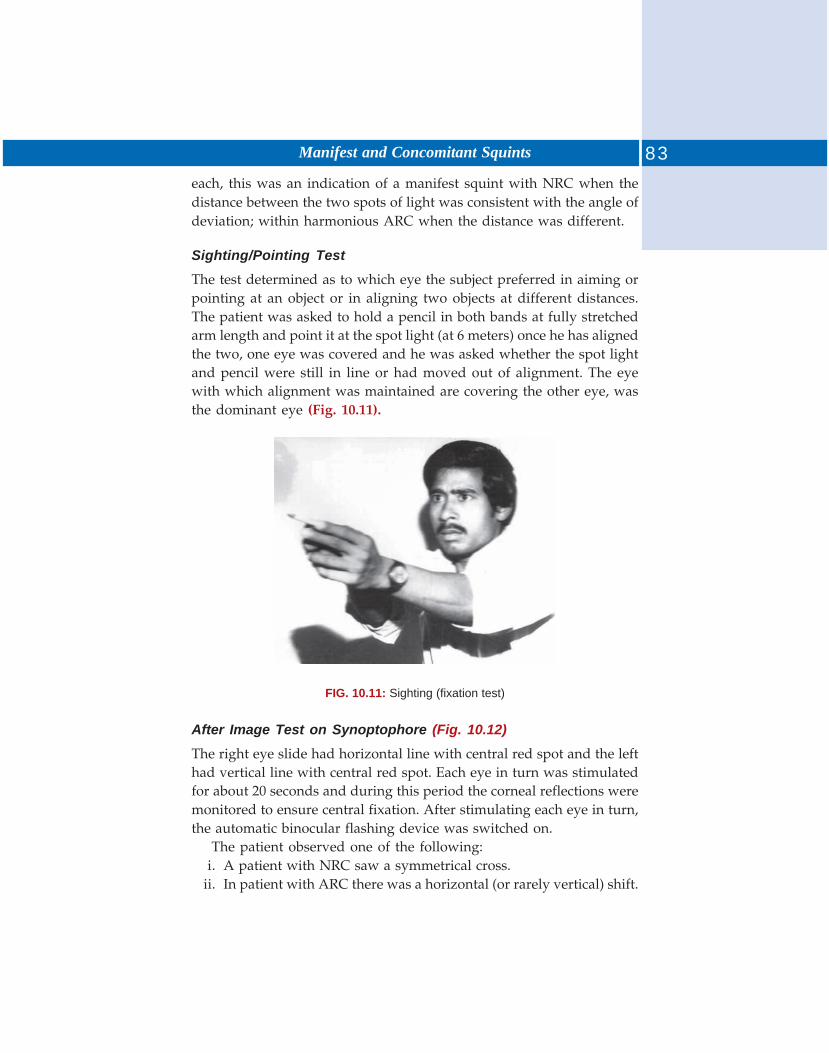
83Manifest and Concomitant Squints
each, this was an indication of a manifest squint with NRC when thedistance between the two spots of light was consistent with the angle ofdeviation; within harmonious ARC when the distance was different.
Sighting/Pointing Test
The test determined as to which eye the subject preferred in aiming orpointing at an object or in aligning two objects at different distances.The patient was asked to hold a pencil in both bands at fully stretchedarm length and point it at the spot light (at 6 meters) once he has alignedthe two, one eye was covered and he was asked whether the spot lightand pencil were still in line or had moved out of alignment. The eyewith which alignment was maintained are covering the other eye, wasthe dominant eye (Fig. 10.11).
FIG. 10.11: Sighting (fixation test)
After Image Test on Synoptophore (Fig. 10.12)
The right eye slide had horizontal line with central red spot and the lefthad vertical line with central red spot. Each eye in turn was stimulatedfor about 20 seconds and during this period the corneal reflections weremonitored to ensure central fixation. After stimulating each eye in turn,the automatic binocular flashing device was switched on.
The patient observed one of the following:i. A patient with NRC saw a symmetrical cross.
ii. In patient with ARC there was a horizontal (or rarely vertical) shift.
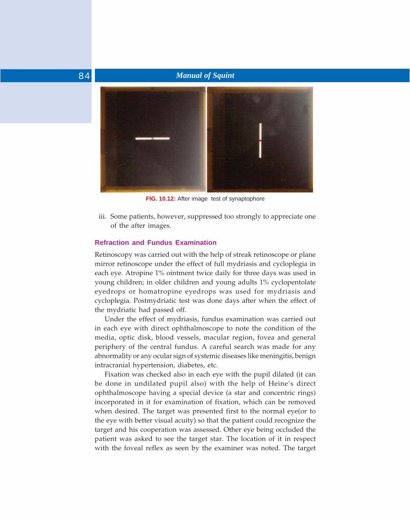
84 Manual of Squint
FIG. 10.12: After image test of synaptophore
iii. Some patients, however, suppressed too strongly to appreciate oneof the after images.
Refraction and Fundus Examination
Retinoscopy was carried out with the help of streak retinoscope or planemirror retinoscope under the effect of full mydriasis and cycloplegia ineach eye. Atropine 1% ointment twice daily for three days was used inyoung children; in older children and young adults 1% cyclopentolateeyedrops or homatropine eyedrops was used for mydriasis andcycloplegia. Postmydriatic test was done days after when the effect ofthe mydriatic had passed off.
Under the effect of mydriasis, fundus examination was carried outin each eye with direct ophthalmoscope to note the condition of themedia, optic disk, blood vessels, macular region, fovea and generalperiphery of the central fundus. A careful search was made for anyabnormality or any ocular sign of systemic diseases like meningitis, benignintracranial hypertension, diabetes, etc.
Fixation was checked also in each eye with the pupil dilated (it canbe done in undilated pupil also) with the help of Heine’s directophthalmoscope having a special device (a star and concentric rings)incorporated in it for examination of fixation, which can be removedwhen desired. The target was presented first to the normal eye(or tothe eye with better visual acuity) so that the patient could recognize thetarget and his cooperation was assessed. Other eye being occluded thepatient was asked to see the target star. The location of it in respectwith the foveal reflex as seen by the examiner was noted. The target
85Manifest and Concomitant Squints
was moved and refixation checked. Confirmation of accuracy of fixationwas also obtained by asking the patient to fixate on different parts ofthe target. Fixation as observed was recorded as foveal, unsteady foveal,erratic or unsteady parafoveal, parafoveal, paramacular, centrocecal,paracecal, divergent and nonfixation. Fixation was also recorded assteady or unsteady. The whole method was repeated for the other eyealso.
Eccentric Viewing vs Eccentric Fixation
1. Eccentric viewing is an intermediate stage between central andeccentric fixation reflex remain oriented towards the fovea, althoughfoveal function is reduced. In eccentric fixation the fixation reflex becomesadjusted to nonfoveal (Paramacular) retinal elements. Eccentric viewingis frequently present in macular retinopathy.
We can differentiate between eccentric fixation and eccentric viewingby visuoscope.
Method: The sound eye is occluded. The examiner projects the visuoscopeasterisk (Star) into (onto) the retinal periphery of the patient. We askthe patient to look directly at the asterisk. Firstly there will be an eyemovement so that the image of the fixation target can form on fovea butthe image will be very dim because foveal function of the patient isreduced (Scotoma, organic lesion). Secondly, the eye will move (again?)so that now the image from fovea can move to peripheral retinal element,where visual acuity may be better than in the fovea. Eccentric viewing ispresent.
2. The first eye movement, displaces the asterisk directly to the fovea.The fixation reflex has adopted itself to peripheral nasal retinal elements.Eccentric fixation is present.
CONCOMITANT SQUINT METHOD OF EXAMINATION
Qualitative Diagnosis of Strabismus
1. Cover test for detection of heterotrophia2. Indirect cover test3. Cover uncover test for detection of heterophoria.
Quantitative Diagnosis of Strabismus
1. Hischberg test2. Prism reflex test of Krimsky
86 Manual of Squint
3. Prism cover test4. Maddox rod test for heterophoria5. Prism dissociation test6. Maddox double prism test for cyclodeviation7. Maddox double rod test for cyclodeviation8. Diplopia test for measurement of ocular deviation.
TREATMENT
The main aim of treatment is to:1. Attain normality of appearance2. To restore binocular single vision in all circumstances.
There are four methods by which a patient can be treated.
Glasses
i. Glasses improve visual acuityii. Lessens or overcome angle of deviation.
Treatment of Amblyopia
It will be discussed in subsequent chapter.
Orthoptic Treatment
The aim is to restore or development of normal function. A number ofpatients who has a weak binocular vision or suppression of the moreametropic eye an effort was make to build binocular vision with orthopticexercises as follows:
Antisuppression Exercises
On cherioscope and chasing and flashing exercises on major amblyoscopewere with the use of simultaneous macular perception slides. It wasgiven in those cases who has complete or partial suppression of moreametropic eye with a view to provide stimulus to the suppressed eye.The exercises were given 10 to 15 minutes daily.
Fusion Exercises
Fusion exercises on major amblyoscope: Fusion exercises were given onthe major amblyoscope with the fusion slides fusion range could beincreased by gradually converging both the tube is of major amblyoscopetill the fusion breaks.
87Manifest and Concomitant Squints
Exercises were give daily or on alternate days for 10 to 15 minutesdepending on the tolerance and convenience of the patient.
Fusion exercises on diploscope: It is based on physiological diplopia andrequires simultaneous use of the eyes.
Home exercises: Home exercises comprising of convergence to near point,(Pencil to nose exercise) and reorganization of physiological diplopiafor near and distance were explained to the patient. Patients wereinstructed to do the exercises almost two to three times daily for 10 to15 minutes.
Operation
Surgery is required for residual angle of squint which is not correctedby glasses in case of accommodative squint. Give preorthoptic exercisein divergent squint to improve the binocular function but it should notbe persisted for long time otherwise convergence spasm will develop,and then, surgery is advocated. The aim of operation is to restore visualaxes to parallelism in all direction of gaze. In neglected cases wheresurgery is carried on for cosmetic reason, one should leave a few degreesof convergence. Since in the passage of time subsequent divergence mayoccur.A. Various weaking operation or extrinsic ocular muscles are:
1. Recession2. Marginal myotomy.
B. Lengthening operation on extrinsic ocular muscles are:1. Simple tenotomy2. Resection.
AMOUNT OF OPERATION
Following empirical rules are useful in chalking out a preoperative plan.1. The larger the deviation, the greater will be the effect of surgery.2. Long-standing deviation (with secondary changes) will require more
surgery then recent deviation.3. More effect is produced per mm of recession or resection in a child
or in patient with small eyes than in an adult or patient with largereyes.
4. Recession is more effective than resection in reducing deviations.
88 Manual of Squint
5. If fusion is present a cure with fusion may be expected, a slight over-correction help obtain the result but if no fusion potential is present,a slight under correction visual produce a stable small angle residualdeviation.
6. The presence of amblyopia makes the result of surgery unpredictableand all such patient should be warned of the possibility of a secondoperation.
7. Esodeviation or exodeviation greater than 50 Δ in a patient withvery poor vision in one eye should be treated with a supramaximalrecession surgery one eye to avoid surgery on better eye.
8. Three muscle surgery for esotropia or exotropia may be required fordeviation greater than 50 Δ but less than 60°.
9. 4 muscle surgery for deviation more than 60°.
Anterior Segment Ischemia
Anterior segment of eye is supplied by 7 anterior ciliary and 2 postciliaryarteries when we do disinsection of the recti, we divide there anteriorciliary arteries and loss of blood to the three muscles (below the age of26 years) and more than 2 muscles in older patient produces some degreeof anterior segment ischemia.
Symptoms and Sign of Anterior Segment Ischemia
Pain, blurring of vision, edema of lid, conjunctiva and cornea, deepanterior chamber with heavy flare, iris atrophy, iris angiography showspoor arterial filling.Treatment: Atropine, topical and systemic steroids.
Faden Operation or Postfixation Suture
It is the weakening procedure on the contralateral synergist muscle of aparalytical lateral rectus. When the muscle is recessed the distancebetween muscle’s origin and insertion is decreased and muscle becomesslack but in Faden operation the muscle is attached to globe althoughwe pass strong posterior fixation suture in the muscle belly so there isno slackness of muscle. Postfixation suture must be strong we can use 3-0 supramid. There are two methods of carrying out Faden’s operation—strong permanent sutures are applied 13mm behind its undetachedinsertion (The appropriate distance of applying posterior fixation suturein case of medial rectus muscle is 13 mm, it is 17 mm in the case of lateralrectus).
First we recess the muscle by 3 mm and then we pass the posteriorfixation suture 13 mm behind the detached insertion.
89Manifest and Concomitant Squints
In Faden operation the effect of recession is increased because allmuscle slackness is taken up by the short length of muscle between thesuture and its point of origin.
This procedure is used on rectus. In DVD (dissociated verticaldivergence) we can use this method to reduce elevation of superior.Rectus by this operation we can increase the effect of recession operationwe can perform this operation in infantile esotropia and nystagmusblockage syndrome.
Adjustable Sutures
Adjustable sutures are useful where it is difficult to predict the result ofconventional recession therapy, e.g. in intermittent.1. Divergence excess type of exotropia2. Vertical muscle palsy3. Cosmetic operation in older patients where there is risk of
postoperative diplopia4. Consecutive exotropia.
The recessed muscle must be sutured in such a way so thatpostoperatively the muscle tendon can be drawn forward or backwardat the time of adjustment (The conjunctival incision has to be left openso that the muscle sutures are easily accessible). This operation isunsuitable in children below 15 years.
ACCOMMODATIONAL SQUINT
Before dealing with accommodational squint proper, it is certain tounderstand the mechanism of accommodation and convergence as wellas their mutual relationship.
Physiology
The association between accommodation and convergence such that wheneach eye undergoes the required amount of accommodation in ordersee a near object. Hence a satisfactory accommodation–convergencesynkinesis results within the occipital cortex and this is an inborn,unconditioned reflex. Accommodation measurement is expressed indiopters and convergence in meter angle. In order to see clearly anobject placed at 1 meter the eye requires one diopter of accommodationas well as 1 meter while of convergence in each eye. Convergencesometimes is also depressed in terms of prism diopters, 1 meter anglebeing equivalent to three prism diopters.
90 Manual of Squint
Under normal conditions, the convergence is influenced not only byaccommodation but also by (i) tone of extrinsic ocular muscles,(ii) proximity of the object and (iii) fusional impulses. Therefore thetotal convergence is composed of the following components.
Accommodative Convergence
It is that part of convergence which is brought about in response to theact of accommodation.
Proximal Convergence
This reflex is stimulated by the sense of nearness of the object and seemsto be independent of accommodation.
Tonic Convergence
It is that convergence which determine position of the visual axis inrelation to each other when eyes are in primary position, fixing a distantobject. It depends on the light that strikes the retina and impulses thatarise in the labyrinths, neck and trunk muscles.
Fusional Convergence
As a result of this reflex the eyes are directed to objects of attention andinterest and are maintained in such a position relative to each other sothat the images of object of fixation fall on the fovea of each eyesimultaneously this reflex exerts a very important influence uponaccommodative convergence. For relationship example, in cases ofsuperable, corrected hypermetropia a greater amount of accommodationis called for, to see a near object clearly. This act of accommodationcould in itself simulate an equal amount of accommodative convergencewhich being excessive would produce a convergent squint. This however,does not happen in all cases of uncorrected hypermetropia becauseparallelism in these cases is thought to be maintained by fusionalconvergence which exerts an influence on accommodative convergenceto nullify the excess of convergence stimulated by excessiveaccommodation. Similarly, in case of uncorrected myopia a reducedamount of accommodation would be needed for fixing a near objectthereby stimulating a lesser amount of convergence. In this case, thefusional convergence comments the deficit in accommodative convergenceand a single binocular vision is maintained. That part of fusionalconvergence which adjusts a deficit of convergence is called positive
91Manifest and Concomitant Squints
relative fusional convergence and that which adjusts the excess ofconvergence is called negative relative fusional convergence. The fusionalconvergence is believed to be mediated through a center in the frontalcortex. If the power to inhibit convergence by fusional reserve (i.e.negative relative fusional convergence) exceeds the excessiveconvergence stimulated by hypermetropia, a squint does not develop.On the other hand, in a case of hypermetropia where accommodativeconvergence exceeds the inhibitory power, manifest convergent squintresults.
Accommodative Convergence/Accommodation (AC/A) Ratio
The amount of accommodative convergence measured in prism dioptersinduced by each diopter of accommodation is called the AC/A ratio.The average value is 3:1 to 5:1 which is usually expressed as 3 to 5 becauseconvergence measured is related to one diopter of accommodation.
Accommodating Squint
Accommodative convergent squint is that squint in which convergentdeviation of the eyes varies according to the amount of accommodationexerted. Due to late development of the ciliary muscle, and child doesnot start taking interest in near objects before the age of two years,squint rarely occurs before the age of 2-2 years although sometimes itstarts at the age of one year.
Classification
Causes in which binocular single vision is present in certain circumstances.
Fully Accommodative Type
Those who when wearing correct glasses enjoy binocular visual acuitybut who when not wearing glasses who a convergent deviation or areduction of their binocular visual acuity if they control the tendency todeviate.
Convergence—Excess Type
Binocular single vision with full binocular visual acuity is present fordistance but there is usually a manifest deviation for near vision evenwith glasses. This type falls broadly speaking into two groups.
Group A: In this group the defect appears to be related to hypermetropia.In these cases some factor is superadded causing overconvergence in
92 Manual of Squint
near vision such a factor may be (a) proximal convergence reflex,(b) defect in the subjective appreciation of the distance of an object fromthe observer, (c) a congenital defect in the action of extrinsic ocularmuscle.
Group B: In this group – (a) the degree of hyper is lower or there maybe no refractive error, (b) additional plus lens do not reduce the manifestdeviation for near vision and may sometimes cause it to increase,(c) There is frequently a vertical defect in addition to manifestconvergence in near vision, (d) The AC/A ratio is markedly higher thannormal, (e) There is marked inability to exercise negative relativepersonal convergence.
Divergence–Insufficiency Type
There is a manifest convergent squint for distance of esophoria for near.It is sometime associated with congenital tropia and following miotictherapy.
Cases in Which Binocular Single Vision is Absent
Partially Accommodative Type
Visual axis are convergent in all circumstances but deviation increasewhere accommodation is exerted and when hypermetropia correctionis removed. This group may be further subdivided:
i. Those patients with normal binocular functionii. Those patients without binocular function
iii. Those who have very weak or anomalous binocular function.
Clinical Investigations
History
When taking the history of squint from the parent it is important tomake sure that the two eyes are not looking in the one direction becausethe word ‘squinting’ is sometimes used to imply ‘screwing up of theeyelids’. One should attempt to discover (a) precisely what the parentor other observer has noticed miss with the child’s eyes and at what agethis anomaly was first noticed, (b) whatever eye is going in or out,(c) whether the squint unilateral or bilateral or each eye, (d) whetherthe squint intermittent or constant, (e) whether squint is increasing,decreasing or stationary, (f) whether squint increases or decreases in
93Manifest and Concomitant Squints
various times of day, and (g) from how long he is using the glasses.Whether angle of squint increases with glasses or it remains the same.
The extent to which various tests need to be carried out tend soonthe characteristics of the condition, age and cooperation of the patientand the duration of the squint allowing tests would cover the examinationrequired in most of the cases.
Refraction and Visual Acuity
Acuity should be tested with and without glasses both for near anddistance.
Refraction: The refractive state of the eye should be carefully determinedunder full cycloplegia so as to uncover total hypermetropia.
Orthoptics Investigations
While doing this test care should be taken to see that accommodation isfully excreted. For this purpose, a small letter for the test type may beused for distance and small letters or picture on the near fixation bar.
Cover Test
Ocular movements: These should be tested with care so as to detect any‘A’ or ‘V’ phenomenon or any vertical anomaly.
Examination with Major Amblyoscope
It is important in increasing the state of binocular function. Themeasurement of the angle of deviation should be undertaken with andwithout glasses.
Measurement of Near Point of Accommodation
There are several methods which can be both uniocular and binocular.
Estimation of the AC/A Ratio
There are several methods which can be employed to measure theAC/A ratio.
Estimation of Negative and Positive Fusional Convergence
As already stated every effort of accommodation is accompanied byaccommodative convergence. If this accommodative convergence isexcessive it is inhibited by negative relative fusional convergence of
94 Manual of Squint
fusional convergence should be made by the following method test isperformed with the patients wearing correcting glasses throughout thetest.
i. The patient is asked to read the smallest possible line of Snellenchart placed at 6 meter distance. The fact that he is fixing binocularlycan be verified by cover test.
ii. Minus spherical lens are inserted before the eyes starting with 0.5Dand then increasing the strength in stages by -0.5D convergent squintappears. The value of greatest minus lens which dermits a clearsingle binocular vision is recorded.
iii. The test is repeated with convex lens similarly and the limit ofclear vision recorded.
iv. The whole test is repeated at 33.3 cm using a small satisfactoryresult for this test with the ability to maintain clear binocular singlevision up to the value of –4.0D for distance and –5.0D sph. For nearvision, this test has been described as a result for relativeaccommodation, it being assured that accommodation can be alteredwhen lens are introduced without any need to inhibit theaccompanying change in convergence. This appears most unlikelyand it is suggested that the term “relative accommodation” isdiscontinued.
Treatment
Although individual cases merit individualized treatment, a general lineof treatment may be described as follows:
Correction of Refraction Error
It should be done at the earliest possible age so that further developmentof the habit of suppression, when accommodation is exerted is prevented.The refraction should be done under complete cycloplegia to correctfull hypermetropia. Over correction of hypermetropia is not desirableregular procedure because it tends insufficiency in later life. Overcorrection has its balance especially in children too young for subjectivetesting provided the over correction secures parallelism of visual axisbut it should never be continued for a prolonged period, any decreasein distant visual acuity on account of this procedure should be explainedto the parents.
95Manifest and Concomitant Squints
Occlusion
Due to late onset of squint and often being intermittent in character it isunusual to find a marked degree of amblyopia in fully accommodativesquint. The inhibition of the fovea in early stage of squint may lead toamblyopia to avoid confusion. The correct spectacle should be wornconstantly and child should be examined at frequent intervals duringinitial stage of occlusion. When partial occlusion is employed the parentand teacher must try to ensure that the child does not give up the glassesotherwise the condition is aggravated by excessive use of accommo-dation. Occlusion may cause than angle of squint to increase.
Orthoptic Treatment
Orthoptic treatment for those cases in which binocular single vision ispresent.
Indications: This treatment is indicated when (i) the clinical shows amanifest deviation when glasses are removed, (ii) he does not appreciatediplopia, (iii) he is unable to straighten his eyes for near fixation.
The treatment may be divided into four stages:1. To overcome suppression, particularly at the convergent angle of
deviation.2. To teach the patient relaxation of accommodation and convergence.3. To teach the patient negative relative fusional convergence and to
improve binocular visual acuity.4. To ensure that the patient has good binocular convergence.
Overcoming Suppression
In giving treatment on the major amblyoscope in order to overcomesuppression when accommodation takes place, the patient should bemade to exert 3 diopters of accommodation either by the insertion of -2D spherical lens in the lens holders of the major amblyoscope, in whichcase he should wear his glasses, or by using lens which are equal to thepatients correction with -3D sphere added. Antisuppression exercisessuch as chasing in and out should be given the use of simultaneousperception slides, with foveal sized fixation picture helps to ensure thatfull accommodation is exerted.
The following exercises may be practiced both in the clinic or at home.
i. Optometric exercises: Wearing red and green glasses the patient isinstructed to practice making a spot light appear alternately red and
96 Manual of Squint
green. If possible, this exercise should being carried out thought thecorrecting lens being worn. At first he may need help by means of rapidalternating covering of the eyes.
ii. Diplopia: Patients who are sufficiently cooperative should be broughtto recognize diplopia when squinting and those who can attain binocularsingle vision may practice recognizing physical diplopia.
The cheiroscope and the pigeon cantonnet stereoscope to valuablefor overcoming suppression.
Teaching Relaxation of Accommodation
At this stage of treatment diplopia should be appropriated so that patientcan instruct how to join the double images to one. However shouldinclude that child should practice maintaining single image. This is anindication to him that his eyes have become straight.
Using a major amblyoscope, the patient should be thought to relaxaccommodation while the angle of the tubes is used to zero or as nearzero as possible. The correction for the patient’s hypermetropia shouldbe reduced by the use of appropriate/lenses inserted into lens holder.Fusion or stereoscopic picture should be used for exercise; the imagewill become increasingly blurred as the visual axes approach parallelism.
When amblyopia has been corrected suppression, the patient has beentaught to control/upon his power of negative relative fusionalconvergence. If this is found to be adequate, treatment/may bediscontinued apart from wearing the glass. Patient should report forregular follow up examinations. The strength of the glasses should bereduced when negative relative fusion convergence seems able to controlthe additional accommodative convergence. If negative relative fusionalconvergence is found to be deficient or patient is further/treatment maybe undertaken to increase the power of inhibiting accommodativeconvergence and increasing negative relative fusional convergence.
Teaching Negative Relative Fusional, Convergenceand Improving Binocular Visual Acuity
There are a variety of instruments which may be used in carrying outthese exercises, such instruments include the major amblyoscope barReader, Holmes and Asher law stereoscopes, remiseparator andstereorgrance cards held in the hand.
With a major amblyoscope exercises may be given during a gradualreduction of the patient’s hypermetropic correction the –1.D sphere are
97Manifest and Concomitant Squints
placed in the lens holders of the instruments during simple fusion slideshe is to describe the picture as accurately as he can taking care that itdoes not become double and that suppression does not occur. When hedoes this, 2.D later – 3D sphere should be substituted and more detailedslides should be used in place of simpler one. Fusion should be maintainedas near to zero as possible and adduction exercises should then bepracticed while patient is asked to maintain a single clear vision.
Teaching Good Binocular Convergence
Simple convergence exercises should be taught and may be practised athome, care being taken to ensure that accurate convergence take placewithout suppression of either eye. The patient should be encouraged tobe aware of physiological diplopia when convergence fails.
Binocular convergence may also be improved by use of sterograme/cards held in hand and by use of a prism bar and also by convergenceexercises using the major amblyoscope.
Miotic Therapy
Miotics are drugs which stimulate accommodation peripherally bycontraction of ciliary muscle and also constrict the pupil. By virtue ofpupil contriction, clearity of vision is improved and by both theseperipheral actions there is a abduction in subjective effort ofaccommodation in order to see clearly. A reduction in the subjectiveeffort of accommodation less accommodation convergence, the resultbeing that clear vision is achieved without the accurance of a manifactconvergent deviation. Miotics are said to be useful for a child also youngfor orthoptic treatment. They are also helpful postoperatively if aconvergent deviation still occurs on accommodation.
Prerequisite
i. Equal visual acuity in either eyeii. Absence of suppression
iii. Presence of fusion with a good range fusioniv. Binocular single vision for distance (with glasses if worn)v. Binocular single vision for near when using the miotics.
The miotics most commonly used are:i. Pilocarpine 1%
ii. DFP (di-isopropyl fluorophosphonate) .005%, 01%iii. PI (Phospholine iodide) 0.06%, 0.125% or 0.25% 0.25%.

98 Manual of Squint
iv. Pilocarpine is instilled three times daily being later reduced as thebinocular visual acuity improves.
Disadvantage
Its action wars off within a few hour. This provides a practical difficultywith children who are at the school all day.
DFP needs only one instillation every 24 hrs. The drops are susceptibleto absorption of moisture so parents should be warned to keep the bottletightly stoppered.
Phospholine iodide is usually instilled once each twenty hour andgiven at night. At first a solution of 0.25% or 0.125% may be used butsubsequently this may be reduced to 0.06%.
It is important that miotic therapy should be continued with orthopticexercises, including home exercises. Throughout the treatment thereshould be regular and careful supervision of the patient in the orthopticdepartment. The patient should be examined to see if there is anyappearance of yet in the pigment layers of iris. In some cases there haveoccurred where the drug has been given over a prolonged period, butthis is lees likely to occur when the strength of drops does not exceed0.25%.
Surgical Treatment
In some case optical and orthoptic treatment to eliminate manifest squintnear fixation, surgical treatment in indicated to achieve binocular singlevision for all distances on fixation. It is important to uncover the fullamount of variation with and without glasses by making the patientexert all accommodation while determining the angle of squint both fornear and distance.
Although on theoretical grounds a bilateral recession of redial rectimight be considered as the operation of choices, recess the medial rectusand resect the ipsilateral medial rectus thus leaving the opposite eye forfurther surgery undertaken in cases of convergence excess type ofbinocular function is good.
If the child is old enough the operation should both loaded andfollowed by appropriate orthoptic exercise in order to achieve maximumbinocular function. In the case of young child surgery may be postponedif the deviation is not frequent and there is no danger of disturbance inthe binocular function.
99Manifest and Concomitant Squints
Criteria for Cure
The patient should have comfortable binocular vision with and withoutglasses for near and distance. If the hypermetropia exceeds + 3D binocularsingle vision should be maintained when correction reduced by 3 diopters.Binocular visual acuity with classes should be equal to visual acutiy ofeach eye or of the weaker eye if it is so.
The patient should be able to bar read N5 with glasses and alsowhere -3D is added to them.
Binocular convergence be 8 cm and should be well-maintained.
Just to Summary the Accommodation Squint
Esotropia (Convergent squint)
i. Accommodational esotropiaii. Nonaccommodational esotropia.
Accommodational ESO is associated with the without (N) accommo-dation. A normal person or one with normal refractive error in order tosee a nearly object, he has to make his lens convex. It is done bycontraction of ciliary muscles.
Accommodative Squint
Relaxation of suspensory ligament. Normally, the lens is kept flattenedby suspensory ligaments.
During accommodation there is contraction of ciliary muscle andrelaxation of suspensory ligament—Lens becomes convex in hyper-metropia which is undercorrected or uncorrected person requires moreaccommodation (over accommodation) so requires over convergence.
Another type of accommodative esotropia.• Not associated with any refractive error. This is because of
neuromuscular abnormality.• Age–2-3 years.• Family history of squint is present.• To start with it is intermittent in nature but later it becomes constant.
It is more for near than for distance.• Cover test in accommodative squint — should use a fixation bar with
a picture over it instead of a torch. This compel the child toaccommodate in order to see properly.
100 Manual of Squint
Treatment of Accommodative Eso
• Refraction under full cycloplegia 1% atropine drop BD × 3 days.
• Give full correction.
• One deviation is corrected the no. of glasses paper step by step whendeviation is absent for distance and present for near bifocal lenseswith increased number for near or give miotic treatment.
– Pilocarpine BD
Or Phosphoeria iodido
Or DFP (rarely).
Action of Mioties
Peripheral stimulation of accommodation and constriction of pupil sothat child sees clearly, central accommodation does not come into play.
Indication of Miotic Theory
i. Small or no refractive errorii. Equal and good visual acuity
iii. When AC/A ratio is increased.
Treatment by surgery:
i. For nonaccommodation part of squint.ii. If amblyopia is present—treatment improves the visual acuity.
iii. Visual acuity poor. Treatment of amblyopia.iv. Improve BF by orthoptic treatmentv. Nonaccommodational squint—surgery is advocated.
EXODEVIATION
Introduction
Generally speaking divergent squint develops due to breakdown ofbinocular reflexes before they are become adequately strong. Ascomparison to convergent squint, divergent squint is less common, theratio being 1:4. The divergent squint is more common in females. Ittends to increase with age. It may pass through latent, or intermittentphase and it may be absent in the morning and tends to increase withfatigue towards the end of the day. Usually, there is no refractive error.Amblyopia and abnormal retinal correspondence are rare in
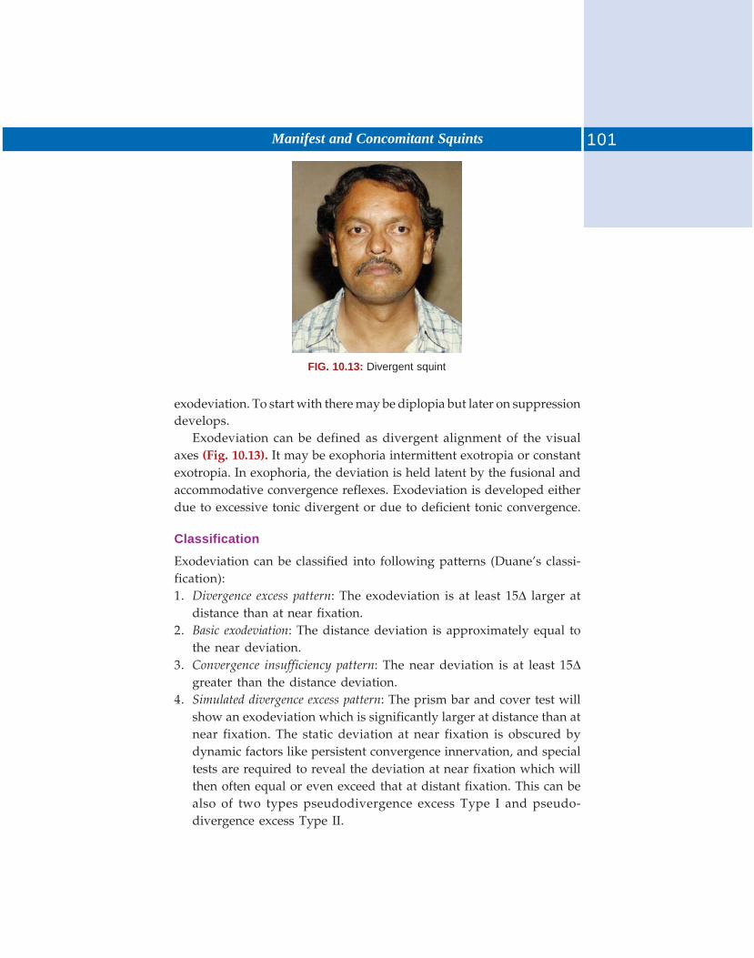
101Manifest and Concomitant Squints
exodeviation. To start with there may be diplopia but later on suppressiondevelops.
Exodeviation can be defined as divergent alignment of the visualaxes (Fig. 10.13). It may be exophoria intermittent exotropia or constantexotropia. In exophoria, the deviation is held latent by the fusional andaccommodative convergence reflexes. Exodeviation is developed eitherdue to excessive tonic divergent or due to deficient tonic convergence.
Classification
Exodeviation can be classified into following patterns (Duane’s classi-fication):1. Divergence excess pattern: The exodeviation is at least 15Δ larger at
distance than at near fixation.2. Basic exodeviation: The distance deviation is approximately equal to
the near deviation.3. Convergence insufficiency pattern: The near deviation is at least 15Δ
greater than the distance deviation.4. Simulated divergence excess pattern: The prism bar and cover test will
show an exodeviation which is significantly larger at distance than atnear fixation. The static deviation at near fixation is obscured bydynamic factors like persistent convergence innervation, and specialtests are required to reveal the deviation at near fixation which willthen often equal or even exceed that at distant fixation. This can bealso of two types pseudodivergence excess Type I and pseudo-divergence excess Type II.
FIG. 10.13: Divergent squint
102 Manual of Squint
Exodeviation can also be of following types:1. Primary exodeviation2. Secondary exodeviation—due to loss of vision in one eye.3. Consecutive exodeviation—due to over correction of convergent
squint.
Investigation
History
Investigations start with the history. In exodeviation, certain points tobe noted during taking history:1. Mode of deviation—Outward deviation suggests exodeviation.2. Age of onset—Age of onset of the majority of the cases of exodeviation
is usually late. That is why retraining is better in case of exodeviations3. Mode of onset—Exodeviations begin as an exophoria which may
deteriorate into intermittent and constant exotropia, as suppressionoccurs.
4. Progression of deviation—The deviation increases in condition ofFatigue or ill-health. Divergence excess type of deviation tends toremain more or less static, whereas with simulated divergence excesstype the near deviation tends to increase. In convergence weaknesspattern, there is a tendency for the deviation to increase.
5. Ocular symptom—In exophoria and intermittent exotropia patientmay complaint of following symptoms:• Blurred vision• Difficulty in focusing• Difficulties with prolonged period of near work, headache• Eyeache• Diplopia• Photophobia• Micropsia.
6. History of using spectacles or prisms: Exodeviation may occur in acquiredmyopia, unilateral anisometropic myopia or in myopic astigmatism.So there may be history of using spectacles for those refractive errors.There may also be history of using base—in prisms in case ofexodeviation.
7. Family history: In exodeviation, there is frequently a family history ofsquint.
103Manifest and Concomitant Squints
Visual Acuity
Visual acuity is tested both uniocularly and binocularly with or withoutglasses. It is tested both for distance and near.
In primary exodeviation, amblyopia is the exception rather than rule,but if the deviation is constant and unilateral, there may be some defectof visual acuity or suppression in the squinting eye.
In intermittent exodeviation, there may be impaired binocular visualacuity due to over exercise of convergence and consequently accommo-dation in order to achieve control of the deviation.
In presbyopia exophoria or intermittent exodeviation may occur.
Refraction
In exodeviation some refraction errors may be detected—like acquiredmyopia, unilateral anisometropic myopia or myopic astigmatism.
Exophoria my be found witha. Bilateral acquired myopia, due to reduction in the demand for
accommodative efforts.b. Presbyopia, as the near point recedes and the bond between
accommodation and convergence is weakened.
External, Examination
To rule our pseudoexodeviation external ovular examination is carriedout. Pseudoexodeviation may be produced by:1. A large positive angle alpha2. Wide interpupillary distance3. Exophthalmos4. A wide palpebral fissure.
Head Posture
Chin elevation – in some cases of exodeviation chin is slightly elevatedin order to favor a more convergent position of the eyes associated withdepression.
Cover Test (Fig. 10.14)
In exodeviation, cover test is performed at 1/3 meter, 6 meters and atfar distance, beyond 6 meters.
Depending on the type of exodeviation the cover test may give thefollowing informations:
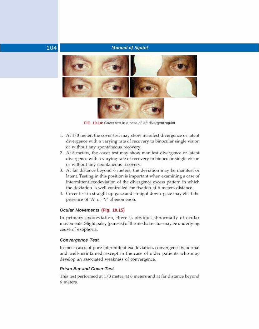
104 Manual of Squint
FIG. 10.14: Cover test in a case of left divergent squint
1. At 1/3 meter, the cover test may show manifest divergence or latentdivergence with a varying rate of recovery to binocular single visionor without any spontaneous recovery.
2. At 6 meters, the cover test may show manifest divergence or latentdivergence with a varying rate of recovery to binocular single visionor without any spontaneous recovery.
3. At far distance beyond 6 meters, the deviation may be manifest orlatent. Testing in this position is important when examining a case ofintermittent exodeviation of the divergence excess pattern in whichthe deviation is well-controlled for fixation at 6 meters distance.
4. Cover test in straight up-gaze and straight down–gaze may elicit thepresence of ‘A’ or ‘V’ phenomenon.
Ocular Movements (Fig. 10.15)
In primary exodeviation, there is obvious abnormally of ocularmovements. Slight palsy (paresis) of the medial rectus may be underlyingcause of exophoria.
Convergence Test
In most cases of pure intermittent exodeviation, convergence is normaland well-maintained, except in the case of older patients who maydevelop an associated weakness of convergence.
Prism Bar and Cover Test
This test performed at 1/3 meter, at 6 meters and at far distance beyond6 meters.
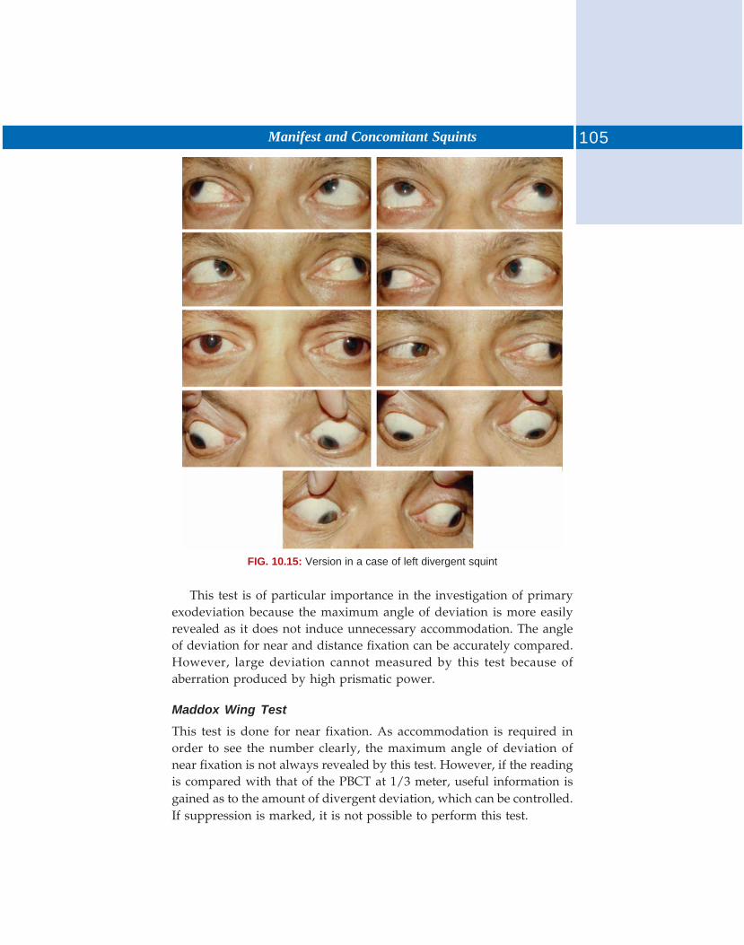
105Manifest and Concomitant Squints
FIG. 10.15: Version in a case of left divergent squint
This test is of particular importance in the investigation of primaryexodeviation because the maximum angle of deviation is more easilyrevealed as it does not induce unnecessary accommodation. The angleof deviation for near and distance fixation can be accurately compared.However, large deviation cannot measured by this test because ofaberration produced by high prismatic power.
Maddox Wing Test
This test is done for near fixation. As accommodation is required inorder to see the number clearly, the maximum angle of deviation ofnear fixation is not always revealed by this test. However, if the readingis compared with that of the PBCT at 1/3 meter, useful information isgained as to the amount of divergent deviation, which can be controlled.If suppression is marked, it is not possible to perform this test.
106 Manual of Squint
Maddox Rod Test
This is done for distance fixation (at 6 meters distance). This test is alsonot possible to perform in the presence of suppression.
If the deviation elicited by Maddox wing test is more than that ofMaddox rod test then it indicated convergence weakness pattern ofexodeviation. If the deviation elicited by Maddox rod test is more thanthat of Maddox wing test then it indicated divergence excess pattern ofexodeviation.
Synoptophore Examination
Suppression occurs at the divergent angle. To estimate binocular functionan attempt should be made in the controlled position as well as at themaximum divergent position:a. With the deviation controlled—Good binocular function with normal
retinal correspondence is usually present. Various devices may benecessary in order to stimulate accommodation and thereby maintainthe controlled position. Such methods include the use of –3.0D sph,lenses before each eye, slides of exercise of convergence to a smalltest object.
b. At the maximum angle of divergence:Estimation on the synoptophore may reveal one of the followingresponses:
i. Simultaneous macular perception with normal retinal corres-pondence. If suppression is dense, this may only be demons-trational with kinetic stimulation.
ii. Lack of retinal correspondence due to gross suppression. Thelion (fixation slide) may be seen to the right or left of the cagebut disappears as the patient tries to superimpose them.
iii. Retinal congruity—The patient is quite unable to related the twoimages. The lion is never seen to approach the cage.
The maximum angle deviation may be difficult to measure if thedeviation is well-controlled. Alternate flashing or occlusion of one eyemay help to elicit the true angle. The use of large simple slides also mayhelp to elicit the angle.
Diplopia Test
In exodeviation, crossed diplopia or suppression of one eye may beelicited.
107Manifest and Concomitant Squints
Special Tests for Exodeviation
In exodeviation, two special tests are carried out:1. Occlusion test2. +3.0D sph, lens test.
Occlusion TestThis test is done to differentiate between true and simulated divergenceexcess pattern. By occlusion, the fusional, the fusional stimuli is removed.After measuring the deviation with prism bar cover test at near anddistant fixation, one eye in covered for 30 to 45 mins and the deviationis increased after occlusion for 30 to 45 mins. In true divergence excessivenear deviation is not influenced by occlusion.
+3.0D Spherical Lens Test+3.0D spherical lenses suspend accommodation and thus suspendaccommodative convergence. In exodeviation with low AC/A ratio, theangle of deviation will increase slightly when measured through +3.0Dspherical lenses. On the other hand, exodeviation with a high AC/Aratio, if the deviation is measured through +3.0D sph lenses, it will increasesubstantially at near fixation.
Management of Exodeviation
Optical Treatment
Refraction should be performed under cycloplegia and the correct glassesprescribed if indicated. If myopia, unilateral or bilateral, is present, itsimportant. Small degrees of hypermetropia or hypermetropic astigmationare best left uncorrected.
Concave lens: Over-correcting concave lenses can be used to stimulateconvergence by inducing accommodation thus aiding control ofexodeviation concave lenses ranging in alternative from 2 to 4 D areadded to the patient’s refractive errors. The main value of concave lensesis to defer the surgical procedure.
Prism: Base in prism can be used to compensate the deviation in childrento allow the continued binocular single vision. Prism has got no curativevalue. It only allows postponement of surgery.
Tinted glass: It is recognized that bright light is a dissociating factor. Byreducing of light entering the eye, tinted glasses can improve the patient’scontrol over exodeviation.
108 Manual of Squint
Surgical Treatment
Operation is usually necessary if a complete cure is to be obtained.
Aim of surgery: The aim of surgery is either to correct deviation or toover correct it slightly, leaving the patient with slight convergence withuncrossed diplopia. This small exodeviation usually disappears duringthe immediate postoperative period and the final results may be moresatisfactory.
Suitable age for surgery: In extreme youth, it is rarely necessary toundertake operation so long as the deviation remains intermittent. Inthe case of a young child, it is better to wait until the age of four or fiveor even later, when cooperation for examination and treatment is morereliable.
Choice of Surgical Procedure
i. In case of true divergence excess pattern, bilateral lateral recessionis the choice of operation.
ii. In basic exodeviation or the simulated divergence excess patterncombination of recession of lateral rectus with resection of medialrectus of the nondominant eye is the preferred choice.
iii. In case of convergence weakness pattern of exodeviation, bilateralmedial recti resection is the operation of choice.
Orthoptic Treatment
If the power of convergence of defective orthoptic exercises to improvethis function are indicated. But if convergence spasm. Preoperativeorthoptic treatment should be confined to the elimination of suppressionand not to the encouragement of convergence.
Preoperative Treatment
If visual acuity of the two eyes is unequal a period of occlusion may beneeded in order to equalize it. Treatment to eliminate suppression atthe divergent angle may be undertaken as follows:
i. By exercise with the synoptophore: Simultaneous perception slideswith foveal sized fixation pictures are used.
ii. By teaching the patient to recognize diplopia with the aid of redand green goggles and spot light.
Convergence exercise is not advised preoperatively because overconvergence may result postoperatively.
109Manifest and Concomitant Squints
Postoperative Treatment
In the postoperative period orthoptic exercise are advised in order tohelp maintain comfortable binocular single vision and to improve therange of fusion. Instruments like Ascher Law stereoscope and thediploscope and Remy separator are particularly useful for the post-operative orthoptic exercises. Stereograms may be used for homeexercises.
Standard for regarding the patient as orthoptically satisfactory incase of primary exodeviations:1. The patient should be symptom-free2. Binocular single vision for near and distance (with or without glasses)
should be as good as the uniocular visual acuity of the less efficienteye
3. There should be no manifest deviation. The cover test for near anddistance fixation should reveal only a small degree of latent deviationwith a rapid recovery to binocular single vision
4. Normal binocular function should be demonstrate with a normalhorizontal fusion range
5. Binocular convergence should be well and easily maintained.
MICROFIXATION SYNDROME (MICROTROPIA)
Microstrabismus is also called by the name of retinal slip, fixationdisparity, esotropia with fixation disparity, strabismus spuriousmicrotropia unilateral anomalous fusion, microtropia.
As the achievement of binocular single vision in patients withstrabismus become more and more important. It became evident thatthere was a small group of patients with residual strabismus which invitedfurther attention. Then very small angle of deviation (8 prism dioptoror less). They had no diplopia and fusional vergence amplitudes weregood microstrabismus is characterized by central suppression.
Scotoma, a very active peripheral binocular vision unharmoniousabnormal retinal correspondence was a common finding slightamblyopia.
Patient with unilateral intraocular lesion also had central suppression.They had straight eyes and peripheral fusion. ‘Fixation disparity’ denotethe inexactness of intersection of visual axes at the point of fixation whilebinocularly fixating. Fixation disparity is a physiologic entity andmonofixation syndrome is a pathological one.
In microtropia, there is a high prevalence of anisometropia.
110 Manual of Squint
Etiology
Monofixation syndrome:1. Primary2. Secondary.
And the presence of central suppression area in the deviated eye:a. Secondary to strabismus—usually esotropes who have achieved
maximum correction, may also be found occasionally in exotropes.b. Secondary to anisometropiac. In cases of unilateral macular lesions.
Microbiology
Anisometropia is another etiological factor responsible for disparity inthe clarity of images. Bais and fusion predisposes to development ofcentral suppression.
In microtropia—an interesting feature in these patients who showdeviation are cover—uncover rest is that the amount of deviation byprism and alternate cover test is greater than that elicited by simultaneousprism and cover test. This is became part of the deviations is madelatent by peripheral fusion which is unmasked by alternate cover test.Simultaneous prism and cover test elicits only manifest deviation.
Presence of binocular facultative scotoma is the one constant featureof monofixation syndrome. The scotoma is demonstrated by binocularperimetry in which the two eyes are dissociated with the help of red-green glasses.
Another test that demonstrates this scotoma is the 4 prism diopter(PD) base out prism test. AO vectographic project. O-chart slide alsogives a quick means of demonstrating the scotoma.
Diagnostic Method
Besides alternate cover test simultaneous prism and cover test, followingare the sensory tests that confirm the diagnosis of monofixationsyndrome.
Monofixation Syndrome
1. Worth form dot-test (Fig. 10.16). It is done for both near (33 cm) anddistance (6 meters). This test often reveals the presence of scotomaand is a quick dated means of evaluating peripheral fusion whendone for distance, the dot 3 mm subtend an angle of 1.25° at thenodal point of the eye and for thus near, approximately 6°. A
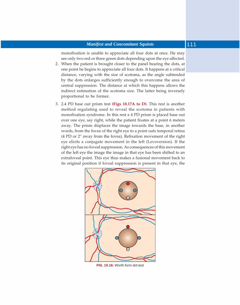
111Manifest and Concomitant Squints
FIG. 10.16: Worth form dot test
monofixation is unable to appreciate all four dots at once. He maysee only two red or three green dots depending upon the eye affected.
2. When the patient is brought closer to the panel bearing the dots, atone point he begins to appreciate all four dots. It happens at a criticaldistance, varying with the size of scotoma, as the angle subtendedby the dots enlarges sufficiently enough to overcome the area ofcentral suppression. The distance at which this happens allows theindirect estimation of the scotoma size. The latter being inverselyproportional to be former.
3. 2.4 PD base out prism test (Figs 10.17A to D). This rest is anothermethod regulating used to reveal the scotoma in patients withmonofixation syndrome. In this rest a 4 PD prism is placed base outover one eye, say right, while the patient fixates at a point 6 metersaway. The prism displaces the image towards the base, in anotherwords, from the fovea of the right eye to a point outs temporal retina(4 PD or 2° away from the fovea). Refixation movement of the righteye elicits a conjugate movement in the left (Levoversion). If theright eye has no foveal suppression. As consequences of this movementof the left eye the image the image in that eye has been shifted to anextrafoveal point. This eye thus makes a fusional movement back toits original position if foveal suppression is present in that eye, the
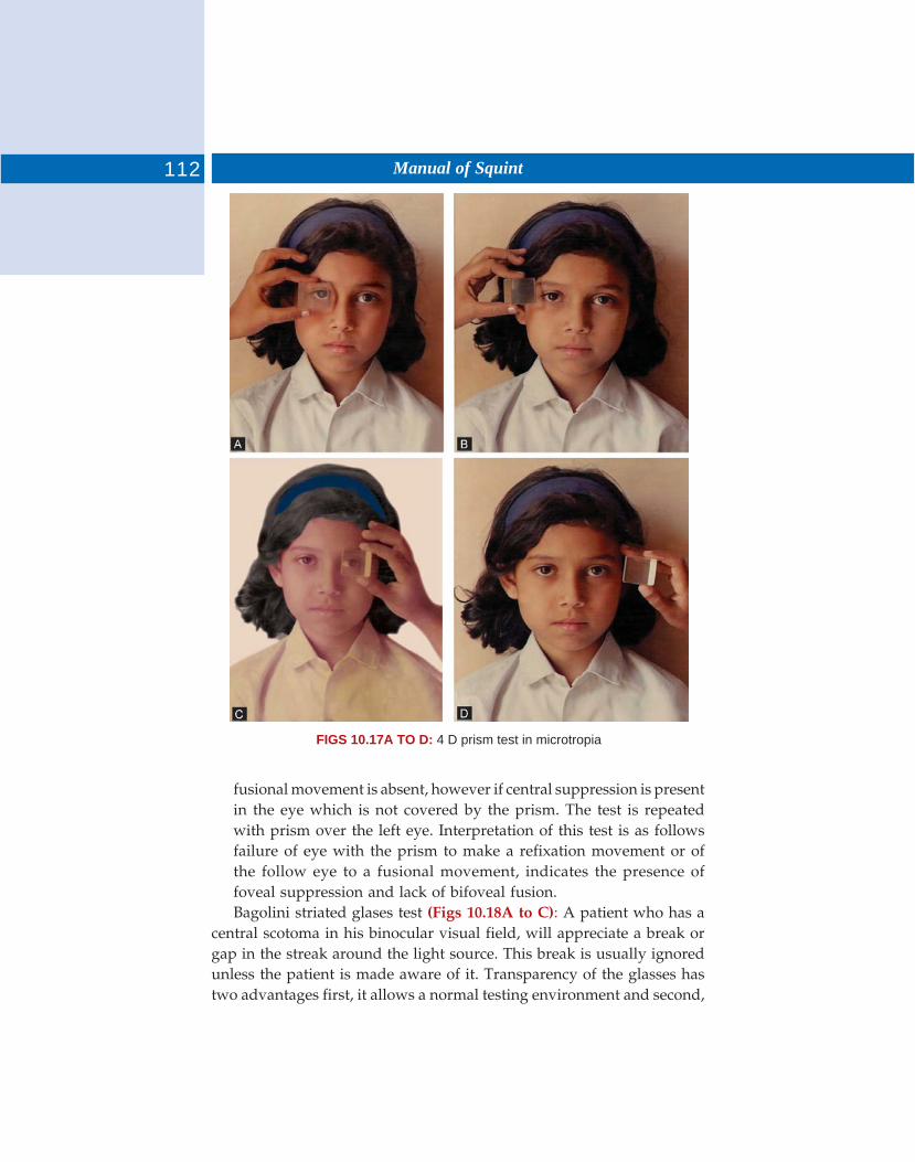
112 Manual of Squint
FIGS 10.17A TO D: 4 D prism test in microtropia
fusional movement is absent, however if central suppression is presentin the eye which is not covered by the prism. The test is repeatedwith prism over the left eye. Interpretation of this test is as followsfailure of eye with the prism to make a refixation movement or ofthe follow eye to a fusional movement, indicates the presence offoveal suppression and lack of bifoveal fusion.Bagolini striated glases test (Figs 10.18A to C): A patient who has a
central scotoma in his binocular visual field, will appreciate a break orgap in the streak around the light source. This break is usually ignoredunless the patient is made aware of it. Transparency of the glasses hastwo advantages first, it allows a normal testing environment and second,
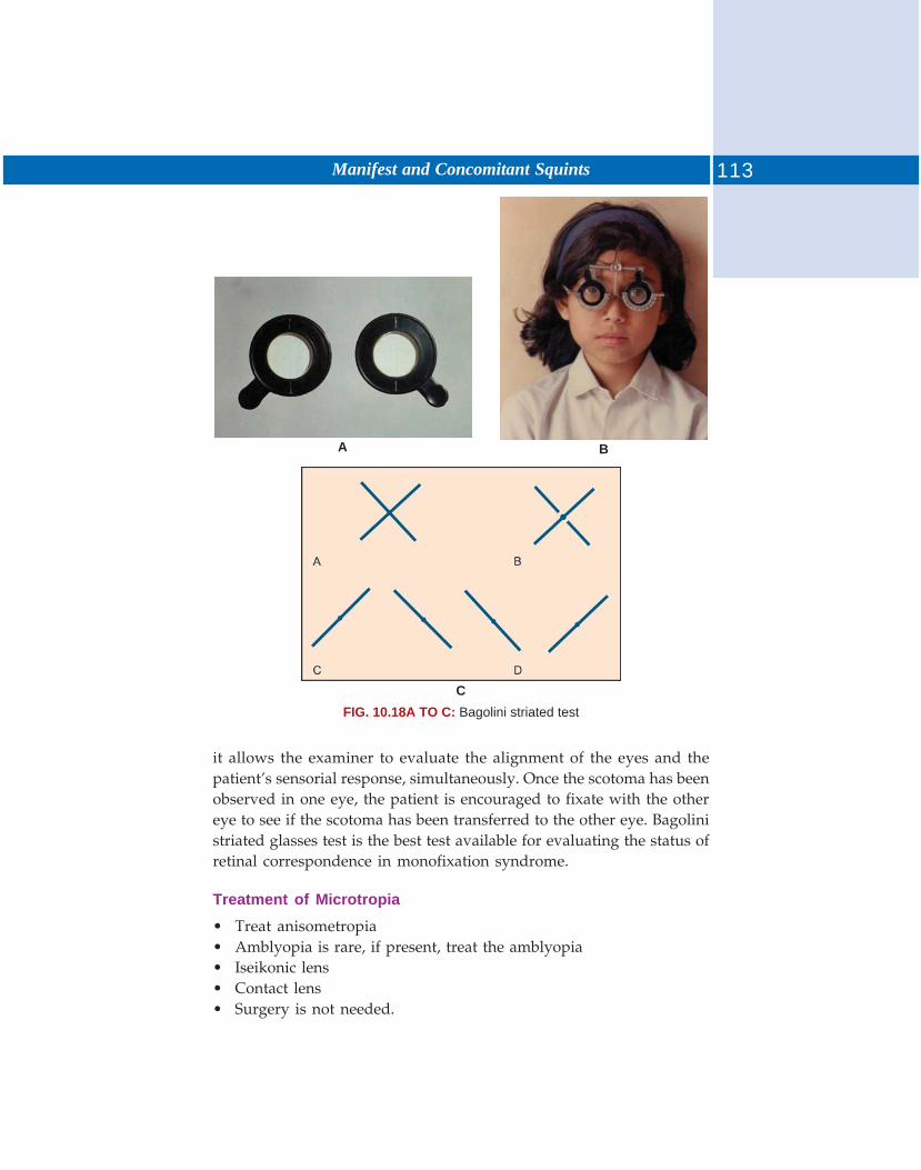
113Manifest and Concomitant Squints
A B
CFIG. 10.18A TO C: Bagolini striated test
it allows the examiner to evaluate the alignment of the eyes and thepatient’s sensorial response, simultaneously. Once the scotoma has beenobserved in one eye, the patient is encouraged to fixate with the othereye to see if the scotoma has been transferred to the other eye. Bagolinistriated glasses test is the best test available for evaluating the status ofretinal correspondence in monofixation syndrome.
Treatment of Microtropia
• Treat anisometropia• Amblyopia is rare, if present, treat the amblyopia• Iseikonic lens• Contact lens• Surgery is not needed.
Paralytic squint is that type of squint in which the eyes move in an inco-ordinated manner so that angle of squint varies in different directionsof gaze and on changing fixation from one eye to the other. This iscaused by the motor imbalance of one or several extraocular muscles.The characteristic features of the paralytic squint are:a. A motor deficiency in the field of action of the paretic muscleb. Diplopia or double visionc. Secondary deviation is greater than the primary deviationd. Compensatory head posturese. Absence of suppression and amblyopia.
ETIOLOGY
Paralysis can be due to:a. Lesion of the nerveb. Lesion of the muscles.
Lesion of the nerve can be at the level of nucleus, nerve roots, nervetrunks or it can be a congenital absence or abnormalities of nerve nucleus.It may be due to:a. Injuryb. Inflammation, e.g. Syphilis, disseminated sclerosis, etc.c. Vascular diseases, e.g. Hemorrhage, thrombosis aneurysms,
arteriosclerosis, etc.d. Neoplasms, e.g. Brain tumore. Toxic, e.g. alcohol, lead, carbon monoxidef. Degeneration, e.g. Chronic nuclear ophthalmoplegiag. Other diseases like, thyroid ophthalmoplegiah. Myasthenia gravis and ocular myopathiesi. Displacement of the visual axis of one eye so that parallelism with
that of the other eye is lost. This may occur in:1. Injuries like blow out fractures causing damage to the supports of
the eyeball.
11 Paralytic Squints
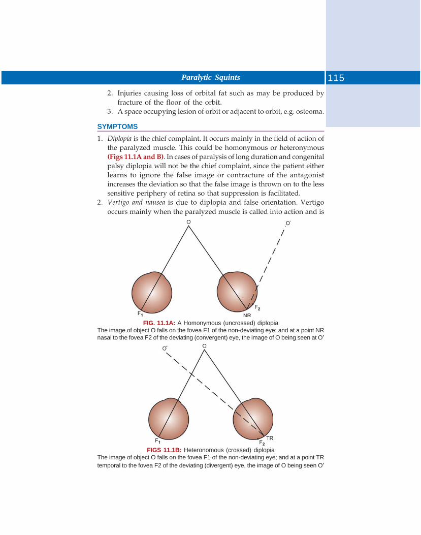
115Paralytic Squints
2. Injuries causing loss of orbital fat such as may be produced byfracture of the floor of the orbit.
3. A space occupying lesion of orbit or adjacent to orbit, e.g. osteoma.
SYMPTOMS
1. Diplopia is the chief complaint. It occurs mainly in the field of action ofthe paralyzed muscle. This could be homonymous or heteronymous(Figs 11.1A and B). In cases of paralysis of long duration and congenitalpalsy diplopia will not be the chief complaint, since the patient eitherlearns to ignore the false image or contracture of the antagonistincreases the deviation so that the false image is thrown on to the lesssensitive periphery of retina so that suppression is facilitated.
2. Vertigo and nausea is due to diplopia and false orientation. Vertigooccurs mainly when the paralyzed muscle is called into action and is
FIGS 11.1B: Heteronomous (crossed) diplopiaThe image of object O falls on the fovea F1 of the non-deviating eye; and at a point TRtemporal to the fovea F2 of the deviating (divergent) eye, the image of O being seen O′
FIG. 11.1A: A Homonymous (uncrossed) diplopiaThe image of object O falls on the fovea F1 of the non-deviating eye; and at a point NRnasal to the fovea F2 of the deviating (convergent) eye, the image of O being seen at O′
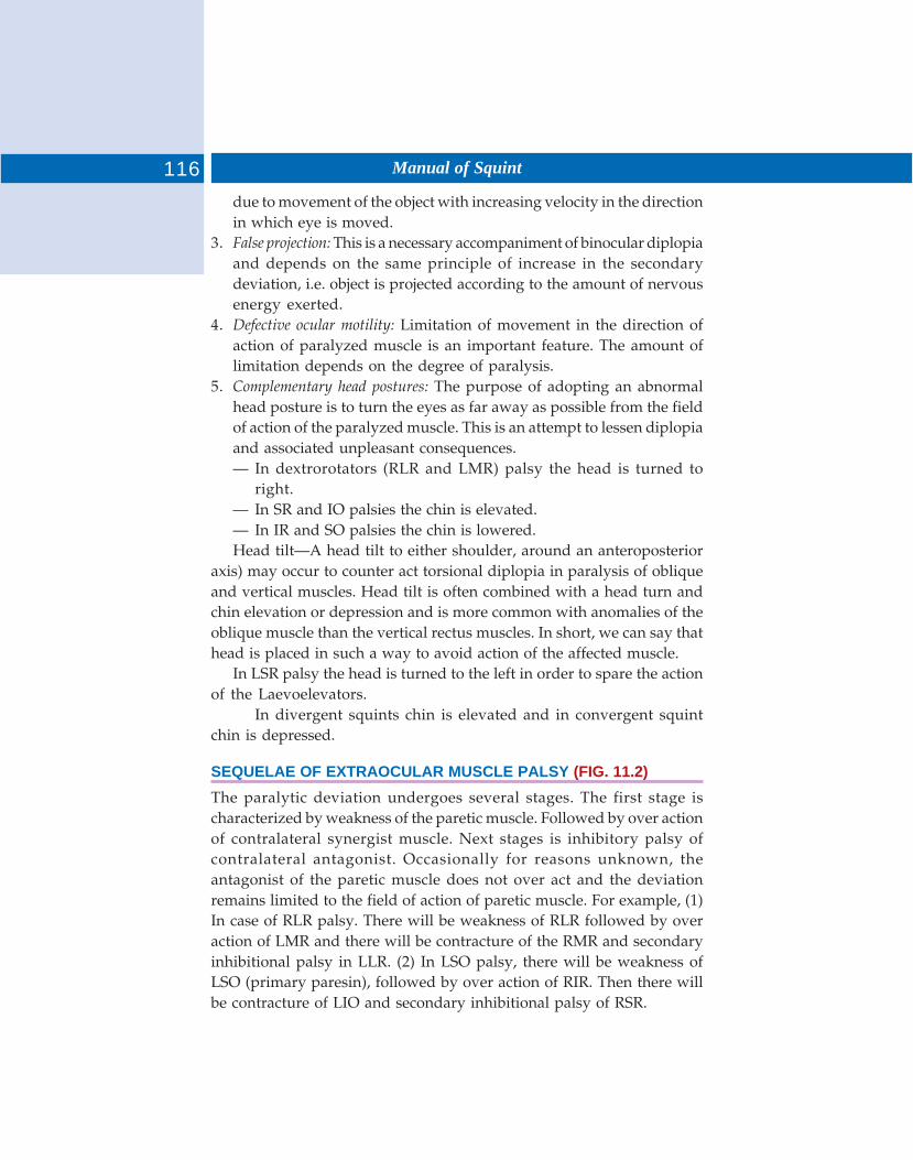
116 Manual of Squint
due to movement of the object with increasing velocity in the directionin which eye is moved.
3. False projection: This is a necessary accompaniment of binocular diplopiaand depends on the same principle of increase in the secondarydeviation, i.e. object is projected according to the amount of nervousenergy exerted.
4. Defective ocular motility: Limitation of movement in the direction ofaction of paralyzed muscle is an important feature. The amount oflimitation depends on the degree of paralysis.
5. Complementary head postures: The purpose of adopting an abnormalhead posture is to turn the eyes as far away as possible from the fieldof action of the paralyzed muscle. This is an attempt to lessen diplopiaand associated unpleasant consequences.— In dextrorotators (RLR and LMR) palsy the head is turned to
right.— In SR and IO palsies the chin is elevated.— In IR and SO palsies the chin is lowered.Head tilt—A head tilt to either shoulder, around an anteroposterior
axis) may occur to counter act torsional diplopia in paralysis of obliqueand vertical muscles. Head tilt is often combined with a head turn andchin elevation or depression and is more common with anomalies of theoblique muscle than the vertical rectus muscles. In short, we can say thathead is placed in such a way to avoid action of the affected muscle.
In LSR palsy the head is turned to the left in order to spare the actionof the Laevoelevators.
In divergent squints chin is elevated and in convergent squintchin is depressed.
SEQUELAE OF EXTRAOCULAR MUSCLE PALSY (FIG. 11.2)
The paralytic deviation undergoes several stages. The first stage ischaracterized by weakness of the paretic muscle. Followed by over actionof contralateral synergist muscle. Next stages is inhibitory palsy ofcontralateral antagonist. Occasionally for reasons unknown, theantagonist of the paretic muscle does not over act and the deviationremains limited to the field of action of paretic muscle. For example, (1)In case of RLR palsy. There will be weakness of RLR followed by overaction of LMR and there will be contracture of the RMR and secondaryinhibitional palsy in LLR. (2) In LSO palsy, there will be weakness ofLSO (primary paresin), followed by over action of RIR. Then there willbe contracture of LIO and secondary inhibitional palsy of RSR.
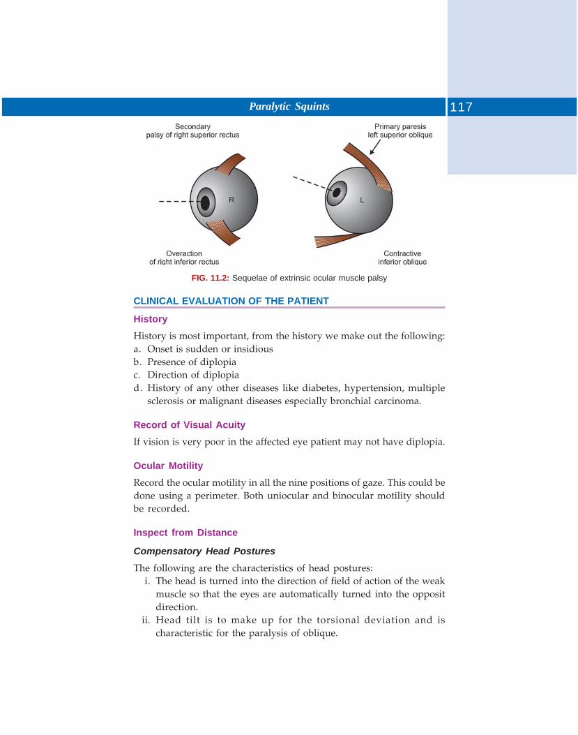
117Paralytic Squints
FIG. 11.2: Sequelae of extrinsic ocular muscle palsy
CLINICAL EVALUATION OF THE PATIENT
History
History is most important, from the history we make out the following:a. Onset is sudden or insidiousb. Presence of diplopiac. Direction of diplopiad. History of any other diseases like diabetes, hypertension, multiple
sclerosis or malignant diseases especially bronchial carcinoma.
Record of Visual Acuity
If vision is very poor in the affected eye patient may not have diplopia.
Ocular Motility
Record the ocular motility in all the nine positions of gaze. This could bedone using a perimeter. Both uniocular and binocular motility shouldbe recorded.
Inspect from Distance
Compensatory Head Postures
The following are the characteristics of head postures:i. The head is turned into the direction of field of action of the weak
muscle so that the eyes are automatically turned into the oppositdirection.
ii. Head tilt is to make up for the torsional deviation and ischaracteristic for the paralysis of oblique.
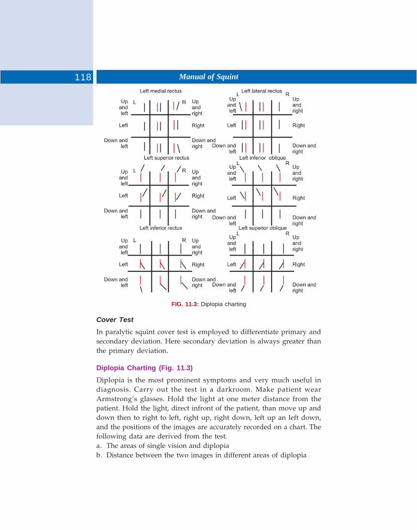
118 Manual of Squint
FIG. 11.3: Diplopia charting
Cover Test
In paralytic squint cover test is employed to differentiate primary andsecondary deviation. Here secondary deviation is always greater thanthe primary deviation.
Diplopia Charting (Fig. 11.3)
Diplopia is the most prominent symptoms and very much useful indiagnosis. Carry out the test in a darkroom. Make patient wearArmstrong’s glasses. Hold the light at one meter distance from thepatient. Hold the light, direct infront of the patient, than move up anddown then to right to left, right up, right down, left up an left down,and the positions of the images are accurately recorded on a chart. Thefollowing data are derived from the test.a. The areas of single vision and diplopiab. Distance between the two images in different areas of diplopia
119Paralytic Squints
c. Whether the images are at the same level or notd. Whether both are erect or one is inclinede. Whether diplopia is crossed or uncrossed.
N.B. False image belongs to the paralyze eye and distance between theimages are maximum in the direction of action of paralyzed muscle.
Hess Charting
This is of both diagnostic and prognostic value.
Advantages
1. Colors of fixation target and indicator are complementary to thegoggles.
2. The method is independent of external illumination.3. It is simple and there cannot be any misinterpretation on the part of
the patient or examiner because only one of the red fixation spotlight is presented at a time.
4. Less difficulty is encountered in testing patients with gross retinalinhibition as the light source from both fixation object and indicatorproduce adequate stimulus.
Interpretation
Compare the charts. The smaller chart indicate the paralyzed side andthe larger the overacting side. In smaller chart, the greatest restrictionindicates the direction of action of paralyzed muscle. In torsionaldeviations the fields have slopping sides.
Fields of Fixation (Uniocular and Binocular) (Figs 11.4 and 11.5)
a. Field of uniocular fixation in that area within which foveal fixation ofa small test object can occur. Its extend corresponds to the limits ofmovements of the eyeball in different direction. This could bemeasured by means of a perimeter. The extend of field in the averagenormal eye is about 45o–50o except in the inner and downwards whereit is limited to some extent by nose.
b. The fields of binocular fixation is that area within which bifovealfixation of a small test object can occur. Its extent is limited partly bythe limits of ocular movements and partly by nose. For the test to beof value the patients cooperation must be good and his binocularvision is strong. The test objects usually employed is 3-5 mm size andcolor is white.
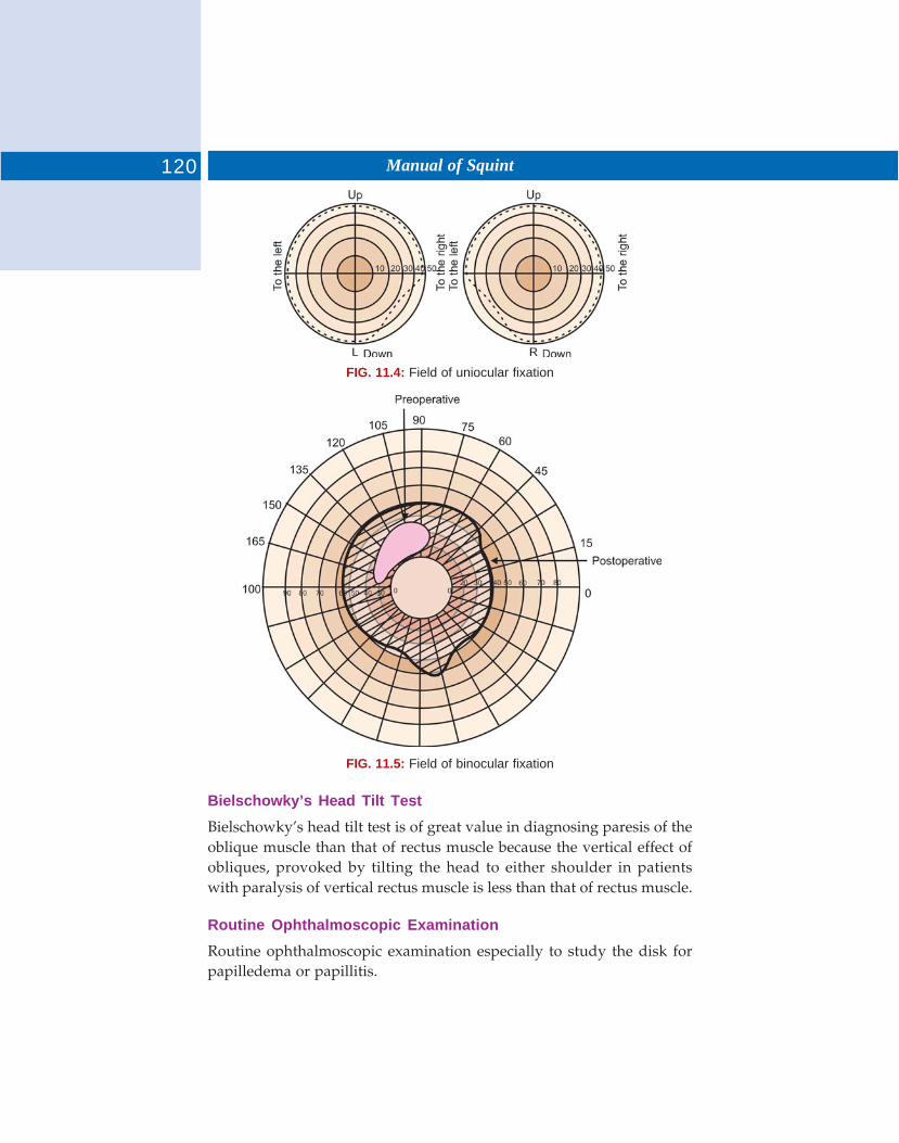
120 Manual of Squint
FIG. 11.4: Field of uniocular fixation
FIG. 11.5: Field of binocular fixation
Bielschowky’s Head Tilt Test
Bielschowky’s head tilt test is of great value in diagnosing paresis of theoblique muscle than that of rectus muscle because the vertical effect ofobliques, provoked by tilting the head to either shoulder in patientswith paralysis of vertical rectus muscle is less than that of rectus muscle.
Routine Ophthalmoscopic Examination
Routine ophthalmoscopic examination especially to study the disk forpapilledema or papillitis.
121Paralytic Squints
Forced Duction Test
This test is performed by anesthetizing the conjunctiva over the insertionof the muscle to be tested. Grasp the insertion of the muscle with atoothed forceps and attempt to rotate the eye in the field of action ofweak muscle.
This is of great value in deciding whether the anomaly of ocularmotility is caused by mechanical factor such as contracture or fibrosis ofa muscle, tightness of muscle following excessive resection, and shrinkageand scarring of the conjunctiva or Tenon’s capsule.
Estimation of Generated Muscle Force
This test is helpful to judge the residual function of an apparently pareticmuscle. The active force generate by muscle can be estimated bystabilizing the eye with a forceps while the patient moves his eyes againstthe resistance. The tug that examiner feels on the forceps is a sign ofresidual function. Absence of the tug is a sign of complete paralysis.
Exaggerated Force Duction Test
It is to estimate the tightness of oblique muscle. For this test eye must beput in the orbit (retroplace the globe) as it is then rocked back and forthby extorting and moving the globe around the tendon to check the tightoblique muscle now.
Differential Intraocular Pressure
The generated muscle could be estimated by comparing into ocularpressure in various positions of gazes and pressure increases as may beas high as 50 mm kg in case of restrictive elements.
Eye Movement Velocity
It may be useful only as an auxiliary diagnostic method, in evaluatingthe paralytic squint the eyes are capable of morning saccades/first eyemovements upto the velocity of 200–5000/sec. In cases of paralysis slowdrifting eye movement, restriction squints have normal saccadic velocitytill the restriction comes into effect.
Electromyography
Electromyography is useful procedure to test the function of each muscleseparately and follow-up study will indicates whether the muscle is inthe process of recovery or not.
122 Manual of Squint
Electro-oculography
Electro-oculography is an auxiliary diagnostic method.
Doll’s Head Phenomenon
Doll’s head phenomenon is tested by turning the head in differentdirections and noting the movement of the eye. This is an importantevidence for the intactness of the oculomotor nucleus in patient withsupranuclear paralysis.
Bell’s Phenomenon
Where in the eyes move upwards on forcible closure of the lids may beof diagnostic importance. For its presence the brainstem pathways mustbe intact even although the frontal bulbar pathway is disrupted. It occurin peripheral fascial palsy in which the lid fail to close when the patientattempts to close the eyes, the lids on the paralyzed side fail to shut anda slight upward movement of globe is observed, Bell’s phenomenon isabsent in nuclear lesion of the 7th cranial nerve which lends support tothe theory that reflex is mediated through lower centers, probably byway of the posterior longitudinal bundle.
Neurological Examination
It is important to determine whether the palsy is nuclear supranuclearand whether there is any focus of irritation or pressure in the course ofthe nerve involved.
Special Tests
Prostigmine/Neostigmine test to rule out myasthenia in cases of transientand intermittent squints.
Investigations for Thyroid Functions
Investigations for thyroid functions, whenever relevant to rule outthyroid ophthalmoplegia.
Testing of Corneal Sensation
Testing of corneal sensation is also done whenever necessary.
Other Investigations
a. Investigations for systemic diseases like diabetes, hypertension,syphilis, etc.
123Paralytic Squints
b. X-ray skullc. X-ray sinus and orbitd. X-ray cheste. Complete hemogramf. CT scanning to ultrasonographyg. Carotid angiographyh. Orbital venography.
TYPES OF PARALYSIS
Depending on the involvement of individual muscles or group of musclesthere can be many form of paralysis.
The main are as follows:
IIIrd Nerve Palsy
a. Complete third nerve palsy — Here only spared muscles are lateralrectus and superior oblique. The eye will be depressed, abductedand intorted. LPS paralysis, causes ptosis as well. In complete IIIrdnerve palsy the intrinsic muscles are also involved causing dilatationof pupil and loss of accommodation.
b. Isolated paralysis of individual muscle, i.e. SR, MR, IR or IO do occur,though rarely.
IVth Nerve Palsy
The movement affected are adduction and depression, superior obliquebeing the muscle paralyzed. The eye may be hypertropic due to overaction of the antagonist. Head will be tilted to the normal side and chinwill be depressed. Patients with bilateral SO palsy will have righthypertropia in left gaze and left hypertropia in right gaze, which increaseson tilting the head on either sides.
VIth Nerve Palsy
Adduction will be affected, lateral rectus being the muscle paralyzed. Thepalpebral fissure may be widened on looking to the side of paralysis(adduction), due to maximal innervational effort. Esotropia may be presentin the primary position. Head is turned over the affected shoulder.
Pseudograefe Sign
During the recovery of the IIIrd nerve palsy, nerve fibers originallyconnected with inferior ractus grow into the sheath of nerves fibers
124 Manual of Squint
supplying the levator muscle so that impulse to look down increase thetonus of the levator, hence there will be retraction of the upper lid indownward gaze, it may be accompanied by contraction of the pupil.
MANAGEMENT
Treatment of paralytic squint is always difficult and must depend oncasual conditions. Every case of paralytic squint should be managedinitially on conservations lines.
The Indications for Therapy
1. Presence of diplopia in the practical field of fixation2. Inability to maintain binocular single vision without anomalous head
posture.Prisms are effective in treating deviations less than 10. for larger
deviations prisms are not usually tolerated for prolonged periods andin such cases surgery becomes unavoidable. Fresnel prisms are tried,but it should be kept in mind that the prisms take away whatever stimulusleft to control strabisms and this may eventually lead to an increase inthe extent of residual deviation.
Treatment of Diplopia
Management of diplopia in a case awaiting spontaneous recovery orsurgery is important.
If the patient does not have BSV in any position of gaze simple eyeshields are given to cover each eye alternately.
If the patient enjoys BSV in certain gazes the occlusion need not becomplete, but it may be so arranged that BSV may be exercised in alimited area, i.e. occlude part of the field of one eye only.
Orthoptic treatment to maintain simultaneous binocular vision, andprevention of suppression is of almost importance.
Surgical Correction
The aim of the surgery in a case of paralytic squint is to abolish diplopiaand to regain comfortable binocular vision in all directions of gaze aswell as to produce a good cosmetic look. Operation is delayed until 6months since the onset of deviation in order to allow spontaneousrecovery and to stabilize the squint.
125Paralytic Squints
Surgical Procedures
Surgery for VIth Nerve Palsy
Sixth nerve palsy can be unilateral or bilateral. It can be aparesis orparalysis.
In unilateral palsy1. See whether contracture is fully developed or not.2. Assess the deviation for near and distance.
i. If the muscle sequent has fully developed, the angle of deviationis same on dextroversion and on levoversion. In such cases youwill have to do a resection of LR along with recession of theipsilateral MR. However if the angle of deviation is small resectionof LR alone may be sufficient.
ii. If the muscle sequelae is partially developed, i.e. there is nocontracture of the MR but there is over action of the contralateralMR in such cases you will have to do LR resection along withrecession of the contralateral MR or alternately a Fadenoperation:1. Here the rectus muscle is first recessed and muscle belly is
anchored to the sclera by one or more circumferentially placedmattress sutures. The muscle is then reinserted in its originalposition.
2. The muscle remain inserted. The edges of the muscle areattached to the globe. The appropriate distance of posteriorfixation sutures behind the normal muscle insertion is MR11-13 mm.
In this procedure the power of the muscle is decreasedalthough its primary position is not altered.
iii. In the case of VIth nerve palsy and the eye is not moving evenupto midline, see whether it is a case o MR contracture or completeloss of function of LR.
This is feasible by:(i) Forced duction test
(ii) Forced generation test(iii) EMG(iv) EOG, etc.If there is MR contracture with LR function, in such cases, do resection
of the LR along with recession of MR.
126 Manual of Squint
In case of contracture of MR with no LR function recess MR at least7 mm with recession of overlying conjunctiva. A transposition operation(reverse Jensens) is also indicated.
In bilateral palsy: It is usual to operate first on the nonfixing eye followedby surgey on the fixing eye at a second stage some weeks later.
In the case of both VIth and IIIrd nerve palsy the VIth nerve palsyshould be treated first (as outlined in Table 11.1).
IVth Nerve Palsy
In IVth nerve palsy the choice of surgery depends on:1. Whether the palsy is unilateral or bilateral2. Whether the sequelae have developed particularly whether there is
overaction of the inferior oblique3. The presence and amount of extorsion and whether this is superable.
In unilateral palsy: The surgery is usually performed and on overactingmuscle, not on the superior oblique itself, because of technical difficulties,and plication on the vertical component is unpredictable as compared tothe effect on cyclodeviation. There is also likelihood of inducing Brown’ssyndrome.
If the muscle sequelae has fully developed, there will be overactionof the contralateral inferior rectus, contracture of the inferior obliqueand secondary underaction of the superior rectus. Vertical deviationwill be same on contralateral up gaze and down gaze. Weakening of IOis the operation of choice.
If torsion is insuperable, recession of IR is contraindicated and aHarada procedure on the affected superior oblique should be performedas a first stage.
According to Payman the surgery to the VIth nerve palsy dependson the degree of abduction possible (Table 11.1).
Divergence paralysis: May be difficult to differentiate from unilateral orbilateral VIth nerve palsy, but this condition is usually comitant. Theesotropia is unchanged or may decrease on lateroversion, unlike withouta VIth nerve palsy, fusional divergence amplitudes are either severallyreduced or absent, causes are head trauma intracranial space occupyinglesions and cerebrovascular accidents.
Harada procedure: The anterior half of the superior oblique tendon isdisinserted and split from the posterior portion along the line of its
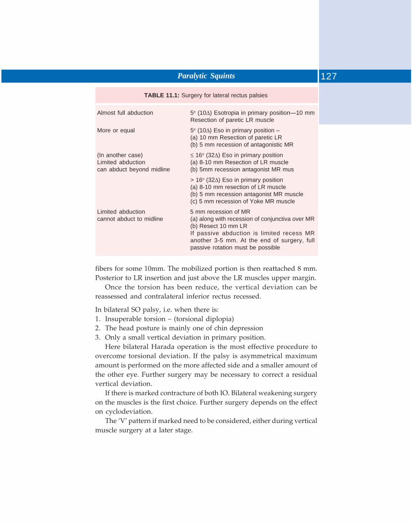
127Paralytic Squints
TABLE 11.1: Surgery for lateral rectus palsies
Almost full abduction 5o (10Δ) Esotropia in primary position—10 mmResection of paretic LR muscle
More or equal 5o (10Δ) Eso in primary position –(a) 10 mm Resection of paretic LR(b) 5 mm recession of antagonistic MR
(In another case) ≤ 16o (32Δ) Eso in primary positionLimited abduction (a) 8-10 mm Resection of LR musclecan abduct beyond midline (b) 5mm recession antagonist MR mus
> 16o (32Δ) Eso in primary position(a) 8-10 mm resection of LR muscle(b) 5 mm recession antagonist MR muscle(c) 5 mm recession of Yoke MR muscle
Limited abduction 5 mm recession of MRcannot abduct to midline (a) along with recession of conjunctiva over MR
(b) Resect 10 mm LRIf passive abduction is limited recess MRanother 3-5 mm. At the end of surgery, fullpassive rotation must be possible
fibers for some 10mm. The mobilized portion is then reattached 8 mm.Posterior to LR insertion and just above the LR muscles upper margin.
Once the torsion has been reduce, the vertical deviation can bereassessed and contralateral inferior rectus recessed.
In bilateral SO palsy, i.e. when there is:1. Insuperable torsion – (torsional diplopia)2. The head posture is mainly one of chin depression3. Only a small vertical deviation in primary position.
Here bilateral Harada operation is the most effective procedure toovercome torsional deviation. If the palsy is asymmetrical maximumamount is performed on the more affected side and a smaller amount ofthe other eye. Further surgery may be necessary to correct a residualvertical deviation.
If there is marked contracture of both IO. Bilateral weakening surgeryon the muscles is the first choice. Further surgery depends on the effecton cyclodeviation.
The ‘V’ pattern if marked need to be considered, either during verticalmuscle surgery at a later stage.
128 Manual of Squint
IIIrd Nerve Palsy
Complete paralysis: In a case of complete IIIrd nerve palsy with contractureof the two remaining active muscles, i.e. SO and LR weakeningprocedures are adopted.1. LR recession by 10mm with recession of the overlying conjunctiva.2. SO tenectomy—this helps to avoid mechanically induced hypotropia.
Strengthening procedures—A superomaximal (up to 8 mm) resection ofMR.
Alternatively—Muscle transportion procedures like Jenson’s procedureare also indicated.
In paresis—It is advisable to correct the horizontal angle initially byresection of MR and recession of LR residual deviation can be treatedconservatively with prisms or by surgery to strengthen the SR or IR.
COMPLETE PARALYSIS OR PARESIS
a. Medial rectus—Resection of MR with ipsilateral or contralateral LRrecession.
b. Inferior rectus palsy—Resection of IR and ipsilateral SR recession.c. Superior rectus palsy—Resection of SR weakening of the IR and IOd. Inferior oblique palsy—Intrasheath tenotomy of ipsilateral SO, a
recession of the contralateral SR depending whether the hypotropiais more marked in down gaze and on the amount of torsionaldeviation.
Weakening Operation
Recession (Rectus muscle weakening operation).
Hang back recession—In this method muscle is disinserted andreattached to its original insertion.
BLOWOUT FRACTURE
A blunt injury to the eye causes increased orbital pressure, and the orbittends to give way at its weakest point, the orbital floor. As a result, theligament of Lockwood drops down and the inferior rectus andoccasionally the inferior oblique are trapped in the fracture. Limitationof action of the superior and inferior rectus muscles and occasionally
129Paralytic Squints
the inferior ethmoidal area may cause simultaneous limitation of lateralrotation. There may also be a relative enophthalmos, or this may developlater as the edema surrounding the fracture subsides or as the orbitalfat atrophies. Damage to the infraorbital nerve with anesthesia of thelower lid and cheek can occur. The eyes be straight in the primaryposition, or the patient may have a hypotropia with diplopia in the upand down positions because of the superior and inferior rectus musclesrestrictions.
Ocular Myopathy
This is a progressive external ophthalmoplegia. There is limitation ofextra- ocular movement and progressive ptosis ultimately the patientends up with a total ophthalmoplegia (it is not usually necessary toperform surgery on the extraocular muscles because the patient’s eyesare straight in the primary position).
Myasthenia Gravis
Myasthenia gravis is a disease characterized by fatigue and weakness ofstriated muscles within the body. This is due to blockage at the neuro-muscular end plate, symptoms of ocular myasthenia gravis includediplopia and ptosis symptoms of myasthenia gravis are less in the morningand they are worse towards the end of the day when patient becomefatigued. Diagnosis is confirmed in most cases by the administration oftensilon intravenously see if there is any improvement in clinical signsor symptoms, tonography is useful on making the diagnosis.
Painful Ophthalmoplegia
Refer to the involvement of one of more of the ocular motor nerves bya chronic granulomatous or nongranulomatous inflammation, usually atthe orbital apex, in the superior orbital tissues, or in the cavernosussinus patient experience pain and involvement of IIIrd, IVth and VIthnerve. If the orbital apex is involved, there may be visual loss frominvolvement of the optic nerve ESR is usually high. Many terms havebeen used to describe the syndrome, including the orbital apexsyndrome, superior orbital fissure syndrome and Tolosa Hunt syndrome.
DOUBLE ELEVATOR PALSY
Double elevator palsy is a common of hyperdeviation. There is a parisesof the superior rectus and inferior oblique muscles in the same eye. The
130 Manual of Squint
inferior oblique shows more restriction than superior rectus. Ptosis andoccasionally Marcus Gunn (Jaw winking) may also be associated. Thepatient will generally walk with the head elevating slightly in order tomaintain binocular vision just below the midline and in the inferior fieldof gaze.
Double elevator palsy probably results from some type of congenitalnuclear lesion, because in the orbit the superior rectus and medial rectus.
Marcus Gunn phenomenon is apparently caused by misdirected nervepathway. In cases with ptosis, surgery for have a risk of exposure Bell’sphenomenon.
Treatment
Surgery is the main stay of treatment in such cases. However, no surgeryis indicated if the patient has binocular vision in a straight ahead position,deviation showing only on locking up and there is no backward headtilt in the primary position.
The surgical approach depends upon the amount of elevation achievedin the affected eye can be elevated above the midline the procedure ofchoice is to weaken yoke muscles, that is, the superior rectus and inferioroblique muscle in the normal eye. The superior rectus 4-5 mm and theinferior oblique 8- mm. In cases where the eye cannot be brought abovethe midtime recession of IR and resection SR muscle in the affected eyeis required. In some of the patient who had undergone surgery on MRfor esotropia, the eye develop a market esotropia and the patient triesto elevate the eye. In the down gaze, the same patient has a largeesotropia. The eye cannot be elevated to midtime in either the adductedor abducted position. Because if this limitation, the condition has beencalled “congenital fibroses syndrome. Such eyes require recession of IRand congenital, the SR is reseated 4-5 mm.
Differential Diagnosis of Ocular vs Congenital Torticollis(Tables 11.2 and 11.3)
The cause of congenital (nonocular) torticollis include:a. Congenital bony malformations of Atlas (Atlas = Ist vertebra), cervical
vertebrae and ribs.b. Malformation of sternomastoid muscles.
131Paralytic Squints
TABLE 11.2: Difference between ocular and congenital torticollis
Ocular Torticollis Congenital torticollis
1. — 1. Marked resistance to passivestraightening of the head
2. — 2. Often, a fibroma mass may be felt inthe contracted sternomastoid muscle
3. Patching (occlusion) of the paretic 3. Patching has no effect on congenitalmuscle can relive ocular torticollis, torticollisprovided no secondary) skeletal ormuscular changes have developedin the neck
TABLE 11.3: Differential diagnosis between congenital and acquired palsy
Congenital Acquired
1. Intermittent diplopia (A)2. Intermittent squint (A)3. Head posture but patient is unaware of this (A)4. Anatomical changes in vertebral column (A)5. Suppression is usually present (A)
TOTAL OPHTHALMOPLEGIA
Inv. of Extrinsic, ocular muscleIntrinsic ocular muscleIPSPtosis+Proptosis+
Ocular movement (A) or restrictedPupillary activity to lightAccommodation and convergence (A)
DOUBLE DEPRESSOR PARALYSIS
Double depressor paralysis is a rare anomaly and it consists of inability todepress the eye from primary position, adduction or abduction. This iscaused by long-standing paralysis of the inferior rectus muscle rather byparalysis of both inferior rectus and superior oblique muscle (Table 11.4).
Botulinum Toxin
In paralytic strabismus, the botulinum induced paralysis of the antagonistmuscle prevents or reduces its contracture during spontaneous recoveryof the paretic muscle. Also, as the initial overaction resulting from theinjection slowly resolves and the eyes approach the primary position,fusion may “lock on” resulting in Orthophora.
132 Manual of Squint
TABLE 11.4: Differential diagnosis between paralytic and nonparalytic squint
Paralytic Nonparalytic
1. Eyes move in such an 1. Eyes move in coordinated mannerincoordinated manner so and angle of squint remain same inthat the angle of squint varies all direction of gazein different direction of gazesand on changing fixation fromone to other eye.
2. Diplopia is a prominent feature 2. No. diplopia3. Nausea and vertigo 3. (A)4. Compensatory head posture 4. (A)5. Onset – sudden 5. Usually in children6. Amblyopia, supervision, ARC, 6. Amblyopia, supervision, ARC,
eccentric fixation absent eccentric fixation present7. Secondary deviation/primary 7. Secondary = Primary deviation
deviation8. Some changes present in 8. Not so
uninvolved muscle9. Palliative treatment is required 9. No palliative treatment
10. Wait for 6-8 months for surgery 10. Immediate it is required
The advantages of botulinum toxin therapy for strabismus are: (1) itcan be performed on an outpatient basis, (2) it carries minimum risk,(3) leaves no scar, (4) can be used for postoperative residual strabismusand (5) can be used when surgery is inappropriate. It has been shown inparalytic strabismus toxin have better chance of recovery than thosethat have not been treated. In lateral rectus palsy contacture of theantagonist medial rectus muscle can be prevented and this should makesubsequent elective surgery easier.
The disadvantages of botulinum toxin therapy for strabismus are:(1) more than two injection is often needed to determine the optimumdose to obtain maximal benefit, (2) alignment changes are not as stableas with surgery and (3) transient partial ptosis and vertical strabismusfrequently occur. There is dramatic relief of symptoms followingtreatment with botulinum toxin.
The other reported complication of botulinum therapy are: (1) scleralperforation, (2) retrobulbar hemorrhages, (3) diplopia, (4) pupillarydilatation, (5) ecchynosis, (6) corneal exposure, (7) ptosis, (8) ectropon,(9) lagophthalmos and (10) chemosis.
Botulinum toxin is curative in some cases of paralytic strabismus, inothers it prevent contracture of the antagonist and should thereforemake subsequent surgery simple.
133Paralytic Squints
APPLIED ANATOMY
Oculomotor Nerve (IIIrd N) (Fig. 12.6)
The oculomotor or the IIIrd cranial nerve innervates all the extraocularmuscles except the lateral rectus and the superior oblique. In addition, itcarries the parasympathetic fibers to the ciliary muscle and the sphincterpupillae. The nucleus of IIIrd nerve is in the form of a complex formedby a number of subnuclei and is situated in the midbrain) at the level ofsuperior colliculi. Below the aqueduct of Sylvius. The subnuclei include:
i. Subnucleus for levator: It lies in the caudal part of the complex and isunpaired, thus supplying levator muscle of the lids of both eyes.Therefore, a lesion confined to this area will lead to ptosis of botheyes.
ii. Subnucleus for superior rectus: This is paired, and supplies the contra-lateral superior rectus muscle. This implies that in a case of IIIrdnerve palsy, if the contralateral superior rectus has been spared,the lesion does not lie in the nucleus.
iii. Subnucleus for medial rectus, inferior rectus and inferior oblique: This isalso a paired group of cells, supplying the corresponding musclesof the same (ipsilateral) side.
iv. Accessory nucleus: Situated posterior to the main mass, it sends pre-ganglionic parasympathetic fibers along the motor fibers, and isrelated to the phenomenon of near reflex, accommodation, andperhaps, convergence (Perlia’s nucleus).
In general, it is rare to see lesions purely localized in the nuclearcomplex. Certain vascular lesion, demyelinating diseases and tumorsmay involve this region.
The efferent fibers from the complex form the fasciculus, which travelthrough the red nucleus and medial part of the cerebral peduncles,emerging from midbrain. Lesions in this zone are caused by the sameconditions as in the case of nuclear complex, and may lead to two well-recognized syndromes:
Benedikt’s syndrome: This is characterized by an ipsilateral IIIrd nervepalsys and a contralateral hemiplegia with tremors. This is the result oflesions of the fasciculus in the red nucleus.
Weber’s syndrome: This caused by the fascicular lesion in the cerebralpeduncles, and is characterized by an ipsilateral IIIrd nerve palsyaccompanied by a contralateral hemiparesis.
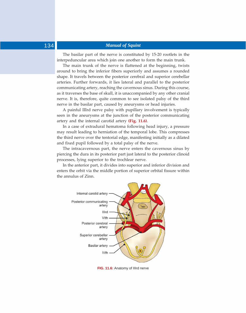
134 Manual of Squint
The basilar part of the nerve is constituted by 15-20 rootlets in theinterpeduncular area which join one another to form the main trunk.
The main trunk of the nerve is flattened at the beginning, twistsaround to bring the inferior fibers superiorly and assumes a roundedshape. It travels between the posterior cerebral and superior cerebellararteries. Further forwards, it lies lateral and parallel to the posteriorcommunicating artery, reaching the cavernous sinus. During this course,as it traverses the base of skull, it is unaccompanied by any other cranialnerve. It is, therefore, quite common to see isolated palsy of the thirdnerve in the basilar part, caused by aneurysms or head injuries.
A painful IIIrd nerve palsy with pupillary involvement is typicallyseen in the aneurysms at the junction of the posterior communicatingartery and the internal carotid artery (Fig. 11.6).
In a case of extradural hematoma following head injury, a pressuremay result leading to herniation of the temporal lobe. This compressesthe third nerve over the tentorial edge, manifesting initially as a dilatedand fixed pupil followed by a total palsy of the nerve.
The intracavernous part, the nerve enters the cavernous sinus bypiercing the dura in its posterior part just lateral to the posterior clinoidprocesses, lying superior to the trochlear nerve.
In the anterior part, it divides into superior and inferior division andenters the orbit via the middle portion of superior orbital fissure withinthe annulus of Zinn.
FIG. 11.6: Anatomy of IIIrd nerve
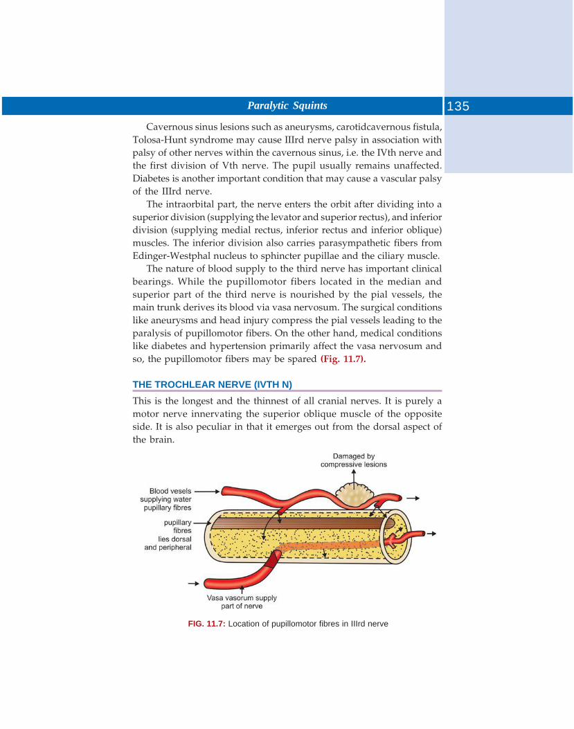
135Paralytic Squints
Cavernous sinus lesions such as aneurysms, carotidcavernous fistula,Tolosa-Hunt syndrome may cause IIIrd nerve palsy in association withpalsy of other nerves within the cavernous sinus, i.e. the IVth nerve andthe first division of Vth nerve. The pupil usually remains unaffected.Diabetes is another important condition that may cause a vascular palsyof the IIIrd nerve.
The intraorbital part, the nerve enters the orbit after dividing into asuperior division (supplying the levator and superior rectus), and inferiordivision (supplying medial rectus, inferior rectus and inferior oblique)muscles. The inferior division also carries parasympathetic fibers fromEdinger-Westphal nucleus to sphincter pupillae and the ciliary muscle.
The nature of blood supply to the third nerve has important clinicalbearings. While the pupillomotor fibers located in the median andsuperior part of the third nerve is nourished by the pial vessels, themain trunk derives its blood via vasa nervosum. The surgical conditionslike aneurysms and head injury compress the pial vessels leading to theparalysis of pupillomotor fibers. On the other hand, medical conditionslike diabetes and hypertension primarily affect the vasa nervosum andso, the pupillomotor fibers may be spared (Fig. 11.7).
THE TROCHLEAR NERVE (IVTH N)
This is the longest and the thinnest of all cranial nerves. It is purely amotor nerve innervating the superior oblique muscle of the oppositeside. It is also peculiar in that it emerges out from the dorsal aspect ofthe brain.
FIG. 11.7: Location of pupillomotor fibres in IIIrd nerve
136 Manual of Squint
The nucleus of trochlear nerve lies in the midbrain at the level ofinferior colliculi, and is in continuation with the nucleus of IIIrd nerve.
The fascicular component is constituted by the axons that curvearound the aqueduct of Sylvius and decussate completely.
The trunk (precavernous part), emerges on the dorsal aspect of themidbrain and curves forward between the posterior cerebral and thesuperior cerebellar runs arteries as does the trunk of IIIrd nerve. Itpierces the dura below the IIIrd nerve to enter the posterior part ofcavernous sinus. The infracavernous part lies in the lateral wall of thesinus below the third nerve and above the first division of Vth nerve. Inthe anterior part, it crosses over the IIIrd nerve and enters the orbitthrough lateral part of the superior orbital fissure, superotemporal tothe annulus of Zinn.
The intraorbital part carries the nerve supply to the superior obliquemuscle.
THE ABDUCENS NERVE (VITH N)
The VIth cranial nerve is motor in function, has a long course andinnervates the lateral rectus muscle of the same side.The nucleus of sixthnerve lies in the middle of pons below the fourth ventricle and is closelyassociated with the fasciculus of the facial nerve. In addition, it is alsoconnected with the pretectal nucleus of both sides as well as the horizontalgaze center in the pontine paramedial reticular formation. Therefore,any lesion affecting the zone of VIth nerve. Nucleus will not cause anisolated paralysis of the ipsilateral lateral rectus muscle, but will beaccompanied by:
i. Paralysis of conjugate movements towards the side of lesionii. Ipsilateral facial nerve palsy, by way of the involvement of fasciculus
of facial nerve.The fascicular part is composed of the axons emerging from the
nucleus, pass forwards through the medial meniscus and the pyramidaltract leaving the brainstem at the junction of pons and midbrain, lateralto the pyramidal prominence. In view of its course through the pyramidaltract and medial meniscus. A lesion in the region of fascicule is likely tocause multiple clinical manifestations in the form of Foville’s syndrome.This is manifested if the lesion is located at the site where the fasciculustraverses the medial meniscus, and shows:• Paralysis of the ipsilateral lateral rectus muscle, paralysis of the lateral
gaze
137Paralytic Squints
• Paralysis of the lateral gaze towards the same side• Facial weakness (damage to the facial nerve nucleus)• Facial analgesia (damage to the sensory portion of Vth nerve)• Homer’s syndrome• Deafness.
Millard-Gubler syndrome. The lesion at the sight of the fasciculuspassing through the pyramidal tract will be characterized by paralysisof the ipsilateral lateral rectus muscle.
Contralateral Hemiplegia
The basilar portion of the nerve, after emerging from the pontomedullaryjunction, passes upwards close to the base of the pons and is crossed bythe anterior inferior cerebellar artery. It runs further upwards on theback of petrous temporal bone near its apex turning sharply at rightangle on the sharp border of the petrous bone before piercing the durato enter the cavernous sinus, lateral to the dorsum sellae. Damage to thebasilar portion may occur in the following situations:
i. Acoustic neuroma: This tumor located at the cerebellopontine anglemay cause damage to multiple cranial nerves, viz. the Vth, VIth,VIIth, and VIIIth nerves. Therefore, paralysis of the lateral rectusin such cases is accompanied by a hearing loss which is the firstsymptom, and loss of corneal sensation which is the first sign (i.e.it precedes the VIth nerve involvement).
ii. Nasopharyngeal tumors: Following the invasion of skull and itsforamina, these tumors can damage the VIth nerve in it basilarcourse.
iii. Raised intracranial pressure (Fig. 11.8): The increased pressure,especially in case of posterior fossa tumors or in benign intracranialhypertention (pseudotumor cerebri) tends to push the braindownwards. Such a movement may damage the VIth nerve onstretching at the sharp border of petrous bone where it makes asharp right angled turn. The nerves may be damaged on both thesides. The resultant VIth nerve palsy, however, has no localizingvalue.
iv. Basal skull fracture: Damage to the VIth nerve (unilateral or bilateral)in this situation is nonspecific, and may be a part of the overalldamage to the brain tissue.
The infracavernous part of the nerve lies below the IIIrd and IVthnerve, as well as the first division of the Vth nerve, placed most medially
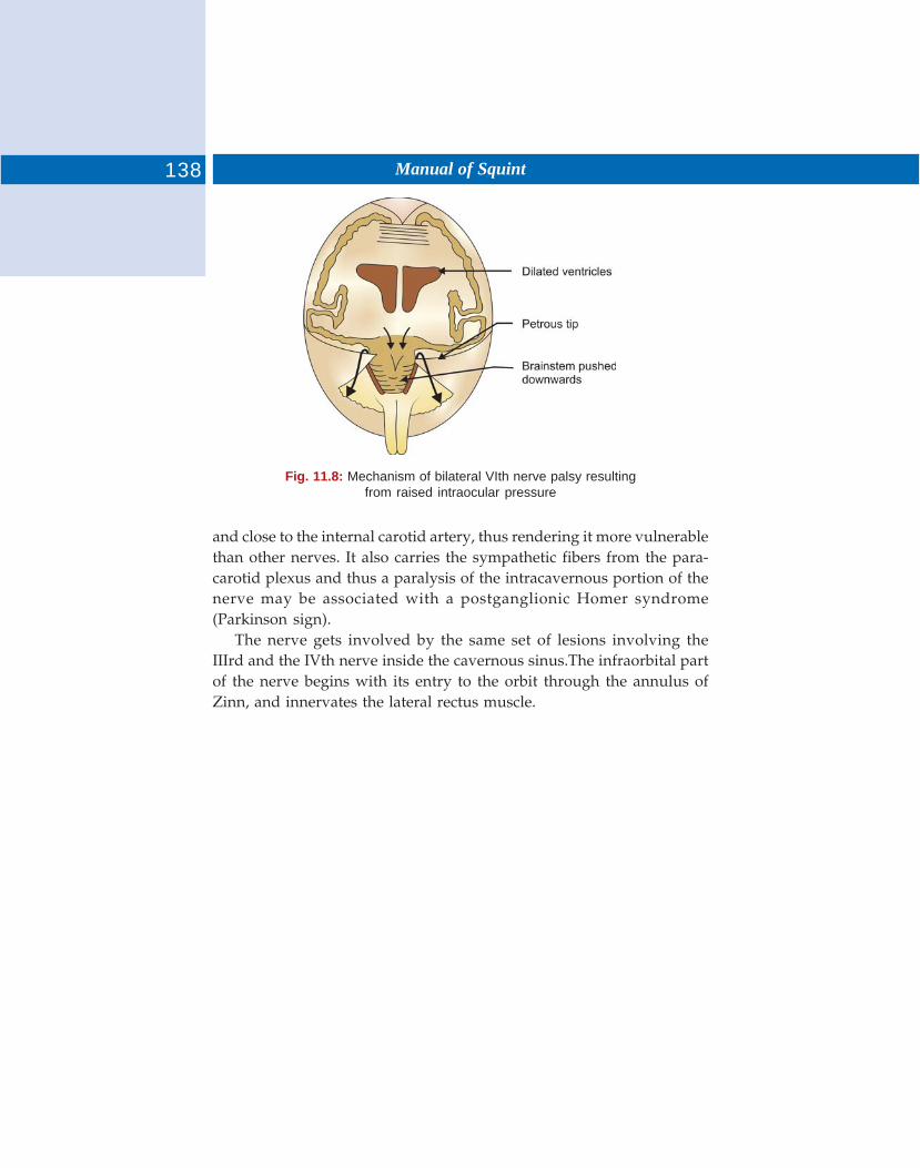
138 Manual of Squint
Fig. 11.8: Mechanism of bilateral VIth nerve palsy resultingfrom raised intraocular pressure
and close to the internal carotid artery, thus rendering it more vulnerablethan other nerves. It also carries the sympathetic fibers from the para-carotid plexus and thus a paralysis of the intracavernous portion of thenerve may be associated with a postganglionic Homer syndrome(Parkinson sign).
The nerve gets involved by the same set of lesions involving theIIIrd and the IVth nerve inside the cavernous sinus.The infraorbital partof the nerve begins with its entry to the orbit through the annulus ofZinn, and innervates the lateral rectus muscle.
As in the case of horizontal deviations, a vertical squint can beconcomitant or incomitant (congenital or acquired). It may express itselfin the form of a hyperphoria, intermittent hypertropia, or a (constant)hypertropia. Depending upon the eye involved, it may be righthypertropia or left hypertropia.
COMITANT VERTICAL DEVIATIONS
It is not uncommon to see such type of deviations, either in an isolatedform or in association with horizontal deviations. The underlying causeis not well-understood. Some form of innervational disturbance may bea likely factor. In many instances, it may be the result of a paralyticincomitant hyperdeviation attaining the character of a comitant deviationover a long period of time. The common features of such deviations areas follows:
i. Intermittent hypertropia is more common than a constant deviationii. Suppression, amblyopia or a vertical anomalous retinal corres-
pondence may be presentiii. Small vertical deviations are typically present in association with
moderate to large horizontal deviationsiv. Many cases demonstrate an under or over action of one or more
cyclovertical muscles.
Treatment
Orthoptic treatment is directed to treat amblyopia. In small deviationsupto 15 prism diopters, prisms are provided to neutralize the deviation.The power of prism is equally divided in two eyes with the base downin front of the hypertropic eye and base up in the other eye.
Surgical treatment is planned on the basis of the amount of deviation,and the presence of associated horizontal deviations. In small deviations
12 Vertical Strabismus
140 Manual of Squint
combined with horizontal deviation, a correction of the vertical elementmay be achieved by shifting the insertion of the horizontal muscle (5-8mm) in the hypertropic eye, or shifting it up (to the same amount) in thehypotropic eye.
In large deviations, a recession of superior rectus of the hypertropicand recession of inferior oblique of the other eye is indicated.
DISSOCIATED VERTICAL DEVIATIONS (DVD)
It is basically a bilateral anomaly characterized by the hyperdeviation ofeither eye when the other eye is fixing. The hyperdeviation isaccompanied by extortion and slight exodeviation. In this situation, ifthe fixing eye is covered, the squinting eye takes up fixation by movingdown and in along with intortion. But, the covered eye does not makeany movement, thus defying the Hering’s law of ocular movements.That is why this type of vertical deviation is prefixed with the term‘Dissociated. It may be associated with any type of strabismus, anyanomaly of binocular vision, nystagmus, or any defect ocular movements.On the other hand, it is seen as an isolated form. The etiology of thisneuromuscular anomaly has not been explained clearly. Features—DVDis characterized by:
i. A spontaneous occurrence of vertical deviation of either eye whenthe patient is fatigued, or when fusion is interrupted by artificialmeans such as covering one eye.
ii. Lack of subjective symptoms in majority of case, an intermittentdeviation being noticed by the onlookers—friends or parents. Somecases may have asthenopic symptoms. Diplopia is rare.
iii. Its frequent association with other forms of strabismus, speciallywith essential congenital exotropia and essential exotropia.
iv. Suppression in the nonfixing eye is present to eliminate diplopia.v. Presence of peripheral fusion if there is no associated horizontal
deviation.vi. Presence of a facultative absolute scotoma, though bilateral,
manifesting in the nonfixing eye.vii. A frequent presence of latent nystagmus in cases of alternate
DVD.
Diagnosis
The diagnosis of DVD is made on a careful assessment of the followingtests.
141Vertical Strabismus
Cover-Uncover Test
In a case of unilateral case of manifest DVD. When the fixing eye iscovered, the deviating eye makes a downward movement unaccom-panied by any downward movement of the uncovered eye.• In a case of alternate DVD, either eye will show elevation under
cover while the fixing eye will move down to take up fixation.• In a case of a latent DVD, the eye under cover elevates but resumes
fixation by making a downward movement. No movements takeplace in the uncovered eye.
Bielschowsky Phenomenon
It is usually present in cases of DVD. It is carried out by covering oneeye, which deviates up under cover. While keeping the cover on, aphotometric neutral filter wedge is placed in front of the fixing eye. Asthe filter wedge is placed before this eye it makes a downward movementwhich increases successively as the density of the filter is made to increase.Conversely, the deviating eye starts moving up successively as the densityif the filter is made to decrease.
Treatment
i. Nonsurgical is of little valueii. Surgical treatment is indicated when the condition presents a
significant cosmetic problem. The following procedures have beenrecommended:
a. Recession of superior rectus: A large recession (7-10 mm) may bedone alone, or a small recession (3-5 mm) may be combined withanchoring of this muscle to the globe by nonabsorbable sutures,12-15 mm behind its insertion (Faden operation).
b. Resection of inferior rectus: This procedure may preferably bereserved for the cases showing a recurrence following largesuperior rectus recessions.
c. Combined operation: This procedure has been recommended forlarge angle DVD particularly in rare cases which have apredominantly monocular vertical deviation with a hypertropiain primary position.
Recession of Inferior Oblique
This procedure which also includes anteriorisation of its insertion, isspecially recommended for patients who have a DVD combined with anoveraction of inferior oblique.
142 Manual of Squint
INCOMITANT VERTICAL DEVIATIONS
Various incomitant deviations caused by extraocular muscle palsies alongwith their sequelae had been dealt in the chapter on Paralytic Squints.However, there are two distinct entities which although causeincomitance, are different from other paralytic squints in many respectsand can be discussed at this stage.
Inferior Oblique Overaction
It is characterized by an upshoot of the eye in adduction. The primaryform is probably caused by some anatomical or innervational anomaly.It is seen either as an isolated anomaly or may be associated withesotropia or exotropia, oftenly having a V-pattern. The secondary formis caused by paresis of either its ipsilateral antagonist muscle (superioroblique), or its yoke muscle (contralateral superior rectus). The primaryform has the following characteristics:
i. Onset is between 2-3 years of ageii. It is frequently bilateral
iii. There is an upshoot or overelevation in the adducted positioniv. Horizontal squints are occasionally associated in the primary
positionv. No head tilt is present
vi. Forced duction test is positive.
Treatment
The preferred surgical plan consists of a recession combined withantriorizaiton of inferior oblique muscle. In this procedure, the inferiormuscle is detached from its insertion and reattached to the sclera nearthe lateral end of lateral rectus muscle. It is combined with the surgeryif there is a concurrent horizontal squint.
Superior Oblique Overaction
It is characterized by a down shoot of the eye in adduction. The etiologyis uncertain. Additional features are as follows:
i. It usually occurs by the age of 2-3 yearsii. It is frequently bilateral though may be asymmetrical at times
iii. It is commonly associated with concomitant esotropia or morecommonly with exotropia
iv. Head tilt is absentv. Forced duction test is positive.
143Vertical Strabismus
Treatment
Treatment is indicated if there is a significant ocular deviation, or thepresence of A-pattern. The recommended treatment consists ofweakening of bilateral superior oblique muscles by way of tenotomy atthe temporal or nasal border of superior rectus.
CYCLODEVIATIONS
Cyclodeviation (torsional strabismus) is of uncommon occurrence andrefers to a misalignment of the eyes along the anteroposterior axis.Depending upon the direction of rotation, it may be classified as:
Excyclophoria or Excyclotropia
Excyclophoria or excyclotropia, when the 12 O’clock point on the corneais rotated temporally.
Incyclophoria or Incyclotropia
Incyclophoria or incyclotropia, when the 12 O’clock point is turn nasally.This type of deviation is caused by an imbalance between the intorters(inferior oblique and superior rectus) and the extorters (superior obliqueand inferior rectus) of the eyeball under the following situations:
i. Paresis or paralysis of a cyclovertical (particularly obliques) muscleii. Complication of surgical procedures on vertical or oblique muscles
iii. Manifestation of certain systemic diseases like Grave 1 disease,myasthenia gravis, etc.
A large majority of the patients are symptom-free. This is because ofthe development of suppression and anomalous retinal correspondenceor on account of some physiological and psychological adaptations.
Treatment is indicated only in symptomatic patients and is alwayssurgical.
DEFINITION
The A and V syndrome consists of an abnormal variation in the amountof horizontal deviation as the eyes move from straight up to straightdown positions of gaze or in other words “there is incomitency in thevertical positions of gaze.” The shape of the letter ‘A’ symbolizes theincreasing convergence down or increasing divergence up is symbolizedby the letter ‘V’.
CLASSIFICATION
Various classifications of the syndrome have been suggested from timeto time. URIST (1951) suggested the following classification:
Group I
Esotropia with bilateral elevation in adduction. In these patients theesotropia is greater for near and in downward gaze than for distanceand upward gaze, a right hypertropia on levoversion and a lefthypertropia on dextroversion. The convergence to near point is good.
Group II
Esotropia with bilateral depression in adduction. In these patients theesotropia is greater for distance and in upward gaze than for near andin downward gaze a right hypertropia is present on to dextroversionand a left hypertropia on levoversion. The convergence to near point isgood to fair.
Group III
Exotropia with bilateral elevation in adduction. In this group theexotropia is greater for distance and in upward gaze than for near andin downward gaze; right hypertropia is present on gaze to the left and
13 A-V and X Syndromes
145A-V and X Syndromes
a left hypertropia on gaze to the right. The convergence to near point isgood.
Group IV
Exotropia with bilateral depression in adduction. In these patients theexotropia is greater for near and is downward gaze than for distanceand upward gaze. A right hypertropia on gaze to the left. The conver-gence to near point is usually poor.
These syndrome has classified in a simple way, as follows:1. V. esotropia—The esotropia is greater below than above.2. A. esotropia—The esotropia is greater above than below.3. V. exotropia—The exotropia is greater above than below.4. A. exotropia—The exotropia is greater below than above.
Thus, he gave the name of ‘A’ and ‘V’ syndrome to verticallyincomitant squint and described A and V esotropia and exotropia. Hedid not define the limits of normal deviation in the up and down positionof gaze. There is a new classification to rectify the previous deficienciesand included even those cases which would otherwise not fit in.
Pure Type
There is a variation in the horizontal strabismus as the patient looks upand down with nonvertical incomitance in any of the nine position ofgaze. This may be further divided into ‘A’ or ‘V’ type of esotropia orexotropia.
Impure Type
Besides the horizontal strabismus which occurs on looking up and down,there is vertical incomitance in one or more of the other positions ofgaze. This group may be further subdivided into 3 patterns.
i. The left eye remains hypertropic on dextroversion and right eyeon levoversion.
ii. The right eye remains hypertropic on dextroversion and left eyeon levoversion.
iii. The same eye hypertropic on both dextro and levoversion.
INCIDENCE
There has been no exact agreement on the frequency of occurrence ofthe ‘A’ and ‘V’ pattern in strabismus. Various authors have given varyingincidence as shown in the Table 13.1.
146 Manual of Squint
CLINICAL PICTURE
The clinical characteristics of patients with A and V pattern shall now beconsidered.
1. V – esotropia – Following features characterize this condition.a. Eso greater below (V)b. May be overaction of oblique, inferior rectus, medial rectusIf there is an abnormal head position, the chin is depressed to bringthe eyes upwards for this is the most favorable position possible tominimize the deviation. Another clinical characteristic which isoccasionally observed is the clumsiness of three patients in goingdownstairs.
2. A – esotropia. This is characterized by:a. Eso greater above than belowb. May be over action of superior rectus or superior obliquec. May be underaction of inferior rectus or inferior oblique.Patient with A exotropia tend to assume the chin up and binocularvision in this particular direction of gaze.
3. V – exotropia. This is characterized by:a. Exo greater aboveb. May be over action of inferior obliquec. May be under action superior rectus or superior oblique.This group comprises those patients who had greater exotropia –constant or intermittent – in upward gaze. If there is an abnormalhead position, it will be a chin up and eyes down position.
4. A – exotropia is characterized by:a. Exo greater below (A)b. May be over action of superior oblique or superior rectusc. May be under action of inferior oblique or inferior rectus.A chin down and eyes up posture may be assumed by same and this
may lead to clumsiness going downstairs.
TABLE 13.1: Incidence of A-V pattern in strabismus
S. No. Author and year Incidence
1. Urist 1951 50%2. Knapp. 1959 12.5%
147A-V and X Syndromes
ETIOLOGY
The exact etiology of this syndrome is still controversial. Various factorsare considered to underline this mechanism of A and V syndromes.
Anatomical Factors
Although anatomical anomalies do not cause A:V syndrome in all cases,there are a number of clinical entities which create true ‘A’ and ‘V’patterns. This in the superior oblique tend on sheath syndrome of Brownit is common to encounter a V pattern. This is attributed to the action ofthe incelastic superior oblique sheath as a grey line forcing divergencein elevation. The adherence syndrome (adherence of lateral rectus withinferior oblique or superior rectus with superior oblique) may create amechanical vergence shift in the vertical fields.
Of considerable interest, although lacking explanation are the patternsassociated with anomalies of the palpebral fissures and facial bones firstpointed out by Urist. There is association of the ‘V’ pattern andoveraction of inferior oblique in patients with mongoloid palpebralfissures.
Innervational Factors
Horizontal Muscle School
Horizontal recti Urist (1951) is believer of this school and the thinks thatA and V syndrome can result from dysfunction of the horizontal rectusmuscles. He feels that basically there is exaggeration of the normaltendency of exodeviation to increase in upward gazes and ofesodeviation to increase in downward gaze. Thus, a defect greater inupward gaze is said to be due to lateral rectus dysfunction; V – exotropiarepresenting overaction of lateral rectus and ‘A’ exotropia representingunder action of medial rectus muscle. The following arguments supportthis theory that A and V syndrome are produced by the dysfunction ofthe horizontal muscles.
i. Correction of the different angle of strabismus that exists in theupward and downward gazes through proper surgical treatmentof the affected horizontal muscles.
ii. Uneven results in the straight up and straight down position ofgaze as a result of the operation mistakenly performed on unaffectedhorizontal muscles.
Urist showed in a case, the effects of incorrect surgical treatmentconsisting of 4 mm recession of both medial recti in ‘A’ esotropia
148 Manual of Squint
that led to an exotropia of 5-10o in looking up and 20o on lookingdown. He suggested that proper surgery in this case for A esotropiawould have been the resection of both lateral recti.
iii. Elimination of vertical deviation after operations on horizontalmuscles only.
iv. Replacement of one syndrome by another with conversion of theapparent under action of the vertical muscle into opposite defectafter an operation on horizontal muscle. Resection of both lateralrecti and recession of medial recti changed the clinical picture intoV – esotropia with moderate spasm of both inferior oblique. It isnoteworthy that by an operation on horizontal muscle only andwith the passage of time the apparent paralysis of both inferioroblique were changed into spasm. This supports that the A –esotropia was of secondary nature.
v. A and V syndrome without abnormalities in the action of verticalmuscle.
Vertical Muscle School
There is another school of thought which believes that the syndrome isthe result of a primary defect in vertical muscles. The opinions however,divided: one group believing the obliques to be it fault while the othergroup believing the defect in vertical rectus to be responsible. Thisabnormality may be manifested through the secondary function ofadduction and abduction of the obliques and vertical recti respective.
Oblique muscle defect: This group feels that in defects greater in the upwardgaze, the inferior obliques are at fault. Thus, V—exotropia would bedue to overacting inferior obliques and A—esotropia due to underactinginferior since the obliques assessory abductors.
Defects greater in the downward gaze are attributed to superioroblique dysfunction. Thus A exotropia is due to overacting superioroblique and V – esotropia due to underacting superior oblique. Jamplosky(1957) is an exponent of this school.
Rectus muscles: This group feels that when defect is greater in the upwardgaze, the superior recti are at fault. Thus V-exo would be due tounderacting superior recti since superior recti are assessory adductors.Similarly, defects greater in downward are attributed to inferior rectidysfunction. Thus, A—exotropia would be due to underacting inferiorrecti and veso would be due to overacting inferior recti.
149A-V and X Syndromes
Combined School
There is yet another school which has varied ideas regarding etiology.Accordingly it is felt that the horizontal recti may be at fault in someinstances and the vertical acting muscles in other or there may becombined dysfunction of both the group of muscles. It is logical sincethe vertical defects appear in gazes where both sets of vertically actingmuscles are working in combination with horizontal muscles or in otherwords a form of synergic dysfunction exists. Some of the authors haveput forward the following clinical arguments in favor of the idea thatthe bilateral vertical deviation are primary and not secondary in A andV syndrome.a. Cases of A and V syndrome without horizontal strabismus in primary
position.b. Cases in which vertical deviation persists after the horizontal muscle
have been operated upon and second operation on the vertical muscleswas needed to correct the vertical deviation.It is possible to come across A and V syndrome without horizontal
strabismus but such cases are very rare. Great majority of cases with Aand V syndrome manifest either as convergent. Dysfunction of the medialrecti and atrophy of the lateral recti. There may be an interventionaloberration whereby the horizontal recti are functional connected to thevertical muscles V tropies the medial recti may be related to the obliqueand the lateral rectus muscle may be similarly related vertical rectusmuscle.
Gobin (1968) felt that the cause of ‘V’ phenomenon may be due tochange in the angle between visual existence muscle axis. For example,He presumes that the ‘A’ incomitance is due to a torsional imbalancebetween the two oblique muscles. This due to a reduction of the anglebetween the superior oblique and visual axis. This is termed asagittalization of the muscles; it increases the vertical and reduces thetorsional action, adding to excyclophoria. This excyclophoria can becompensated by a contraction of the intorsional muscles and an inhibitionthe extorsional muscles, i.e. a contraction of the superior rectus andsuperior oblique and inhibition of the inferior rectus and inferior oblique.This change in contraction of the vertical muscle produces a depressionin adduction and an ‘A’ variation of the horizontal angle of squint.
The ‘V’ incomitance is also due to lack of torsional balance betweenthe obliques, caused in this case by a legitimization of the inferior oblique.
150 Manual of Squint
The angle between his muscle and the visual axis is reduced, and thisresults in a decreased extorsional and an increased vertical action leadingto encyclophoria. This encyclophoria of the eyes can be compensatedfor by a contraction of the extorsional muscles and an inhibition of theintorsional muscles, i.e. a contraction of the inferior rectus and inferioroblique and inhibition of the superior rectus and superior oblique.
X-incomitance is due to a torsional imbalance between the obliqueon the one hand and the vertical rectus on the other. This may be due toa sagittalization of both the oblique, whereby their torsional action isdecreased and there up and excyclophoria on looking down. On lookingup the extorsional muscles will be contracted and the intorsional muscleswill be contracted and the extorsional muscles inhibited, i.e. on elevationthere is contraction of the inferior oblique and inhibition of the superiorrectus and on depression there is contraction of the superior obliqueand inhibition of the inferior rectus.
DIAGNOSIS
Electromyographic Studies
In V type exotropias the electrical activity of lateral recti increased andthat of medial recti reciprocally inhibited in upward gaze. In A typeexotropias the identical changes occurred in downward gaze.Corresponding alternations appear in A and V exotropias. He thereforeconcluded that the horizontal recti must play part in the varying angleof strabismus.
The more presence of increased activity of muscle may on by reflectthe new position of eye rather than tell us why the eye moved to thisposition. For example, if the eye of an exophore is covered that eye willdeviate under cover and the electromyogram will show increasedactivity of the lateral rectus of that eye. This does not mean that anabnormally overactive lateral rectus caused exophoria. It simply meansthat eyes are moved by the eye muscle and that in this case the deviatedeye was pulled outwards by its lateral rectus muscle.
Electromyographically at the deviating eye experiences innovationalshift in both vertical and horizontal muscle as it moves into the obliqueposition. This shift of horizontal innovation is not seen in the fixatingeye which simple rotates in a vertical meridian. He therefore concludedthat the horizontal muscles also exert an influence upon the pattern andthese patterns are influenced my both vertical as well as horizontalmuscle.
151A-V and X Syndromes
Method of Testing
1. A vertical imbalance should be noted whether or not it is bilaterallyis symmetrical equal and in accordance with the theoretical pattern.Facial bony pattern and shape should be noted as.
2. Full refractive correction should done because uncorrected ametropiamay produce variable findings.
3. It is important to control accommodation by having the patient fixatea small letter or picture to eliminate accommodation with plus 3.00 Dsphere for near measurements an ‘A’ and ‘V’ pattern may be stimulatedby difference in accommodation convergency in various direction ofgaze.
4. Although demonstration of the ‘A’ and ‘V’ pattern for distance andnear is sufficient to make the diagnosis but for sake of completeness,midline measurement in up gaze and down gaze be made for bothdistance and near. More accurate would be to use synaptophore forthe measurement at 25° upward gaze and 25° of downward gaze todemonstrate. A and V pattern which may be misused just bymeasuring the angle for squint by Hirschberg’s method.In order to diagnose a clinically ‘V’ pattern there much be a difference
of 15 prism diopters between up gaze and down gaze. Similarly in orderto make a diagnosis of a clinically significant ‘A’ pattern there must be a10 diopter difference between up gaze and down gaze.
It has been known that eyes tend to diverge in upward gaze and tocoverage in downward gaze. The fact that we ordinarily look up indistance vision and look down in near vision has been given importancein this phenomenon.
Because of this mild built in V-pattern the above limit for A and Vhave been suggested.
Difference in the Pattern
Neither all patients with an ‘A’ and ‘V’ pattern have a demonstrablevertical dysfunction nor do all patients with vertical dysfunction displayan ‘A’ and ‘V’ pattern. For example, we have V-esotropias withoveraction of the inferior oblique, V esotropia with no discernible muscledysfunction and V esotropia with under action of the inferior oblique.
Although these are V–esotropias they manifest in or different fashionand the survival approach that would cure one type may be quitedisappointing in the others.
152 Manual of Squint
Importance of Version
Versions are of special importance in the diagnosis of ‘A’ and ‘V’ patternoveraction and underactions of vertical muscles require moreconsideration here than in tropias which are horizontally comitant invertical gaze.
Tests for Fusion
It should be done on all cases. It is important to discover whether thereis single binocular vision present and in what field of gaze. Obviously,fusion should be preserved it possible for the sake of future stability.Everyone is concerned with how the eyes look, we must be equallyconcerned with how the eyes see.
Role of Orthoptic Examination
A detailed orthoptic examination in all such cases is essential for properdiagnosis and aid in management. Lee’sor Hess screening wheneverpossible should always be done to find associated condition of variousmuscles in an attempt to find the etiological factor which may have abearing the treatment.
TREATMENT
It is surgical and only surgical. There are no nonsurgical means by whichthese conditions can be influenced directly. It should be apparent thatthere is a variety of approaches to the surgical treatment of the ‘A’ and‘V’ pattern.
I. The most commonly performed operations for strabismus arerecession or resection operations on the horizontal muscle whichmight influence the A and V pattern.
II. While doing so their insertions may be transplanted upward ordownwards thus changing the mechanical advantage of muscleswhich might influence the A and V pattern.
III. Since vertical recti are secondary adductors weakening orstraightening of their action might increase or decrease theiradducting effect thus influencing the A or V pattern.
IV. To modify the adducting affect of vertical recti their insertions.May be moved usually to enhance adduction or temporarily todiminish adduction.
V. The oblique muscles are secondary abductions. Their weakeningshould, therefore reduce abduction and their strengthen enhanceabduction.
153A-V and X Syndromes
These approaches do not always produce the expected results asextraocular muscles act in pairs and groups and so operation on an muscleinfluences the other. For these reasons the applicability of theseapproaches must be treated against actual experience with varioussurgical procedures.
Horizontal Recti
The lateral rectus are attacked if the defect is greater upward. Thus inV-exodeviation there will be weakened while in A eso they would bestrengthened. If the defect is greatest below the medial recti are attackedin an identical manner, i.e. in V-esodeviation they would be weakenedwhile in A-exodeviation they would be strengthened.
With these there is a definite risk of over correction in the field ofleast deviation if the field of greatest deviation is fully corrected — a ‘V’esodeviation can at times be converted into a V-exodeviation.
In an effort to minimize this, supra or infraplacement of the horizontalmuscles insertion is tried. These are done in an effort to alter the functionmore in one vertical position than in other. There is some disagreementas to the direction in which placement should be done. One groupinfraplaces the medial recti to obtain increased action in upward gaze.While the other group will supraplace these for a similar purpose.
The horizontal recti should be placed in the direction if increasedfunction is the aim. For example, in V- eso the medial recti will be recordedand placed downwards in an effort to lessen more adduction in thedownward gaze where as in A – exo the medial recti will be resectedand supraplaced in an effort to achieve increased adduction in the lowergaze as compared to upward gaze. The amount of displacement mayvary from a few mm to a full width of tendon, on the severity of thevertical incomitance and individual surgeons choice. This type of surgeryis suitable for cases in which there are no vertically overaction orunderacting muscles.
Vertical Muscle School
As we know whenever there is an over action of inferior oblique, thereis ‘V’ pattern both is eso and exotropia. Weakening of inferior oblique istherefore the operation of choice. This would reduce the ‘V’ pattern byincreasing an esotropia in the upper field of fixation and by decreasingan exotropia in the same field. A paradical effect, i.e. a reduction in theesotropia or an increase in the exotropia may sometimes be seen.
154 Manual of Squint
Occasionally, the combined efforts in the upper or lower field may be sogreat as to charge V esodeviation or V exodeviation into A eso or Aexodeviation.
In A esodeviation inferior oblique would be a strength by resectingand advancing them. The superior oblique are utilized if the defect isgreatest in the downward gaze. Thus in A exodeviation they would beweakened and in V – esodeviation they would be strengthened.
Vertical Recti
This school utilized the secondary function of adduction of the verticalrecti as the basis of their surgical approach. Superior recti are attackedwhen the imbalance is greatest in upward gaze. Thus in V – exodeviationthe superior recti would be strengthened and in an A – eso they wouldbe weakened.
A second maneuver is available here as compared to supra- andinfraplacement of the horizontal recti. It consists of nasal placement ofvertical recti insertions when increased adduction is required andtemporal placement when decreased adduction is aimed at. There is nocontroversy regarding this as in horizontal recti. The amount of placementmay vary. The average is about 5 to 7 mm. In extreme cases the musclecould be moved midway between its formal insertion and the horizontalrectus insertion. This may be combined with a resection or recessiondepending upon the severity of the defect and the opinion of theindividual surgeon. The resection and recession should be done withgreat caution here.
Combined School
Since according to this group either the horizontal or vertical recti are atfault, so the surgical approach also varies. When there is no verticalmuscle component, they follow the techniques of horizontal recti schoolusually doing a weakening or strengthening procedure combined withsupra- or infraplacements. If there is a demonstrable symmetrical verticaldefect, they attack either of the vertical muscles. But there is a unilateralor asymmetrical vertical defect or one does not fit into theoretical pattern,this vertical component will be handled by standard vertical surgery.
In addition to the A and V patterns, some other pattern have alsobeen described.1. X- phenomenon. This consist of two components.
a. Horizontal incomitance: The angle of deviation is more convergentin the primary position than on looking up or down.
155A-V and X Syndromes
b. Vertical incomitance: In looking up there is elevation in adductionand looking up there is elevation in adduction and looking downthere is depression in adduction. In symmetrical X phenomenonthe incomitance is equal in elevation and depression. In theasymmetrical types the relative divergence is less on looking upthan is looking down. (XA–phenomenon) or the relativedivergence is great on looking up than on looking down (XV–phenomenon).
2. Y–phenomenon–In this case the eyes are orthophoric in primaryposition and in downward gaze but show an increased divergencein upward gaze. The reverse phenomenon and have called it aninverted Y–pattern.
There are a group of certain congenital disorder of ocular motility inwhich the pathology is usually in the musculofascial system of the orbital.These anomalies have certain feature in common.
CLINICAL FEATURES
1. Gross limitation of ocular movements in one or more direction ofgaze with small angle of deviation with orthophoria in the primaryposition.
2. Some limitation of ocular movement observed in the direction oppositeto that of the main limitation.
3. Some retraction of the affected eyeball and narrowing of palpebralfissure when the eye is rotated in a certain direction usually oppositeto that of main limitation of movement and there is some wideningof palpebral fissure in the direction of main limitation of movement.
4. Forced duction test is positive. But if the primary affected muscle isone of the recti then the rigidity of the affected muscle cannot betested by forced duction test.
FORCED DUCTION TEST
Indications
To assess the degree of a paresis of muscle or the presence of acontracture, fibrosis or incarceration. This can be done by possibledegrees of rotation of the globe by various methods.
Forced Duction Test of Goldstein
Through the anesthetized conjunctiva near the limbus, the tendon of themuscle is grasped by toothed forceps while another forceps grasps the
14 MusculofascialAnomalies
157Musculofascial Anomalies
belly of offending muscle. If the forceps cannot be made to approachone another easily then there is rigidity of this muscle or its sheath or itis incarcerated. Then forced duction test is positive.
Forced Duction Test of Scott
The conjunctiva near the limbus over the attachment of the paretic muscleis held by nontoothed forceps and while it is held steadily, an attempt ismade to move the eye in field of action of the muscle so that it powercan be assessed. The eye can also be moved away from the field ofaction of the muscle in a simple paresis. There is a full excursion of theglobe in this direction, but this is limited in the presence of abnormalrigidity of the muscle or its sheath.
DIVISION
Pathology of congenital musculofascial anomalies can be divided asfollows:
Congenital Anomalies in the Insertions or Tendons of Muscles
Fibrosis of the Muscles
Congenital anomalies in the insertions or tendons of muscles.1. Absence or hypoplasia of muscles: Most common: inferior rectus2. Fusion of muscles: Due to
i. Defective cleavage in developing mesodermii. Fusion of tendons fusion of fascial sheath, e.g. Adherence
syndrome3. Fibrous bands4. Abnormal insertions.
Commonest Cause of Congenital Defects in Ocular Motility
Fascial anomalies: Resulting from thickening of intermuscular membraneand the incorporation of latter into muscular fibre resulting in obstructionto adequate contraction and relaxation.
FIBROTIC RETRACTION OF MUSCLE
Duane’s Retraction Syndrome
This was initially described by Stilling (1887) and Turk (1896). Hencealso known as Stilling–Turk-Duane’s syndrome. This is most commontype of musculofascial anomaly seen more commonly in females.
158 Manual of Squint
Etiology
Myogenic factors1. Producing retraction
a. Posterior insertion of medial rectusb. Presence of a flat, broad, tendinous band attaché behind the
insertion of medial rectusc. Fixation of the globe by a fibrotic lateral rectus muscle.
2. Producing narrowing of palpebral aperturea. Ptosis as a passive process secondary to retraction of the globe.
3. Producing vertical movementsa. Oblique muscle overaction to compensate for an ineffective lateral
rectus muscleb. Overaction of vertical recti to compensate for the ineffective medial
rectusc. Cocontraction of horizontal recti augmenting their vertical actiond. Resistance offered by optic nerve in the direction of retraction
causing a vertical movement of the globe.
Neurogenic factors1. Electromyography reveals absence of electrical activity in lateral rectus
muscle on attempted abduction. This paradoxical behavior of lateralrectus could be due to innervation of the muscle by IIIrd nerve insteadof VIth nerve.
2. Cocontraction of the horizontal recti could be cause of retraction ofthe globe.
3. Abnormal synergistic action between the medial rectus and superiorand inferior recti or oblique muscles have also been foundelectromyographically which may explain the vertical movements insome cases.
4. At present it is believed that Duane’s retraction syndrome is aninnervation disturbance of muscular or supranuclear origin ratherthan a structural anomaly.
Acquired Duane’s Retraction Syndrome
It has been reported following head injury or with brainstem tumor.Iatrogenic Duane’s syndrome following removal of dermolipoma hasalso been reported.
159Musculofascial Anomalies
Inverse Duane’s Retraction Syndrome
Characterized by restriction of adduction and retraction on abductionDegeneration of medial rectus muscle in medial orbital has beenimplicated as its cause.
Type I: Most commonCharacterized by:1. Marked restriction or total absence of abduction2. Normal or mildly restricted adduction3. In the primary position, straight or slightly esotropic eyes4. Narrowing of palpebral aperture with some degree of ptosis on
adduction5. On abduction, there is widening of palpebral aperture.
Type II: Characterized by:1. Limitation of adduction and there is retraction on attempted
adduction2. Normal or mildly limited abduction3. Eyes may be orthophoric or there may be esophoria/esotropia.
Type III: Characterized by:1. Gross restriction of adduction2. Slight limitation of abduction3. Retraction on attempted adduction.
Patients with Duane’s syndrome are orthophoric in primary positionor they may be adopt a suitable head posture to enjoy uniocular singlevision. There may be upshoot or down shoot of the eye in adduction.
Associated Congenital Anomaly
1. Perceptive deafness with associated speech disorder2. Iris stroma dysplasia3. Pupillary abnormalities4. Cataract5. Persistent hyaloid arteries6. Choroidal colobomas7. Crocodile tears8. Goldenhar’s syndrome9. Klippel-Feil anomaly
10. Cervical spina bifida11. Labyrinthine deafness
160 Manual of Squint
Duane’s syndrome associated with deafness and Klippel-Feil anomalyconstitute.
Wildervanck’s Syndrome
TreatmentNo treatment.a. When eyes are straight in primary positionb. No amblyopia.
Indications for surgerya. Cosmetic—abnormal head postureb. Manifest squint interfering with binocular functions.
Surgery
A. When esotropia is present: Recession of the medial rectus of affectedeye accompanied by a free tenotomy of any abnormal bands.
Recession of the medial rectus of normal eye may be necessary as asecondary procedure.
B. When exotropia is present: Ipsilateral lateral rectus recession withcontralateral medial rectus resection can be done. Lateral rectus resectionmust never be done as it has little effect on the angle of deviation and itfurther aggravate retraction. Recession of lateral rectus has a beneficialeffect on retraction with no worsening of abduction weakness.
Surgeries intended to reduce the esodeviation usually aggravateadduction weakness.
C. Posterior fixation suture of the contralateral medial rectus or ipsilaterallateral rectus (Faden procedure) has also been advocated.
Disadvantage of Surgery
Full range of ocular movement can never be achieved.
Vertical Retraction Syndrome
• Rare congenital condition• Vertical recti are involved.
Characterized by limitation of movement of affected eye on elevationor depression associated with retraction of globe and narrowing ofpalpebral fissure.
161Musculofascial Anomalies
The affected eye thus shows hypotropia on looking upwards andoutwards and hypertropia on looking downwards and outwards whileorthophoria may be only be evident on depression.
Forced duction test indicates that the lesion is due to the restrictionof movement of the affected muscle itself.
Superior Oblique Sheath Syndrome of Brown
First described in 1950’s by Brown.
Etiology
1. Here the sheath of the superior oblique tendon is congenitallyshortened. Forced duction test becomes dramatically negativefollowing stripping of the sheath. Some evidence have shown fibrousconnections between tendon and sheath.
2. Thickening of tendon sheath has also been noticed, e.g. unusualmanifestation of rheumatoid arthritis.
3. Tendon anomalies: Cases have been reported in whom repeated attemptto elevate the adducted eye result in a sudden release of resistance.The eye regains full motility. There are cases in whom intermittentBrown’s syndromes is present which disappears with a click—superior oblique click syndrome. All these cases points towards somekind of resistance in the path of the tendon.
4. Anomalies of inferior oblique muscle: Anomalies of sheath of inferioroblique muscle have been implicated. Case report of fibrous bandsbetween inferior oblique and lateral wall are available. Also cases ofBrown’s syndrome following blow out fracture of the orbital floorhas been reported.
5. Paradoxical innervation of the superior oblique muscle has beenhypothesized by some authors, who belived that superior obliqueloses its ability to relax and subsequently goes into contracture.
Acquired Brown’s Syndrome
It has been reported following:1. Blunt injury to the eye2. Following superior oblique tucking procedure (strengthening
procedure)3. Secondary to inferior oblique palsy
162 Manual of Squint
Clinical Features
1. Usually found in children2. More often unilateral and usually sporadic3. Shows dominant tendency when inherited4. Eyes usually straight or hypotropic in primary position5. Limited right elevation in adduction6. Normal right elevation in abduction7. No or minimal superior oblique overaction8. Positive forced duction test on elevating the globe on adduction9. Down shoot in adduction
10. Anomalous head position with idiopathic head tilt and chin up toopposite side to compensate for some excyclotropia.
Differential Diagnosis
1. Inferior oblique palsy: Forced duction test is negative, i.e. there is noresistance to elevation in adduction. Also there is no depression onadduction, normal action of ipsilateral superior oblique andcontralateral superior rectus and there is no V-phenomena.
2. Orbital floor fracture: There is restriction of elevation not only inadduction, but also in direct elevation.
Treatment
Treatment of Brown’s syndrome yields unsatisfactory results.Spontaneous recovery is known in acquired cases.
Indication for Surgery
1. Presence of primary position hypotropia and an anomalous headposture
2. When torticollis becomes a cosmetic problem.
Procedure
Dissection and stripping of tendon sheath is a logical solution.
Superior Oblique Tenotomy
Many result in symptoms of palsy necessitating secondary surgery usuallyinferior oblique recession of the same side or inferior rectus recessionagain on the same side.
Strabismus Fixus
In this congenital anomaly, one or both the eyes are fixed in either theconvergent position caused by fibrous tightening of the medial recti
163Musculofascial Anomalies
(convergent strabismus fixus) or fibrosis of lateral recti (divergentstrabismus fixus).
No horizontal movement is possible. Patient developed a variablehead posture. There is no diplopia, suppression is frequent, usually noamblyopia as the patient tends to use both eyes alternately. Binocularfunctions are poor.
Divergent strabismus fixus and vertical strabismus fixus are rare.
Treatment
Liberal recession of medial recti along with recession of conjunctiva andTenon’s capsule. Abduction beyond midline can never be achieved. Asa general rule the patient compensates for the loss of movements of theeye with free rotation of this head.
Fibrosis of the Extraocular Muscles
Rare familial disorder involving most or all extraocular muscles.
Clinical Features
1. Downwards fixation of both eyes2. Severe ptosis3. Perverted convergence on attempted elevation or on looking to the
side.Inheritance is autosomal dominant.
Differential Diagnosis
1. Blow out fracture of the orbital floor.2. Brown’s syndrome3. Double elevator palsy4. Endocrine myopathy
Treatment
By complete inferior rectus tenotomy. Ptosis surgery may be done.
Adherence Syndrome
Sheaths of lateral rectus and inferior oblique muscles may be adherentcausing pseudoparalysis of lateral rectus. Adhesions between superiorrectus and superior oblique may present with pseudoparalysis of superiorrectus.
Treatment
By lysis of these adhesions.
Normal retinal correspondence is said to exist in a person when retinalelements in the two eyes (for example, the two foveae) which shouldhave a common visual direction, activity prove to possess it. Normalretinal correspondence is the most fundamental fact in binocular vision.It is based on the anatomic and physiologic organization of the organ ofvision, it is not acquired in the course of individual development.
On the other hand abnormal retinal correspondence (ARC) is therearrangement of the common visual direction of retinal elements of thetwo eyes, corresponding retinal elements loosing then common visualdirection and is said to exist when the fovea of one eye is usedsimultaneously with a retinal area other than fovea of the squinting eye.It is a functional sensory adaptation consequent to strabismus to avoiddiplopia and confusion and to restore some form of binocular vision.This adaptation is brought about by an inherent desire for some form ofbinocular vision and to avoid diplopia and confusion that would other-wise take place. It is due to an interpretation or adaptation at a corticallevel abnormal retinal correspondence is a binocular condition. In thepresence of ocular deviation with normal retinal correspondence withbinocular function diplopia results, as the protection is correct to thealtered position of the eye. But in abnormal retinal correspondence, inspite of the ocular deviation the projection is straight ahead and nodiplopia results. Objective localization in the space does not coincidewith the subjective localization, while dealing with a care of abnormalretinal correspondence, in every subjective test the projection is correctin spite of the deviation and there are constant discrepancies betweenthe objective and subjective visual space. Abnormal retinalcorrespondence was first coined by Chavsse in 1939. The other termused for it are anomalous correspondence, anomalous projection andanomalous binocular function. ARC may be harmonious orunharmonious. It is said to be harmonious when the angle of anomaly is
15 Abnormal RetinalCorrespondence
165Abnormal Retinal Correspondence
equal to the angle of deviation and unharmonious when the angle ofanomaly is less than the angle of the squint. Harmonious Abnormal RetinalCorrespondence, this is a type of abnormal retinal correspondence in whichthe area of the retina of the squinting eye stimulated by an object, duringthe fixation of that object by the fovea of the nonsquinting eye, assumesan abnormal type of projection, so that image of the object is appreciatedin a straight head direction. Unharmonious Anomalous RetinalCorrespondence, this is a type of abnormal retinal correspondence in whichthe area of the retina of squinting eye stimulated by an object, duringfixation of that object. By fovea of the nonsquinting eye, assumes anabnormal type of projection, so that image of the object is appreciated tothe side of the fixation object in a direction opposite to that of the directionof the squint. Incidence of ARC varies quiet significantly dependingupon the method of assessment. It varies from 0.6 to 92%, which in itselfbetrays any agreement on it incidence.
DEVELOPMENT OF ABNORMAL RETINAL CORRESPONDENCE
There have been various views on the development of ARC varioustheories put forward by different people. According to empirical theoryretinal correspondence was acquired and not congenital and that welearn the relationship of the retinal points to each other by means of eyemovement, and that macular correspondence is formed by the correlationof sight and touch, the two macula’e being the most sensitive points ofvision and therefore directed towards the object we use to notice. Themore often we repeat this process the more fixed is the relationship ofthe retinal. The visual directions determined by the spatial value of theretinal elements are not fixed in space absolutely. They change with theposition of the eyes and are only fixed relative in the visual direction ofthe fovea. Which is termed the principle visual direction. In normal retinalcorrespondence the fovea of the fixating eye and the extramacularelement of the deviated eye have common visual direction. It impliesthat here is single vision with there two, originally disperate retinalelements. It is apparent therefore that abnormal retinal correspondencerepresents an adaptation of the sensory apparatus of the eyes to theabnormal position of the eyes. According to another theory innate theoryfor development of ARC is based on this assumption that local sign ofeach retinal element and the type of retinal correspondence are presentsince birth. There is some connection between each retinal area with thecortical area and corresponding retinal elements in each retina have
166 Manual of Squint
connection with the same cortical area, and fusion is due to the excitationof single cortical area by the two separate corresponding retinal elementswith this hypothesis the phenomenon of stereopsis cannot be explainedbecause stereopsis or depth perception is perceived by stimulation ofdisparate retinal elements.
Harmonious anomalous correspondence is said to exist as the shift invisual directions to such an extent that it has completely effect the angleof squint. It the shift in visual direction is less extensive than would berequired by the duration then unharmonious anomalous correspondenceis said to exist. In the development of an anomalous correspondence theangle of anomaly gradually increases until it finally reaches and amountequal to that of the angle of squint. Before this final stage of harmoniouscorrespondence, the stage of unharmonious correspondence wasdeveloping. All cases of abnormal retinal correspondence has harmoniousrelationship to start with and the unharmonious state develops becauseof slight change in the angle of squint. The concept of gradual attemptsby the patients to achieve a harmonious type of ARC as a result of theprogressive and purposive change in the degree of unharmoniouselement is widely accepted.
Relationship of ARC with the Age of Onset of the Squint
Anomalous correspondence is found more commonly in persons in whomthe deviation of the visual axes arose early in life ARC is more prevalentwhen the onset of squint is before 4years of age as compared to afterthis age. This anomalous, adaptation requires individual adoptability aswell as time for it to become deeply rooted. The younger the patient isat the time of onset of squint, the more readily and more speedily itdevelops.
Relationship of ARC with the Type of Squint
ARC is more common in esotropia than in exotropia. Anomalouscorrespondence is for more prevalent in the alternating type.
In paralytic squints the sensory adaptation are rare. It may be presentonly in those cases where the paralysis has occurred in the very earlychildhood or is congenital and a long duration of time has elapsed orthey have developed a secondary concomitance. In microtropiaanomalous correspondence is a rule and is a primary and hereditarydefect.
167Abnormal Retinal Correspondence
Relationship of ARC with Angle of Squint
With very small angle, ARC is the rule. Harmonious anomalouscorrespondence prevails in patients with low degree of strabismus 30 orless whereas suppression is the rule in larger duration.
Relationship of ARC with Suppression and Amblyopia
Anomalous correspondence and suppression coexist in patients withconcomitant strabismus. Harmonious ARC prevails in patients of lowdegree of squint, while suppression is the rule in duration larger than30°.
DIAGNOSIS OF ABNORMAL RETINAL CORRESPONDENCE
Well-established abnormal retinal correspondence is a serious obstacleto the recovery of the normal binocular vision and preexisting abnormalretinal correspondence also influence the postoperative results so it isvery important to diagnose the state of retinal correspondence. For thispurpose, various tests are listed in Table 15.1.
TABLE 15.1: Ovarian tests for ARC
• Synoptophore test• The after image test• Bagolini’s striated glasses test• Worth Fur-dot test• Bifoveal correspondence test• Projected after image test• Maddox wing test• Maddox rod test• Diplopia test• Double image test• Mirror screen test• Phase difference haploscopy• Projection or polorized test• Hallden test• Vertical prism test of Fitton• The screen test• The congruouce test of Tschermak.
Synoptophore Test
The patient was seated before the synoptophore and the instrumentadjusted according to the patient’s height and interpupillary distance.Depending upon the patient’s visual acuity, foveal, parafoveal, macularor paramacular slides were used. First the objective angle was measured
168 Manual of Squint
by flashing the tubes alternately and observing for any movement ofeither eye. The tubes were manipulated in a way that movement of eyeswas eliminated on alternate flashing. The objective angle was read onthe scale when the objective angle was measured patient was asked toput the lion into the cage. The angle at which the patient was able to putthe lion into the cage was taken as the subjective angle and the differencebetween the two was angle of anomaly.
If the subjective angle was equal to the angle measured objectivelythan the cases were said to have normal retinal correspondence. Thenthe angle of anomaly (i.e. the difference between the subjective and theobjective angle) was equal to the objective. Angle, they were said tohave harmonious abnormal retinal correspondence. If on the other hand,the angle of anomaly was less than the objective angle but more thanzero, then the cases were supposed to have unharmonious abnormalretinal correspondence. The cases who were able to see the two slidesimultaneously but could not put the lion into the cage, then the angle ofcrossing was taken as the subjective angle.
Bagolini’s Striated Glasses Test
First the angle of the squint was measured by the prism bar cover test.Then the patient was asked to fix a spotlight at a distance of 30 cm andthen at a distance of 6 meters as the case may be after asking the patientto wear the trial frame, Bagolini’s striated glasses were put on beforeeach eye. The glasses were put in a manner so that striations of the glassin front of one eye was at right angle to the striations of the glass in theother eye. If a patient with a manifest squint saw a spotlight crossed inthe center by two lines, the patient was supposed to have harmoniousabnormal retinal correspondence. If he saw two spots, each intersectedby a separate line then appropriate prisms, base in or base out in exotropesand esotropes respectively, were placed in front of one eye till the patientstarted seeing one spotlight with two lines crossing at right angle toeach other. The strength of the prism required to produce this effectwas noted. If the strength of the prism was equal to the object angle ofthe squint then patient was supposed to have normal retinal corres-pondence. If the strength of the prism was less than the objective anglethen the presence of unharmonious abnormal retinal correspondencewas declared suppression of one eye was indicated when patient couldappreciated only one spotlight with one line crossing it at a time.
169Abnormal Retinal Correspondence
After-Image Test
This test was conducted on major amblyoscopes. The test was carriedout in a semi-darkroom. The tubes were set at zero opal filters wereremoved and the specially designed slides, one with a horizontal slit,another with a vertical slit and each having a red spot in the center forfixation, were inserted in the slide slot. Now each eye was stimulated inturn for twenty seconds (to eliminate the suppressions) eye and this eyewas stimulated in the end, i.e. after the nonsuppressing eye wassimulated. During the flashing patient was instructed to fix at the centralred spot on the slide and not to move the eyes. Simultaneously a watchwas kept on the patient’s eyes. After each eye was flashed, opal filterswere reinserted slides were removed and binocular flashing device wasswitched as to facilitate the after image. Now patient was told to look atthe illuminated white screen and was asked to draw that he sees. Ifpatient was as complete cross is indicated that patient has normal retinalcorrespondence. If the patient sees the incomplete cross. Then the patientwas declared to be having abnormal retinal correspondence. If the patientsees only one line it indicated that patient has suppression of the othereye.A. Normal localization in NRCB. Abnormal localization in esotropesC. Abnormal localization in exotropes.
Worth’s Four Dot Test
Four dot test can be utilized for diagnosing the state of retinalcorrespondence. If the patient sees four dots in presence of manifestsquint if indicates abnormal retinal correspondence. The patient wearsred glass before the right eye and green glass before the left eye. At adistance of 6 meters he sees three red dots with right eye and two greenwith the other eye and 4 (2 red, 1 green, 1 white) with both eyes. If thesquinting patient sees four dots only it means he is having harmoniousabnormal retinal correspondence but it is unusual for this anomalousbinocular response to be appreciated at the normal distance of the test.If diplopia occurs so that five lights are appreciated, then the prisms areput in front of one eye base out or base in according to the type ofdeviation to eliminate diplopia. If the strength of the prism required toeliminate diplopia is equal to the angle of squint measured objectivelythen there is normal retinal correspondence. If it is less, there isunharmonious abnormal retinal correspondence.
170 Manual of Squint
Projected after Image TestBoth pupils of the patient were fully dilated and then a graticule havingslit was put vertically in the project scope and then it was projected onthe eye for twenty seconds. Now the graticule was rotated by 90°, i.e.kept horizontally and it was projected on the fovea of the leading eyefor next twenty seconds. Then the patient was asked to look at theintermittently illuminated white screen to facilitate the after images andasked to draw the after images. If patient drew complete cross, he wasdeclared to have normal retinal correspondence. If on the other hand hedrew after image symmetrical cross or incomplete cross the patient wasdeclared as having abnormal retinal correspondence. There patients whoappreciated only vertical or horizontal after image, had suppression ofone eye.
Bifoveal Correspondence TestThis is one of the most dissociating tests to determine retinalcorrespondence. In this test one eye was dilated with drosyn and thepatient was asked to fix a small spotlight situated at 6 meters distancewith the other eye.
Then by the projectoscope, using the after graticule, this star wasprojected on the fovea of the dilated eye and the patient was asked if hecan see the star superimposed over the spotlight. If so, then normalretinal correspondence was declared in that case. If the patient unableto see the star over the spotlight but away from the spotlight thenabnormal retinal correspondence was declared. If patient was unable tosee the star and spotlight simultaneously, it indicated suppression ofone eye.
Maddox Wing TestMaddox wing test is also a useful test for binocular projection at a neardistance while liking through the two holes, left eye sees the figureswhile the right eye sees arrow. If in the presence of squint the allowpoints at zero it shows that harmonious type of abnormal retinalcorrespondence is present. If the measurement of the angle by Maddoxwing is less than in measured objectively, it means there is unharmoniousabnormal retinal correspondence.
Maddox Rod TestWhile we are determining the retinal correspondence the Maddox rodtest records the subjective response of the patient, while the objective
171Abnormal Retinal Correspondence
angle is determined by the prism and convertest, when objective responseis zero in presence of manifest squint, shows there is harmoniousabnormal retinal correspondence. If subjective response is less than theobjective angle means unharmonious type. If it is equal to the objectiveone there is normal correspondence.
Diplopia Test
In diplopia test, the patient is asked to fix a small light source. Beforethis objective angle of squint is determined by the prism cover test or bysynoptophore. Then a red glass is placed in front of one eye and greenin front of the other eye. He will as a rule readily see two light, one redanother white. In normal correspondence the direction and amount ofseparation of two light corresponds to the amount of deviation. If thereis unharmonious abnormal retinal correspondence then the separationof the two image will be smaller than angle of squint. There would beno separation in harmonious type of abnormal retinal correspondence.Diplopia test is easily performed, even in small children, but patientoften shows suppression on this test and this difficulty can be overcometo come extent by:a. Putting red filter in front of eye which patient habitually used for
fixationb. A prism may be placed base up or base down (5 to 10 prism diopter)
in front of one eye. This displaces the image above or below theregion of elective suppression.
MANAGEMENT
Through ARC represents an abnormal reflex development which iscapable of considerable degree of fixity and is an obstacle to thedevelopment or NRC and true fusion but it does provide a from ofbinocular cooperation that leads to a reasonable range of fusionalamplitude (through anomalous) often and a harmonious ARC is often ahighly satisfactory substitute for the normal sensory relationship of thetwo eyes through perverted and inferior to bifoveal fixation among tothe poor resolution provided at the eccentric retinal point and thus throughARC cannot contribute significantly to the binocular image to give riseto “fine stereopsis” but when the deviation is small as in “microtropia”it does give rise to a ”coarse stereopsis” never the less if the eyes are infavorable position from the cosmetic point of view, a harmonious ARCprovides a useful degree of binocularity which makes the developmentof dense amblyopia less likely and tends to stabilize the position of eyes
172 Manual of Squint
throughout life so that a consecutive divergence in esotropia becomes ararity. If ARC is found to be present it is desirable to discover the extentto which it has developed fixity. It may be unstable and variable andthus amenable to treatment or it may be stable, constant and difficultyto eradicate. It is necessary to test the patient’s power of fusion and iffusion pictures can be joined without difficulty the range of fusion canbe tested on provided both control are visible presence of a range offusion in a patient with ARC indicates a well-grounded perverted reflexwhich has been conditioned by time and usage in much the same way asa normal reflex. Presence of week stereoscopic vision demonstrable withlarge slides which requires peripheral fusion usually indicates aharmonious type of ARC. A well-established harmonious ARC carries apoor prognostic significance for treatment when treating patients withARC it is essential to realize that not only one is aiming to overcome anabnormal reflex but one that has become accepted as correct with regardto the body although ocularly incorrect, so aim must be at establishingnormal projection (binocular and uniocular) not only in relation to eyesbut also to the body. Before reestablishing NRC it is essential to eradicatethe ARC present which can be persued with two fold aims of treatment,viz (i) the further development and consolidation of ARC is preventedby obstructing abnormal binocular stimulus, which can be achieved byuse of occlusion, orthoptic and surgical treatment as also by prismtherapy, (ii) to attempt overcoming the present ARC and to restore normalcorrespondence and projection, (iii) when the decision is to consolidatethe condition of ARC , attempts are made to make ARC usally alternativeto normal binocular function by suitable orthoptic exercises the aim oftreatment should be to improve the anomalous fusion range, to increaseperipheral(anomalous) stereoscopic sense and to develop power ofbinocular convergence.
Treatment Modalities Available and their Scope
Occlusion Therapy
An ARC is a binocular condition so occlusion of one or other eye as apassive from of therapy serves the dual purpose of presentation of ARCby arresting its onward progress by interrupting the continuedstimulating of noncorresponding points and curatively it also serves toreduce the stability of ARC, and to get better results it requires veryoften to be combinded with other form of treatment of ARC andcontinued uninterruptly and discarded only if it fails to serve its purpose
173Abnormal Retinal Correspondence
after being given reasonably long period of trial. The place of occlusionin preoperative period is accepted by many surgeons to be helpful inbreaking the ARC, but postoperative occlusion is advocated when theresidual squint is seen to gradually increase in time in an attempt toreestablish the previous angle of anomaly.
Orthoptic Treatment
Stage Ia. Full correction of refractive error under complete cycloplegiab. As already discussed, conventional occlusion to avoid stimulation of
noncorresponding points and to stimulate anatomical fovea.
Stage II: Stimulation of the fovea once the vision in the amblyopic eyewas sufficient to appreciate the foveal slide on the synoptophore, thepatient was given stimulation of normal fovea by stimulating at theobjective angle.
Method of stimulating at the objective angle: The objective angle was measuredby the simultaneous foveal perception slides and the tubes were set atthe objective angle. The automatic flashing device was switched on infront of the amblyopia eye stimulation was given from 10 to 15 minutes.(A careful observation is necessary to avoid any chance of using theabnormal retinal points. This can be done by drawing the patient’sconcentration to the fixation object by taping the picture in the fixingeye now and then and checking the objective angle).a. Bi-kinetic retinal stimulation was performed using simultaneous foveal
perception slides, deeping the tubes at the objective angle and thepatient looking straight ahead, he was asked to superimpose thepicture.
b. Appreciation of an after-image of symmetrical cross. This was donetwice a day for 15-20 minutes for each sitting even while theconventional occlusion was being continued. As a result of the abovestimulation, the angle of anomaly was reduced neared to normal.
c. In cases of gross amblyopia, appreciation of Hadinger’s brushes washelpful both in improving the visual acuity and strengthening thenormal fovea.A cross after image was produced fixing eye and the Hadinger’s
brush placed in front of the amblyopic eye. Patient was advised to super-impose the brush in the center of the cross after image. Failure toappreciate the brush is due to any organic lesion of the macular area.
174 Manual of Squint
Stage III: It is the stage where the objective and subjective angles areequal. At this stage antisuppression and fusional exercise were givenusing simultaneous foveal perception slides or Mayou slides in thesynoptophore. Chasing, and in and out exercises were carried out.Fusional reserve and fusional range were exercised by fusional slides offoveal size with vertical controls.
Home Exercises
Cherioscopic tracing was advised as a home antisuppression exercisesand also Kodak Wratten Gelating Filter Red No. 92 (wavelength of whichis 600 u) was used in front of the amblyopic eye along with the continuedconventional occlusion which allowed only the stimulation of the maculararea. This gave the best result in improving the visual acuity andstrengthening the anatomical fovea.
Prismotherapy
Principles and aims of prism therapy in ARC. They are: (i) to secure astate of sensory orthropsia of the eye so that simultaneous stimulationof the fovea and other corresponding points of the two retinal is attendedin natural condition of seeing in everyday life, but treatment periodmay extend between 2-8 months of even 18 months, requires frequentcareful observation during this period with frequent change of powerof prism correction, (ii) to foster the development and consolidation ofbifoveal relationship, it very often requires to be combined with otherrequires to be combined with other measure like orthoptics and surgerywhen indicated to obtain a final motor balance between the eyes toensure that fusion is maintained through the creation of sensoryorthotropsia by prismotherapy is unlikely to be followed by aspontaneous elimination of the deviation except perhaps in small angleesotropia, (iii) As a rule, the prismatic correction is applied equally bydividing the power between the two eyes the base of will obviously onthe type of the strength of prismatic correction depends on the judgmentwhether to correct exactly or over correct the deviation.
Indications and contraindications of prismotherapy: Elimination of amblyopiaif present is an essential prerequisite for effective prismotherapy.Prismotherapy is also useless in (i) congenital squint, alternating ofuniocular, when the innate capacity for fusion is even potentially absent,(ii) in the presence of intractable amblyopia, (iii) when for any reason asatisfactory state of motor balance cannot be obtained by any means.
175Abnormal Retinal Correspondence
It is suitable in young children with sensory mechanism relatively plasticor in when the ARC has not been firmly entrenched and it is often usefulalso in tacking a persistent postoperative paradoxical diplopia in an adultif functional cause for it can be ruled out preoperative prismotherapymay appear more reasonable to establish bifoveal vision except whenthe squints more than 35 to 40D or in the presence of marked degree ofincomitances in the horizontal or vertical plane so that angle of deviationis variable. Postoperative prismotherapy is helpful to secure and promotebifoveal stimulation during the transitory period after surgery or therelieve persistent post-operative diplopia in adults.
Surgical Treatment of ARC
It should be emphasized that the best treatment of anomalous retinalcorrespondence is surgery. Identifying the presence of ARC inform thesurgeon that the patient will experience diplopia immediately aftersurgery presuming the eye alignment has been changed sufficiently tomove the image of the object of regard outside the suppression scotoma.Postoperative paradoxical diplopia is more readily accepted in childrenthan in adults.
In cases where the angle is large, the preliminary surgical correctionof the angle helps easy carrying out of the orthoptic treatment usuallysurgery was performed into cases when the presence of ocular deviationwas obvious and there were complains of diplopia and abnormal headposture. Results may follow surgical treatment of a case of esotropiawith ARC (i) NRC with binocular single vision in which case, the retinalcorrespondence should be consolidated by further orthoptic treatment(ii) there may be a small residual deviation but with development ofNAC. Further orthoptic treatment and/or further surgical treatmentshould be undertaken within a short interval of time in order to eradicatethe deviation, (iii) the ocular appearance may be improved but ARCmay be still present. Although no further orthoptic treatment is necessary.Such cases need not be kept under observation. If there is indicationthat NRC can be developed, further orthoptic treatment should beconsidered immediately, (iv) tertiary correspondence may occur in whichcase no further treatment is indicated, (v) the deviation may revert tothe original angle of squint. Orthotic treatment including occlusion isindicated for a limited period and it may be necessary.
Postoperatively a course of orthoptic exercises were carried out inimproving the binocular functions and fusional reserve.
176 Manual of Squint
The term amblyopia is derived from 2 Greek words Aubus meaning bluntand Wu meaning vision, i.e. blunt vision.
Amblyopia is a condition of diminished visual acuity which isnot associated with any structural abnormality or disease of media,fundi or visual pathway and which is not overcome by the correctionof the refractive error. It is generally accepted that uniocular amblyopiais present if the best corrected vision in one eye is at least 2 linespoorer on the Snellen chart than the other eye and no organic pathologyis seen.
CLASSIFICATION
Chavasse (1939) classified amblyopia mainly into two groups.
Amblyopia of arrest: This occurs due to the deviation of the eye duringplastic period of macular development, i.e. from birth to six years, sothat the macular development in the deviated eye is arrested.
Amblyopia of extinction: This occurs when the visual acuity is alreadypresent but is lost through inhibition and disuse. This portion ofamblyopia can be recovered, if the treatment is instituted the right time.
It is felt that since information above the retinal correspondence is ofmore value than any other thing in a case of amblyopia. So, amblyopia isclassified according to the type of retinal correspondence. Accordinglycases of amblyopia has been classified as follows:
I. Amblyopia with strabismus and normal retinal correspondence.II. High grade amblyopia with strabismus and abnormal retinal
correspondence.III. Moderate amblyopia with strabismus and abnormal retinal
correspondence.IV. Amblyopia with strabismus and mixed retinal correspondence.
Mixed retinal correspondence means functional association in which
16 Amblyopia
177Amblyopia
correspondence is not firmly established so that it may vary withthe method or time of testing.
V. Amblyopia with strabismus due to anisometropia, with, normalretinal correspondence.
VI. Amblyopia without demonstrable strabismus due to spontaneousreduction of a manifest deviation.
VII. Amblyopia which is usually due to congenital cataract and is saidto be classical type of amblyopia exanopsia. Amblyopia iscustomarily divided into following groups or categories:
Congenital Amblyopia
It may involve one or both eyes. It can be of the following three types:a. Organic amblyopia
Receptor amblyopia or due to central hemorrhageb. Amblyopia secondary to nystagmus
i. Latent nystagmusii. Manifest micronystagmus
c. Amblyopia secondary to congenital achromotopia.
Ametropic Amblyopia
It occur in one or both eyes in children and adults who have significantrefractive errors and have not worn their glasses previously. The visionremained poor and the retina continued to function at a subnormal levelsfor years. There cases are mostly of high hypermetropia or astigmatism.When prescribed spectacles in adult age, they rarely show an immediateimprovement of vision. However, a regular use of correct spectacles formonths or years may bring about a marked visual improvement withoutany other therapy.
Anisometropia Amblyopia
In there cases one eye has got a normal or near normal visual acuity andthe other has a high refractive error most commonly hypermetropia,some times a high astigmatism and occasionally in high myopia wherethe far paint is so close to the eyes that it is not practical to use the eyeeven for close work. These cases specially that of the hypermetropic orastigmatic group mostly maintain straight eye and therefore a central orfoveal fixation and at least some binocular function. In anisometropiathe visual objects imaged on the two foveas are identical but of differentsharpness, or if the anisometropia is corrected of different size
178 Manual of Squint
(aniseikonia). Inhibition thus occurs to keep a blurred image from themore ametropic eye from interfering with perception of sharp imagefrom the fellow eye. The retinal images of different nature percent anobstacle to fusion therefore, anisometropia may be quite often associatedwith secondary strabismus.
Strabismus Amblyopia
In this condition there is a reduced visual acuity in one eye in patientswith strabismus or a history of strabismus without ophthalmoscopicallydemonstrable anomalies of the fundus.
It is an active suppression of the reception of stimuli from certainparts of the retina by the brain. This means that the higher visual centersand related areas responsible for the reception of stimuli and perceptionof an image start disregarding the stimulesent by one or more areas ofretina usually there are such two areas in the squinting eye, i.e. themacula which is responsible for confusion and another more peripheralarea which is responsible for diplopia. This process of active inhibitionof stimuli in brain is facultative in early stage but becomes obligatory inlate stage. Continued suppression of the stimuli from the macular arealeads to amblyopia. Which it seems reasonable to is a result of pureactive inhibition in early stages but there in a combination of both activeand passive processes in late or well-established cases of amblyopia, i.e.a factor of disuse becomes superimposed upon the continued and constantactive inhibition, in a matter of months or more usually years (5). StimulusDeprivation Amblyopia (Amblyopia Exanopsia) — If occurs in childrenwith congenital total cataract, complete ptosis of corneal opacitis, etc.The amblyopia is because of lack of stimulation during the formativeperiod. The exact nature of the lesion is not clear, head tilting is knownabout the site and mechanism of this defect but this amblyopia differs atleast clinically from amblyopia in strabismus and anisometropia by itsirreversibility and occasional bilateral occurrence. Generally visual out-come is less favorable in children with complete congenital cataract thanin those with partial congenital cataract, even when the ocular mediaare perfectly transparent postoperatively. In some of there cases fovealhypoplasia has been assure to be the cause for the visual loss,especiallyif poor vision is associated with microophthalmos or other congenitalanomalies. In other the history clearly indicates that disuse may haveindeed been a fact in producing amblyopia.
179Amblyopia
Meridional Amblyopia
Visual acuity in astigmatism may vary according to the degree ofastigmatism while astigmatism of lower degree does not affect visualacuity, higher degree may be associated with reduced vision, whichmay be correctable with correction of the refractive error. If, however,if may remain uncorrected for a long time, it may be associated withamblyopia. Any of there subjects may have better vision in one meridianthan in the other corresponding to the axis of astigmatism even afterwearing full optical correction. This condition is called Meridionalamblyopia. The condition has been known clinically for a long time. Inmeridional amblyopia, contrast sensitivity function is reduced, neuralelement that process spatial frequencies are affected by meridionalamblyopia. Retinal sensitivity has also been found to be reduced in theamblyopia meridian in cases of meridional amblyopia. Meridionalamblyopia also found in subjects who developed astigmatism secondaryto soft tissue anomalies of the orbit in their early life.
HERIDITY IN AMBLYOPIA
Amblyopia which so often accompanies concomitant squint and refractiveerrors is not genetically determined but because it is only a secondarycharacteristic inheritance may be associated only because of inheritanceof myopia, hypermetropia or concomitant squint.
Ocular Dominance
Dominance may be defined as physiological pre-eminence, priority orpreferential activity of one of any anatomically similar bilateral pair ofstructure in the body for example the hands, the feet, the eyes, ears andtwo cerebral hemisphere, ocular dominance is the term used for thephysiological superiority of one type of an individual over the other eyeduring binocular through both eyes are anatomically and opticallyidentical. The dominant eye possesses a greater sense of clarity, sharpnessof outline and detail and refinement of discrimination. The role of oculardominance in amblyopia, squint has gained importance. In treatment ofamblyopia, cases with crossed dominance showed greater benefit fromocclusion because the amblyopic eye is encouraged to be used better.The functional status of the fellow dominant in amblyopia also needsattention. It has been pointed out that the fellow dominant eve is alsonot a normal eye as compared to control normal eye with binocularfixation.
180 Manual of Squint
The changes related to ocular dominance can be demonstrated moreclearly in layer IV of the striate cortex.
VISUAL ACUITY IN AMBLYOPIA
From a practical clinical stand point, a difference in vision of two lineson visual acuity chart is frequently used as a diagnostic criteria foramblyopia. Neutral density filters produce a profound reduction in visionin eyes with central retinal lesions and glaucoma whereas “the vision ofthe eyes with functional amblyopia is not reduced by such filters andoccasionally even slightly improves.
Many patients with amblyopia are capable of discriminating rathersmall visual acuity symbols when they are presented singly against auniform ground, whereas when there symbols are presented in a row,as on a visual acuity chart, they must be larger for a patient to be able torecognize them with amblyopia eye. Thus, most amblyopia eyes seen tohave two. Acuities, which could be designated as line acuity, or ‘Snellen’acuity and single E acuity. This is known as crowding phenomenon. Atthe completion of treatment, presence or absence of crowdingphenomenon has significant prognostic value.
ACCOMMODATION IN AMBLYOPIA
Visual acuity in near fixation is better than in distance fixation in a numberof amblyopics. There is improvement in the fixation pattern of theamblyopia eye in downward gaze and there is a weakness ofaccommodation of the amblyopia eye compared with that of the normallysighted fellow eye. The third of amblyopic patients have reducedaccommodation in their defective eye. Accommodation in a case ofamblyopia was very low both for near and distance as compared withcontrol cases. However, it improved significantly immediately oftencompletion of treatment by penalization. Retinal threshold andsensitivity—There is a decreased sensitivity of the foveal cones inamblyopia. Flicker fusion threshold of the foveal area of patients ofamblyopia is considerably depressed. A functional defect of the fovealcones would be responsible for a reduced visual acuity.
Phenomenon of Contest
The ability of strabismus amblyopia eye to differentiate contours invarying degree of luminance of the background and found that higherdegree of contrast than normal is required at ordinary levels of
181Amblyopia
illumination but in a dim light no significant difference between theability of the two eyes are demonstrable. In organic amblyopia retinabehaves normally in this respect.
Pupillometer Anomaly
Macular area has the highest pupillometer sensitivity in normal eyes,whereas in amblyopia eyes the pupillometer sensitivity was greaterperipherally than centrally studies have also shown that pupil of theamblyopic eye on an average is 0.5 mm larger than the normal eye.
Dark Adaptation
There is slight delay in the dark adaptation of the amblyopic eye due toraised, threshold of the rods. But it has been shown by various workersthat the visual acuity of an amblyopic eye may increase almost to thelevel usually achieved by the normal eye, in dim illumination andsometimes there may even be a slight improvement in the vision of theamblyopic eye when the illumination is reduced. This shows that thedefect lies in the photopic vision restricted to the cones in the centralarea while the peripheral retina remains normal.
Color Sense
Usually, it is normal but minor defects may occur in severe cases.
Light Sense
There is no difference is the light sense of the amblyopia and of thenormal fellow eye. The amblyopic eye yields the typical responses andtypical differences between center and periphery expected from normaleye. The entire apparatus of simple light perception is virtually normalin the amblyopic eye.
Fixation Movement—Patterns
In dark adopted states, the amblyopic eye exhibits remarkably steadycentral fixation, even if the eye in ordinary conditions showed eccentricor wordering fixation.
Normally, a saccadic movement occurs when an eye changes fixationpoint. In conditions of light this saccadic movement is irregular withfine oscillations in an amblyopic eye while in dark adaptation theamblyopic eye moves normally.
182 Manual of Squint
Central fixation in constantly squinting eye may be lost in infant within6 and 8 weeks 5 and 6 months, at 18 months of age and so on, but afterthe age of 6 it is never lost eccentric fixation (nonfoveolar) and no fixationnonfoveolar fixation may be parafoveolar (between the foveola and themacula), paramacular (outside but close to the edge of the macular) orperipheral some where between the edge of the macula and the diskand occasionally even beyond the disk, eccentric fixation develops onthe basic of an anomalous retinal correspondence and the eccentricfixation area becomes associated with the principle visual directionMechanism of development of eccentric fixation is a form of sensoryand motor adaptation of visual processes in strabismus.
Amblyopic Scotoma
To avoid confusion and diplopia during simultaneous activity of thetwo eyes, two types of suppression occur:
i. Suppression of the area of the retina of the squinting eye which hassame projection as the fixing fovea.
ii. Suppression of the area of the retina of the squinting eyecorresponding to the angle of squint to allow free play to the fixingfovea at the fixation point.
ERG1: Studies showed no consistent difference in the wave formsbetween normal and amblyopic eyes.
VER: Shows slight reduction of amplitude in amblyopia and that too notconsistent with the degree of amblyopia. Latency has been shown to beincreased in the amblyopic eyes.
All these visual responses of the amblyopic eye do not imply that theamblyopic mechanism is retina itself. The abnormality arises from thefact that the fovea of the amblyopia eyes in themselves give normalscotopic responses at photopic luminance level.
SCREENING OF AMBLYOPIC-STERCO-ACUITY
I. Stero test: Sometimes it may not be possible to differentiate betweenamblyopia and heterotropia due to some false visual clues in thistest.
II. Random dot E test: To overcome false result in Titmus test. Thistest is simple to perform and gives a pass or fail response. It canalso be quantituted by increasing the testing distance from thepatient.
183Amblyopia
III. TNO test: Provide quick assessment and some further clues inamblyopia.
Out of all there tests, Random test is more reliable than othermethods.
IV. Dynamic stereopsis test: This device is easier to use and interpretcorrectly than random dot stereogram.
On the basis of value of contrast images on stereopsis and normalbinocular vision there is a relationship between stereopsis and binocularcortical neurons. Recovery of binocularity has been minimum.
PATHOGENESIS OF AMBLYOPIA
There is a considerable diversity of opinions regarding the seat ofinhibition in a case of amblyopia.1. The retina: In some amblyopia eyes there is a malorientation of retinalreceptors. Frequent retinal hemorrhages of neonates defects of retinalganglion cells, sustained or cells in the area central is of the retina, providethe physiological basis of high visual acuity. Amblyopia is a functionalloss of ‘X’ cells due to inappropriate stimulations of the fovea byhabitually blurred images during the critical period of development. Ithas been supported by experiments on kittens with surgically producedsquint or penalization. In unilateral amblyopia as in uniocular squint oranisometropia, the X-cells would be inadequately stimulated as bothproduce blurred images. Amblyopia retinal ganglion cells cause a slightin ocular dominance in the cortex.
No changes in the size of the parafoveal or peripheral retinal ganglioncells were found in the eye, which was sutured. Only one case, wheresuture was left-from one to two year of age showed decrease in thedensity of the parafoveal ganglion cells but the peripheral portion werenormal. Form there experimental studies we can reclassify amblyopia intwo basic types, abnormal binocular interaction amblyopia and stimulusdeprivation with abnormal binocular interaction amblyopia from visiondeprivation is the exclusive cause of unilateral amblyopia as caused byunilateral congenited cataract, corneal opacities, bilateral high hyper-metropia. Unilateral amblyopia in cases of strabismus, anisometropiaand unilateral cataract is caused by a combination of visual deprivationand abnormal binocular interaction.
2. According to another view the seat of inhibition in amblyopia is incentral nervous system. Relative lowering of the pupillary responses tolight in amblyopia eyes suggested that inhibition originated in the cortex
184 Manual of Squint
and was projected so as to suppress the activity of retina. In amblyopiavision the entire apparatus of light perception and spatial localization isnormal, while form vision suffers, particularly in bright light. Thecomplete removal of occipital lobes results in virtually complete loss ofpattern and object vision with little loss in the capacity to react to lightto discriminate brightness.
Thus, pattern vision is cortical and the other visual function aresubcortical. Amblyopia, therefore appears to be the cortical inhibitionof the highest function of pattern vision without improvement of thelower functions of the light sense and spatial projection. There isdecreased sensitivity of the foveal cones. Flicker-Fusion threshold ofthe foveal area of patients with amblyopia is considerably depressed.When fluctuation of retinal illuminance with change of pupil diameterare prevented with an artificial pupillary aperature the critical flickesfrequency for the center of the field is lower in amblyopia eye than inthe normal eye. It is observed that reduction of foveal cones sensitivitywas much less than the reduction of visual acuity in such eyes. Therefore,it appears in probable that functional defect of the foveal cones wouldbe responsible for a reduced visual acuity. In case of amblyopia, there isa possible existence of visual agnosis. Amblyopia eye is not at its bestunder photopic condition but it shows a relative improvement of itsfunction its function under mesopic condition. Light adopted amblyopiceyes were characterized by unsteadiness and jerky movement duringfixation. The unsteadiness and jerky movements were seen to disappearentirely when the amblyopic eyes were dark adopted. Amblyopic eyeshowed relatively improved or normal function under reducedillumination in contrast to pathologically amblyopic eye suggesting thatmechanism which operates in strabismus amblyopia must differ fromthat in the presence of organic lesion.
The spatial summation (functions) of the amblyopic eyes at the foveawas considerably higher in the light adopted state than in the normalfellow eyes. There is a high contrast requirement of the amblyopic eyeat high luminances clearly differing from normal eye. A studyexperimental amblyopia in retinas of cat showed atrophy in the corpusgeniculation and functional disturbances in the cortex. By cutting onemedial rectus in kitten, alternating strabismus developed. These kittendeveloped functional disturbances. The number of cortical cells drivenby both eyes decreased from 80 to 20%. This would seen to indicate thatthe seat of suppression is to be found in the cerebral cortex.
185Amblyopia
The functions integuity of the visual system may depend not only onthe adequacy of afferent impulse activity but also an interrelationshipand possible interaction of the input received by one eye.
Optometer responses and adjustments resulting from visual stimuleapparently take place after relay of visual impulses, from area 17 (striatearea) to area 18 + 19 (parastriate area). If there is rivalry between thetwo eyes requiring suppression the conflict must be resolved at thislevel, and as a result in some instances an altered fixation or optometerresponse pattern will also emerge in the final resolution of the conflict.It has been shown experimentally in monkeys that no inhibitory patternleader into area 17 proper, therefore the incoming visual impulse muchreach area 18 and 19 before any adjustment, either motor or sensory,can take place. All other cortical association pathways having to do withvision and visual responses channel into this area as well.
In EEG, with alternate stimulation of the normal and amblyopic eyesthere is a characteristic difference. The normal response is blocking ofthe alpha rhythm and this occurred when the normal eye was stimulated.When the amblyopic eye was stimulated, no suppression or alpha activityoccurred when the vision was below 20/70. They concluded that these,difference in cerebral response would place the seat of the defect inhemispheres rather than in the retina. There is abnormal alpha rhythmin amblyopia.
The recent finding from a study of experimental amblyopic in therhesus monkey, where amblyopia of varying degree was produced byunilateral lid closure of artificial esotropia, suggested that only a smallnumber of neurons were driven from the deprived or the deviated eyewith less severe amblyopia many neurons received input from that eye.Invisual cortex normally 80% of the cells in the visual cortex are binocular,i.e. they respond to stimule from either eye and 20% cells are monocular.Number of binocular driven cells in visual cortex were significantlydecreased as also the monocular driven cells that could be activatedthrough the deprived eye.
3. The optic nerve-amblyopia may be caused by occurrence ofhemorrhages in the optic nerve in the newborn or by delay in normalprocess of mylenization of the nerve fibers.
4. Lateral geniculate body is other possible site suggested of visualpathway for the site and cause of amblyopia. Amblyopia, which doesnot improve might be due to atrophy of disuse in the cells of lateral
186 Manual of Squint
geniculate nucleus which depend on both retinal and cortical activityfor then continued vitality. There is a significant reduction of cell sectionareas in all layers of lateral geniculate nucleus that received input fromthe deprived or esotropic eye cell, shrinkage is observed in the lateralgeniculate nucleus with the number of cortical neurons that respondedto stimulation from the deprived or esotropic eye. In strabismus thecells sizes were decreased only in the binocularly in everted portion ofLGK and no effect was seen on monocular interaction of the soleamblyogenic factor in cases of squint.
INCIDENCE OF AMBLYOPIA
The incidence of amblyopia is high enough in general population to posean important economic problem. In uniocular visual loss 66% cases aredue to amblyopia. The incidence of strabismus with amblyopia is 2.2%.
TREATMENT OF FUNCTIONAL AMBLYOPIA
Various methods of treatment have been advocated in amblyopia.1. Correction of refractive error2. Occlusion3. Red filter treatment4. Penalization5. Prisms6. Pleoptics7. Pharmacologic therapy8. Minimal occlusion9. CAM vision—stimulator treatment
10. Rapid Autoflashing11. Levodopa with minimal occlusion12. Orthoptic treatment.
Correction of Refractive Error
One of the most important steps in the management of any case ofamblyopia is retinoscopy under full cycloplegia and prescription ofsuitable glasses wherever indicated. However, there are certain specificindication are:
1. Accommodation squint in strabismus amblyopia2. Anisometropic amblyopia3. Ametropic amblyopia.
187Amblyopia
In children below the age of 5 years, full objective correction asdetermined by retinoscopy should be prescribed. Above that age wherefull cooperation of the patient is available, the power of the glassesprescribed should be such that it gives the maximum correction of squintand the best visual acuity for distance near.
Occlusion
The concept behind patching of good eye (Conventional occlusion) is toforce the amblyopic eye to develop normal visual acuity by constantuse. Conventional is not advocated in cases of eccentric fixation becauseit may intensify and establish eccentric fixation. In such cases, inverseocclusion is advised conventional occlusion of the fixating eye during allworking hours, regardless of the fixation behavior of the amblyopiceye. However, care should be taken to avoid occlusion amblyopia whenoccluding at ages between birth and the age five years. Occlusion of thesound eye has been carried out a 3 to 1 or 1 to 1 basis in first year orsecond year of age to prevent occlusion amblyopia (i.e. amblyopia eye isoccluded every fourth or fifth day) occlusion is continued until visualacuity is equal in both eyes. If there is no improvement after a three orfour month period of constant treatment it is discontinued. Instead ofpatch soft contact lens (occluder) can be used.
Advantages of Occlusion of Amblyopia Eye
1. Occlusion of the amblyopic eye accustoms the child to wearingocclusion before there is frightening loss of vision.
2. The spatial localization of the retina of the squinting eye becomemore normal under the occlusion, so that occlusion is changed to thefixing eye, the chance of false projection or uniocular deplopia aregreatly reduced.
3. Visits to clinic need not be frequent. These difficulties of transport,finance and use of the orthoptic test’s time are overcome.
Disadvantages
1. The treatment is very lengthy. Parents and child find the wearing ofocclusion for long tedious and unless there is a noticeable improvementthe patients are noncooperative.
2. The final visual improvement is frequently not dramatic, particularlyif prolonged occlusion on the fixing eye has been carried outpreviously.
188 Manual of Squint
3. For the best results it is still necessary to start occlusion, under 5years of age.Therefore, the present trend to prescribe occlusion of the sound eye
in cases of children below the age of six years whether eccentric fixationexists or not. Above that age, squinting eye should be occluded in thepresence of eccentric fixation. This would cut down the total time ofocclusion and duration of treatment, if successful.
Practical Application of Recent Concept
i. Occlusion therapy acts by removing the inhibitory stimulate to theamblyopic eye that arise from stimulation of fixing eye concept ofocclusion amblyopia should not mean that occlusion therapy isdangerous up to 6 years of age. One the contrary, it is more effectivewhen started at the younger age.
ii. Occlusion should as a rule, always be complete and constant duringall working hours. Partial occlusion may cause abnormal binocularinteraction.
iii. Prolonged occlusion at early age may cause occlusion amblyopia,so eye may be patched on 3 to 1 day rhythm during first year oflife, 4 to 1 day during second year and late on it is prolonged withfrequent checkups. During the period when patch is removed fromfixing eye, amblyopic eye should be patched, so that abnormalbinocular interaction do not become active.
iv. In correction of gross anisometropia contact lenses should be given,to reduce the risk of aniseikonic amblyopia.
Red Filter Treatment
For treatment of amblyopia with eccentric fixation, Brinker and Kotz(1963) suggested occlusion of the sound eye and application of red filterthat excludes wavelength shorter than 640 mm, on the spectacle framebefore the amblyopic eye.
Principle
The retina contains the light sensitive cells, the rods and cones. Thefovea consists only of cones and concentration of cones decreases towardsthe periphery while the concentration of rod increases. If a patient hasgot foveal fixation he has cone fixation. But if the fixation is eccentricbeyond the immediate parafoveal region, he may be assured to haverod fixation. If light is prevented from stimulating rod but it is allowed
189Amblyopia
to stimulate cones, then presentably the patient will fix with the area ofthe retina having greatest concentration of cones, i.e. fovea.
A filter is used which is of such optical qualities that is transmits onlylight, which stimulates the cones, and no light to which the rods mightbe sensitive, such a filter is the Kodak No. 92 Red-Wratten-filter.
Advantages
i. This filter method requires less time of both the orthoptist and thepatient, attendances being weekly instead of daily.
ii. Cooperation is required from the patient apart from wearing thefilter occlusion. This enables treatment to be carried out on veryyoung children.
iii. No extensive equipment is necessary.
Disadvantages
i. This red filter is not suitable to be worn for long time as it quicklybecomes scratched and cracked; a substitute filter has howeverbear found in the Huby-Kodalaid filter which closely resemblesthe Kodak in optical qualities but is made of strong material.
ii. Many patients will not tolerate the total occlusion of good eye, it istherefore suggested that the red filter may be worn for increasingperiod each day and that the eccentrically fixing eye should betotally occluded at other time.
Treatment should be continued for nine months to one yearbefore a case is considered incapable of being improved with redfilter.
Penalization
Penalization (means punishment or inhibition) defines as cycloplegia withatropine and over correction of the fixating eye with spectacles. Theprinciple of the penalization is to blue the near vision of the fixating eyeby atropine so that the patient uses amblyopic eye for near work.
There are following methods of penalization:A. Penalization for nearB. Penalization for distanceC. Total penalizationD. Selective penalizationE. Alternating distance penalization.
190 Manual of Squint
Advantages
i. Penalization is essentially a kind of mild or partial occlusion of thegood eye which have cosmetic benefit and avoids occlusionamblyopia.
ii. It is applicable in early cases of amblyopia and in cases of amblyopiawhere visual acuity is better than 20/200.
iii. It is helpful in cases of amblyopia with latent component ofcongenital hystagmus.
However, the inhibitory influence of the sound eye is not eliminatedin penalization.
Minimal Occlusion
Here the child wears a totally opaque patch for only 20-30 minutes aday, during which he plays some kind of visually demanding game whichdemands much concentration. The task is as fine and difficult as he isable to undertake.
CAM Vision—Stimulator Treatment
Principles—This is a physiologically bases new method of treatment foramblyopia. The new technique consisted of occlusion of the functionaleye for only 7 to 10 minutes, during which the amblyopia, eye views avery powerful stimulus of slowly rotating high contrast square wavegatings of the highest spatial frequencies appreciated by the patient.
CAM vision stimulator is neutrophysiologically based. All cells inthe visual cortex of the cat and monkey are specifically sensitive to theorientation of a bar, edge or grating stimulus. This discovery caused arevolution in the field of amblyopia research and attempts were madeto show that human visual system was similarly organized. There are atleast two different types of ganglion cells in the cat retina. They showedthat ‘X’ type neuron behaves linearly. The finding that some of thehardwars of the visual system was behaving sufficiently linearly, led tofurther new work in psychophysics and neurophysiology. Long exposiveto a grating of given spatial frequency (Number of cycle per degree ofvisual angle) and orientation reduces the sensitivity of the visual systemat that spatial frequency and orientation. By this indirect method theywere able to define spatial frequency sensitivity of individual channelsor discrete set of channels. We can treat visual neurons tuned filters forspatial frequencies in same way as auditory physiologist treat auditoryneurons as turned filters for sound frequencies. Some amblyopia have
191Amblyopia
decreased visual acuity for optotypes, but can exhibit normal contrastsensitivity function. This means that neurons for detecting each spatialfrequency present and able to signal the presence of a grating targetwith normal sensitivity. On the basis of these observation a stimulusshould be employed to activate each set of neurones in turn, and theobvious way of doing this is to rotate a high contract black and whitestriped pattern (grating) slowly through 360°. This would then activateall orientationally selective neuron in turn. Further, grating patterns ofdifferent spatial frequency would have to be rotated to activate each setof size dependent neurons.
CAM vision stimulates is an instrument developed one principlesevolved by Professor Fergns Campbell, at Cambridge University. Itconsists of a box like device on which an appropriate grating due car, beplaced over a lurn plate immediately behind a transparent plastic plate.The grating disk can be rotated at the rate of one revolution per minute,after connecting the instrument to electric supply. There are seven highcontrast square wave gratings, circular in shape and of different spatialfrequencies. Before starting the treatment the patient was shown theseries of the gratings, after covering his normal eye. The widest stripewas presented first and he was asked to indicate the orientation of gratingby pointing in the direction of the times. Thus the finest stripe he couldsee was determined. During the treatment, patient was shown the gratingin sequences from the level spatial frequency to the highest specialfrequency he could see.
With normal eye occluded, patient was seated in front of the CAMvision stimulator. The first grating was placed on the turn table andthan transparent plate was placed over it. The patient was engaged inpencil games (i.e. to draw pictures, circles or squares) on the plate, wherethe grating was relating. The patient was asked to hold his head as faraway from the apparatus, as he could (preferably 28 cm). This procedurehelped to concentrate fixation on the underlying stimulus, patient wasalso monitored constantly for his eye position and alertness to ensure?A high level of attention to the task one grating was rotated for 1-2minutes and then next grading was placed over the turn lable. The totaltreatment session lasted for 10-14 minutes. Such patient received thegrating stimulation individually. The patient was then sent home withhis normal eye unoccluded.
Such treatment was administered on daily basis and visual acuitywas assessed at weakly internals. The treatment was stopped if no
192 Manual of Squint
improvement occurred at the end of 7 sessions. If vision improved, thetreatment was continued till no further improvement occurred.
It may be the intense nature of visual tasks and concentrated eyehand coordination performed by the patient, which leads to theimprovement of vision. Grading stimulation is slightly better thanocclusion in improving visual acuity in anisometropic amblyopia withcentral fixation. CAM stimulation is a treatment of choice, it the long-term sound eye occlusion cannot be performed for any reason.
Autoflashing
By rapid flashing stimulation on synaptophore, stimulation of soundeye and minimal occlusion of the sound eye.
Usually, there is improvement in distant visual acuity in all patientsranging from one to three times on Snellen’s chart. Clement Clarksynaptophase (Model 2051) is usually used. It has got an automaticflashing device attached to its base one or both of the tubes can beintermittently illuminated. Patient was made to sit in front ofsynaptophore and his normal eye was occluded. Foveal/Paramacularperception slight was put in front of the amblyopia eye, depending uponits visual acuity lamp in tube in front of the patient was rapidly flashedafter putting the dial setting on RAPID, and the patient was asked toconcentrate on the target. The session lasted for 15 minutes and thenthe occluder before the normal eye was removed. Treatment wasadministered on daily basis visual acuity was assessed at the end ofseven sessions. Treatment was stopped if there was no improvement. Incases, showing improvement, treatment was continued fill no furtherimprovement in visual acuity occurred.
Prism
Use of prism is not much popular. Prisms have been used in combinationwith, occlusion therapy for the treatment of amblyopia with eccentricfixation. Usually ophthalmological use several prism that it base in foresotropia and base out for exotropia along with patient occlusion ofsound eye with the help of neutral density fill.
Pleoptics (Gr. Pleos, full, Gr. Optikos, pertaining to sight)
Bangerter (1946) coined the term pleoptics which included all treatmentof amblyopia by whatever method, including conventional, collision.Principle of Bangerter’s method of treating amblyopia with eccentric
193Amblyopia
fixation is to dazzle the eccentrically fixation retinal area with brightlight while protecting the fovea, followed by intermittent stimulation ofthe macula with flashes of light, under direct observation of the therapist.Cuppers (1956, 1961) in his approach to treat eccentric fixation, attemptedto reestablish, at least temporarily, the physiologic superiority of thefovea over retinal periphery with a modified ophthalmoscope(Euthyscope), fovea is protected with a black mask, retinal peripheryincluding the area used for eccentric fixation is dazzled with bright light.A negative after-image is provoked and enhanced by flickering moreillumination. The treatment is complimented by fixation exercised using.Haidinger brushes (coordinator) or a combination of Haindinger bruchesand after images. However, this method is not possible. In its patientsunder 6 years of age as sustained concentration and cooperation isrequired.
This is a great controversies of over the efficiency of pleoptictreatment.
Pharmacologic Therapy
In some cases of strabismus amblyopia, there is improvement with smalldose of strychnine. There is relatively good evidence that neuronal inhibitand within the visual cortex is mediated by inhibitory neurotransmitter,gamma-aminobutyric acid. The reveal of certain affects of visualdeprivation can be observed by intravenous injection of Bicuculline andby enhancing neuronal plasticity by activating central norepinephrinesystem. Thus neurochemical reactivation of dormant visual connectionsor protection of the visual system against amblyopia may thus one dayreverse or prevent amblyopia.
Levodopa/Carbidopa for Childhood Amblyopia
The neurotransmitter dopamine (DA) is involved in several visualfunctions. Visual deprivation decreases retinal DA concentration inchicken monkeys. In animals with deprivation amblyopia several studiessuggest that neurotransmitters are involved in visual cortical plasticityand can release partial visual acuity in adult cats. By an action on D1 orD2 receptors. DA influences receptive field properties of retinal neurons,gap junction between horizontal cells, light adaptive movement betweenrods and cones and appears also to be involved in visual informationprocessing to the brain. In human, light DA contents have been defectedin amacrine and interplexi form cells. A physiological visual evoked
194 Manual of Squint
potential and contact sensitivity in Parkinson’s disease, which ischaracterized by a general dopamine deficiency, further more, levodopaadministration increases the ERG- b-wave, selectively changes theamplitude of oscillatory potentials. An association between functionalchannels in the visual pathway (i.e. amblyopia) and neurotransmitter inthe activity is strongly suggested by literature. From deprivation ofchickens and occlusion of newborn infant monkey decreased retinal DAconcentration. Other studies demonstrated that catecholamines and otherneurotransmitters such as GABA, acetylcholine and glut a mats areinvolved in neuronal plasticity in deprivation amblyopia and can restorepartial visual acuity in adult cats. It has been seen dopamine is presentin the human retina paid also appears to involved in visual informationprocessing; to brain, the dopaminergic effect cannot be localized to aspecific part of the visual pathway. Levodopa, with a fixed dosecombination of peripheral decarboxylase inhibitor (e.g. carbidopa) cantemporarily improve visual acuity, contrast sensitivity and decreasescotoma size in amblyopia eye of children and adult.
The traditional treatment for amblyopia is ecclusion of the dominanteye and forced use of the amblyopia eye, when occlusion is firstimplemented on a child with active amblyopia, the success of occlusiontherapy is dependent on compliance and, from a clinical perspective,compliance depends on the child’s initial visual acuity in the amblyopiceye children with deep amblyopia, say worse than 20/100 are less likelyto comply with occlusion than children with mild amblyopia when thechild with deep amblyopia has his dominant eye occluded, he does nothave any functional vision with the amblyopic eye, find it difficult towatch television, play grasses or do close work or home work. Iflevodopa/carbidopa can be used to improve visual acuity such thefunctional vision can be achieved by the amblyopic eye then compliancecould be increased and success of occlusion therapy might be improved.It is believed that levodopa/carbidopa could be tolerated by childrenwith amblyopia and support the possibility that levodopa/carbidopacould be used to augment occlusion therapy, older children and evenadults could be benefitted with levodopa/carbidopa therapy 3 weeksof part time occlusion combined with levodopa/carbidopa can yieldimproved children and amblyopia adults, levodopa therapy is veryencouraging and warrants further study.
195Amblyopia
Orthoptic Treatment
Immediately after completing one or the other form of the abovetreatment, the patient is given fusion exercises, The session lasts for 10minutes as is administered on the daily basis fill the fusional amplitudesincreases.
Home Exercises
During the period of treatment for amblyopia, patient is also asked todo some home exercises to stimulate vision in the amblyopic eye. Theseconsisted of watching television, threading the needle and to readthrough a passage of newsprint of appropriate size at proper workingdistance. He is asked to spend about twenty-thirty minutes daily forthese exercises.
In general, vision is a sensory function upon which depends the naturalposition of the objects that surrounds us. The spatial relationship ofobjects is known to us in two way through perceptive and stereoscopicsense. Perceptive sense is based on relative size, shape and positioningof the images of various subjects. Thus, if there is disparity in relativeimage size and shape, there will be defective spatial localization of theobject.
The difference in relative size and shape of ocular image is termed asaniseikonia, that is abnormal unequal monocular perceptual images.
In equality of image was taken as a problem in producing defectivebinocular vision and defective spatial localization in the past also. Minusand plus spherical and cylindrical lenses effect on ocular images. Theywere of the opinion that there effects are produced in cases ofanisometropia and could be eliminated by the constant use of glasses.Size of the retinal image could be equalized with proper correction ofanisometropia by placing the lenses 15 mm from the cornea.
It is generally believed that if equal images could be achieved thereis relief in symptoms, but if uncorrected, it causes squint and amblyopia.Later it was observed that aniseikonia is independent of any refractiveerror as it was seen in emmetropia also but in large number of cases itwas present with anisometropia and there was relief from symptomsafter correction with iseikonic lenses. Aniseikonia causes no trouble incongenital or in developmental cases, “but disturbances may developwhen the refraction is corrected in adults.
CAUSES OF ANISEIKONIA
Aniseikonia can be due to optical, anatomical or central causes.
17 Aniseikonia
197Aniseikonia
Optical Causes
Aniseikonia is most commonly due to anisometropia. The basic imagesand the corrected images vary in size according to whether the basicimages are axial or refractive, whether they are corrected with minus orplus lenses. In axial refractive error there is increase or decrease in imagesize by 2% for every diopter in hypermetropia and myopia respectively,whereas in refractive aniseikonia there is increase or decrease in size ofthe image of about 0.5% per diopter.
Anatomical Causes
Neuroanatomy of retinal receptor mechanism (Rods and cones) effectsthe retinal images. If the cones are crowded the image will be shortenedand if they are separated it will be larges. If also depends on distributionof neural receptors in retina. Aniseikonia may be found in patients afterdetachment operation, macular lesions and certain corneal scars.
Central Causes
In cases of emmetropia, aniseikonia may be present which suggests thataniseikonia is not always the result of anisometropia but probably thebrain perceives asymmetrically in these cases. Thus it also depends oncertain psychological factors of the perspective mechanism especiallywith simultaneous perception and with previous perceptual habits andknowledge. In such cases either patient is having low threshold orhypersensitivity.
Physical factors like asymmetric convergence, physical character ofthe object like size, shape, position and distance of the object also affectsthe retinal images.
CLASSIFICATION OF ANISEIKONIA
Aniseikonia may be physiological and abnormal or anomalous.
Physiological Aniseikonia
A slight difference in size and shape of the retinal images of the twoeyes occurs normally and this retinal disparity is because of lateralseparation of the eyes and is responsible for stereoscopic interpretationof space. This discrepancy is compensated psychologically and does notgive rise to symptoms. Aniseikonia can be produced in two normal eyesby attending the luminance of an object presented to one of the eyes onthe two equal objects the brighter will appear to be larger.
198 Manual of Squint
Abnormal or Anomalous Aniseikonia
Etiologically it may be:
Optical
When aniseikonia is because of optical phenomenon, known as opticalabnormal aniseikonia. It may be:a. Inherent: It depends on difference in dioptric system of the two eyes,
e.g. anisometropia.b. Acquired: It depends on the correcting lenses worn, their power
position, thickness and form.
Anatomical
It depends upon the density of the retinal mosaic, i.e. distribution ofrods and cones, and perhaps other factors at the perceptual levelconcerned with the simultaneous perception of the two visual images, amatter about which little is known. Aniseikonia is classified as:
i. Normal physiological aniseikonia: As described by Duke Elder (1970)ii. Abnormal aniseikonia: He proposed the following classification to
abnormal aniseikonia depending upon the axis in which aniseikoniaexists.
a. Meridional axis 180o
b. Meridional axis 90°c. Overalld. Cyclo type due to oblique cylinderse. Asymmetric type.
According to another school of thought aniseikonia may be classifiedas:
i. Physiological or normal: As described by Duke Elder (1970)ii. Inherent: Which exists with emmetropia or isometropia and can be
considered as anatomic congenital or inherent typeiii. Induced: This type of aniseikonia which is induced by the correction
of anisometropia and also that type of aniseikonia which is inducedby changes in base curve or thickness, or by distance of the lensfrom eye.
The image size of difference in image sizes are overall symmetricalor meridional, the retinal image of one eye is symmetrically longer orsmaller in one meridian than of the other or the retinal image of one eyeis symmetrically larger in one meridian than that of the other or the
199Aniseikonia
retinal image of one eye is symmetrically larger in one meridian andsmaller in another than that of the other eye.
Asymmetrical when there is difference in shape.a. A progressive increase or decrease in size across the visual
axis with plus or minus lenses.b. Irregular distortion of the image or the combination of above.
Easy and comfortable fusion of the two retinal images demands thatthere is as equal as possible in brightness, from and size when ananiseikonia is present but as the last requirement is not fulfilledaniseikonia, therefore, is an obstacle to fusion. If the centers of the imagesare fused, the peripheral margins are not and vice versa. However, centralfusion is mostly commonly affected in aniseikonia due to predominanceof fovea in binocular vision. If the aniseikonia is very small, the difficultyis negligible, but it is large say 4.5% or more, the patient will suppresspart of the image of one eye, making fusion difficult, or suppression,amblyopia and deviation may supervene. There is a tremendouscontroversy on tolerance of aniseikonia. It is generally believed that 5%aniseikonia is physiological while even 20% may be tolerated, whileeven 3% may produce symptoms. There is variance in tolerance inaniseikonia by individual patients.
Aniseikonia also affects localization depending upon if the aniseikoniais horizontal or vertical or both, effect on stereopsis may also occur.However, these may disappear. When the patient is adjusted to thecorrection a, conversation occur physiologically, through the aniseikoniabasically may remain the same.
OPTICS
Spectacle Magnification
It is defined as the ratio of the retinal image size in the correctedametropic eye to that in the uncorrected eye, having reference to anobject at infinity. The spectacle magnification is always greater than unityfor a convex lens and less than unity for a concave lens. A concept that acorrecting lens placed at the anterior focal point of an eye does not alterthe size of the retinal image is also a misconception.
Magnification with Contact Lenses
As the correcting lens approach the eye, the magnification approachesunity. This, of courses, is the case when considering a contact lens. The
200 Manual of Squint
contact lens greatly affects the size of the image. The retinal image sizeis greater or lesser than unity in hypermetropia and myopia respectively,when corrected with glasses. Since contact lens are worn in contact withthe cornea, they reduce the retinal image size in hypermetropia andincrease it in myopia in comparison with the glasses.
MEASUREMENT OF ANISEIKONIA
Various methods of measuring aniseikonia are described from time totime which, are as follows:
Clinical Instrument
A clinical instrument for the measurement of aniseikonia was essentiallya heploscope and is original eikonometer of Amas. The principle of theinstrument was simply by presenting two images, one to each eye, in areflecting stereoscopes. Fusion was prevented by employing dissimilarobjects of the same size of such a design the discrepancies between themcould not be readily assessed. The magnitude of difference in size of theocular images is determined by employing a series of ‘C’ power lensesthat magnify the size of image.
Horopter Apparatus
The principle of this apparatus the same as above. There are similarobjects in the field of view which fuse and dissimilar object determinablelateral distances which do not fuse. It differ, however, in that the similarobjects are at the point of fixation, while the dissimilar are images onperipheral retina and the position of the dissimilar objects can variedlaterally to each other. Patient maintaining his fixation coincides theeach line with solid lines by moving the handle. The position of wiregives the distance of corresponding retinal points from fixation point atthe particular peripheral angle. Many peripheral angles are taken andimage disparity in horizontal meridian is determined.
Standard Eikonometer
The target used is composed of four pairs of lines arranged round acentral fixation mark. The central fixation mark is seen by both eyes, thelight from the even number line is polarized in one direction and thelight from the odd number line is polarized to other direction through
201Aniseikonia
polarizing filter. Any abnormality in conceding the lines will reveal theaniseikonia. Meridional aniseikonia can also be measured.
Space Eikonometer
The best technique so far devised is of space eikonometer. This is basedupon the fact that when the incongruity of the ocular image differs fromnormal, anomalous spatial localization must necessarily result. If spatiallocalization is removed from accessory aids and uniocular clues and thepatient has to rely solely upon desparities of the image of the two eyes.Any image desparity is measured by neutralizing the displacement ofimage with iseikonic lenses. The sensitivity of the instrument is up to0.05 percent of the image desparity.
Maddox Rod Test
Two Maddox rods were placed before the two eyes to obtain a binocularimage of two vertical streaks from two muscle lights, the streaks willappear at unequal distances from the observer if aniseikonia is presentat the axis of 90°. If aniseikonia is at 180° then three lights will appear,on in center of each line. Thus, aniseikonia in horizontal meridian can beknown space eikonometer is the most satisfactory technique. Here targetslides for amblyoscope bases on Ames vertical and cross with the subjectfusing the two slides while the arms of synaptophore are slowly divergedabout 0.250 at each interval. At certain point the image will break as thelines on the side of the eye receiving larger image will occur as fusionbreak. This should be repeated for confirmation. In front of the eyereceiving small image as determined as 5% aniseikonic lens is placedand the test is repeated, if the result is revised then the 5% lens is reducedgradually untill a neutral point is reached, when the breaking of thelines occur at the same time and to the same amount on each slide. Tomeasure aniseikonia over 5% to axis 1.05 slide is used with 5% lens andtest is repeated.
Aniseikonia
Symptoms are both subjective and visual. Patients with difference inimage size may feel visual symptoms as well as asthenopic symptoms.Aniseikonis interferes with the smooth functioning of visual discomfort,eye stain, burning, itching, blurred vision, diplopia fixation difficulty
202 Manual of Squint
and squint. Fusion mechanism demands adjustment of visual axes of thetwo eyes so that images, fall, not on exactly corresponding point but onnon-corresponding areas thus eyes are whipped up to accurate focusingfixation and fusion.
Patients may develop neurotic symptoms like tenseness, irritability,vertigo, headache and exhaustion. Patient may be having gastricdisturbance like gastritis, nausea, vomiting and indigestion are alsoassociated symptoms in aniseikonic patients.
Large number of cases show a partial and local suppression whichtakes place only at fovea, patient often feels slanting of the surface andups and down on waling aniseikonia and may be compensated after theuse of glasses.
Small differences in size of the retinal image of the two eyes are notgenerally appreciated and it is likely that these do not impair binocularvision. As a general rule differences upto 5% can be compensated by theplasticity of visual perceptive mechanism but such compensation mayimpair the effectiveness of depth perception. Stereopsia markedlyimproves after correction of such disparity when the difference is inexcess of this and or compensatory power is poor binocular vision becomesdifficult or been impossible. Suppression and amblyopia may develop atan early stage in such cases. If however, binocular vision has alreadybeen well-established and sudden marked aniseikonia may be introduced(as in monocular aphakia) diplopia and other consequences may developwhich have to be appropriately dealt with tolerance to aniseikonia canalso be helpful in maintaining the binocular vision and preventingamblyopia.
Abnormal difference between the size and/or shape of the ocularimages in a horizontal direction deranges the apparent position ofobjects in visual field. This causes an apparent horizontal rotation ofthe visual field and may affect the fusion process. There much as fusionbecause in general depth perception from the disparity of images ineach eyes.
In higher degrees, aniseikonia causes imperfect binocular vision, butin lower degrees in earlier life it causes eye strain. It was accordinglyobserved in the investigation on aniseikonia and fusion that largemajority of cases with convergence insufficiency had poor tolerance toaniseikonia and those with better fusion had better tolerance. Graduallyincreasing aniseikonia steudily reduced fusion range and affected fusionultimately.
203Aniseikonia
MANAGEMENT
Small degree of aniseikonia may be corrected by iseikonic lenses. Theselenses cause magnification without introducing appreciable refractivepower by changing the beam of rays passing through them. Magnificationin one or in all meridian can be given in iseikonic lenses to suit theregular aniseikonia.
The fact that as we approaches the modal point of the eye, themagnification approaches unity, prompted the idea of contact lenses.Contact lenses reduce aniseikonia considerably and binocular functioncan also improve with contact lenses.
Aniseikonia is disturbing in monocular saphakia even with contactlenses though binocular functions are fairly good residual aniseikoniahas been affectively reduced by the prescription of a combination ofover powered contact lens and minus spectacle glass. It is tolerated welland improves the binocular function and therefore prevents onset ofamblyopia.
204 Manual of Squint
Rhythmic rapidity to and fro movement of the eyes is called nystagmus.Type of nystagmus described based one certain characteristics like rate(rapid or slow), amplitue (corse or fine), direction (horizontal, verticalor rotational), and type of movements (pendular or jerky). Nystagmusis pendular where eye movements in each direction are equal. On theother hand it is called jerky when there is slow component in one directionand fact component in opposite direction.
SPECIFIC TYPES
I. Congenital: It is usually present at birth but may be noticed by parentswithin a few months. Most commonly it is a horizontal nystagmus.Acromatopia and hypophasia of the optic nerve may be present.Usually the nystagmus reduced is of totally absent in a certaindirection of gaze the patient’s null point. Visual acuity is best testedat null point.Latent nystagmus is type of congenital jerk nystagmus appearingon attempted fixation, when other eye is covered. Rapid irregularrandom eye movement in all direction of gaze are seen in cases oflaber’s congenital amaurosis or hypoplastic optic nerve. Headnodding and head taking is seen more frequently in congenitalthat acquired nystagmus.Spasmus nutans is a condition featuring nystagmus, head noddingand toticallie with onset between the age of 4 to 12 months.
II. Down beating nystagmus: When fast phase of nystagmus isdownwards.
III. Up-beating nystagmus: When fast phase of nystagmus is upwards.This is a pattern of ocular movements in which the one eye elevateswhile the other depresses, usually accompanied by intorsion andextorsion on elevation and depression respectively.
18 Nystagmus
205Nystagmus
IV. Periodic alternating nystagmus: It is jerky type of nystagmus whichshows rhythmic change in direction and amplitude.
V. Physiological nystagmus: There 2 forms of phenomenon-optokinenystagmus and caloric nystagmus. Optokinetic nystagmus resultwhen a person gazed at a succession of object-moving fast in onedirection for example, looking at the outside object, through awindow of a fast moving train-rail road nystagmus. The eyes followone object slowly and then return quickly to fixate at the next object.When warm or water is irrigated in the external auditory canal,convention correct are produced in the semicircular canal resultingin calonic nystagmus.
In cold water irrigation the induced has a fast phase in the directionopposite ear. If warm is used the fact phase will be on the same side.

AAbducens nerve 136
contralateral hemiplegia 137acoustic neuroma 137basal skull fracture 137nasopharyngeal tumors 137raised intracranial pressure 137
Abnormal retinal correspondence 164development 165diagnosis 167
after-image test 169Bagolini’s striated glasses test 168bifoveal correspondence test 170diplopia test 171Maddox rod test 170Maddox wing test 170projected after image test 170synoptophore test 167Worth’s four dot test 169
management 171treatment 172
home exercises 174occlusion therapy 172orthoptic treatment 173prismotherapy 174surgical treatment of ARC 175
Abnormalities of binocular vision 27mechanism 27
anisometropia and eccentric fixation31
binocular vision and anisometropia28
relationship between anisometropiaand amblyopia 30
relationship with squint 30vision in anisometropia 29
Accommodation ratio 32Accommodational squint 89
classification 91convergence—excess type 91divergence–insufficiency type 92fully accommodative type 91
clinical investigations 92cover test 93
Index
estimation of the AC/A ratio 93examination with major amblyo-
scope 93history 92measurement of near point of
accommodation 93orthoptics investigations 93refraction and visual acuity 93
physiology 89accommodative convergence 90fusional convergence 90proximal convergence 90tonic convergence 90
Actions of extraocular muscles 10Amblyoplia 176
accommodation in amblyopia 180dark adaptation 181phenomenon of contest 180pupillometer anomaly 181
classification 176ametropic amblyopia 177anisometropia amblyopia 177congenital amblyopia 177meridional amblyopia 179strabismus amblyopia 178
heridity in amblyopia 179ocular dominance 179
incidence of amblyopia 186pathogenesis of amblyopia 183screening of amblyopic-sterco-acuity
182treatment of functional amblyopia 186
autoflashing 192CAM vision—stimulator treatment
190correction of refractives error 186minimal occlusion 190occlusion 187orthoptic treatment 195penalization 189pharmacologic therapy 193pleoptics 192prism 192red filter treatment 188
visual acuity in amblyopia 180

208 Manual of Squint
Aniseikonia 196causes 196
anatomical causes 197central causes 197optical causes 197
classification 197abnormal or anomalous aniseikonia
198physiological aniseikonia 197
management 203measurement 200
clinical instrument 200horopter apparatus 200Maddox rod test 201space eikonometer 201standard eikonometer 200
optics 199magnification with contact lenses
199spectacle magnification 199
Applied anatomy of paralytic squint 133Assessment of binocular functions (on
synoptophore) 80after image test on synoptophore 83Maddox wing test 81
Bagolini’s striated glass test 82near point of accommodation 82near point of convergence 81Worth’s four dot test 82
refraction and fundus examination 84sighting/pointing test 83simultaneous macular perception
(SMP) 80A-V and X syndromes 144
classification 144clinical picture 146electromyographic studies 150
difference in the pattern 151importance of version 152method of testing 151role of orthoptic examination 152tests for fusion 152
etiology 147incidence 145treatment 152
combined school 154horizontal recti 153vertical muscle school 153
BBenedikt’s syndrome 133Binocular vision 16Blowout fracture 128
myasthenia gravis 129ocular myopathy 129painful ophthalmoplegia 129
CComplete paralysis or paresis 128Concomitant squint 60
angle of deviation 77corneal 80flashing method 79Hirschberg’s method 77prism bar cover test 78prism bar reflection test (Krimsky’s
test) 79subjective angle of deviation 80synoptophore 79
classification 60etiological causes 63
central obstacles 63motor obstacles 63optical obstacles 63sensory obstacles 63
general features 64method of examination 66
history 66ophthalmological examination 67orthoptic examination 73systemic examination 66
sequelae of events 65symptoms 64
cyclotropia 65Concomitant squint method of examination
85qualitative diagnosis of strabismus 85quantitative diagnosis of strabismus 85treatment 86
Convergence insufficiency 51Convergence paralysis 56Convergence spasm 56
DDander’s law 7Double depressor paralysis 131Double elevator palsy 129
209Index
EEdinger-Westphal nucleus 5Esophoria 43Exodeviation 100
classification 101basic exodeviation 101convergence insufficiency pattern
101divergence excess pattern 101simulated divergence excess pattern
101investigation 102
convergence test 104cover test 103diplopia test 106external examination 103head posture 103history 102Maddox rod test 106Maddox wing test 105occlusion test 107ocular movements 104prism bar and cover test 104refraction 103special tests for exodeviation 107synoptophore examination 106visual acuity 103
management 107optical treatment 107orthoptic treatment 108surgical treatment 108
postoperative treatment 109preoperative treatment 108
Exophoria 42Extraocular muscles 2
anatomy 2nerve supply 5
FFibrotic retraction of muscle 157
acquired Brown’s syndrome 161clinical features 162differential diagnosis 162indication for surgery 162procedure 162strabismus fixus 162superior oblique tenotomy 162treatment 162
acquired Duane’s retraction syndrome158
adherence syndrome 163treatment 163
Duane’s retraction syndrome 157fibrosis of the extraocular muscles 163
clinical features 163differential diagnosis 163treatment 163
inverse Duane’s retraction syndrome159
superior oblique sheath syndrome ofBrown 161
etiology 161vertical retraction syndrome 160Wildervanck’s syndrome 160
GGrades of binocular vision 18
fusion 19central 19peripheral fusion 19
simultaneous perception 18foveal perception 18macular perception 18paramacular perception 18
stereopsis 19Gradient method 37
HHering’s law of equal innervation 14Heterophoria 38
classification 39esophoria 39exophoria 39hyperphoria 40
etiology 39investigations 44
history 44ophthalmic examination 44
symptoms 40treatment 49
basic orthoptic treatment 51orthoptic treatment 49
LListing’s law 7
210 Manual of Squint
MMaddox rod and Maddox wing test 44Major ablyoscopic method 35
graphic methed 36holoscopic method 36method of fixation disparity 36
Manifest squint 59classification 59
Mechanisms of binocular vision 16central mechanisms 18motor mechanisms 17
anatomical factors 17physiological (or dynamic) factors
17sensory mechanisms 16
retinal correspondence 16retinal sensitivity 16visual pathway 17
Methods for determination of ratio 33fixation-desparity method 33gradient method 33graphic method 33haloscopic method 33heterophoric method 33
Microfixation syndrome 109diagnostic method 110etiology 110microbiology 110
Monofixation syndrome 110Musculofascial anomalies 156
clinical features 156division 157forced duction test 156
indications 156
NNystagmus 204
specific types 204congenital 204Down beating nystagmus 204periodic alternating nystagmus 205physiological nystagmus 205up-beating nystagmus 204
OObstacles to vision at various ages from
birth to infancy 26Ocular movements 6
ductions 7abduction 7adduction 7excycloduction (extorsion) 8incycloduction (intorsion) 8infraduction 8supraduction (Sursumduction) 8
involuntary 10psychoptic reflexes 10static reflexes 10statokinetic reflexes 10
neurological control 6physiology 7
primary position 7secondary position 7tertiary position 7
vergences 9convergence 9divergence 9
versions (conjugate movements) 8dextrocyclovesion 9dextrodepression 9dextroelevation 9dextroversion 8infraversion 8levocycloversion 9levodepression 9levoelevation 9levoversion 8supraversion 8
voluntary 10convergence 10dextroversion and levoversion 10oblique parallel movements 10supraversion and infraversion 10
PParalytic squints 114
etiology 114symptoms 115
complementary head postures 116defective ocular motility 116diplopia 115false projection 116vertigo and nausea 115
Pseudodivergent strabismus 57Pseudoesotropia 57Pseudohypertropia 58Pseudostrabismus 57
211Index
RRecording of visual acuity 22Role of hereditary 43Role of refractive errors 40
SSensory adaptation in heterophorias 43Sequelae of extraocular muscle palsy 116
clinical evaluation of the patient 117Bell’s phenomenon 122Bielschowky’s head tilt test 120differential intraocular pressure
121Doll’s head phenomenon 122electromyography 121electro-oculography 122estimation of generated muscle
force 121exaggerated force duction test 121eye movement velocity 121fields of fixation 119forced duction test 121hess charting 119history 117inspect from distance 117investigations for thyroid functions
122neurological examination 122ocular motility 117record of visual acuity 117routine ophthalmoscopic examina-
tion 120special tests 122testing of corneal sensation 122
management 124indications for therapy 124surgical correction 124surgical procedures 125treatment of diplopia 124
types of paralysis 123IIIrd nerve palsy 123IVth nerve palsy 123VIth nerve palsy 123
Sherrington’s law of reciprocal innervation15
Snellen’s test 36Strabismus 1
TTenon’s capsule 4Total ophthalmoplegia 131Trochlear nerve 135
VVertical strabismus 139
comitant vertical deviations 139treatment 139
cyclodeviations 143excyclophoria or excyclotropia 143incyclophoria or incyclotropia 143
dissociated vertical deviations (DVD)diagnosis 140treatment 141
incomitant vertical deviations 142inferior oblique overaction 142superior oblique overaction 142
Vision in various refractive errors 24anisometropia 25astigmatism 25hypermetropia 24myopia 25
Visual acuity 20angular 22cortical 22
WWeber’s syndrome 133