Embed Size (px)
Citation preview
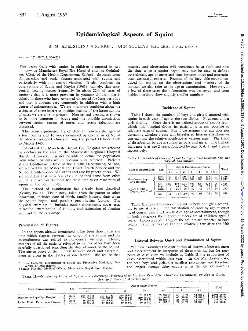
334 5 August 1967
Epidemiological Aspects of Squint
A. Al. ADELSTEIN,* M.D., D.P.H.; JOHN SCULLYt M.B., CH.B., D.P.I., D.O.M.S.
Brit. med. 7., 1967, 3, 334-338
This paper deals with squint in children diagnosed in two
clinics-the Manchester Royal Eye Hospital and the Ophthal-mic Clinic of the Health Department, Salford-discusses some
demographic and social factors associated with squint andparticularly with non-central viewing. It also confirms theobservations of Scully and Naylor (1961)-namely, that non-
central viewing occurs frequently (in about 220/0 of cases ofsquint) ; that it is more prevalent in younger children, parti-cularly in those who have remained untreated for long periods;and that it appears very commonly in children with a highdegree of anisometropia. We are now more confident about theestimates of these interrelationships because of the larger sampleof cases we are able to present. Non-central viewing is shownto be more common in boys; and the possible associationsbetween squint, season of onset, and recent infection are
discussed.The records presented are of children between the ages of
a few months and 10 years examined by one of us (J. S.) at
the above-mentioned clinics during the period October 1960to March 1965.
Patients at the Manchester Royal Eye Hospital are referredby doctors in the area of the Manchester Regional HospitalBoard. However, it is not possible to define the populationfrom which patients would necessarily be referred. Patientsat the Ophthalmic Clinic of the Health Department, Salford,are referred by the Maternal and Child Health Service and theSchool Health Service of Salford and also by practitioners. We
are confident that very few cases in Salford came from otherclinics, and we can therefore use these data to estimate rates ofsquint in the community.The method of examination has already been described
(Scully, 1961a). The history, taken from the parent or otherinformant, includes date of birth, family history, date whenthe squint began, and possible precipitating factors. Thephysical examination includes ocular movements, cover test,refraction, examination of fundus, and estimation of fixationwith aid of the visuscope.
Presentation of Figures
In the papers already mentioned it has been shown that thetime which elapses between the onset of the squint and itsascertainment was related to non-central viewing. Hence,mothers of all the patients referred to in this paper have beencarefully questioned regarding the date of onset of the squint.The age at onset or the interval between onset and ascertain-
ment is given in the Tables as one factor. We realize that
* Senior Lecturer, Department of Social and Preventive Medicine, Uni-versity of Manchester.
Senior Hospital Medical Officer, Manchester Royal Eye Hospital.
memory and observation will sometimes be at fault and thatthe time when a squint began may not be easy to define;nevertheless, age at onset and time between onset and ascertain-ment are useful criteria. Because of the inevitable error intro-duced by relying on the observations and memory of themothers we also refer to the age at examination. However, ina few of these cases the information was destroyed, and some
Tables therefore show slightly smaller numbers.
Incidence of Squint
Table I shows the numbers of boys and girls diagnosed withsquint in each year of age at the two clinics. Boys outnumbergirls slightly. Since there is no defined group of people fromwhich this hospital draws its patients it is not possible tocalculate rates of squint. But if we assume that age does not
determine whether a case will be referred here or elsewhere we
can examine the relative incidence at various ages. The trendof distribution by age is similar in boys and girls. The highestincidence is at age 2 years, followed by ages 3, 4, 1, and 5 years
successively.
TABLE I.-Number of Cases of Squint by Age at Asceriainment, Sex, andPlace of Examination
Age at Examination (years)Place of Examination Sex E-
<1 1- 2- 3- 4- 5- 6+
Boys 47 211 365 315 222 115 91Manchester Royal Girls 38 201 308 284 206 108 104Eye Hospital l l
All 85 412 673 599 428 223 195Boys 10 49 54 40 54 37 41
Salford Health Girls 5 25 56 52 44 37 34Department Clinic
L All 15 74 110 92 98 74 75
'i-otai
1,3661,249
2,615285253
538
Table II shows the cases of squint in boys and girls accord-ing to age at onset. The distribution of cases by age at onset
is, of course, different from that of age at ascertainment, thoughin both categories the highest numbers are of children aged 2years. However, about 16 O(, of the squints are reported to havebegun in the first year of life and relatively few after the fifthyear.
Interval Between Onset and Examination of Squint
We have examined the distribution of intervals between onset
and ascertainment in categories of three months, but for pur-poses of discussion we include in Table II the proportion of
cases ascertained within one year. In the Manchester data,for both boys and girls, the smallest percentage and therefore
the longest average delay occurs when the age of onset is
TABLE II.-Number of Cases of Squint and Percentage Ascertained within One Year After Onset (in parentheses) by Age at Onset,Sex, and Place of Ascertainment
I ~~~~~~~~~~Ageat Onset (Years)
Place of Ascertainment Sex
A
_ ___r___Total< 1 1- 2- 3- 4- 5- 6+
Manchester Royal Eye Hospital Boys 229 (53-7) 303 (71 3) 340 (85 0) 261 (83-5) 169 (88 2) 60 (81-7) 42 (85-7) 1,404 (76 9)Girls 203 (44-8) 266 (78 2) 299 (82 6) 239 (91-6) 162 (90-7) 66 (90-9) 59 (89-8) 1,294 (79-2)
Salfford Health Department Clinic.. Boys 36 (58-3) 53 (88-7) 64 (68-8) 47 (72-3) 40 (80-0) 26 (80-8) 22 (86-4) 288 (75-7)
{ Girls 22 (45-5) 47 (6881) 49 (83-7) 56 (80-4) 41 (87-8) 24 (917) 18 (83-3) 257 (78-2)
BkrrisHMEDICAL JOURNAL
on 12 March 2022 by guest. P
rotected by copyright.http://w
ww
.bmj.com
/B
r Med J: first published as 10.1136/bm
j.3.5561.334 on 5 August 1967. D
ownloaded from
5 August 1967 Squint-Adel
under 1 year. The percentage of cases ascertained within oneyear of onset tends to rise as the age at onset increases, andafter the age of 2 years more than 80 % of cases are ascertainedwithin one year. In the data from Salford the trend of thelength of interval between onset and ascertainment is muchthe same as in the Manchester data, though somewhat irregularowing to relatively smaller numbers.The differences between the sexes with respect to the interval
between onset and ascertainment may now be considered. TheManchester data show that when the age at onset is under 1year a higher percentage of boys than of girls are ascertainedwithin one year, and when the age at onset is after 1 yearthe girls have the shorter interval-that is, the percentage ofgirls ascertained is higher than that of boys within one year ofonset for each age except for age 2. The differences, however,are small and attain statistical significance (P<0.05) in onlyone age group. The Salford data show the same trends exceptthat in boys ascertainment is quicker when onset is in the firsttwo years, and only later is the position reversed.
Type of Squint
The number of cases diagnosed in the Manchester Royal EyeHospital according to the type of squint and the percentage ofeach type at each age group of onset are shown in Table III.(The number of children at Salford was too small formeaningful examination in these categories.)
TABLE III.-Number of Cases by Type of Squint, and Percentage ofEach Type by Age at Onset (Manchester)
Type of Squint<1 1-
Age at Onset (years)
2- 3- 4- 5- 6+
Right convergent squint 22 7 32-5 33 9 44-0 39-9 40-5 46-5 35-2 950Left ,, ,, 31-3 40-4 43-5 37-6 48-6 41-2 33-7 40-0 1,078Alternating ,, ,, 37-7 22-2 18-8 15-2 10-0 15-1 15-8 20-5 553Right divergent ,, 2-5 1-2 0-8 0-2 - 0-8 - 0 9 25Left ,, ,, 1-9 1-6 1-4 1-0 0-6 - 3-0 1-3 36Alternating ,, ,, 3 9 2 1 1 6 2-0 0-9 2 4 1 0 2 1 56
Total number .. .. 432 569 639 500 1331 1126 101
The overall total shows that convergent squint is morecommon on the left side than on the right (40% and 35.2%respectively). As the age of onset increases the percentage ofalternating squints diminishes-from 38% when the onset isin the first year to about 15 % after the age of 3.
TABLE IV.-Percentage of Children with Non-central Viewing Accordingto Age at Onset, Interval Between Onset and Ascertainment, and Sex.(The Number of Children with Squint in Whom a Diagnosis ofFixation was Made is Shown in Parentheses)
MonthsBetweenOnset andAscertain-
ment
Boys .. {
0-67-2425+
Age at Onset of Squint (years)
<2 | 2 + 3 4+ All Ages
Manchester Royal Eye Hospitai26-7 (101) 22-7 (379)39-2 (153) 31-6 (152)45-1 (71) 51-7 (29)
14-4 (195) 20-9 (675)31-3 (64) 34-7 (369)50-0 (12) 47-3 (112)
Allintervals 36-6 (325) 26-6 (560) 19-9 (271) 27 9(1,156)0-6 16-5 (97) 14-9 (349) 8-6 (220) 13-1 (666)7-24 26-9 (104) 21-1 (114) 26-7 (60) 24-5 (278)
Girls * 25 + 28-1 (64) 43-5 (23) 66-7 (6) 34-4 (93)
Allintervals 23-4 (265) 17-7 (486) 13-6 (286) 18-0 (1,037)
Salford Health Department Clinic0-6 23-3 (30) 23-7 (59) 9 4 (53) 18-3 (142)7-24 38-9 (18) 40-6 (32) 19-4 (31) 32-1 (81)
Boys .. 5+ 66-7 (9) 80-0 (10) 66-7 (3) 72-7 (22)
Allintervals 35-1 (57) 34-7 (101) 14-9 (87) 27-8 (245)
0-67-24
Girls . . 25 +
All intervals
11-1 (18) 21-3 (61) 9-7 (62) 14-9 (141)40-0 (10) 22-2 (18) 10-5 (19) 21-3 (47)45-5 (11) 40-0 (10) 33-3 (3) 41-7 (24)
'stein and Scully BRMSHMEDICAL JOURNAL 335
Central Viewing According to Age at Onset, Interval BetweenOnset and Diagnosis, and Sex of the Child
When the child does not co-operate a decision on centralviewing cannot be made, and it is not surprising that theproportion of cases in which no decision is arrived at is largerin younger children. Among the Manchester children whosesquint began in the first year of life 47% of boys and 44%of girls could not be diagnosed for fixation. Most of thesechildren were, of course, examined before the age of 2. Theproportion of cases not diagnosed for fixation diminishes asthe age of onset increases, and when the onset occurs after theage of 4 hardly any cases remain undiagnosed.
Table IV shows the percentage of children with non-centralviewing (among those cases in which a decision on centralviewing could be made) according to age of onset, intervalbetween onset and diagnosis, and sex. The association of eachof these factors with non-central viewing can be examinedindependently.
Data from Manchester
Non-central Viewing and Interval Between Onset and Diag-nosis.-The percentage of children with non-central viewingincreases as the interval between onset and diagnosis increasesthe trend is present in each age of onset and in both sexes.
Fixation and Age at Onset of Squint.-This relation is bestexamined among those children in whom the interval betweenonset and ascertainment was less than six months-that is, thoserepresented in the first row for each sex in Table IV. Theseinclude most of the cases of squint, and the distribution of delayis likely to be similar within each group by age at onset.Among these children non-central viewing is most frequent inthose with the youngest age at onset, and the rate decreasesprogressively as the age at onset increases. In each section ofthe Table there is a clear difference between the group with theyoungest onset age and that with the oldest. When the delaybetween onset and ascertainment is 7 to 24 months the gradientof the percentage of non-central viewing is not distinct in eachof the two rows, but there is a clear difference in proportionof non-central viewing between the children with the youngestand those with the oldest age at onset. This trend of decreaseof non-central viewing with increasing age at onset does notemerge in the group showing the longest interval between onsetand ascertainment-more than 25 months. However, it seemsreasonable to suppose that the different trend has arisen becausein this group there is a considerable difference between the aver-age intervals in the three groupings of age at onset; of thosechildren whose onset age is below 2 some have had a com-paratively long delay, whereas in those in whom the age atonset is over 4 years the interval is short. The cases are toofew in number for a more detailed analysis of this open-ended category.
Non-central Viewing According to Sex.-The boys have ahigher proportion with non-central viewing than the girls ineach category of age at onset and in each category of intervalbetween onset and diagnosis (except one in which the numbersare small). Thus, on examining the column which refers toonset under 2 years of age and comparing the data of the boyswith those of the girls, in each category of interval we have thefollowing comparisons of pairs of percentages: 26.7, 16.5 ; 39.2,26.9; and 45.1, 28.1. In general the Salford data follow thesame pattern, though there are exceptions which are to beexpected because of small numbers. Table IV presents the casesaccording to age at onset, and, since they are actually examinedat varying times after onset, the data are presented in Table Vwith age at diagnosis as the variable in place of age at onset.This Table shows that two factors-interval after onset andsex-are, as shown previously, independently related to thefrequency of non-central viewing. However, age at ascertain-
c
28-2 (39) j 23-6 (89) 10-7 (84) 19-3 (212)-I--I-
on 12 March 2022 by guest. P
rotected by copyright.http://w
ww
.bmj.com
/B
r Med J: first published as 10.1136/bm
j.3.5561.334 on 5 August 1967. D
ownloaded from
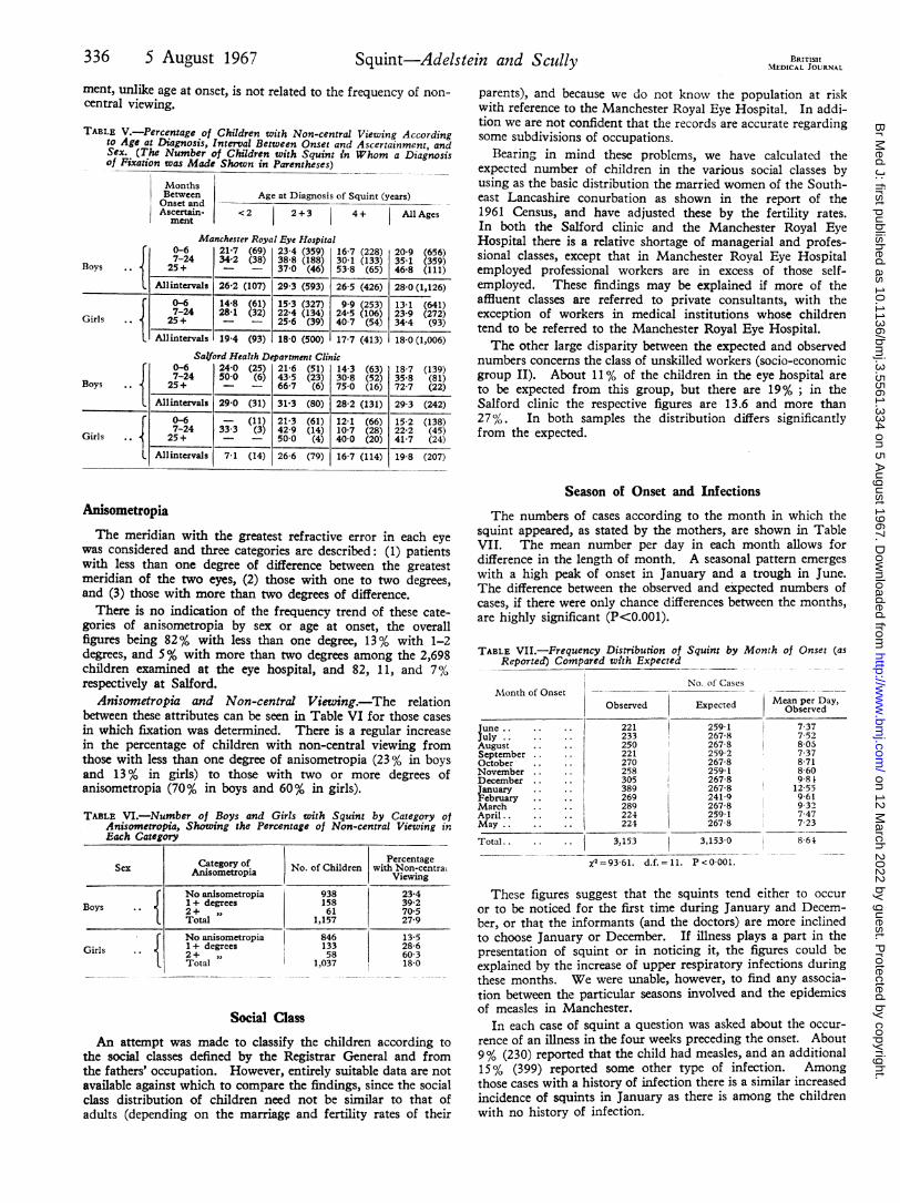
ment, unlike age at onset, is not related to the frequency of non-central viewing.
TABLE V.-Percentage of Children with Non-central Viewing Accordingto Age at Diagnosis, Interval Between Onset and Ascertainment, andSex. (The Number of Children with Squint In Whom a Diagnosisof Fixation was Made Shown in Parentheses)
MonthsBetween Age at Diagnosis of Squint (years)Onset andAscertain- <2 2+3 4 + All Ages
Boys .. {
0-67-2425+
Manchester Royal Eye Hospital21-7 (69) 23-4 (359) 16-7 (228) 209 (656)34-2 (38) 38-8 (188) 30-1 (133) 35-1 (359)
- 37-0 (46) 53-8 (65) 46-8 (111)
28 0 (1,126)
r -6 14-8 (61) 15-3 (327) 9-9 (253) 13-1 (641)7-24 28*1 (32) 22-4 (134) 24-5 (106) 23-9 (272)Girls .. 25 + - - 25*6 (39) 40 7 (54) 34-4 (93)
L Allintervals 19-4 (93) 18-0 (500) 17-7 (413) 18-0 (1,006)
Salford Health Department Clinic06 240 (25) 21-6 (51) 14-3 (63) 18-7 (139)7-24 50 0 (6) 43-5 (23) 30 8 (52) 35-8 (81)
Boys .. 25+ - - 66-7 (6) 75-0 (16) 72-7 (22)
Allintervals 29 0 (31) 31-3 (80) 28-2 (131) 29-3 (242)
Girls .. {
0-67-24
25 +
Allintervals
- (11) 21-3 (61) 12-1 (66) 15-2 (138)33-3 (3) 42-9 (14) 10-7 (28) 22-2 (45)
50-0 (4) 40-0 (20) 41-7 (24)
7-1 (14)[ 26-6 (79) 16-7 (114)
BRITISHMEDICAL JOURNAKL
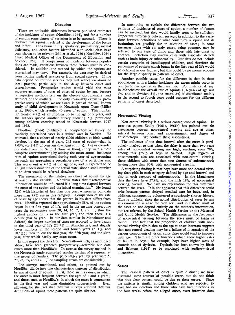
parents), and because we do not know the population at riskwith reference to the Manchester Royal Eye Hospital. In addi-tion we are not confident that the records are accurate regardingsome subdivisions of occupations.
Bearing in mind these problems, we have calculated theexpected number of children in the various social classes byusing as the basic distribution the married women of the South-east Lancashire conurbation as shown in the report of the1961 Census, and have adjusted these by the fertility rates.In both the Salford clinic and the Manchester Royal EyeHospital there is a relative shortage of managerial and profes-sional classes, except that in Manchester Royal Eye Hospitalemployed professional workers are in excess of those self-employed. These findings may be explained if more of theaffluent classes are referred to private consultants, with theexception of workers in medical institutions whose childrentend to be referred to the Manchester Royal Eye Hospital.The other large disparity between the expected and observed
numbers concerns the class of unskilled workers (socioeconomicgroup II). About 11% of the children in the eye hospital areto be expected from this group, but there are 19% ; in theSalford clinic the respective figures are 13.6 and more than27%. In both samples the distribution differs significantlyfrom the expected.
19-8 (207)
Anisometropia
The meridian with the greatest refractive error in each eye
was considered and three categories are described: (1) patientswith less than one degree of difference between the greatest
meridian of the two eyes, (2) those with one to two degrees,and (3) those with more than two degrees of difference.There is no indication of the frequency trend of these cate-
gories of anisometropia by sex or age at onset, the overallfigures being 82% with less than one degree, 13% with 1-2degrees, and 5% with more than two degrees among the 2,698children examined at the eye hospital, and 82, 11, and 7%respectively at Salford.
Anisometropia and Non-central Viewing.-The relationbetween these attributes can be seen in Table VI for those casesin which fixation was determined. There is a regular increasein the percentage of children with non-central viewing fromthose with less than one degree of anisometropia (23% in boysand 13% in girls) to those with two or more degrees ofanisometropia (70% in boys and 60% in girls).
TABLE VI.-Number of Boys and Girls with Squint by Category ofAnisometropia, Showing the Percentage of Non-central Viewing inEach Category
Sex Category ofAnisometropia
Season of Onset and Infections
The numbers of cases according to the month in which thesquint appeared, as stated by the mothers, are shown in TableVII. The mean number per day in each month allows fordifference in the length of month. A seasonal pattern emergeswith a high peak of onset in January and a trough in June.The difference between the observed and expected numbers ofcases, if there were only chance differences between the months,are highly significant (P<0.001).
TABLE VII.-Frequency Distribution ofReported) Compared with Expected
Month of Onset
June.July . . ..
AugustSeptemberOctober
November
December
January .February .. .MarchApril..May.
Total.
PercentageNo. of Children with Non-centrai
Viewing
No anisometropia 938 23-4Boys 1+ degrees 158 39 2
1 2+ ,, 61 70-5Total 1,157 27-9
GirlsNo anisometropia1 + degrees2+ ,Total
84613358
1,037
13-528-660 318-0
Social Class
An attempt was made to classify the children according tothe social classes defined by the Registrar General and fromthe fathers' occupation. However, entirely suitable data are notavailable against which to compare the findings, since the socialclass distribution of children need not be similar to that ofadults (depending on the marriage and fertility rates of their
Squint by Month of Onset (as
No. of Cases
Observed Expected
221233250221270258305389
259 1267-8267-8259 2267-8259-1267-8267?8
269 241 9289 267-8224 259 1224 267 8
3,153 3,153-0
X2 = 93-61. d.f. =1 1- P <0-001-
Mean per Day,Observed
7-377 528057-378-718 609 8112-559-619327-477-23
These figures suggest that the squints tend either to occuror to be noticed for the first time during January and Decem-ber, or that the informants (and the doctors) are more inclinedto choose January or December. If illness plays a part in thepresentation of squint or in noticing it, the figures could beexplained by the increase of upper respiratory infections duringthese months. We were unable, however, to find any associa-tion between the particular seasons involved and the epidemicsof measles in Manchester.
In each case of squint a question was asked about the occur-rence of an illness in the four weeks preceding the onset. About9% (230) reported that the child had measles, and an additional15% (399) reported some other type of infection. Amongthose cases with a history of infection there is a similar increasedincidence of squints in January as there is among the childrenwith no history of infection.
336 5 August 1967 Squint-Adelstein and Scully
Allintervals 26-2 (107) 29-3 (593) 26-5 (426)
.1 Il-1I-
i!~~~~~~~~
F
on 12 March 2022 by guest. P
rotected by copyright.http://w
ww
.bmj.com
/B
r Med J: first published as 10.1136/bm
j.3.5561.334 on 5 August 1967. D
ownloaded from
5 August 1967 Squint-Adelstein and Scully BRITISH 337MEDICAL JOURNAL 337
Discussion
There are noticeable differences between published estimatesof the incidence of squint (Nordl6w, 1964), and for a numberof reasons some degree of variation is to be expected. There isevidence that squint is related to the development of the foetusand infant. Thus brain injury, spasticity, prematurity, mentaldeficiency, and other factors identified with social class havebeen shown to be relevant (Miller et al., 1960 ; Nordldw, 1964 ;Chief Medical Officer of the Department of Education andScience, 1966). If comparisons of incidence between popula-tions are made, variations between these factors must be con-sidered. In addition, the method by which the squints areascertained may vary. For example, the data may be derivedfrom routine medical services or from special surveys. If thedata depend on routine services they will reflect variations oflocal practice, particularly in the delay between onset andascertainment. Prospective studies would yield the mostaccurate estimates of rates of onset of squint by age, becauseretrospective methods rely on the observations, memory, andattitudes of the mothers. The only reasonably extensive pros-pective study of which we are aware is part of the well-knownstudy of child development in Newcastle upon Tyne (Milleret al., 1960), which revealed 40 cases of squint. This numberrepresented 4.7% of all children up to the age of 5 years, andthe authors quoted another survey showing 5 % prevalenceamong children entering school in Newcastle between 1947and 1952.Nordl6w (1964) published a comprehensive survey of
routinely ascertained cases in a defined area in Sweden. Heestimated that a cohort of nearly 6,000 children at birth woulddevelop, by the age of 6 years, 242 cases of squint-that is,4.03% (or 2.6% of constant divergent squints). Let us considerour data from the Salford clinic as though they were almostcomplete ascertainment; by adding the mean annual incidencerates of squints ascertained during each year of age-groupingwe reach an approximate prevalence rate of a particular age.This works out as 4.3 % at the age of 6, and is expected to bea slight underestimation of the actual state, since a small numberof children would be referred elsewhere.The assessment of the relative incidence of squint by age
at onset is also variable. Nordldw states that "retrospectiveinformation would be influenced if a long time elapsed betweenthe onset of the squint and the initial examination." He found52 % with histories of less than one year, whereas in our datamore than 75 % are in this category. Comparison of the rateof onset by age shows that the pattern in his data differs fromours. Nordl6w reported that approximately 38% of the squintsbegan in the first year of life, and in the ensuing consecutiveyears the percentages were 20, 14, 16, 7, 1, and 1; thus thehighest proportion is in the first year, and then there is adecline year by year. In our data (similar in Manchester andSalford) the largest number of cases are reported to have begunin the third year of life (23.7% in Manchester), and slightlylower numbers in the second and fourth years (21.1% and18.5%); then follow the first year, the fifth year, and the sixthyear, after which hardly any cases occur.
In this respect the data from Newcastle-which, as mentionedabove, have been gathered prospectively-resemble our datamuch more than Nordldw's. In essence the survey method inthe Newcastle study comprised regular visiting of a representa-tive group of families. The percentages year by year were 5,27, 25, 25, and 17. (The sampling errors are considerable.)The surveys mentioned, and others, as pointed out by
Nordlbw, divide into two characteristic, patterns of distributionby age at onset of squint. First, those such as ours, in whichthe onset is most frequent between the ages of 1 and 4 years;and those, such as Nordldw's, in which the onset iscommonestin the first year and then diminishes progressively. Evenallowing.for the fact that different surveys adopted differentdefinitions of squint these patterns are quite distinct.
In attempting to explain the difference between the twopatterns of incidence of onset of squint, a number of factorscan be invoked, but they would hardly seem to be sufficient.Important differences between surveys, in addition to the varia-tions between definitions of what constitutes a squint and itsbeginning, are possibly due to selection of cases; in someinstances those with an early onset, being younger, may bereferred to one type of clinic and those with late onset toanother. Selection may involve cases with associated defectssuch as brain injury or subnormality. Our data do not includecertain categories of handicapped children, and therefore thepercentage of squints which began in the first year of life wouldbe deficient in our figures ; but this could by no means accountfor the large disparity in patterns of onset.
Another possible cause for the difference is that in thosepopulations with a higher incidence the excess might occur inone particular age rather than another. For instance, if, say,in Manchester the overall rate of squints at 6 years of age was5°% and in Sweden 3%, the excess 2% if distributed mainlyin the second to fourth years could account for the differentpatterns of onset described.
Non-central Viewing
Non-central viewing is a serious consequence of squint. Inprevious papers Scully (1961a, 1961b) has pointed out theassociation between non-central viewing and age at onset,interval between onset and ascertainment, and degree ofanisometropia. We confirm these associations.The influence of the time interval after the onset is parti-
cularly marked, so that when the delay is more than two yearsrates of non-central viewing are high, reaching even 70%among this group of boys in Salford. High degrees ofanisometropia also are associated with non-central viewing,those children with more than two degrees of anisometropiahaving more than 60% with non-central viewing.A surprising finding is that boys have more non-central view-
ing than girls in each category defined by age and interval andalso in each category of anisometropia. In the Manchesterdata the boys have 27.9% and the girls 18% of non-centralviewing, overall. We have no explanation for this differencebetween the sexes. It is not apparent that this difference couldarise because parents delayed medical care for boys, and, inaddition, subsequently volunteered a spuriously shorter history.This is unlikely, since the actual distribution of cases by ageat examination is alike for each sex; and in Salford most ofthe cases do not depend entirely on the mother's intervention,but are referred by the School Health Service or the Maternaland Child Health Service. The differences in the frequencyof non-central viewing between the sexes must be taken asfound. The fact that the proportion of children with non-central viewing diminishes as the age at onset increases suggeststhat non-central viewing may be a failure of integration of thevarious components of vision, since these would tend to improvewith age. There are other functions which show higher ratesof failure in boys ; for example, boys have higher rates ofenuresis and of dyslexia. Dyslexia has been shown by Birchand Belmont (1966) to be associated with auditory-visualintegration.
Season
The seasonal pattern of onset is quite distinct; we havediscussed some sources of possible error, but do not thinkthat this clear pattern could be due to these errors. Sincethe pattern is similar among children who are reported tohave had no infection and those who have had infections inthe four weeks before the alleged onset, overt infection does
on 12 March 2022 by guest. P
rotected by copyright.http://w
ww
.bmj.com
/B
r Med J: first published as 10.1136/bm
j.3.5561.334 on 5 August 1967. D
ownloaded from
338 5 August 1967 Squint-Adelstein and Scully
not explain the trend. We can only speculate on factors suchas subclinical infections, or length of daylight.
SummaryWe report an analysis of 3,243 cases of squint examined at
a health department clinic and an eye hospital. The estimatedprevalence of squint at the age of 6 years is 4.3%, similar toestimates in other surveys in England. Incidence of onset ishighest in the third year of life, and, if this distribution reflectsthe incidence in the community, it represents one characteristicpattern of distribution. Another pattern has been describedin which the highest rate of onset is in the first year anddiminishes rapidly thereafter. Non-central viewing occurs in23% of the cases of squint in which this function could beassessed. Non-central viewing is much more frequent in boys,in children in whom the onset is early, in those who have had
the squint for a longer period, and in those with high degreesof anisometropia. There is evidence that squints begin moreoften in winter, with a peak incidence in January.
We are indebted to Mrs. Anne Fish for her painstaking andcareful handling of the data, 'and to Professor C. I. Phillips forhis advice. We wish to thank the surgeons at the Manchester RoyalEye Hospital for making available for examination cases under theircare.
REFERENCESBirch, H. G., and Belmont, Lilian (1966). E.E.N.T. Digest, vol. 28,
No. 12, p. 47.Chief Medical Officer of the Department of Education and Science (1966).
The Health of the School Child. H.M.S.O., London.Miller, F. J. W., Court, S. D. M., Walton, W. S., and Knox, E. G.
(1960). Growing Up in Newcastle-upon-Tyne. London.Nordlow, W. (1964). Acta ophthal. (Kbh.), 42, 1015.Scully, J. (1961a). Brit. med. Y., 2, 1610 .
(1961b). Brit. 7. Ophthal., 45, 741.and Naylor, E. J. (1961). Brit. med. 7., 2, 81.
Progestin Therapy of Breast Cancer: Comparison of Agents
BASIL A. STOLL,* M.R.C.S., F.F.R.
Brit. med. J., 1967, 3, 338 -341
Over recent years many synthetic orally active progestationalagents have been introduced to clinical practice. The multi-plicity of such agents presents a problem of choice to theclinician considering progestin therapy in breast cancer. Asimilar problem applies also to uterine cancer. Various authors,each using a different progestin, have reported their results insmall series of breast cancer cases (Jonsson et al., 1959 ; Lewinet al., 1959; Douglas et al., 1960; Baker and Kelley, 1960;Jolles, 1962; Bucalossi et al., 1963 ; Stoll, 1965). The trialsare summarized in Table I, and from the results it is obviousthat there is a place for progestin therapy in breast cancer. Toour knowledge no basis has yet been shown for statements thatthe presence of breast cancer contraindicates the use of pro-gestins because they may possibly stimulate the tumour(Klawans, 1965).
TABLE I.-Reports of Clinical Response in Breast Cancer After ProgestinAdministration
Author Progestin Clinical Response in
Jonsson et al. (1959) .. Bromoketoprogesterone 7 of 34Lewin et al. (1959) .. Norethindrone 5 22Douglas et al. (1960) .. Norethisterone
oenanthate 1 12Baker and Kelley (1960) .. Norethynodrel 4 20Jolles (1962) Hydroxyprogesterone
caproate 3 9Bucalossi et al. (1963) .. Medroxyprogesterone 11 30Stoll (1965) .. .. Norethisterone acetate
Medroxyprogesterone 3 16
Nevertheless, there still remains the evaluation of the mostefficacious agent and the most suitable stage of the disease for itsadministration. The choice of suitable agent is made moredifficult by differences in side-effects, particularly in the inci-dence of gastrointestinal upset, biochemical evidence of liverdamage, and signs of masculinization after prolonged use. Forthis reason it was thought valuable for one observer, usingstandardized criteria, to compare examples of each class ofprogestin in the treatment of advanced breast cancer. Thisreport is of a limited pilot trial of this nature.
Material
A total of 72 postmenopausal women with advanced breastcancer were treated by synthetic oral progestins selected torepresent the three major groups in use-that is, 19-nortesto-sterone derivatives, 17at-hydroxyprogesterone derivatives, andthe testosterone derivatives. The initial daily dosage given wasin general six times that used in oral contraceptive practiceand is noted in Table II. Cases were not randomized, but wereallocated on a chronological basis as' agents became available fortrial.As well as objective evidence of tumour regression, special
note was taken of side-effects from each drug-mainly theimmediate ones, but also in a few cases the long-term ones.Special investigations carried out in the majority of patientsincluded liver-function studies and serial cytohormonal assess-ment of the vaginal smear, both before and during treatment.In some of the later cases glucose-tolerance tests were carriedout after at least four weeks of hormone treatment.
All 72 patients had undergone either a natural menopause orcastration. They were suffering from advanced breast cancer,beyond the control of both surgery and radiotherapy. Allpatients had measurable evidence of progressing soft-tissuelesions regularly measured, and photographed when appropriate.Simultaneous treatment of the part under observation was notpermitted by any method except local dressings. Patients wereselected for drug trial only if the disease was progressing inactivity as recorded in serial observations. Previous hormonetherapy had been discontinued for at least two months andcastration carried out at least six months previously.
Nathanson's (1952) classical criteria of objective responsefrom hormone therapy in breast cancer were used. A responseindicates observation of at least 50% regression in size of acces-sible lesions, while, at the very least, all existing inaccessiblelesions remain static in activity and no new lesions appear. Ofthe 72 patients, there were 65 who received the hormones fortwo months or longer and were assessed for tumour regression.The patients who discontinued the drugs before completion oftwo months' treatment are excluded from the analysis as having* Cancer Institute, Melbourne, Australia. Address requests for reprints
to 54 Princes Park Avenue, London N.W.l 1.
on 12 March 2022 by guest. P
rotected by copyright.http://w
ww
.bmj.com
/B
r Med J: first published as 10.1136/bm
j.3.5561.334 on 5 August 1967. D
ownloaded from


![9887basem aldeek some%final epidemiological aspects about cancer in king saudi[1]1 [autosaved]](https://img.pdfslide.us/doc/110x75/558438bad8b42a77068b4c31/9887basem-aldeek-somefinal-epidemiological-aspects-about-cancer-in-king-saudi11-autosaved.jpg)