Embed Size (px)
Citation preview
SQUAMOUS CELL CARCINOMA OF THE ESOPHAGUS
GENERAL THORACIC SURGERY
CHAPTER 143
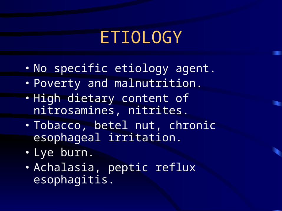
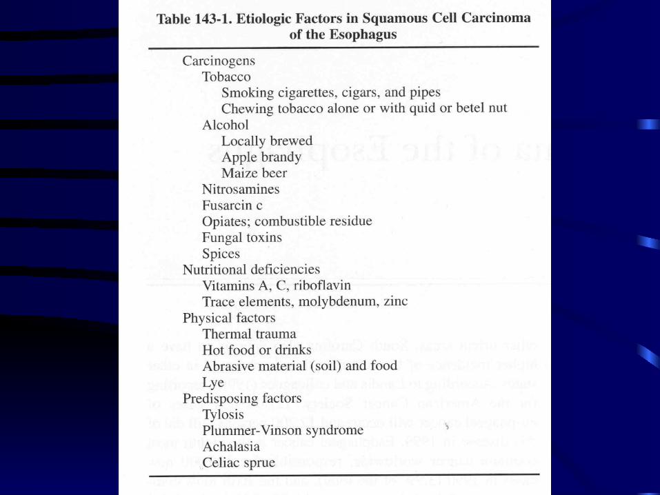
ETIOLOGY
• No specific etiology agent. • Poverty and malnutrition. • High dietary content of nitrosamines,
nitrites. • Tobacco, betel nut, chronic esophageal
irritation.• Lye burn. • Achalasia, peptic reflux esophagitis.
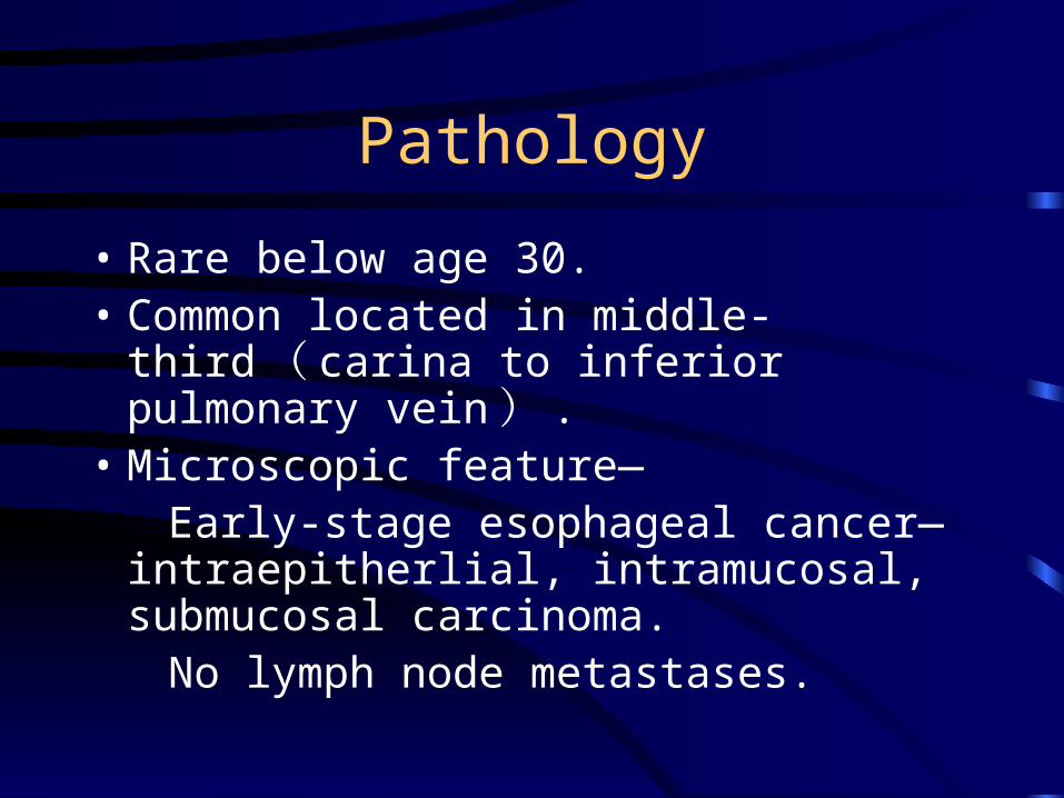
Pathology
• Rare below age 30. • Common located in middle-third ( carina
to inferior pulmonary vein ) .• Microscopic feature— Early-stage esophageal cancer—
intraepitherlial, intramucosal, submucosal carcinoma.
No lymph node metastases.
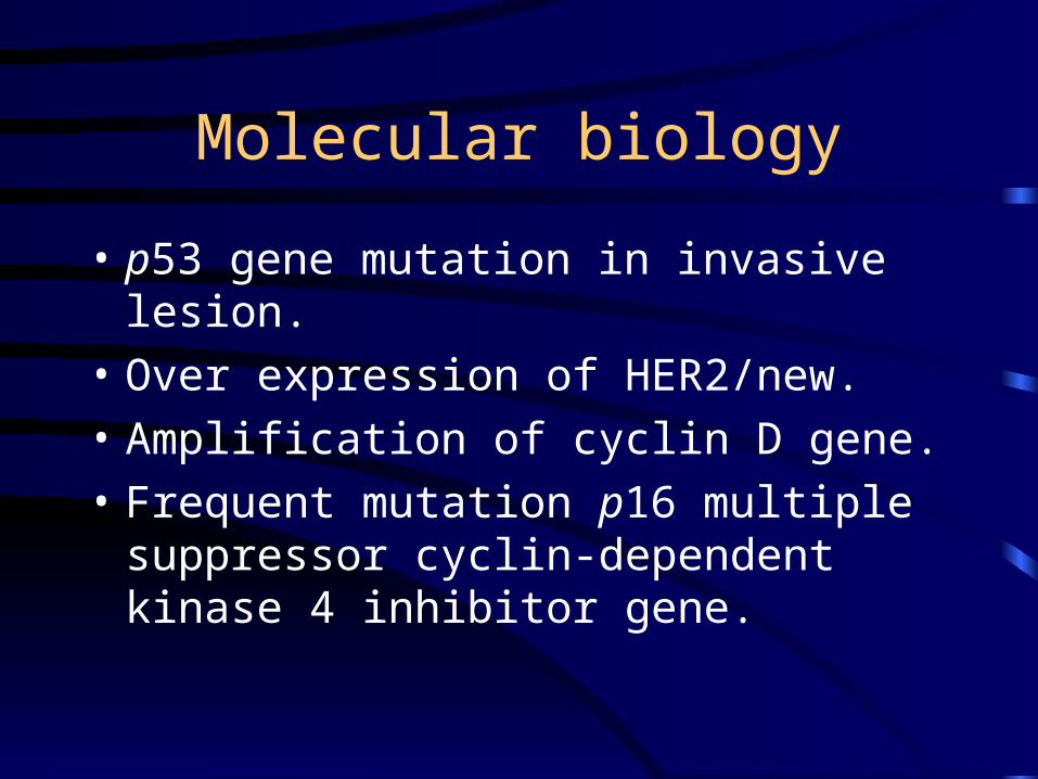
Molecular biology
• p53 gene mutation in invasive lesion.
• Over expression of HER2/new.
• Amplification of cyclin D gene.
• Frequent mutation p16 multiple suppressor cyclin-dependent kinase 4 inhibitor gene.
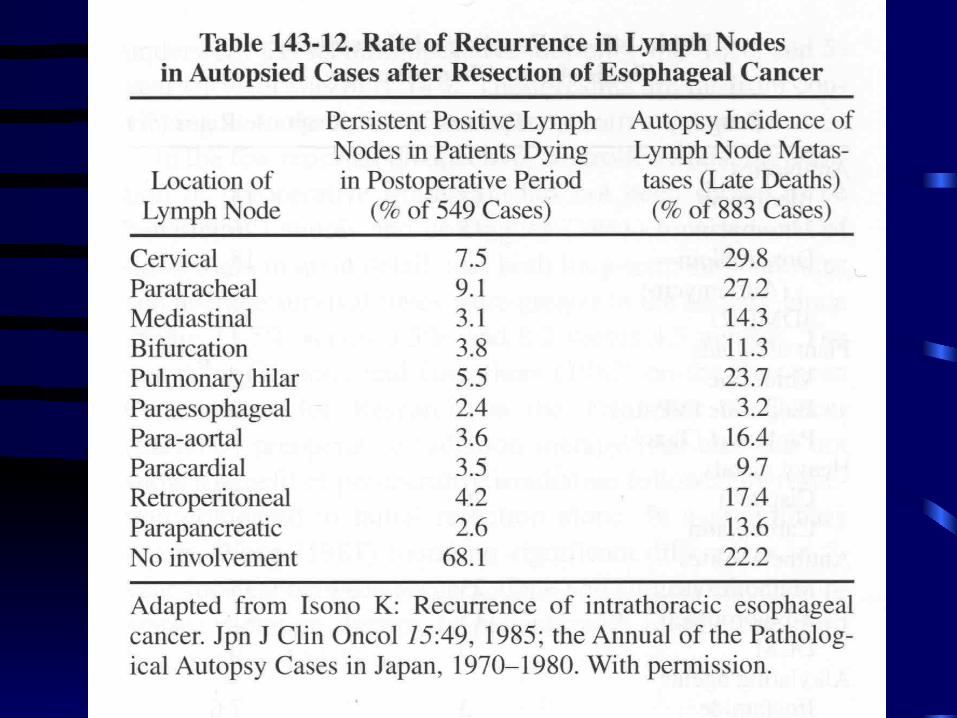
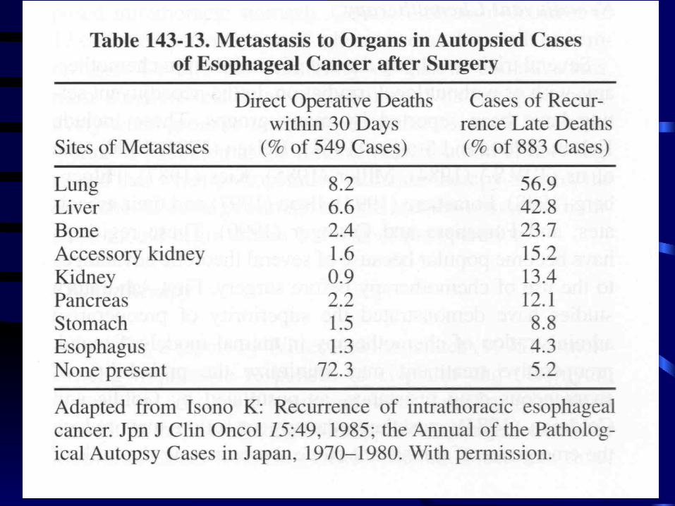
Metastases
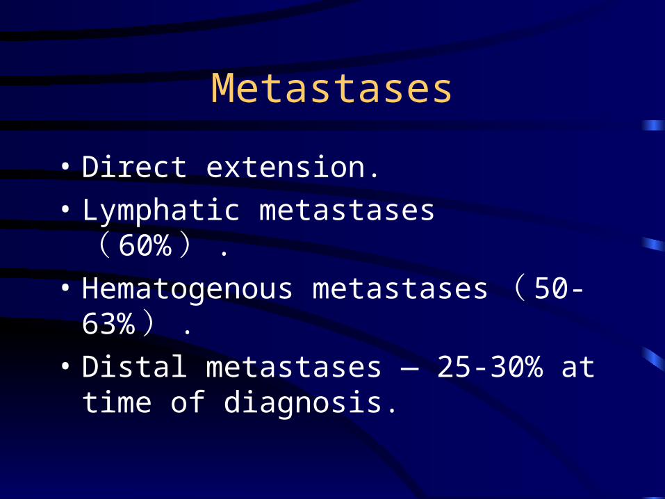
• Direct extension.
• Lymphatic metastases ( 60% ) .
• Hematogenous metastases ( 50-63% ) .
• Distal metastases — 25-30% at time of diagnosis.
Metastases
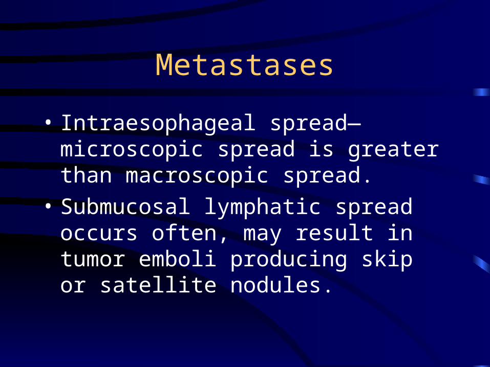
• Intraesophageal spread—microscopic spread is greater than macroscopic spread.
• Submucosal lymphatic spread occurs often, may result in tumor emboli producing skip or satellite nodules.
Metastases
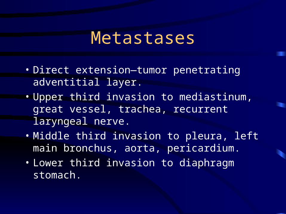
• Direct extension—tumor penetrating adventitial layer.
• Upper third invasion to mediastinum, great vessel, trachea, recurrent laryngeal nerve.
• Middle third invasion to pleura, left main bronchus, aorta, pericardium.
• Lower third invasion to diaphragm stomach.
Metastases
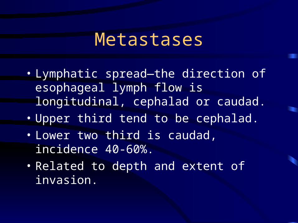
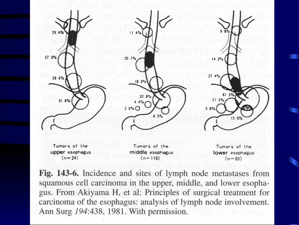
• Lymphatic spread—the direction of esophageal lymph flow is longitudinal, cephalad or caudad.
• Upper third tend to be cephalad.
• Lower two third is caudad, incidence 40-60%.
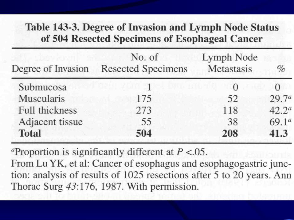
• Related to depth and extent of invasion.
Lymph node station
• 1 — the paraesophageal lymph node.
• 2 — periesophageal, celiac perigastric lymph node.
• 3 — the distal subdiapgragm or supraclavicular, lateral thoracic region.
Clinical manifestation
• s/s — infrequently at early stage. • Retrosternal discomfort, pain sensatin of
frication, burning. • Slow passage of food during swallowing. • Progressive dysphagia — first solid food,
then soft food, then liquid. • Melena, hematemess, anemia, weight loss,
hoarseness, hiccough, cachexia.
Diagnostic studies
• Cytologic screening
• Upper GI series
• CT
• Endoscopy
• Endoscopic ultrasonography
• Bronchoscopy: evaluation the tracheal or bronchial invasion.
Cytologic screening
• Screen asymptomatic people in high-incidence area.
• Obtain smear of esophageal mucosa with abrasive balloon catheter.
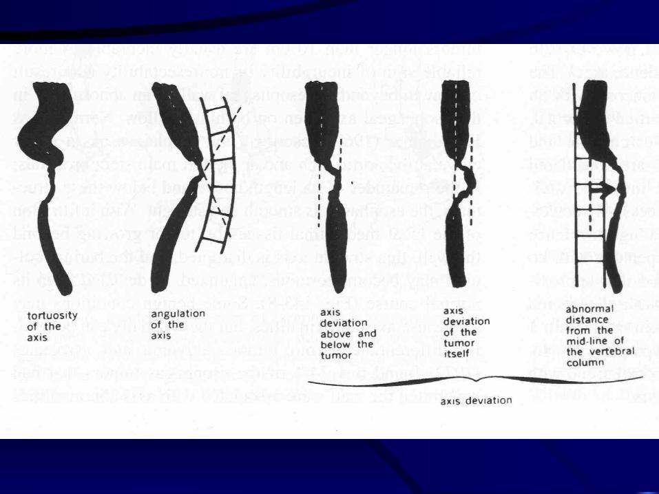
Upper GI series
• Diffucult in demonstration the early lesion
• Length of lesion, not correlate with degree of tumor penetration.
• Longer than 10 cm is incurable.
• Esophageal axis, 74% tumor penetrated wall associated with axis abnormalities.
• Demonstration tracheoesophageal fistula.
CT• Four stage — I — intralumonal mass without wall thickening. II — wall thickening. III — tumor spread into adjacent tissue. IV — distal meatastases. • Identified lymph node. • Aortic invasion: loss fat planes and contact less
hen 45 degree — invasion unlikely; exceed 90 degree — invasion real possibility.
• Invasion to pericardium is difficult to detect.
Endoscopy
• Essential in all patients. • Biopsy should be done in all cases. • Positive diagnosis 90%. • If no lesion—mucosal stain— Toluidine blue stain — the tumor cell not
the normal nucosa. Lugol’s solution stain — the normal cell
not the tumor cell.
Endoscopic ultrasonography
• —detailed studies the structure of esophageal wall and periesophageal tissue.
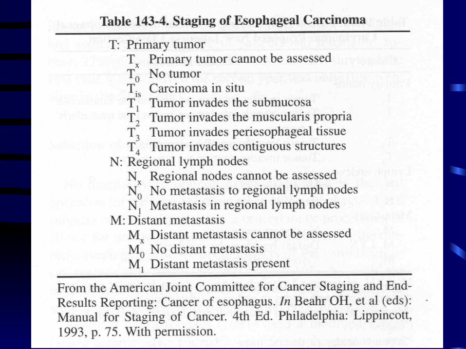
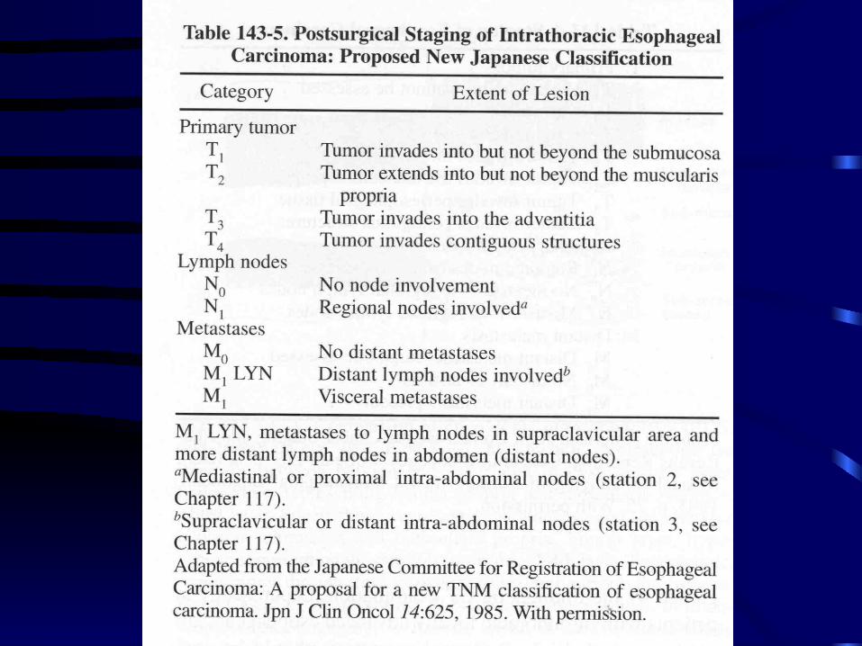
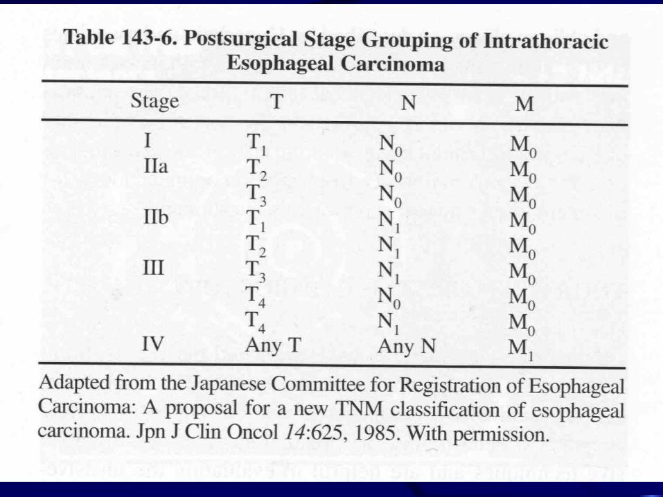
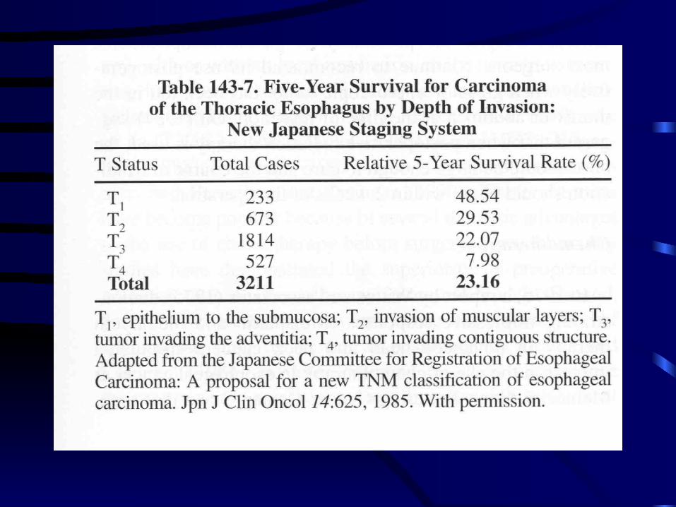
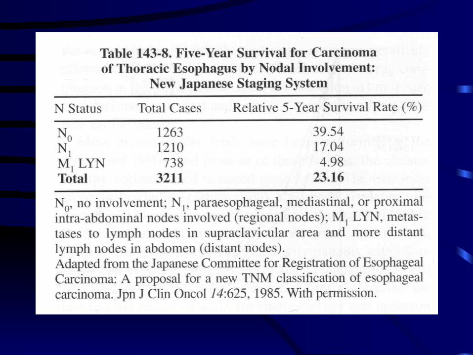
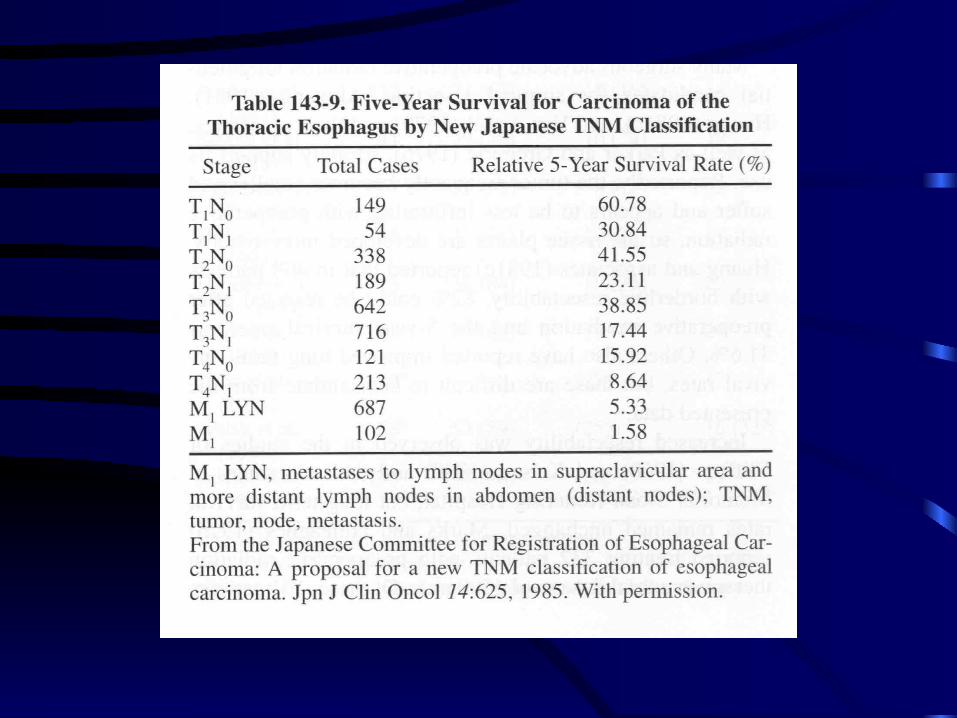
Staging
• —TNM system.
Surgical therapy
• Transthoracic, trandhiatal esophagectomy.
• Reconstruction.
• Respectability rate—45%-56%.
• Morbidity—most respiratory complication.
• Mortality—0.8%-12%.
• Surgical result—long-term survival is poor.
Resection plus adjuvant therapy
• Preoperative radiation therapy
• Postoperative irradiation
Preoperative radiation therapy
• Tumor became smaller and softer.
• Less infiltrating tissue plane develop.
• Increase respectability.
• Long term survival unchanged.
Postoperative irradiation
• No survival advantage.
• Significant reduction in local recurrence.
• High incidence of complication related the transposed intrathoracic stomach.
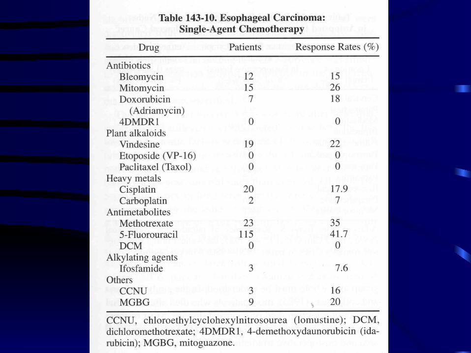
Chemotherapy
• Response rate — 40-60%.
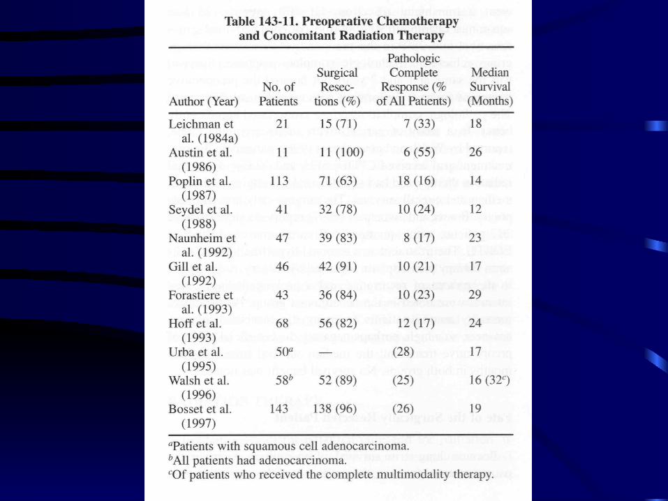
• Neoadjuvant chemotherapy.
Fate of surgically resected patient
• Survive more than 5 year tend to have the follow prognostic factors—
Small tumor less than 5 cm long.
No invasion to advantia.
No lymph node involvement.
Age younger than 60 year.
Women.
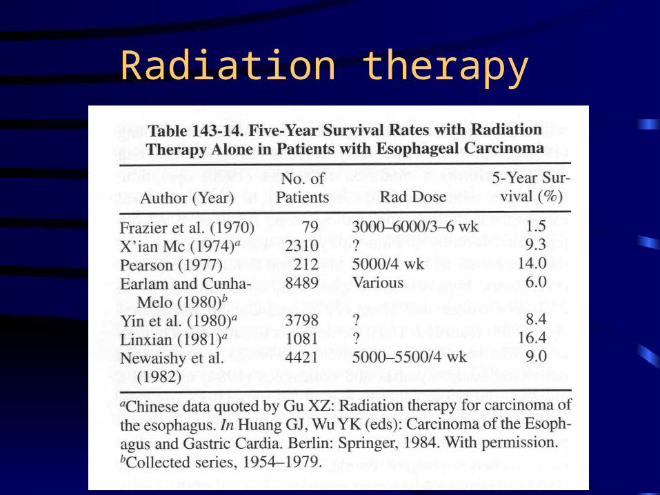
Radiation therapy


![Presence of Porphyromonas gingivalis in esophagus and its ... · oma [20, 25]. Since esophageal squamous cells are histo-logically similar to oral squamous cells and esophageal infection](https://img.pdfslide.us/doc/110x75/60bf33469a25d95dc16b0f67/presence-of-porphyromonas-gingivalis-in-esophagus-and-its-oma-20-25-since.jpg)