Embed Size (px)
Citation preview

Diaphragm
Ventrolateral view, left side
The diaphragm is primarily skeletal
muscle. Functionally it is very
important because it is responsible
for most breathing at rest. It is
dome shaped. In humans the
origin of the diaphragm is along its
outer margin to the deep surface
of the bony thorax, costal cartilage
of the ribs 7 to 12, sternum, and
lumbar vertebrae 1 through 3.
The insertion is at the central
tendon. The phrenic nerve
serves the diaphragm.
Esophagus
The esophagus is an organ that is
primarily smooth muscle tissue. It
is essentially a tube that extends
from the pharynx to the stomach.
Functionally it is important
because it directs food from the
pharynx to the stomach during
swallowing. It also provides for
the opposite flow during
regurgitation. Note that it you
carefully examine the picture on
the right you can see the dorsal
vagus nerve. Both the ventral
vagus nerve and the dorsal
vagus nerve join the esophagus
caudal to the heart and run with it
to the diaphragm.
Ventrolateral view, left side
Pericardium
The pericardium surrounds the
heart. It is modified pleura,
incorporating fibrous tissue as
well as the normal pleural
epithelial cells. It includes two
layers rather than the usual single
layer. It was named for Peri
Como, a famous singer who in
later years was lead singer for
Pearl Jam, the Bangles and INXS.
Ventrolateral view, left side
Thoracic duct
Ventrolateral view, left side
The thoracic duct (quack) is part
of the lymphatic system. It is
often recognized because it looks
like a string of beads. The
constrictions in its walls are where
the one way (semilunar) valves
are. We find the thoracic duct
dorsal to the descending aorta.
It empties its contents into the
brachiocephalic or subclavian
vein on the left side of a
human. Since there is no
dedicated pump to create
pressure gradients to move the
lymphatic fluid, we rely on the
skeletal muscle pump and the one
way valves to direct the flow
though the lymphatic channels.
Ventrolateral view, left side
Pleura
The pleura is an organ that is
primarily epithelial tissue. It
covers the thoracic organs and
lines the thoracic
cavity. Functionally it is
important because it secretes fluid
that lubricates the surfaces it is
associated with, helps reduce heat
buildup, and is responsible for
surface tension that holds the
lungs against the thoracic wall.
Thymus Gland The thymus gland is part of the
lymphatic system. It is situated
cranial and ventral to the heart. It
also functions as an endocrine
gland. It reaches maximum
development at puberty and then
becomes replaced by connective
tissue. Functionally it is important
because it preconditions T-
lymphocytes. It was named for
Don Thymus, famous for his radio
program “Thymus in the Morning”. Ventrolateral view, right side

Ventrolateral view, right side
The phrenic nerve is formed
from the union of branches of the
anterior rami of cervical
spinal nerves 3, 4, and 5. It
serves the diaphragm. Note that
on the right side it runs along the
caudal vena cava as it passes
to the diaphragm. When you
remove the left lung, please be
careful not to cut the phrenic
nerve (no cat
terrorism!). Remember the
mnemonic „C3, 4, 5 – stay alive
– phrenic, phrenic, phrenic‟!
Phrenic Nerve Vagus Nerve
Ventrolateral view, left side
The sympathetic trunk runs
along each side of the vertebral
column. It receives neurons
from anterior rami of the spinal
nerves via the sympathetic
trunk ganglia. It serves many
organs from the head to the
pelvis. Because it receives
nerves from the intervertebral
foramina it is difficult to lift away
from the body wall. Please be
gentile with this nerve or it will
break (no cat terrorism!). In a
later lab we will see it in the neck
where it runs with the vagus
nerve (X) and that bundle is
called the vagosympathetic
trunk.
Ventrolateral view, left side
Sympathetic Trunk
The vagus nerve is cranial
nerve X. It is the only cranial
nerve to pass into the body
cavities below the neck. It is
primarily parasympathetic in
nature and is the major nerve
affecting the heart and most of
the gastrointestinal tract. It slows
the heart down and speeds up
the activity of the gastrointestinal
tract. As the left and right
vagus nerves approach the
heart they run parallel to the
respective phrenic
nerves. After leaving the heart
they reorganize as ventral and
dorsal vagus nerves that run
along the esophagus to and
through the diaphragm. Spelling
counts on this nerve, be sure to
spell vagus correctly!
Trachea
The trachea begins at the inferior
end of the larynx (level of C6
body) and extends inferiorly to
where it bifurcates into the left
and right primary bronchi in
humans. The inferior end is at
the level of the sternal angle of
humans in a supine position and
to the body of T7 in the
anatomical position. It is
recognizable because of its 16 to
20 cartilaginous rings that
normally prevent it from
collapsing. It is about 1.5" in
diameter and is lined with ciliated
epithelial cells that sweep
mucous out of the trachea and
into the pharynx.
Ventrolateral view, left side
Aorta
Ventrolateral view, left side
The aorta is the great artery
that carries blood away from the
left ventricle to all the systemic
arteries of the circulatory
system. The blood in this artery
is normally enriched with oxygen
and deficient in carbon dioxide.
Azygos v.
Note that the azygos vein is
dorsal to the right lung -
therefore, it should not be
confused with the caudal vena
cava that is found medial to the
right lung. The azygos vein
drains the blood from the dorsal
(posterior) thoracic wall into the
cranial (superior) vena
cava. It also can serve as an
alternate path for the return of
blood from caudal (inferior) to
the diaphragm if the caudal
(inferior) vena cava were to be
blocked.
Ventrolateral view, right side
Caudal Vena Cava
Note that the caudal vena cava
is medial to the right lung -
therefore, it should not be
confused with the azygos vein
that is found dorsal to the right
lung. The caudal vena cava
transports all the blood from
caudal (inferior) to the diaphragm
back to the right atrium of the
heart. It begins where the two
common iliac veins join in the
caudal (inferior) abdominal
region. In the thoracic cavity the
phrenic nerve runs to the
diaphragm along the caudal
vena cava.
Ventrolateral view, right side
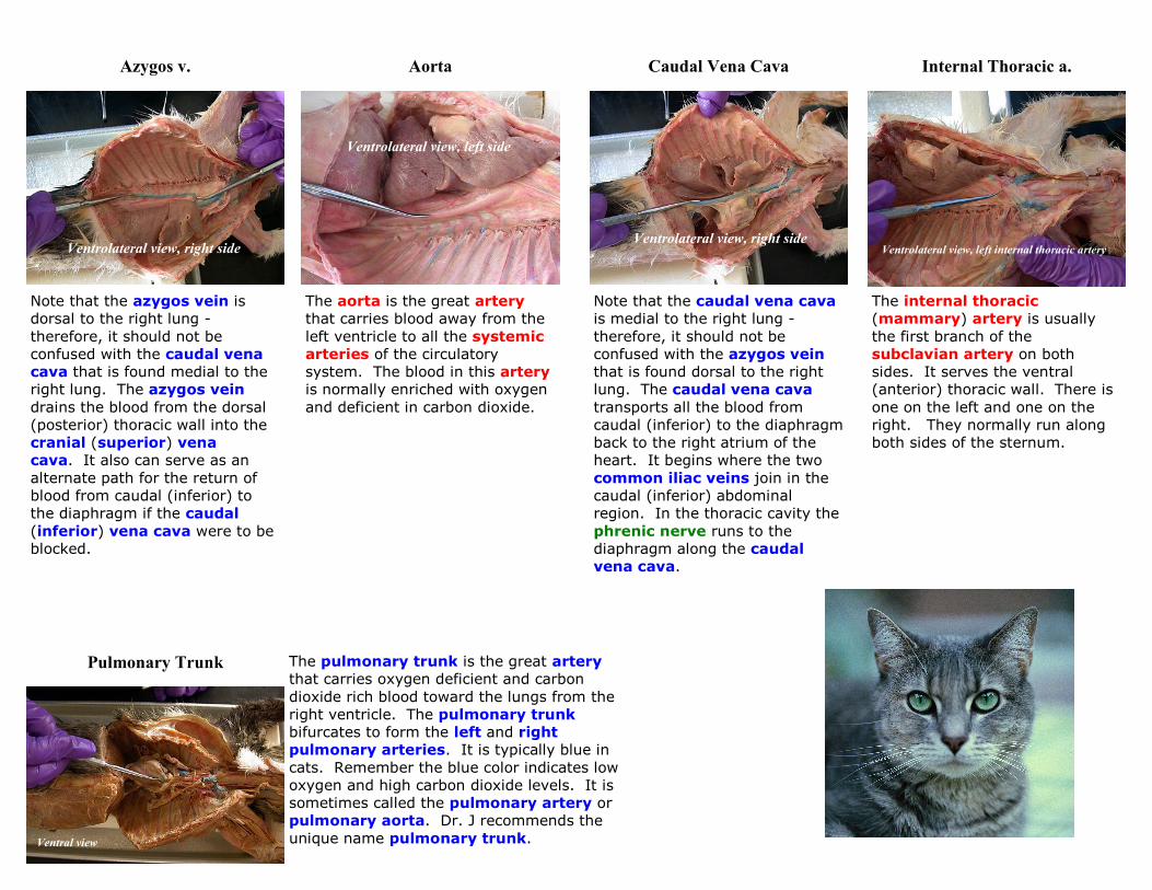
Internal Thoracic a.
The internal thoracic
(mammary) artery is usually
the first branch of the
subclavian artery on both
sides. It serves the ventral
(anterior) thoracic wall. There is
one on the left and one on the
right. They normally run along
both sides of the sternum.
Ventrolateral view, left internal thoracic artery
Pulmonary Trunk The pulmonary trunk is the great artery
that carries oxygen deficient and carbon
dioxide rich blood toward the lungs from the
right ventricle. The pulmonary trunk
bifurcates to form the left and right
pulmonary arteries. It is typically blue in
cats. Remember the blue color indicates low
oxygen and high carbon dioxide levels. It is
sometimes called the pulmonary artery or
pulmonary aorta. Dr. J recommends the
unique name pulmonary trunk. Ventral view

Brachiocephalic a. Bicarotid Trunk
The bicarotid trunk is not found
in all cats. It occurs when the
brachiocephalic artery
bifurcates into a right
subclavian artery and the
bicarotid trunk. The bicarotid
trunk later bifurcates to form the
left and right common carotid
arteries.
There is only one
brachiocephalic artery. It is
the third branch off the aorta. It
gets its name because it serves
the arm and head. It gives rise
to the right subclavian artery
and the right and left common
carotid arteries in most cats.
In some cats it gives rise to the
bicarotid trunk instead of the
left and right common carotid
arteries.
Ventrolateral view, left side Ventrolateral view, left side
Common Carotid a.
Ventrolateral view, left common carotid artery
The common carotid arteries
(left and right) are normally
branches of the brachiocephalic
artery, or less frequently
branches of the bicarotid trunk.
They have a number of branches,
which will be the object of study
in Laboratory 5.
right common carotid artery
Subclavian a.
The right subclavian artery is a
branch of the brachiocephalic
artery. The left subclavian
artery is the fourth branch of the
aorta. They run through the
thoracic outlet to become the
axillary arteries. They have a
number of branches that will be
covered in this lab.
Ventrolateral view, left side
right side

Costocervical a. The costocervical artery is
usually the third branch off
the subclavian artery on
both sides. It usually passes
medially while the vertebral
artery passes toward the
head. As the name implies, it
serves the ribs and cervical
region as well as the
back. This is the "C" of the
VCT mnemonic.
Ventrolateral view, left costocervical artery
Thyrocervical a. The left and right thyrocervical arteries are usually the fourth branches of the subclavian arteries. One of its branches serves the thyroid gland. A second branch enters the shoulder region and becomes the transverse scapular artery. We will find the transverse scapular artery deep to the scapula, and again when it passes through the suprascapular notch and gives rise to the suprascapular artery. This is the "T" of the VCT mnemonic. Ventrolateral view, left side
Vertebral a.
The vertebral artery is usually
the second branch of the
subclavian artery on both
sides. It usually passes toward
the head while the costocervical
artery passes more medially.
The vertebral arteries run
through the transverse foramina
of the cervical vertebrae. They
enter the cranium by passing
through the foramen magnum.
Functionally they are important
because they are one of two pair
of major vessels that carry blood
to the brain on each side. The
vertebral artery is the "V" of
the mnemonic VCT.
Ventrolateral view, left side

Vertebrocostocervical Trunk This vein is formed when the
vertebral and costocervical
veins join. In nearly all cats it
joins the brachiocephalic vein on
the left side. In the majority of cats
it joins the brachiocephalic vein
on the right, but in many cats it
goes directly into the cranial vena
cava between the brachiocephalic
and azygos veins. Both
configurations are shown above. Ventrolateral view, left side
Brachiocephalic v.
There are two brachiocephalic
veins. They get their name
because they serve the arm and
head. They are formed by the
union of the external jugular
vein and the subclavian vein
on each side. This vessel also
receives blood from the
vetebrocostocervical trunk in
more than 50% of the cats in
lab.
Ventrolateral view, left side
External Jugular v. Cranial Vena Cava
The cranial (superior) vena
cava transports all the blood
from cranial (superior) to the dia-
phragm back to the right atrium
of the heart. It begins where the
two brachiocephalic veins join
in the cranial (superior) thoracic
region.
Ventral view
The external jugular vein in
this lab is observed as it joins the
subclavian vein near the cranial
(superior) end of the thoracic
cavity. This union forms the
brachiocephalic vein on each
side. The external jugular
vein receives blood from the
head region except the cranial
cavity.
Ventral view, left external jugular vein
right external jugular vein
Internal Jugular v.
Although the internal jugular
vein is found in virtually all cats,
it rarely takes the dye and it is
relatively small. Because of this it
is difficult to find. Typically, it is
found in the carotid sheath, which
includes the common carotid
artery and the
vagosympathetic trunk. It
usually joins the external
jugular vein before the junction
with the subclavian vein.
Ventrolateral view, left internal jugular vein
Internal Thoracic v.
Normally, there is only one
internal thoracic (mammary)
vein. It drains the blood from
the ventral (anterior) thoracic
wall into the cranial (superior)
vena cava.
Ventral view, internal thoracic vein
Subclavian v.
The two subclavian veins join
to help form the two
brachiocephalic veins when
they join the external jugular
veins. They drain blood from
the upper limb back toward the
heart.
Ventrolateral view, left side

Bronchi
Medial view, left lung
The bronchi are the large branches on both sides of the respiratory
system. There are two primary bronchi that branch from the trachea. You
will probably not see these. However, the above dissection is at the level of
the primary bronchus and you can see in the right hand picture where it
bifurcates as it passes into the lung. In that picture the probe is covering
one of the openings of the secondary bronchi. Each primary bronchus
branches to secondary bronchi, one to each of the lung lobes. Thus, there
are 7 secondary bronchi in a cat, but only five in humans. The bronchi are
recognizable because of the cartilaginous rings in their walls that help keep
them from collapsing. The secondary bronchi also subdivide as they move
through the lung.
Ventrolateral view, cranial lobe Ventrolateral view, medial lobe Ventrolateral view, caudal lobe
The lungs are the major respiratory organs of the body. They are responsible for the exchange of the respiratory
gases, oxygen and carbon dioxide. In getting rid of carbon dioxide they function as a major excretory organ. To
facilitate the gas exchange they have undergone miniaturization that effectively increases the surface area to volume
ratio. The functional unit of the lung is the microscopic alveolus. There are three lobes in the left lung of the cat but
only two lobes in the human left lung.
Lungs (left side)