Embed Size (px)
Citation preview
1
Skeletal System Anatomy:Bones, Joints and Ligaments
Scott RiewaldUnited States Olympic Committee
KIN 856 – Physical Bases of CoachingSkeletal Anatomy
The Skeletal System
Bone is dynamic with living cells that continually remodel bone tissue Bones of the skeletal system provide the internal framework of the body Responds through adaptation to specific demands placed upon it through training and conditioningSpecific protocols of loading and unloading these tissues cause unique adaptations to bones, ligaments, tendons, and cartilage.
KIN 856 – Physical Bases of CoachingSkeletal Anatomy
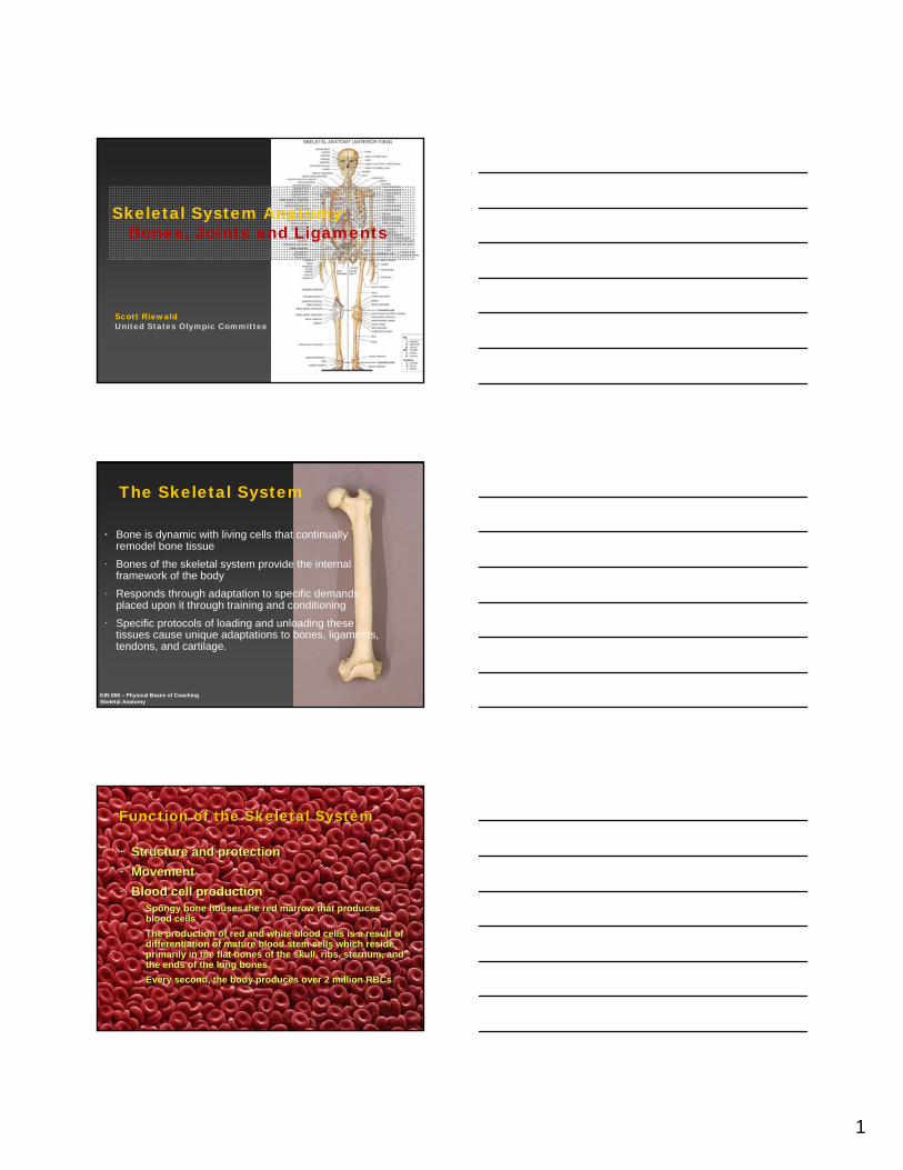
Function of the Skeletal SystemFunction of the Skeletal System
Structure and protectionStructure and protectionMovementMovementBlood cell productionBlood cell production
–– Spongy bone houses the red marrow that produces Spongy bone houses the red marrow that produces blood cells blood cells
–– The production of red and white blood cells is a result of The production of red and white blood cells is a result of differentiation of mature blood stem cells which reside differentiation of mature blood stem cells which reside primarily in the flat bones of the skull, ribs, sternum, and primarily in the flat bones of the skull, ribs, sternum, and the ends of the long bones. the ends of the long bones.
–– Every second, the body produces over 2 million Every second, the body produces over 2 million RBCsRBCs
2
KIN 856 – Physical Bases of CoachingSkeletal Anatomy
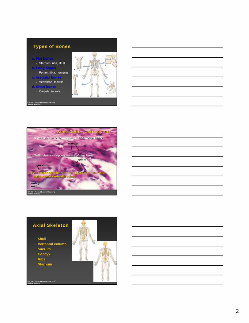
Types of Bones
a. Flat bonesa. Flat bonesSternum, ribs, skull
b. Long bonesb. Long bonesFemur, tibia, humerus
c. Irregular bonesc. Irregular bonesVertebrae, maxilla
d. Short bonesd. Short bonesCarpals, tarsals
KIN 856 – Physical Bases of CoachingSkeletal Anatomy
Bone Cells Bone Cells -- OsteocytesOsteocytesOsteoclastsOsteoclasts –– reclaim calcium or damaged bonereclaim calcium or damaged bone
OsteoblastsOsteoblasts –– deposit matrix of new bonedeposit matrix of new bone
Both responsible for remodeling that can occur with Both responsible for remodeling that can occur with training and skeletal loading.training and skeletal loading.
KIN 856 – Physical Bases of CoachingSkeletal Anatomy
Axial Skeleton
SkullSkullVertebral columnVertebral columnSacrumSacrumCoccyxCoccyxRibsRibsSternumSternum
3
KIN 856 – Physical Bases of CoachingSkeletal Anatomy
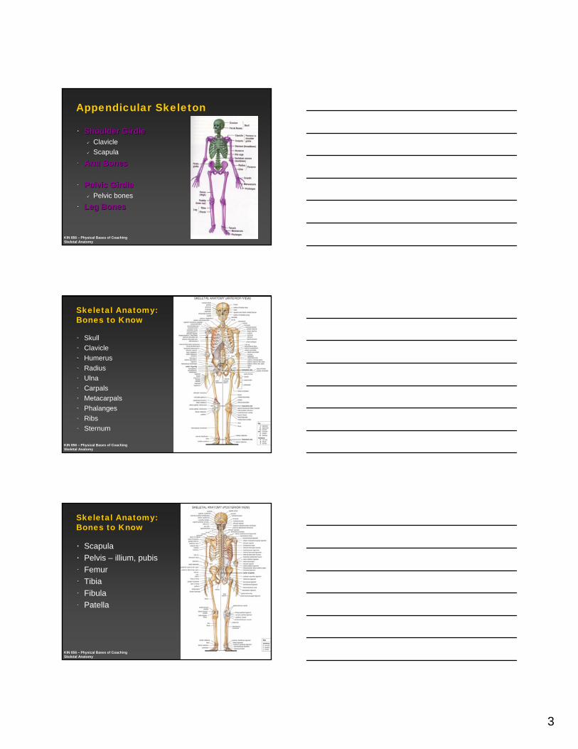
Appendicular Skeleton
Shoulder GirdleShoulder GirdleClavicleScapula
Arm BonesArm Bones
Pelvic GirdlePelvic GirdlePelvic bones
Leg BonesLeg Bones
KIN 856 – Physical Bases of CoachingSkeletal Anatomy
Skeletal Anatomy:Bones to Know
SkullClavicleHumerusRadiusUlnaCarpalsMetacarpalsPhalangesRibsSternum
KIN 856 – Physical Bases of CoachingSkeletal Anatomy
Skeletal Anatomy:Bones to Know
ScapulaPelvis – illium, pubisFemurTibiaFibulaPatella
4
KIN 856 – Physical Bases of CoachingSkeletal Anatomy
Bones of the Foot
TalusCalcaneousMetatarsalsPhalanges
KIN 856 – Physical Bases of CoachingSkeletal Anatomy
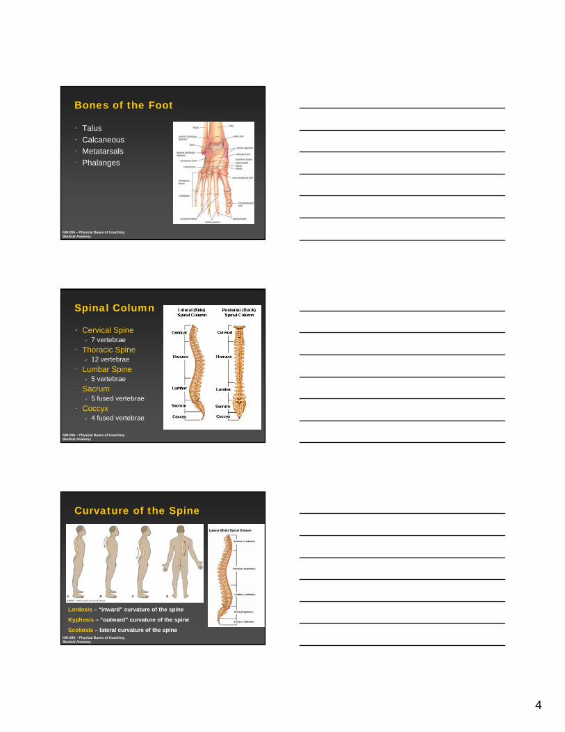
Spinal Column
Cervical Spine7 vertebrae
Thoracic Spine12 vertebrae
Lumbar Spine5 vertebrae
Sacrum5 fused vertebrae
Coccyx4 fused vertebrae
KIN 856 – Physical Bases of CoachingSkeletal Anatomy
Curvature of the Spine
Lordosis – “inward” curvature of the spine
Kyphosis – “outward” curvature of the spine
Scoliosis – lateral curvature of the spine
5
KIN 856 – Physical Bases of CoachingSkeletal Anatomy
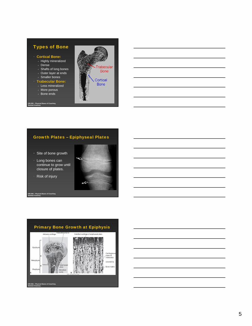
Types of Bone
Cortical Bone: Cortical Bone: Highly mineralizedDenseShafts of long bonesOuter layer at endsSmaller bones
TrabecularTrabecular Bone:Bone:Less mineralizedMore porousBone ends
KIN 856 – Physical Bases of CoachingSkeletal Anatomy
Growth Plates – Epiphyseal Plates
Site of bone growth
Long bones can continue to grow until closure of plates.
Risk of injury
KIN 856 – Physical Bases of CoachingSkeletal Anatomy
Primary Bone Growth at Epiphysis
6
KIN 856 – Physical Bases of CoachingSkeletal Anatomy
Joint Classifications
Fibrous/ Synarthrosis (immoveable)Dense connective tissue/ collagene.g. Skull sutures, distal radio-ulnar, pelvis
Cartilagenous/ Amphiarthrosis (partially moveable)
Connected by cartilagee.g. ribs
Synovial/ Diarthrosis (freely moveable)Have a joint capsule/ bursa with synovial fluidMost joints in the body
KIN 856 – Physical Bases of CoachingSkeletal Anatomy
KIN 856 – Physical Bases of CoachingSkeletal Anatomy
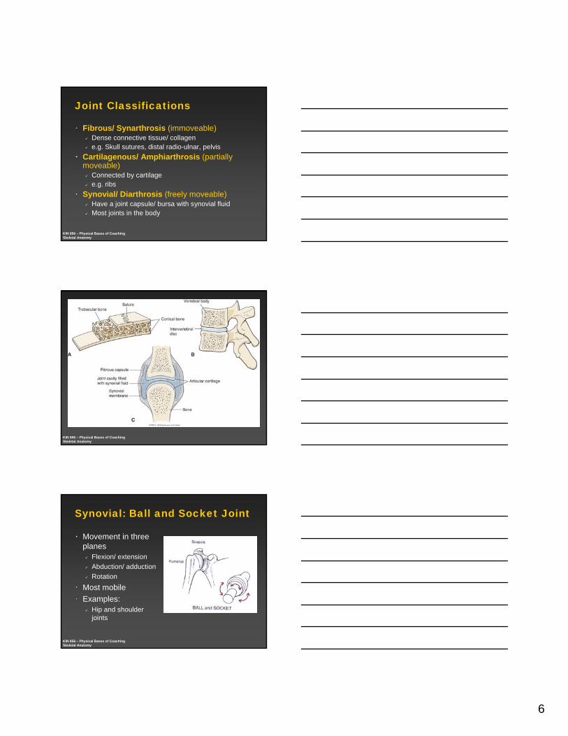
Synovial: Ball and Socket Joint
Movement in three planes
Flexion/ extensionAbduction/ adductionRotation
Most mobileExamples:
Hip and shoulder joints
7
KIN 856 – Physical Bases of CoachingSkeletal Anatomy
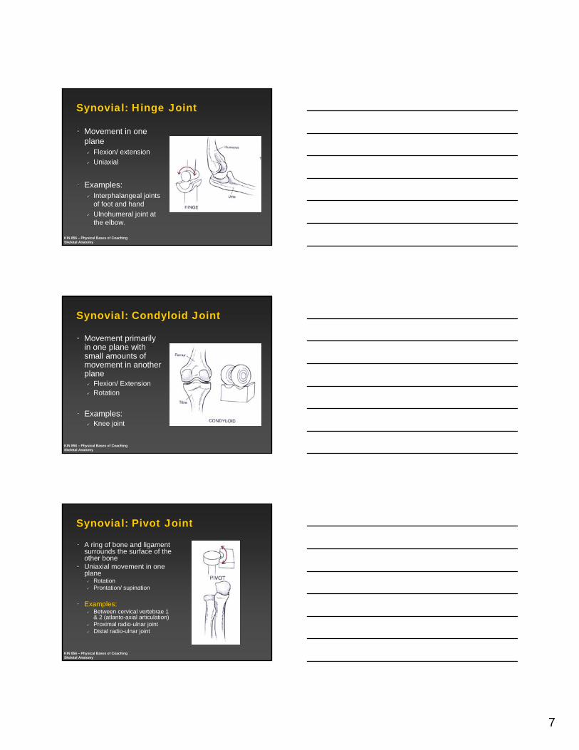
Synovial: Hinge Joint
Movement in one plane
Flexion/ extensionUniaxial
Examples: Interphalangeal joints of foot and handUlnohumeral joint at the elbow.
KIN 856 – Physical Bases of CoachingSkeletal Anatomy
Synovial: Condyloid Joint
Movement primarily in one plane with small amounts of movement in another plane
Flexion/ ExtensionRotation
Examples: Knee joint
KIN 856 – Physical Bases of CoachingSkeletal Anatomy
Synovial: Pivot Joint
A ring of bone and ligament surrounds the surface of the other bone Uniaxial movement in one plane
RotationProntation/ supination
Examples:Between cervical vertebrae 1 & 2 (atlanto-axial articulation)Proximal radio-ulnar jointDistal radio-ulnar joint
8
KIN 856 – Physical Bases of CoachingSkeletal Anatomy
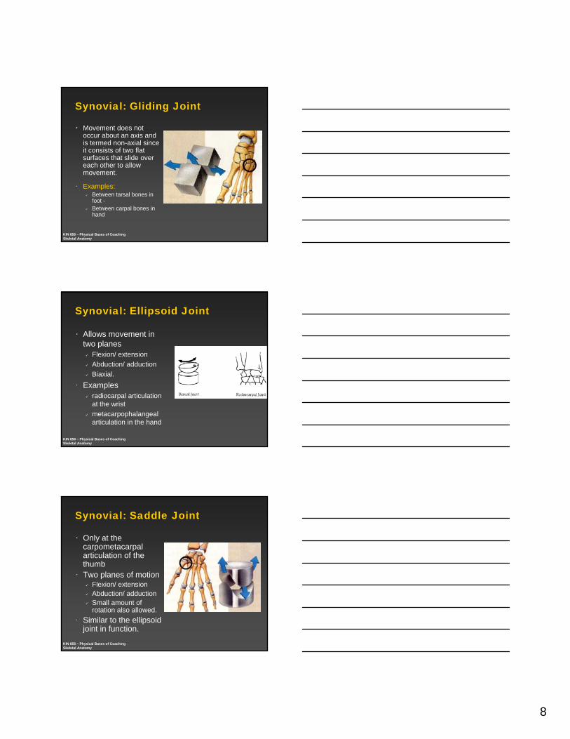
Synovial: Gliding Joint
Movement does not occur about an axis and is termed non-axial since it consists of two flat surfaces that slide over each other to allow movement.
Examples:Between tarsal bones in foot -Between carpal bones in hand
KIN 856 – Physical Bases of CoachingSkeletal Anatomy
Synovial: Ellipsoid Joint
Allows movement in two planes
Flexion/ extensionAbduction/ adductionBiaxial.
Examples radiocarpal articulation at the wrist metacarpophalangealarticulation in the hand
KIN 856 – Physical Bases of CoachingSkeletal Anatomy
Synovial: Saddle Joint
Only at the carpometacarpalarticulation of the thumbTwo planes of motion
Flexion/ extensionAbduction/ adduction Small amount of rotation also allowed.
Similar to the ellipsoid joint in function.
9
KIN 856 – Physical Bases of CoachingSkeletal Anatomy
Wolff’s Law
“The densities, and to a lesser extent, the sizes and shapes of bones are determined by the magnitude and direction of the acting forces applied to bone.”
SpecificitySpecificity - The body will adapt to the stresses placed upon it – as long as those stresses are reasonable and not excessive.
KIN 856 – Physical Bases of CoachingSkeletal Anatomy
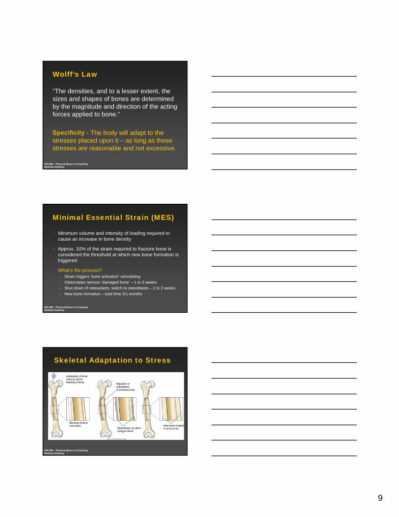
Minimal Essential Strain (MES)
Minimum volume and intensity of loading required to cause an increase in bone density
Approx. 10% of the strain required to fracture bone is considered the threshold at which new bone formation is triggered
What’s the process?Strain triggers ‘bone activation’ remodelingOsteoclasts remove ‘damaged bone’ – 1 to 3 weeksShut down of osteoclasts, switch to osteoblasts – 1 to 2 weeksNew bone formation – total time 3½ months
KIN 856 – Physical Bases of CoachingSkeletal Anatomy
Skeletal Adaptation to Stress
10
KIN 856 – Physical Bases of CoachingSkeletal Anatomy
Appropriate Training Stimuli
‘Dynamic loading’ – rapid loadingAxial loading/ higher impact forces
Higher Frequency loadingDirectional specificity
Small gains in bone density can produce large strength improvements
More, shorter workouts as opposed to longer ones
Takes 6-8 hours for bone to recover ability to lay down new bone
KIN 856 – Physical Bases of CoachingSkeletal Anatomy
Skeletal Adaptations to Loading
KIN 856 – Physical Bases of CoachingSkeletal Anatomy
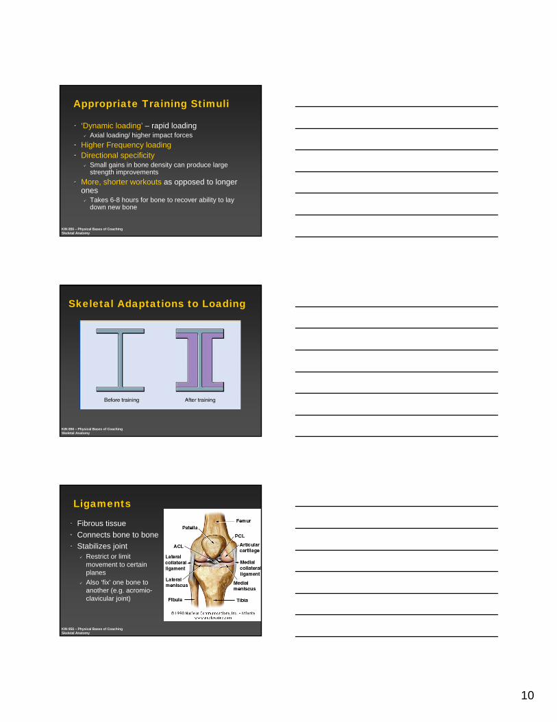
Ligaments
Fibrous tissue Connects bone to boneStabilizes joint
Restrict or limit movement to certain planesAlso ‘fix’ one bone to another (e.g. acromio-clavicular joint)
11
KIN 856 – Physical Bases of CoachingSkeletal Anatomy
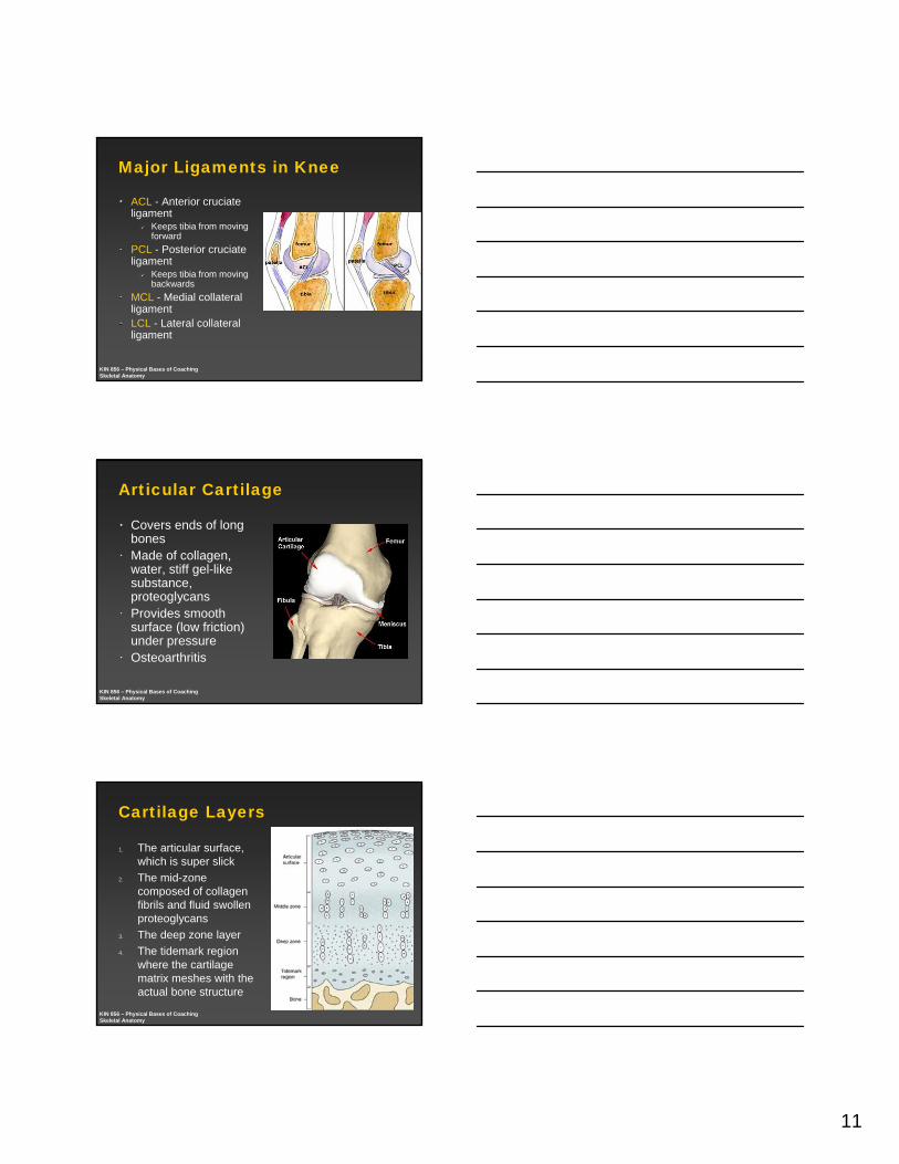
Major Ligaments in Knee
ACL - Anterior cruciateligament
Keeps tibia from moving forward
PCL - Posterior cruciateligament
Keeps tibia from moving backwards
MCL - Medial collateral ligament LCL - Lateral collateral ligament
KIN 856 – Physical Bases of CoachingSkeletal Anatomy
Articular Cartilage
Covers ends of long bonesMade of collagen, water, stiff gel-like substance, proteoglycansProvides smooth surface (low friction) under pressureOsteoarthritis
KIN 856 – Physical Bases of CoachingSkeletal Anatomy
Cartilage Layers
1. The articular surface, which is super slick
2. The mid-zone composed of collagen fibrils and fluid swollen proteoglycans
3. The deep zone layer 4. The tidemark region
where the cartilage matrix meshes with the actual bone structure
12
KIN 856 – Physical Bases of CoachingSkeletal Anatomy
Skeletal Health - Osteoporosis
Loss of bone densityPostmenopausal womenMen and women >70Young athletes with eating disorder (e.g. anorexia)
Higher bone density in ‘active years’ leads to higher bone density later in lifeFemale athlete triadDEXA scan
KIN 856 – Physical Bases of CoachingSkeletal Anatomy
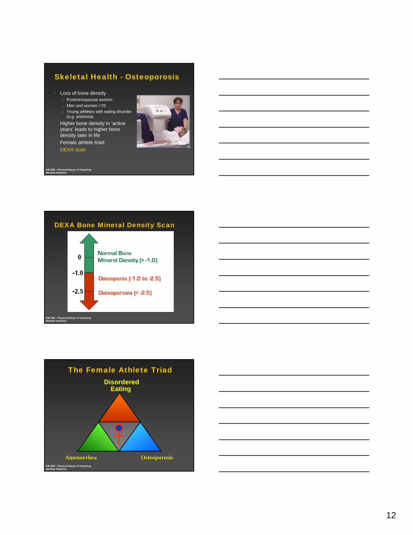
DEXA Bone Mineral Density Scan
KIN 856 – Physical Bases of CoachingSkeletal Anatomy
The Female Athlete TriadDisordered
Eating
AmenorrheaAmenorrhea OsteoporosisOsteoporosis