Embed Size (px)
Citation preview
The Skeletal System
The Skeletal System
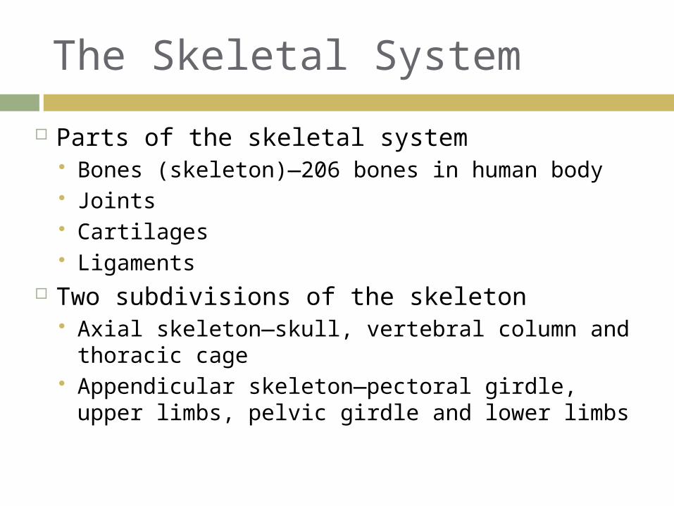
Parts of the skeletal system Bones (skeleton)—206 bones in human body Joints Cartilages Ligaments
Two subdivisions of the skeleton Axial skeleton—skull, vertebral column and
thoracic cage Appendicular skeleton—pectoral girdle,
upper limbs, pelvic girdle and lower limbs
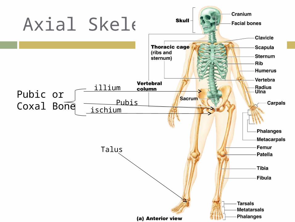
Axial Skeleton
Pubic or Coxal Bone Pubis
illium
ischium
Talus
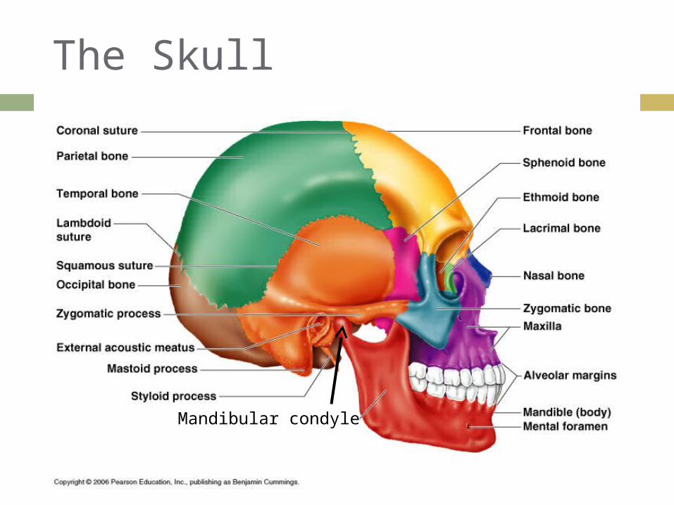
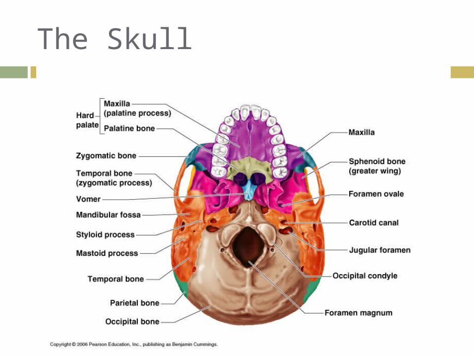
The Skull
Mandibular condyle

Axial Skeleton—Skull
Two sets of bones Cranium Facial bones
Bones are joined by sutures
Only the mandible is attached by a freely movable joint
The Skull
Axial Skeleton—Skull
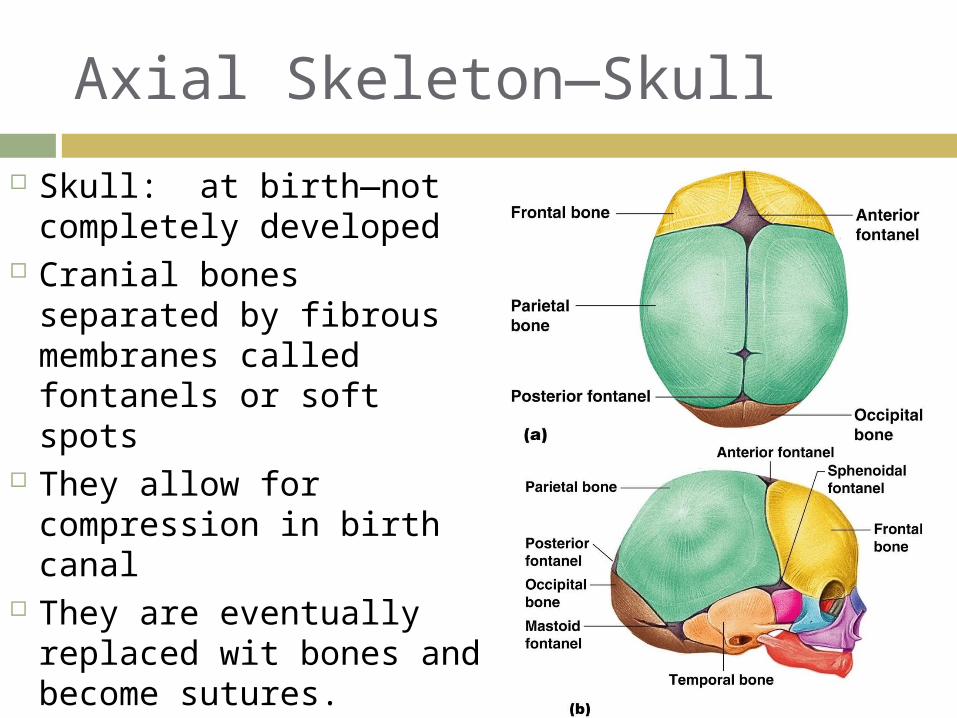
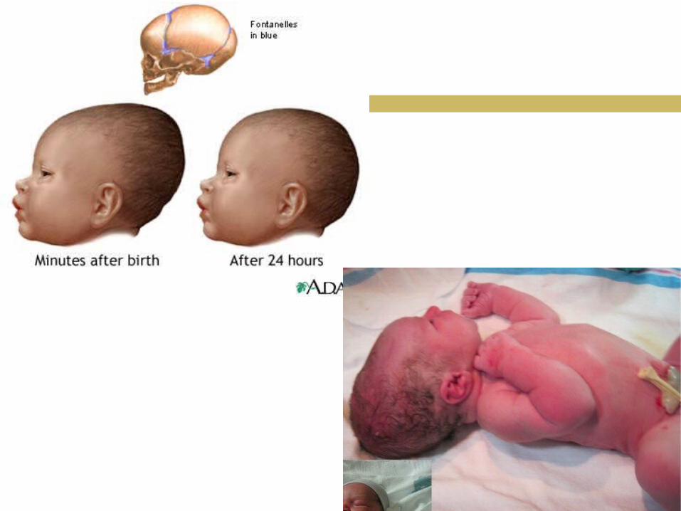
Skull: at birth—not completely developed
Cranial bones separated by fibrous membranes called fontanels or soft spots
They allow for compression in birth canal
They are eventually replaced wit bones and become sutures.
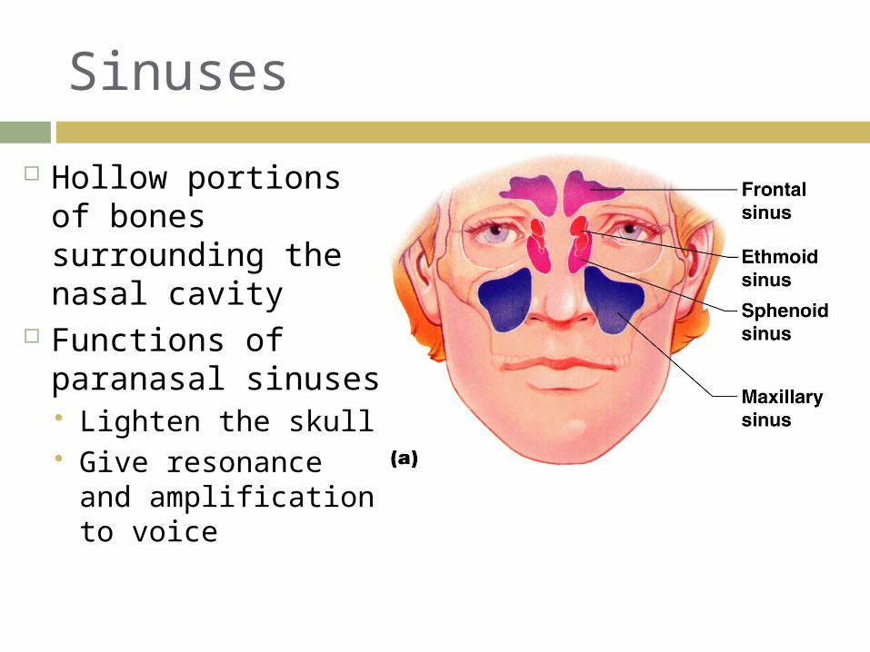
Sinuses
Hollow portions of bones surrounding the nasal cavity
Functions of paranasal sinuses Lighten the skull Give resonance
and amplification to voice
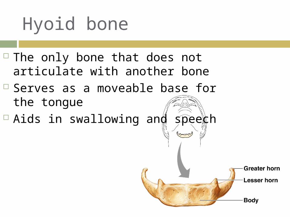
Hyoid bone
The only bone that does not articulate with another bone
Serves as a moveable base for the tongue
Aids in swallowing and speech
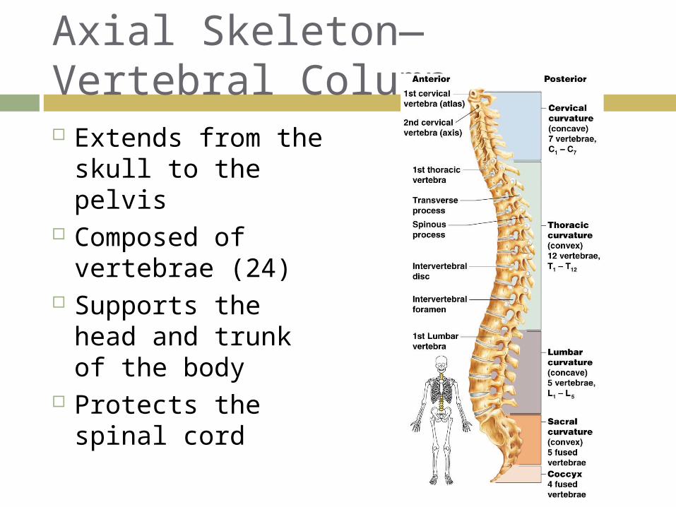
Axial Skeleton—Vertebral Column Extends from the
skull to the pelvis Composed of
vertebrae (24) Supports the
head and trunk of the body
Protects the spinal cord
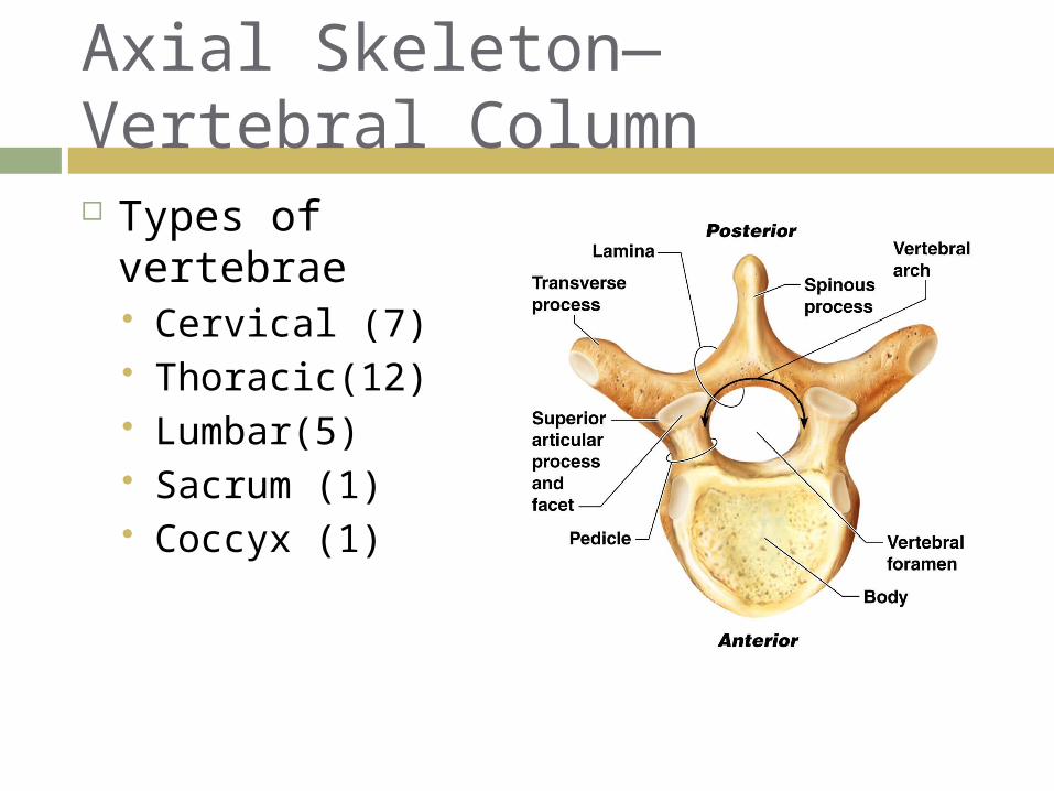
Types of vertebrae Cervical (7) Thoracic(12) Lumbar(5) Sacrum (1) Coccyx (1)
Axial Skeleton—Vertebral Column
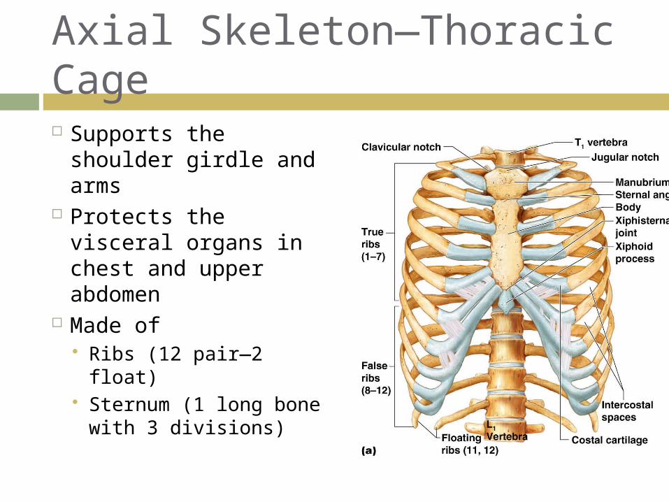
Supports the shoulder girdle and arms
Protects the visceral organs in chest and upper abdomen
Made of Ribs (12 pair—2 float) Sternum (1 long bone
with 3 divisions)
Axial Skeleton—Thoracic Cage
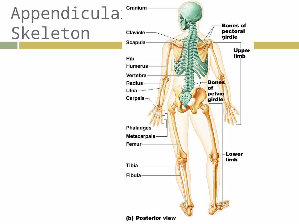
Appendicular Skeleton
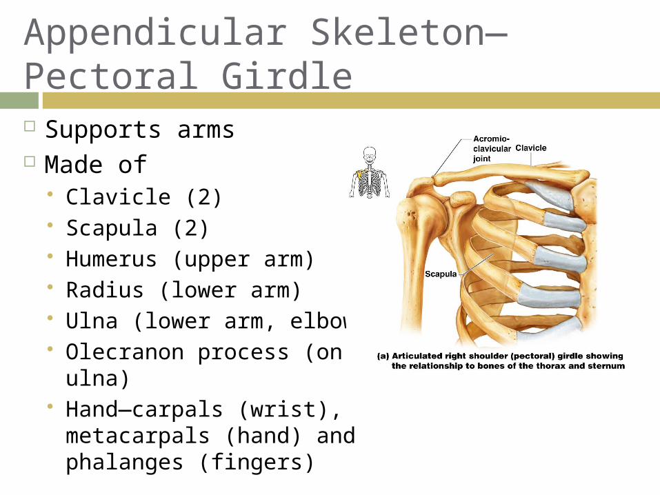
Appendicular Skeleton—Pectoral Girdle Supports arms Made of
Clavicle (2) Scapula (2) Humerus (upper arm) Radius (lower arm) Ulna (lower arm, elbow) Olecranon process (on
ulna) Hand—carpals (wrist),
metacarpals (hand) and phalanges (fingers)
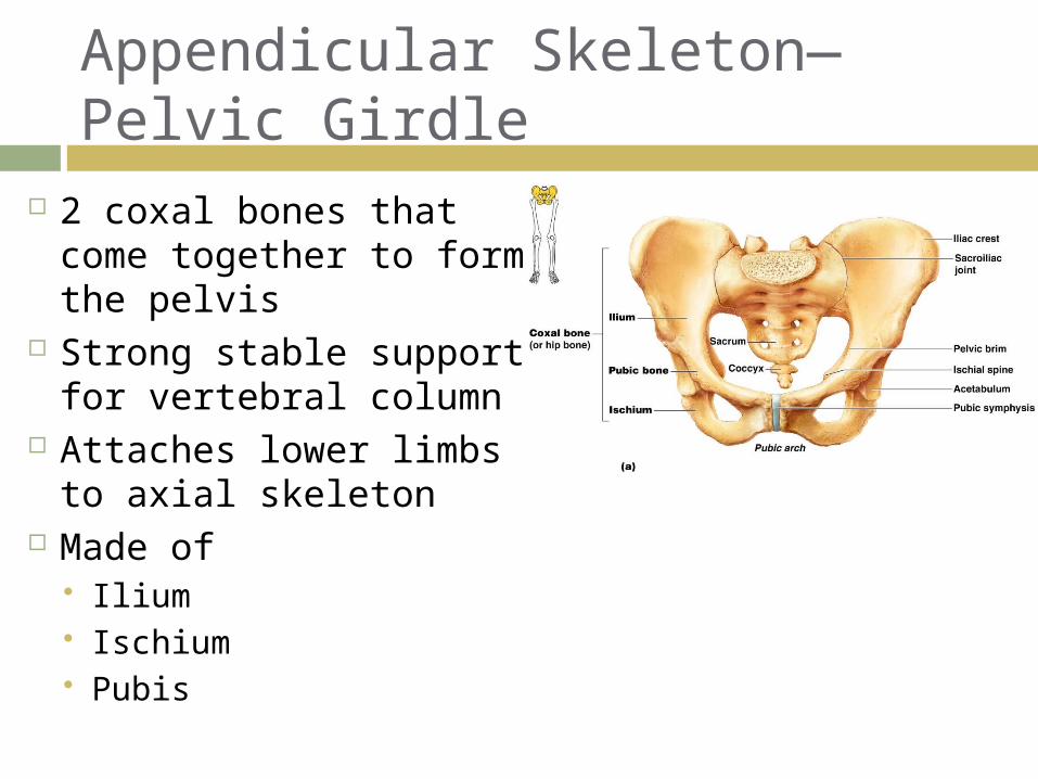
Appendicular Skeleton—Pelvic Girdle
2 coxal bones that come together to form the pelvis
Strong stable support for vertebral column
Attaches lower limbs to axial skeleton
Made of Ilium Ischium Pubis
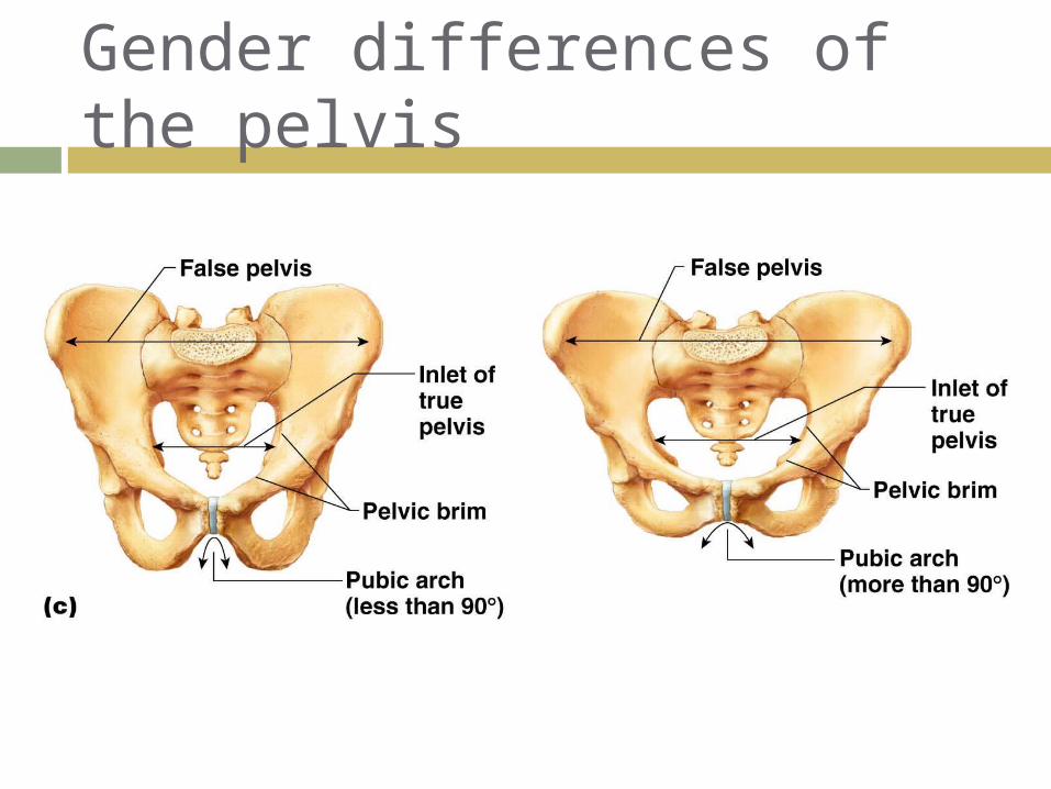
Gender differences of the pelvis
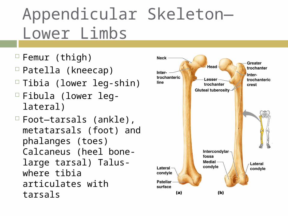
Femur (thigh) Patella (kneecap) Tibia (lower leg-shin) Fibula (lower leg-
lateral) Foot—tarsals (ankle),
metatarsals (foot) and phalanges (toes) Calcaneus (heel bone-large tarsal) Talus-where tibia articulates with tarsals
Appendicular Skeleton—Lower Limbs
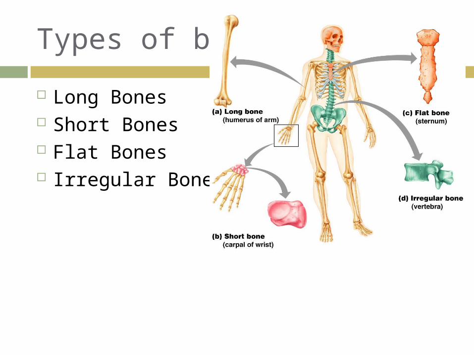
Types of bones
Long Bones Short Bones Flat Bones Irregular Bones
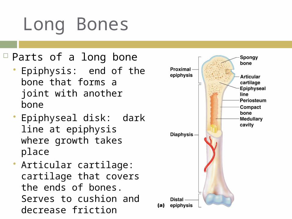
Long Bones
Parts of a long bone Epiphysis: end of the
bone that forms a joint with another bone
Epiphyseal disk: dark line at epiphysis where growth takes place
Articular cartilage: cartilage that covers the ends of bones. Serves to cushion and decrease friction
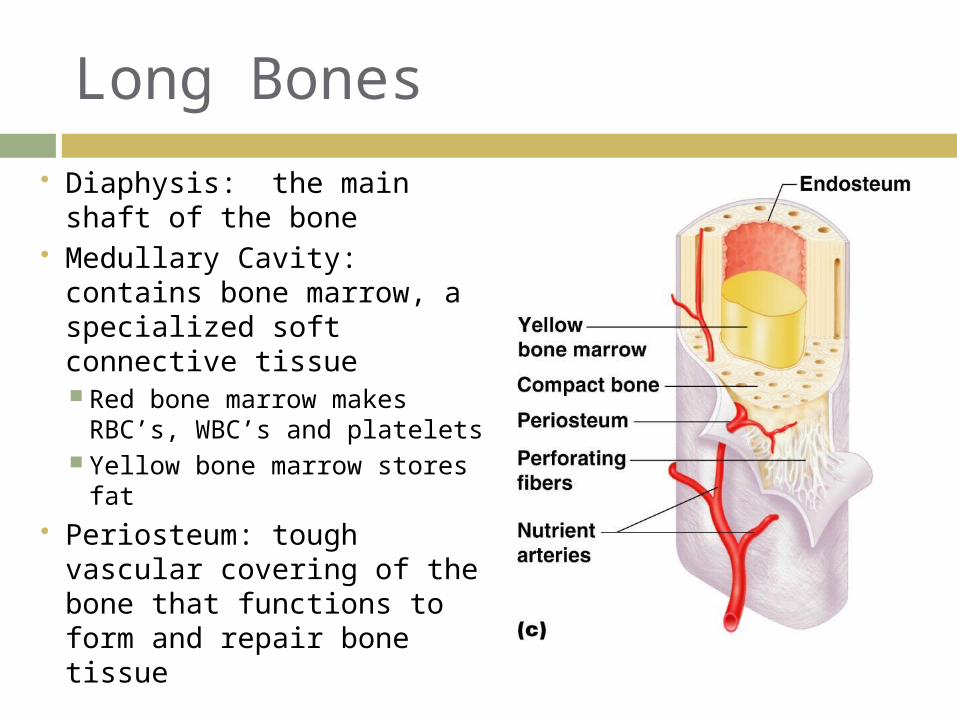
Long Bones
Diaphysis: the main shaft of the bone
Medullary Cavity: contains bone marrow, a specialized soft connective tissue Red bone marrow makes
RBC’s, WBC’s and platelets Yellow bone marrow stores
fat Periosteum: tough vascular
covering of the bone that functions to form and repair bone tissue
Long Bones
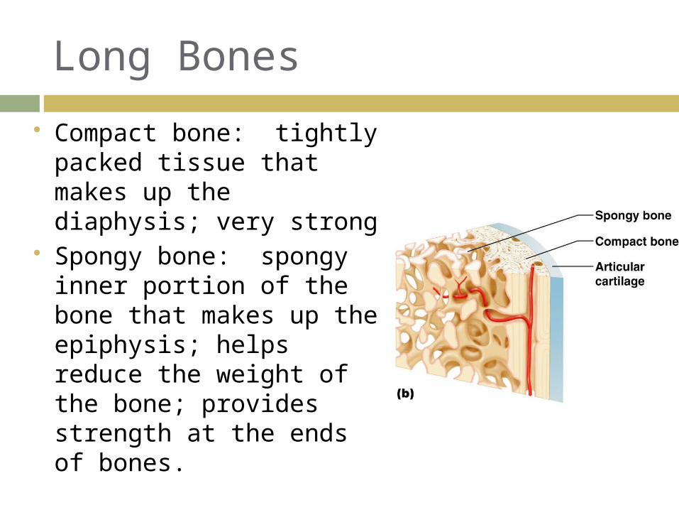
Compact bone: tightly packed tissue that makes up the diaphysis; very strong
Spongy bone: spongy inner portion of the bone that makes up the epiphysis; helps reduce the weight of the bone; provides strength at the ends of bones.
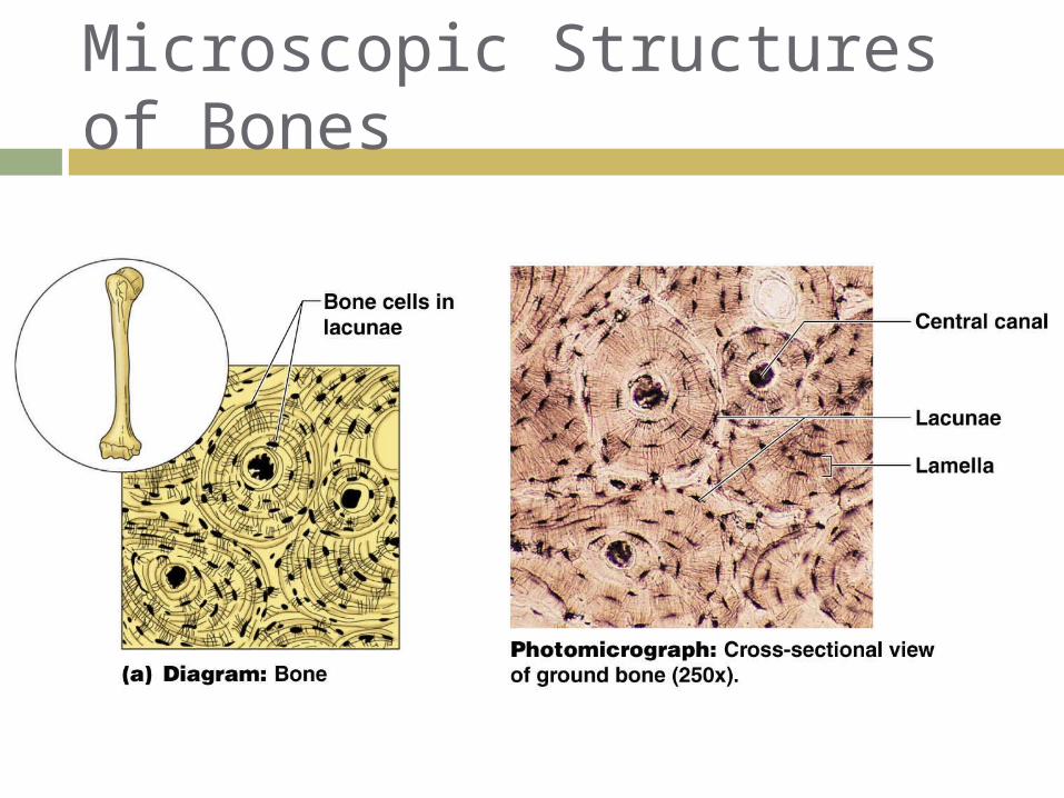
Microscopic Structures of Bones
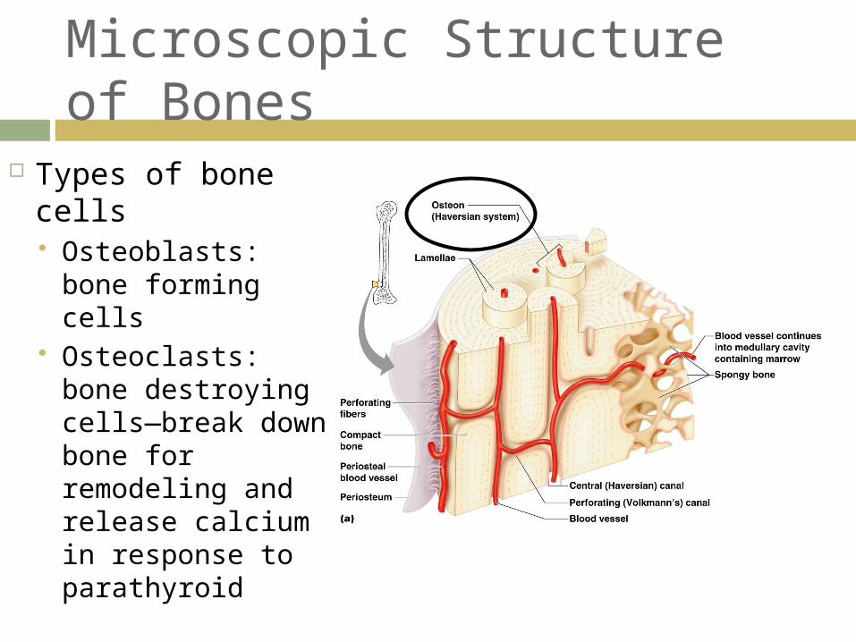
Microscopic Structure of Bones
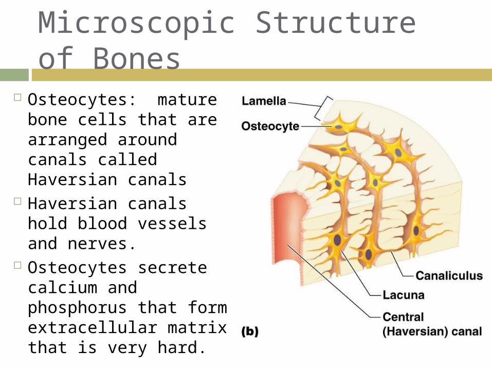
Osteocytes: mature bone cells that are arranged around canals called Haversian canals
Haversian canals hold blood vessels and nerves.
Osteocytes secrete calcium and phosphorus that form extracellular matrix that is very hard.
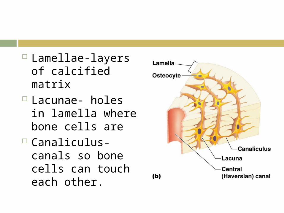
Lamellae-layers of calcified matrix
Lacunae- holes in lamella where bone cells are
Canaliculus-canals so bone cells can touch each other.
Microscopic Structure of Bones
Types of bone cells Osteoblasts:
bone forming cells Osteoclasts:
bone destroying cells—break down bone for remodeling and release calcium in response to parathyroid
Formation of the Human Skeleton In embryos, the skeleton is primarily
hyaline cartilage During development, much of this
cartilage is replaced by bone Cartilage remains in isolated areas
Bridge of the nose Parts of ribs Joints
Bone Growth (Ossification)
Epiphyseal plates allow for lengthwise growth of long bones during childhood New cartilage is continuously formed Older cartilage becomes ossified
Cartilage is broken down Enclosed cartilage is digested away, opening
up a medullary cavity Bone replaces cartilage through the action of
osteoblasts
Bone Growth (Ossification)
Bones are remodeled and lengthened until growth stops Bones are remodeled in response to two
factors Blood calcium levels Pull of gravity and muscles on the skeleton
Bones grow in width (called appositional growth)
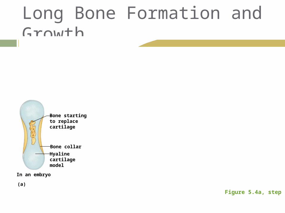
Long Bone Formation and Growth
Figure 5.4a, step 1
Bone startingto replacecartilage
In an embryo
Bone collar
Hyalinecartilagemodel
(a)
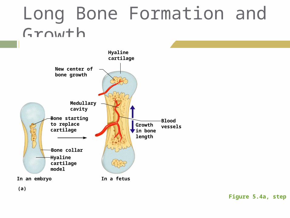
Long Bone Formation and Growth
Figure 5.4a, step 2
Bone startingto replacecartilage
In a fetusIn an embryo
Growthin bonelength
Bloodvessels
Hyalinecartilage
New center ofbone growth
Medullarycavity
Bone collar
Hyalinecartilagemodel
(a)
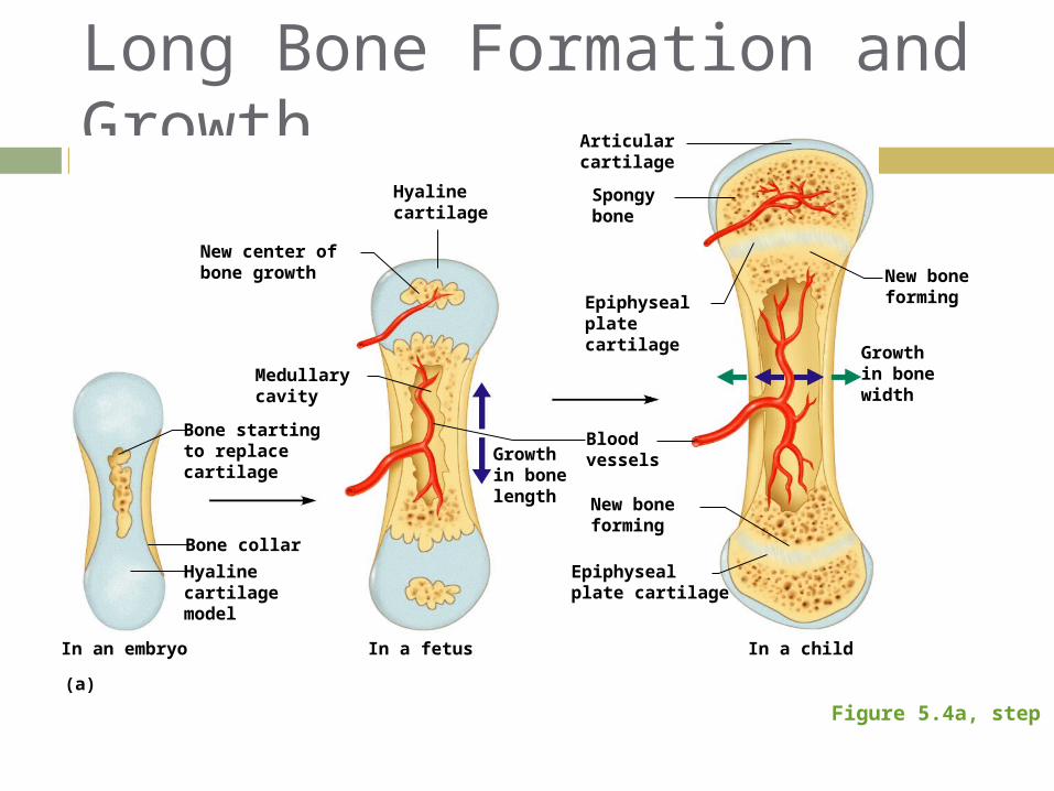
Long Bone Formation and Growth
Figure 5.4a, step 3
Bone startingto replacecartilage
Epiphysealplatecartilage
Articularcartilage
Spongybone
In a childIn a fetusIn an embryo
New boneforming
Growthin bonewidth
Growthin bonelength
Epiphysealplate cartilage
New boneforming
Bloodvessels
Hyalinecartilage
New center ofbone growth
Medullarycavity
Bone collar
Hyalinecartilagemodel
(a)
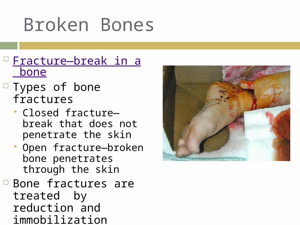
Broken Bones
Fracture—break in a bone
Types of bone fractures Closed fracture—
break that does not penetrate the skin
Open fracture—broken bone penetrates through the skin
Bone fractures are treated by reduction and immobilization
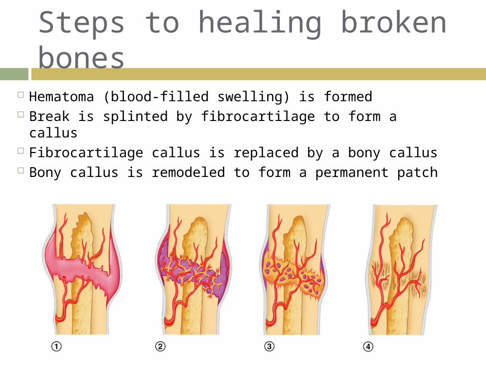
Steps to healing broken bones
Hematoma (blood-filled swelling) is formed Break is splinted by fibrocartilage to form a callus Fibrocartilage callus is replaced by a bony callus Bony callus is remodeled to form a permanent patch
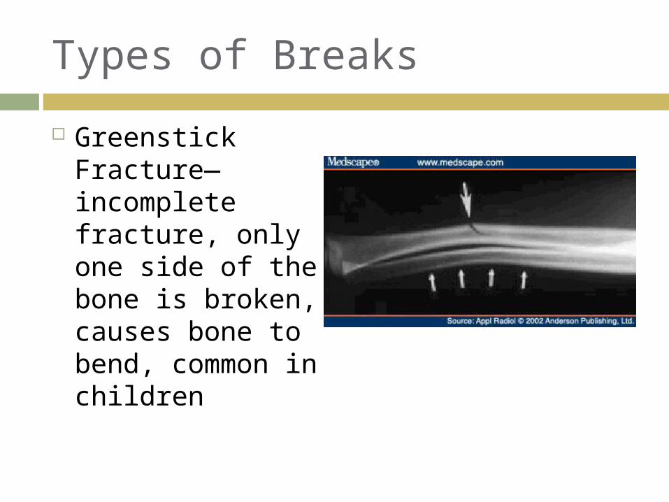
Types of Breaks
Greenstick Fracture—incomplete fracture, only one side of the bone is broken, causes bone to bend, common in children
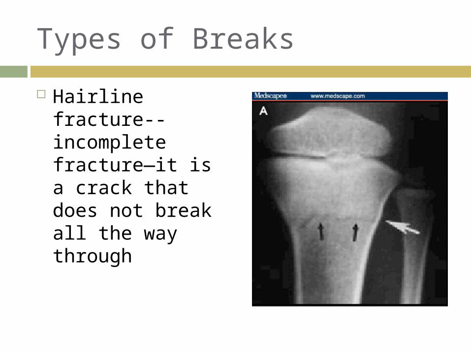
Types of Breaks
Hairline fracture-- incomplete fracture—it is a crack that does not break all the way through
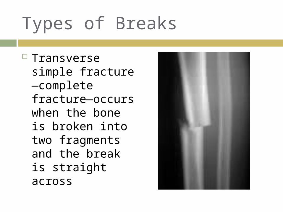
Types of Breaks
Transverse simple fracture—complete fracture—occurs when the bone is broken into two fragments and the break is straight across
Types of Breaks
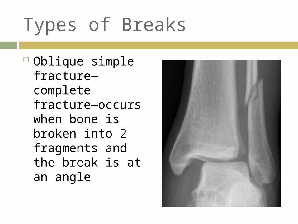
Oblique simple fracture—complete fracture—occurs when bone is broken into 2 fragments and the break is at an angle
Types of Breaks
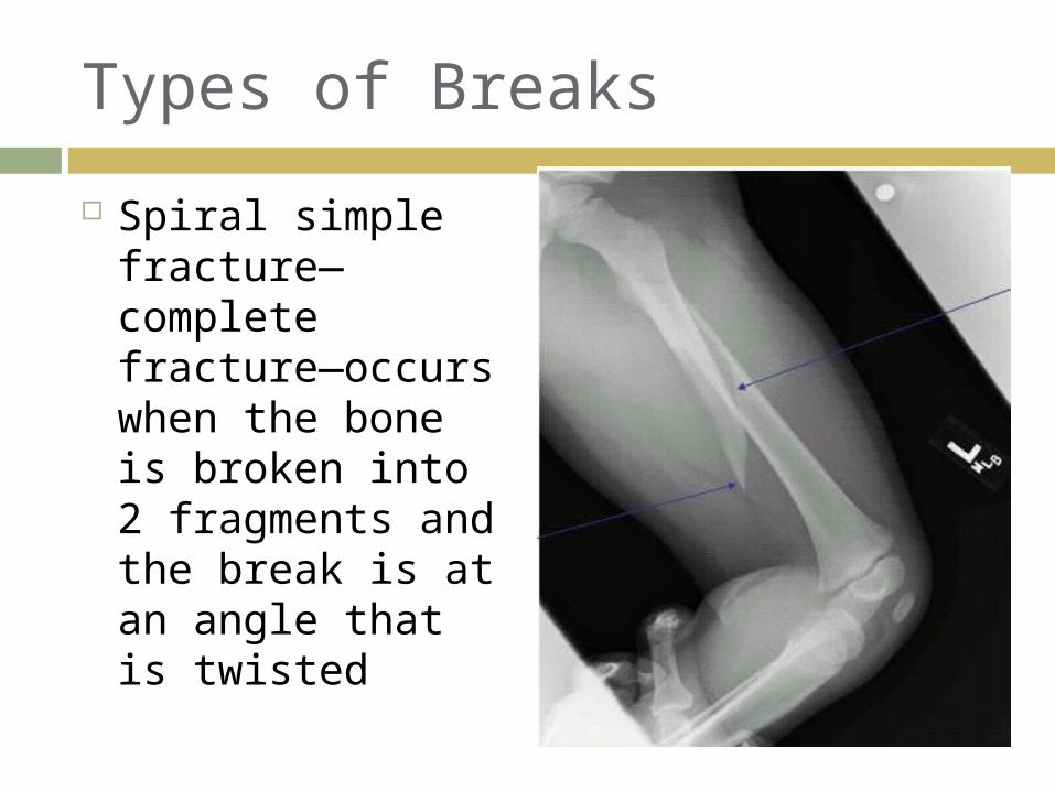
Spiral simple fracture—complete fracture—occurs when the bone is broken into 2 fragments and the break is at an angle that is twisted
Types of Breaks
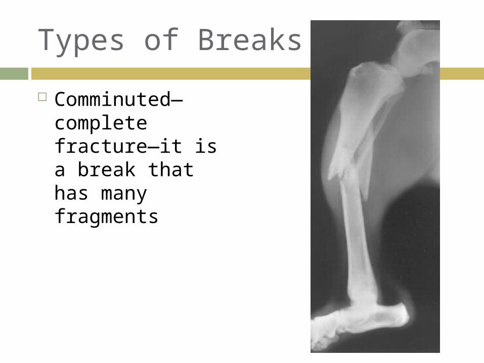
Comminuted—complete fracture—it is a break that has many fragments
Types of Breaks
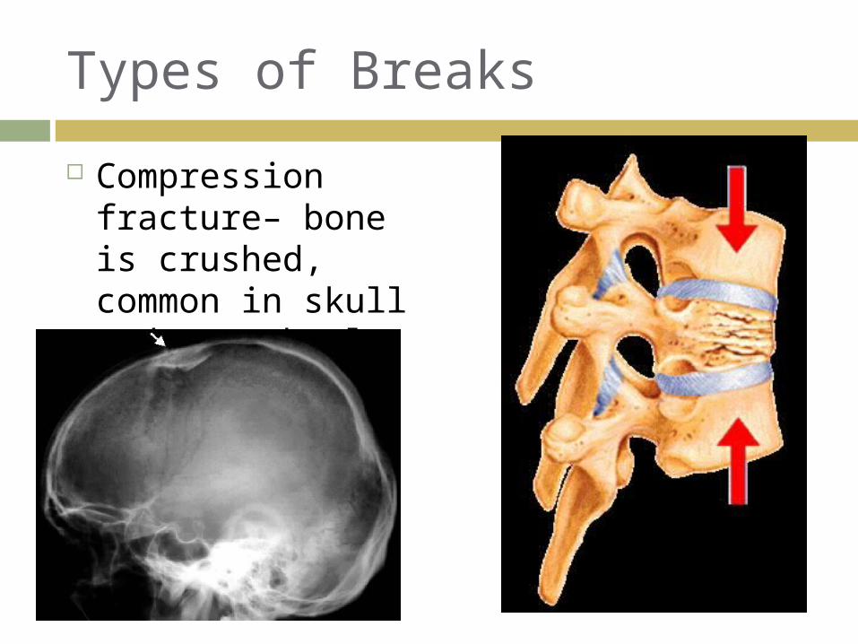
Compression fracture– bone is crushed, common in skull and vertebral column