Embed Size (px)
Citation preview

JOURNAL OF BACTERIOLOGY, Apr. 1993, P. 2436-2444 Vol. 175, No. 80021-9193/93/082436-09$02.00/0Copyright © 1993, American Society for Microbiology
Sequence of the Gene Coding for Ammonia Monooxygenasein Nitrosomonas europaea
HUGH McTAVISH,"2 JAMES A. FUCHS,3 AND ALAN B. HOOPER'*Department of Genetics and Cell Biology, 1 Graduate Program in Biochemistry,2 and Department of
Biochemistry,3 University ofMinnesota, St. Paul, Minnesota 55108
Received 9 November 1992/Accepted 5 February 1993
Nitrosomonas europaea, a chemolithotrophic bacterium, was found to contain two copies of the gene codingfor the presumed active site polypeptide of ammonia monooxygenase, the 32-kDa acetylene-binding polypep-tide. One copy of this gene was cloned, and its complete nucleotide sequence is presented. Immediatelydownstream of this gene, in the same operon, is the gene for a 40-kDa polypeptide that copurifies with theammonia monooxygenase acetylene-binding polypeptide. The sequence of the first 692 nucleotides of thisstructural gene, coding for about two-thirds of the protein, is presented. These sequences are the first sequencesof protein-encoding genes from an ammonia-oxidizing autotrophic nitrifying bacterium. The two proteinsequences are not homologous with the sequences of any other monooxygenase. From radioactive labelling ofammonia monooxygenase with ['4C]acetylene it was determined that there are 23 nmol of ammoniamonooxygenase per g of cells. The k.a of ammonia monooxygenase for NH3 in vivo was calculated to be 20 s-l.
Nitrosomanas europaea is an autotrophic gram-negativebacterium of soils and fresh water that obtains all of itsenergy for growth from the oxidation of ammonia to nitrite.This oxidation occurs in two steps. Ammonia is first oxi-dized to hydroxylamine by ammonia monooxygenase(AMO) (8):
NH3 + 02 + 2e- + 2H+ -- NH20H + H20
Hydroxylamine is then oxidized to nitrite in an energy-yielding dehydrogenase reaction by the periplasmic enzymehydroxylamine oxidoreductase (1, 9). The acceptor for thefour electrons generated in the latter reaction is the tetra-heme cytochrome C554, which is thought to carry the elec-trons in two-electron steps (2). Two of the four electrons aredestined for an oxidative electron transfer chain and cyto-chrome aa3 terminal oxidase (5); the other two must be usedin the AMO reaction.
Little is known about AMO. It can be irreversibly inhib-ited by acetylene, a suicide substrate for many monooxyge-nases, and inhibition with [14C]acetylene radioactively labelsa 28-kDa polypeptide that is associated with the membranefraction (16). AMO is also inhibited by metal chelators, suchas thiourea and allylthiourea (10, 20). The fact that thesechemicals bind several metals (28), with a preference forcopper, has been considered suggestive evidence that AMOmight contain copper (3, 30).AMO activity has been demonstrated with a crude mem-
brane fraction (35, 36), but no further purification has beenaccomplished. The electron donor for AMO in vivo isunknown, although with the crude membrane fraction it wasfound that the addition of cytochrome C554 stimulated am-monia oxidation activity (35), indicating that cytochromeC554 can serve as at least an indirect electron donor for AMO.AMO is significant because of the key role that it plays in
the nitrogen cycle and because it is able to degrade a widerange of hydrocarbons and halogenated hydrocarbons (14,39, 40). The study of AMO is also important as a model forthe related enzyme methane monooxygenase. Methane mono-
* Corresponding author.
oxygenase exists in a soluble form and a membrane form inthe methanotrophic bacteria, with the membrane form beingmore widespread (3). It is thought that AMO and themembrane form of methane monooxygenase are probablyhomologous enzymes because, in addition to both beingmembrane enzymes, they have very similar substrate spec-ificities (both enzymes can oxidize both ammonia and meth-ane) and similar inhibitor profiles (3, 4). Also, the membranemethane monooxygenase may, like AMO, be a copper-containing enzyme (27, 31, 34).
In this paper we report the partial purification of theacetylene-binding polypeptide of AMO and the cloning andsequencing of the gene for this polypeptide. We also reportthe cloning and partial sequence of a second gene in thisoperon which codes for a polypeptide that copurifies withthe acetylene-binding polypeptide.
MATERIALS AND METHODS
Bacterial growth. N. europaea was grown in continuousculture (23) in a 55-liter fermentor on medium containing (perliter) 1.33 g of Na2HPO4, 0.11 g of KH2PO4, 4 g of(NH4)2SO4, 3 ml of a solution containing 68 g ofMgCl2- 6H20 per liter and 3.08 g of CaCl2 2H20 per liter,and 1 ml of a solution containing 1.34 g of FeSO4 .7H20 perliter, 1.0 g of CUSO4* 5H20 per liter, and 1.6 -g of Na2EDTAper liter. The pH was continually titrated to 7.87 with 50%(wt/vol) K2CO3. The flow rate of medium through thefermentor was 27 liters/24 h. After flowing out of thefermentor, cells were stored at 40C until concentration bytangential flow across a Pellicon cassette filter unit (MilliporeCorp., Bedford, Mass.) and collection by centrifugation.
Electrophoresis, protein sequencing, and amino acid analy-sis. Analytical sodium dodecyl sulfate (SDS)-polyacrylamidegel electrophoresis (PAGE) was done on 0.75-mm-thickminigels as described by Porzio and Pearson (26), with themodification that a 5% stacking gel containing 125 mMTris-HCl (pH 6.8) was added. Preparative SDS-PAGE topurify the AMO acetylene-binding polypeptide for proteinsequencing was done by using a single lane across the top ofa 1.5-mm-thick minigel; it was done in the same way as
2436
on February 20, 2020 by guest
http://jb.asm.org/
Dow
nloaded from
AMO GENE SEQUENCE 2437
analytical SDS-PAGE except that Tricine replaced glycine inthe buffers in order to maintain a lower pH during electro-phoresis, which reduces N-terminal modification that blocksprotein sequencing (25). After electrophoresis a slice of thegel was stained to determine the position of the 27-kDapolypeptide. This band was then cut out of the unstained geland electroeluted at 150 V for 3 h in a Centrilutor apparatus(Amicon Corp., Danvers, Mass.) with 20 mM HEPES (N-2-hydroxyethylpiperazine-N'-2-ethanesulfonic acid)-NaOHbuffer (pH 7.8). The sample was then concentrated with aCentricon 10 microconcentrator (Amicon), washed fourtimes with water, and concentrated to a volume of 80 ,ul. Itwas then subjected to Edman degradation with a model 470gas phase sequencer (Applied Biosystems, Inc.) with on-linephenylthiohydantoin (PTH) analysis.For amino acid analysis, the acetylene-binding polypep-
tide was purified in the same way, except that it was elutedfrom the gel slice by diffusion in 5 mM Tris-HCl (pH7.5)-0.1% SDS. The purified samples were sealed under avacuum in 6 N HCl-1.5% phenol, hydrolyzed at 110'C for 24h, and analyzed with a Beckman model 6300 amino acidanalyzer equipped with a 12-cm column by using HiRezNa-E, -S, and -D buffers.The 40-kDa polypeptide that copurifies with the AMO
acetylene-binding polypeptide was sequenced by Edmandegradation after SDS-PAGE and blotting of the gel onto anImmobilon 0.45-pm-pore-size polyvinylidene difluoridemembrane filter (Millipore), using the procedure describedby Matusdaira (24).
Radioactive or fluorescent labelling ofAMO. [14C]acetylenewas generated from Ba14CO3 as described by Hyman andArp (12). Fluorescein isothiocyanate was coupled to propar-gylamine as follows. Seven volumes of 142 mM fluoresceinisothiocyanate in dimethyl sulfoxide was mixed with 3volumes of 400 mM proparglyamine hydrochloride-250 mMNa2CO3 in water, and the preparation was allowed to reactat room temperature for 2 h. To label AMO, the resultingfluorescein thiocarbamoylpropargylamine (FTCP) (100 FM)was incubated with a 2% (wet wt/vol) suspension of cells in50 mM NaPO4 (pH 7.5).DNA techniques. N. europaea genomic DNA was pre-
pared by the following procedure. Cells (1 g) were digestedwith lysozyme (15% [wt/vol] cells and 1.5 mg of lysozymeper ml in 50 mM Tris-HCl [pH 8.0]-10 mM EDTA-12.5%sucrose) for 30 min at 37°C and lysed with 0.4% SDS. Theextract was treated with DNase-free RNase (9 ,ug/ml) for 10min at 37°C and then with proteinase K (0.3 mg/ml) for 40min and then extracted with phenol, phenol-chloroform, andchloroform at room temperature; the DNA was precipitatedwith 150 mM NaCl-65% ethanol and redissolved in TE (29).Agarose gel electrophoresis of DNA was carried out in
Tris-acetate-EDTA buffer as described by Sambrook et al.(29). The gels were blotted onto ICN Biotrans+ nylonmembranes by using the manufacturer's improved transferprotocol.
Oligonucleotides were obtained from Oligos Etc., Wilson-ville, Oreg. For probing of Southern blots, 25 pmol ofoligonucleotide was end-labelled with T4 polynucleotidekinase and 12.5 ,ul of 5'-[-y-32P]ATP (7,500 Ci/mmol; 10mCi/ml). Southern blot filters were prehybridized and hy-bridized in standard solutions (29), except that 10% (wt/vol)polyethylene glycol (molecular weight, 8,000) was includedin the hybridization solution. After hybridization each mem-brane was rinsed twice in medium-stringency buffer (0.5xSSPE, 0.2% SDS; 1x SSPE is 150 mM NaCl, 1 mM EDTA,and 10 mM NaPO4, [pH 7.4]) and then washed for 30 min in
medium-stringency buffer or high-stringency buffer (0.1 xSSPE, 0.2% SDS).A genomic library was created from N. europaea DNA
partially digested with Sau3AI to yield fragments that wereapproximately 10 to 20 kb long. This DNA was ligated intolambdaGEM-11 BamHI arms (Promega Corp., Madison,Wis.), packaged into phage with Packagene obtained fromPromega Corp., and transfected into Eschenchia coliKW251.A size-fractionated library of KpnI fragments approxi-
mately 1.6 kb long was created with the pUC119 vector.DNA was size fractionated by agarose gel electrophoresisand then purified from each gel slice with a Prep-A-Gene kit(Bio-Rad Corp., Richmond, Calif.). Following digestion,vector DNA was treated with alkaline phosphatase beforeligation to the insert. DNA was transformed into transform-able E. coli DHSaMCR purchased from Bethesda ResearchLaboratories, Gaithersburg, Md.Lambda and plasmid libraries were plated and screened as
described by Sambrook et al. (29). Amplification and purifi-cation of lambda DNA and plasmids were done as describedby Sambrook et al. (29).Dideoxy sequencing of single- and double-stranded DNAs
was carried out with a Sequenase kit obtained from U.S.Biochemicals, Cleveland, Ohio.Homology searches. Protein sequences were used to search
for homologous proteins in the GenBank, EMBL, andSwissProt data banks with the FastDB program in theIntelligenetics software package. The IFind program wasused for alignments with specific proteins.
Purification of AMO acetylene-binding polypeptide. N.europaea cells (10 ml of a 20% [wt/vol] suspension in 50 mMNaPO4 [pH 7.5]) were broken by three freeze-thaw cycles. Afew grains of pancreatic DNase was added to reduce viscos-ity. The membranes were sedimented by centrifugation in aSorvall GSA rotor at 20,000 x g for 15 min at 4°C. The pelletwas washed three times in 50 mM NaPO4 (pH 7.5) andresuspended in 70 ml of the same buffer. A 23-ml volume ofprechilled 20% (wtlvol) Triton X-100 in water was added,and the solution was incubated at 4°C for 15 min and thencentrifuged at 25,000 x g for 10 min at 4°C to sediment theinsoluble material. The supernatant was loaded onto DEAE-Sepharose CL-6B column (2 by 10 cm) equilibrated with 37.5mM NaPO4 (pH 7.5)-0.5% Triton X-100. The eluate wascollected. The AMO polypeptides were precipitated byadding to the eluate an equal volume of 40% (wt/vol)polyethylene glycol (molecular weight, 3,500), incubatingthe preparation on ice for 40 min, and centrifuging thepreparation in a GSA rotor at 25,000 x g for 15 min.
Nucleotide sequence accession number. The DNA sequencereported here has been deposited in the GenBank data baseunder accession number L08050.
RESULTS
Purification of AMO acetylene-binding polypeptide. Thefirst step in purifying the presumed active site polypeptide ofAMO was to label it with either a radioactively labelled orchromophorically labelled suicide substrate ([14C]acetyleneand FTCP, respectively). FTCP was synthesized by reactingpropargylamine with fluorescein isothiocyanate, whichleaves an acetylene functional group attached to a fluores-cein label. Both of these suicide substrates were found tospecifically inhibit oxidation of ammonia in whole cellswithout affecting oxidation of hydroxylamine (data notshown), which indicates that they are specific for AMO.
VOL. 175, 1993
on February 20, 2020 by guest
http://jb.asm.org/
Dow
nloaded from
2438 McTAVISH ET AL.
AE2a)I)
B
1 2
I
2 1
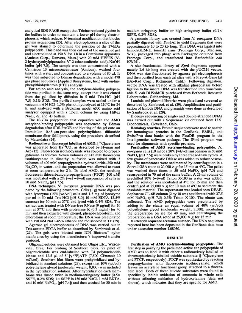
FIG. 1. (A) Coomassie-stained SDS-PAGE gel of DEAE-Sepharose eluate of Triton X-100-solubilized membrane supernatantfrom cells labelled with FTCP. Lane 1, 7 pug; lane 2, 28 pug. Themolecular weights of the markers were (from the top to the bottom)66,000, 45,000, 36,000, 29,000, 24,000, 20,100, and 14,200. (B)Photograph of the unstained gel shown in panel A when it was
illuminated with long-wavelength UV light.
After cells were labelled in this way, the label was
followed during purification (see Materials and Methods) byoptical absorbancy or radioactivity. DEAE-Sepharose chro-matography of the Triton X-100-solubilized membranesyielded just two major polypeptides (Fig. 1). When cells hadbeen treated with FTCP, the smaller of these two polypep-tides, migrating at 27 kDa, was labelled with the fluorescentchromophore (Fig. 1). When the cells had been treated with[14C]acetylene, this polypeptide was radioactive (data notshown).
N-terminal amino acid sequence of acetylene-bindingpolypeptide. The labelled AMO polypeptide was separatedfrom the other major polypeptide in this preparation bySDS-PAGE and was electroeluted from the polyacrylamidegel slice, and the N-terminal sequence was obtained byEdman degradation. The amino acid sequence obtained inthis way was SIFRTEEILKAAKMPPEAVCM. Quantita-tive data support the idea that the sequence determined inthis way was indeed the sequence of the acetylene-bindingpolypeptide rather than a contaminant. The maximum num-
ber of moles of amino acid removed during the cycles ofsequencing approximately matched the number of moles ofacetylene bound to the polypeptide (determined by its levelof radioactivity) (Table 1). This preparation was calculatedto have 461 pmol of bound acetylene prior to undergoing fourcycles of concentration and buffer exchange with water on a
Centricon-10 centrifugal ultrafiltration membrane. Duringsequencing, the yields of some amino acid residues are
greater than the yields of other amino acid residues becausethe former are more resistant to the harsh chemistry of theEdman degradation procedure. Thus, the minimum estimate
TABLE 1. Amounts of different amino acids found during eachcycle of Edman degradation sequencing of the AMO
acetylene-binding polypeptidea
Cycle Residue Amt (pmol)
1 Ser 1152 Ile 923 Phe 1624 ?5 Thr 1146 Glu 1647 Glu 2048 Ile 1579 Leu 293
10 Lys 22911 Ala 30812 Ala 34313 Lys 23814 Met 13015 Pro 19116 Pro 19817 Glu 12418 Ala 29519 Val 7620 ?21 Met 30
a Assuming one acetylene molecule per polypeptide molecule, the prepa-ration was calculated, from its level of radioactivity, to have 461 pmol ofacetylene-binding polypeptide prior to being concentrated by ultrafiltrationand then sequenced.
for the amount of polypeptide is the amount obtained for theamino acid residue whose yield is greatest, in this case 343pmol of alanine in cycle 12 (Table 1). Allowing for sequenc-ing yield loss due to blockage and termination during theprevious cycles of the Edman degradation procedure, andpossibly allowing for some sample loss during the concen-tration and buffer exchange step, this value is in goodagreement with the value of 461 pmol of polypeptide that wecalculated if we assumed 1 bound acetylene molecule perpolypeptide molecule.
Cloning and sequencing of the AMO gene. The chemicallydetermined N-terminal amino acid sequence of the 27-kDaAMO polypeptide was used to design a degenerate oligonu-cleotide probe for the AMO gene matching the amino acidsequence TEEILKAAKMP. This oligonucleotide, AMO oli-gonucleotide 1, had the sequence 5'-ACI-GA(A,G)-GA(A,G) -ATI - (C,T)TI -AA(A,G) - GCI - GCI -AA(A,G) -ATG -
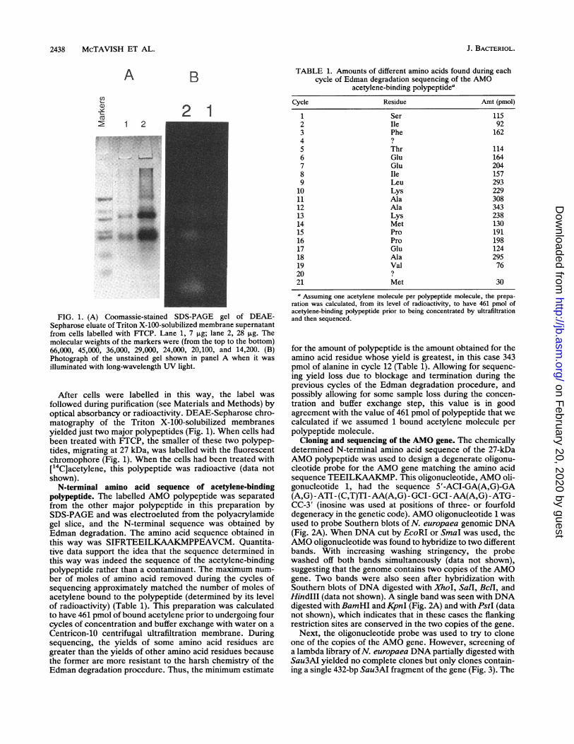
CC-3' (inosine was used at positions of three- or fourfolddegeneracy in the genetic code). AMO oligonucleotide 1 wasused to probe Southern blots of N. europaea genomic DNA(Fig. 2A). When DNA cut by EcoRI or SmaI was used, theAMO oligonucleotide was found to hybridize to two differentbands. With increasing washing stringency, the probewashed off both bands simultaneously (data not shown),suggesting that the genome contains two copies of the AMOgene. Two bands were also seen after hybridization withSouthern blots of DNA digested with XhoI, Sail, BclI, andHindIII (data not shown). A single band was seen with DNAdigested with BamHI and KpnI (Fig. 2A) and with PstI (datanot shown), which indicates that in these cases the flankingrestriction sites are conserved in the two copies of the gene.
Next, the oligonucleotide probe was used to try to cloneone of the copies of the AMO gene. However, screening ofa lambda library ofN. europaea DNA partially digested withSau3AI yielded no complete clones but only clones contain-ing a single 432-bp Sau3AI fragment of the gene (Fig. 3). The
J. BACTERIOL.
.V,I-7"!.
on February 20, 2020 by guest
http://jb.asm.org/
Dow
nloaded from
AMO GENE SEQUENCE 2439
A
kb :~M CD
B
m Xq Go
FIG. 2. (A) Autoradiograph of a Southern blot of N. europaeacut with four restriction enzymes and electrophoresed through a0.6% agarose gel. The blot was hybridized with radiolabelled AMOoligonucleotide 1 and after hybridization was washed in high-stringency buffer at 42'C for 30 min. (B) Autoradiograph of the sameSouthern blot filter used in panel A, except that this filter washybridized to radiolabelled AMO oligonucleotide 2. After hybridiza-tion the filter was washed in high-stringency buffer at 520C for 30min.
nucleotide sequence of this fragment matched exactly thechemically determined N-terminal amino acid sequence ofthe polypeptide.
This Sau3AI fragment included a KpnI site, so an attemptwas made to clone a KpnI fragment that would contain theremainder of the gene. A 27-mer oligonucleotide, AMOoligonucleotide 2, corresponding to bases 380 to 406 of thesequence shown in Fig. 3, was synthesized, radiolabelled,and used to probe the same Southern blot filter shown in Fig.2A (Fig. 2B). This oligonucleotide appeared to hybridize tothe same one or two bands as AMO oligonucleotide 1 exceptin the KpnI lane. The KjpnI result was expected since theKpnI site at bases 366 to 371 (Fig. 3) lies between the bindingsites of the two AMO oligonucleotides. This result confirmsthat there are two copies of the AMO gene and that thehybridization of AMO oligonucleotide 1 to two bands wasnot due to fortuitous hybridization of the oligonucleotide toa second, unrelated sequence.AMO oligonucleotide 2 hybridized to a 1.6-kb KpnI frag-
ment (Fig. 2B), so a plasmid library of size-fractionated1.6-kb KpnI-digested genomic DNA was created andscreened with AMO oligonucleotide 2. This experimentidentified several clones, six of which were analyzed by
agarose gel electrophoresis and Southern blotting. All sixwere found to have identically sized 1.6-kb KpnI inserts thathybridized with AMO oligonucleotide 2 and that matchedthe size of the KjnI fragment from genomic DNA thathybridized with oligonucleotide 2.One of these clones was sequenced completely. The
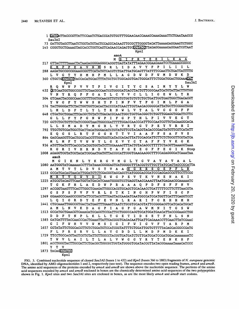
resulting sequence, combined with the sequence of the432-bp Sau3AI fragment described above, is shown in Fig. 3.The Sau3AI and KpnI clones overlapped at bases 366 to 432(Fig. 3), and in this overlap region their sequences matchedexactly. Collectively, the sequence of the two clones shownin Fig. 3 includes an open reading frame at positions 249 to1079 that should code for a polypeptide having a molecularweight of 31,861; this is approximately the molecular weightof the AMO acetylene-binding polypeptide determined bySDS-PAGE, which was 27,000. We designate this geneamoA.Amino acid composition of the acetylene-binding polypep-
tide. To confirm that the sequence shown in Fig. 3 was notthe sequence of a contaminant and that amoA is indeed thegene for the acetylene-binding polypeptide, a purified prep-aration of the polypeptide was analyzed quantitatively foramino acid composition (Table 2). The radiolabelled acety-lene-binding polypeptide was purified as described above.From its level of radioactivity and the specific activity of the['4C]acetylene, the sample analyzed in the experimentshown in Table 2 was calculated to contain 200 pmol ofacetylene and, assuming one bound acetylene molecule perpolypeptide molecule, 200 pmol of polypeptide. From thisinformation and from the sequence of amoA, we predictedthat the sample contained 46.2 nmol of the amino acidsanalyzed. (Trp and Cys were excluded from the analysisbecause they were destroyed in the acid hydrolysis; Gly wasexcluded because glycine was in the electrophoresis bufferand was not completely removed from the sample prior tothe amino acid analysis.) We observed 40.13 nmol of theseamino acids, 87% of the predicted amount. Table 2 alsoshows that the relative mole percentages of the differentamino acids agreed reasonably well with the mole percent-ages predicted from the sequence of amoA.
Second gene in the amo operon. Downstream from amoA asecond open reading frame was found, which continuedthrough the remainder of the 1,516-bp cloned KpnI fragment(Fig. 3). The 40-kDa polypeptide that copurified with theacetylene-binding polypeptide had been sequenced by Ed-man degradation. Its N-terminal sequence, ?GE?SQEP-FL?M?TVQWYDI, exactly matched the sequence of theprotein encoded by the second open reading frame in theAMO clone (Fig. 3). The fact that the gene for this copuri-fying polypeptide immediately follows the gene for theacetylene-binding polypeptide is evidence that this polypep-tide is involved in AMO activity, so we designated the geneamoB. The partial sequence of amoB shown in Fig. 3encodes 26.346 kDa (not including the hypothesized cleavedleader sequence) of the polypeptide that has an apparentmolecular weight as determined by SDS-PAGE of 40,000.
Analysis ofamoA and amoB sequences. The start codon foramoA must be the GTG beginning at base 249. GTG, ratherthan the usual ATG, is the start codon in 7% of the genes ofE. coli (7). The evidence that this GTG is the start codonincludes the fact that it immediately precedes the codon forthe first amino acid of the mature protein and the fact that itis preceded by a sequence that matches reasonably well theconsensus sequence for prokaryotic ribosome binding sites.Also, the nearest other possible in-frame start codon is theGTG beginning at base 182 (Fig. 3). However, between this
VOL. 175, 1993
on February 20, 2020 by guest
http://jb.asm.org/
Dow
nloaded from
2440 McTAVISH ET AL.
1 aADTTACCCGTTATTCCAATCTGACCGATGTGGTTTGGAACAACCAAAGCAAAGAAATTCTGAATAACCGSau3AI
73 GATTGTAGCTTAATCTGGTATGTACTCCAGGCAGAAGTTGCGCTTGGGGTACATTAAAAGAGGAAGTCTGGC145 CGGGTGCTGAAAGTAGCACCTGGTCAGTCAGAACCAGAGTGC GTAC TAGAGGAAAAGAGGAATCGTAAT
KpnIamoAM S I F R T E E I L K A A
217 GTTACTTTTAAACTATAAGAGGGGAGGGCAGGGTGAGTATATTTAGAACGGAAGAAATCCTGAAAGCGGCCK M P P E A V H S R L I D A V Y F P I L I I L
288 AAGATGCCGCCGGAAGCGGTTCATATGTCACGTCTGATTGATGCAGTTTATTTTCCAATTCTGATTATTTTGL V G T Y H M H P M L L A G D W D F W M D W K D
360 CTGGTGTACACCACATGCACTTTATGCTGCTGGCAGGTGACTGGGATTTCTGGATGGACTGGAAAMKpnI Sau3AI
R Q U W P V V T P I V G I T Y C S A I M Y Y L W432 OGTCAATGGTGGCCGGTTGTAACGCCAATCGTGGGGATCACCTACTGTTCGGCAATCATGTATTACTTGTGG
V N Y R Q P F G A T L C V V C L L I G E W L T R504 GTCAACTACCGCCAACCGTTTGGTGCAACGTTGTGTGTGGTGTGTCTGCTGATTGGTGAGTGGCTGACACGT
Y W G F Y W W S H Y P I N F V T P G I M L P G A576 TACTGGGGATTCTACTGGTGGTCACACTACCCCATCAACTTCGTAACACCGGGCATTATGCTTCCGGGTGCG
L M L D F T L Y L T R N W L V T A L V G G G F F648 CTGATGCTGGACTTCACGCTGTATCTGACACGCAACTGGCTGGTGACGGCTCTGGTTGGAGGTGGATTCTTC
G L L F Y P G N W P I F G P T H L P I V V E G T720 GGTCTGCTGTTCTATCCGGGTAACTGGCCGATTTTTGGACCAACCCATTTGCCAATCGTTGTAGAAGGCACA
L L S M A D Y M G H L Y V R T G T P E Y V R H I792 TTGCTGTCGATGGCTGATTACATGGGACATCTGTATGTTCGTACAGGTACACCCGAGTATGTTCGTCATATT
E Q G S L R T F G G H T T V I A A F F S A F V S864 GAGCAAGGTTCACTGCGTACCTTTGGTGGTCATACCACAGTTATTGCAGCATTCTTCTCTGCGTTCGTATCA
M L M F T V W W Y L G K V Y C T A F F Y V K G K936 ATGTTGATGTTCACCGTATGGTGGTATCTTGGAAAAGTTTACTGTACAGCCTTTTTCTACGTTAAAGGTAAA
R G R I V H R N D V T A F G E E G F P E G I K Stop1008 AGAGGTCGTATCGTACATCGCAATGATGTTACCGCATTCGGTGAAGAAGGCTTTCCAGAGGGGATCAAATAA
amoBM G I K N L Y K R G V M G L Y G V A Y A V A A L
1080 AATGGGTATCAAAAACCTTTATAAACGGGGAGTGATGGGGCTTTACGGTGTTGCTTATGCAGTAGCCGCATTAA M T V T L D V S T V A A lH G E R S Q E P F L R
1153 GCCATGACAGTAACATTGGATGTCTCGACGGTAGCAGCTCATGGGGAGCGATCCCAGGAGCCGTTCCTTCGGM R T V Q W Y D Il K W G P E V T K V N E N A K I
1225 ATGCGTACAGTTCAATGGTATGACATCAAATGGGGTCCTGAGGTAACCAAAGTTAATGAGAACGCAAAAATCT G K F H L A E D W P R A A A Q P D F S F F N V
1297 ACCGGTAAGTTTCACTTGGCTGAAGATTGGCCACGTGCAGCTGCACAACCTGATTTTTCCTTCTTTAACGTAG S P S P V F V R L S T K I N G H P W F I S G P
1369 GGAAGCCCAAGCCCGGTATTCGTACGTTTGAGTACCAAGATCAATGGCCATCCATGGTTCATTTCAGGTCCTL Q I G R D Y E F E V N L R A R I P G R H H M H
1441 CTGCAAATTGGCCGTGACTATGAGTTTGAAGTCAATCTGCGTGCACGTATTCCGGGACGTCATCACATGCATA M L N V K D A G P I A G P G A W M N I T G S W
1513 GCCATGCTGAACGTCAAAGATGCAGGTCCGATTGCTGGGCCAGGTGCATGGATGAACATTACCGGAAGCTGGD D F T N P L K L L T G E T I D S E T F N L S N
1585 GATGATTTTACCAACCCACTGAAATTATTGACGGGTGAGACAATTGATTCAGAAACCTTCAACTTATCGAACG I F W H V V W M S I G I F W I G V F T A R P M
1657 GGTATATTCTGGCACGTTGTCTGGATGTCCATCGGTATTTTCTGGATTGGTGTTTTTACAGCACGGCCGATGF L P R S R V L L A Y G D D L L M D P M D K K I
1729 TTCCTGCCACGTAGTCGTGTATTGTTGGCCTACGGTGATGATCTGTTGATGGATCCGATGGACAAAAAAATCT W V L A I L T L A L V W G G Y R Y T E N K H P
1801 ACCTGGGTACTTGCCATTCTGACGCTGGCGCTGGTATGGGGTGGATACCGTTATACGGAAAATAAACATCCCY T V
1873 TACACEGCKpnI
FIG. 3. Combined nucleotide sequence of cloned Sau3AI (bases 1 to 432) and KpnI (bases 366 to 1883) fragments ofN. europaea genomicDNA, identified by AMO oligonucleotides 1 and 2, respectively (see text). The sequence encodes two open reading frames, amoA and amoB.The amino acid sequences of the proteins encoded by amoA and amoB are shown above the nucleotide sequence. The portions of the aminoacid sequences encoded by amoA and amoB enclosed in boxes are the chemically determined amino acid sequences of the two polypeptidesshown in Fig. 1. KpnI sites and two Sau3AI sites are enclosed in boxes, as are the most likely amoA and amoB start codons.
J. BACTERIOL.
on February 20, 2020 by guest
http://jb.asm.org/
Dow
nloaded from

AMO GENE SEQUENCE 2441
TABLE 2. Quantitative amino acid analysis of the["4C]acetylene-binding polypeptide purified by SDS-PAGE
and comparison with the amino acid compositionpredicted from the sequence of amoA
Amino No. of residues nmol nmol mol% mol%acid(s) predicted from observed predicted observed predictedgene sequence
Val 23 3.8 4.6 9.4 10.0Ala 15 3.0 3.0 7.5 6.5Leu 28 4.7 5.6 11.8 12.1Ile 18 2.8 3.6 7.1 7.8Pro 15 2.2 3.0 5.4 6.5Phe 21 3.5 4.2 8.8 9.1Met 12 1.8 2.4 4.6 5.2Tyr 17 2.7 3.4 6.8 7.5Ser 8 2.0 1.6 5.1 3.5Thr 20 2.6 4.0 6.4 8.7His 9 1.3 1.8 3.3 3.9Lys 7 1.6 1.4 4.1 3.0Arg 12 2.0 2.4 5.0 5.2Glu 10Gln 3Glu + Gln (13) 2.8 2.6 7.1 5.6Asp 8Asn 5Asp + Asn (13) 3.1 2.6 7.7 5.6Gly 26Trp 14Cys 4
Total 40.1 46.2
codon and the codon for the first amino acid residue of themature protein is an in-frame stop codon. Thus, there are nopossible start codons other than the GTG at bases 249 to 251.There are three in-frame ATGs upstream from amoB,
beginning at bases 1081, 1114, and 1156, plus a GTG at base1111. Of these, the most likely start codon is probably theone beginning at base 1081, since it is preceded by the bestribosome binding site of the candidates and it almost imme-diately follows the stop codon for amoA. If this is the startcodon, the gene would encode a 37-amino-acid leader se-quence, which might direct insertion of the polypeptide inthe plasma membrane. The amoA gene product apparentlyhas an internal signal sequence.No significant level of overall amino acid identity was
observed between the amoA or amoB gene products and anyof the gene products in the protein and DNA sequence databases. Alignments with particular gene products were alsoexamined in order to detect possible regions of local homol-ogy. However, alignment of amoA and amoB with theproteins encoded by the soluble methane monooxygenasegene cluster (32, 33), the bacterial integral membrane mono-oxygenases alkane hydroxylase and xylene monooxygenase(17, 18, 37), and the copper-containing monooxygenasestyrosinase (11, 21) and dopamine B-hydroxylase (38) re-vealed no regions of significant local homology.Hydropathy plots of the amoA and amoB gene products
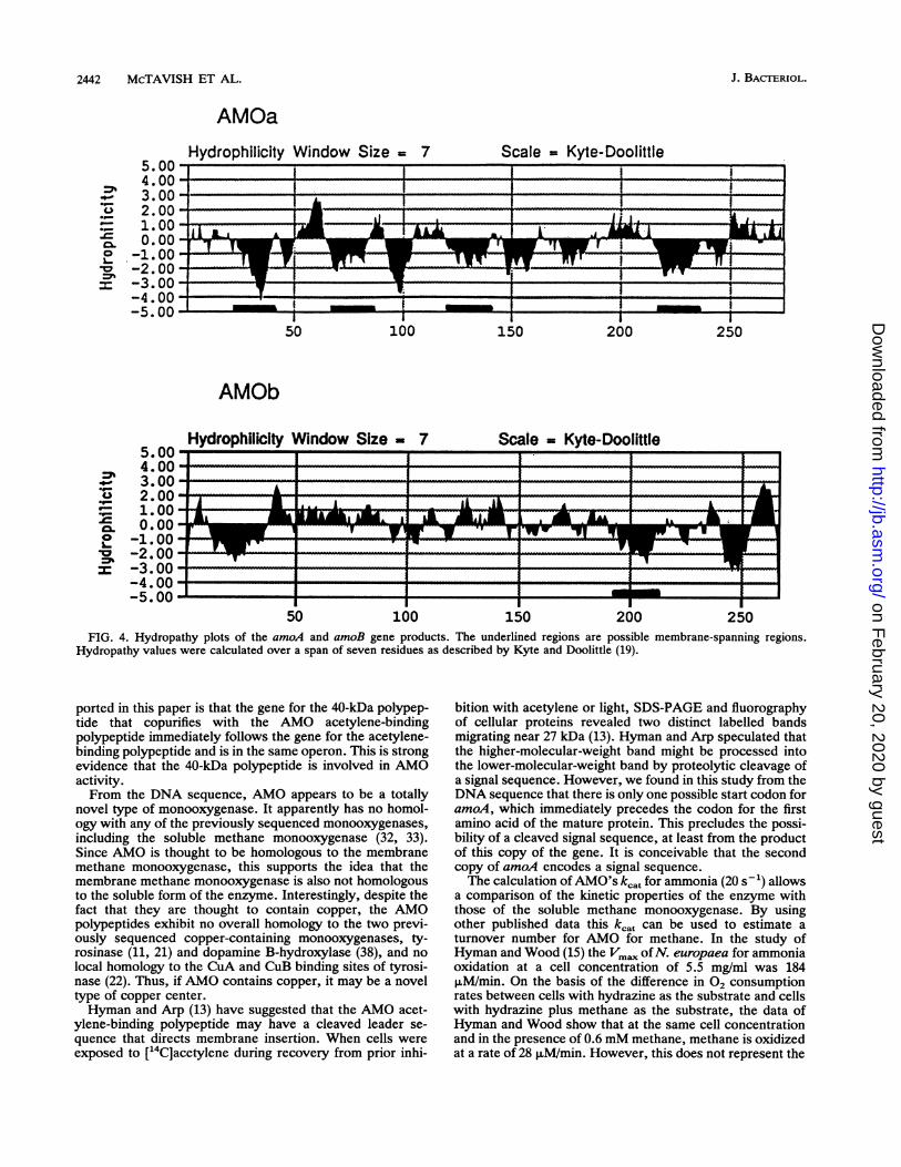
(Fig. 4) show that the amoA gene product, the acetylene-binding protein, is very hydrophobic and contains fourpotential membrane-spanning regions. The partial sequenceof the amoB product is less hydrophobic but still contains atleast one possible membrane-spanning region in addition to aquite hydrophobic hypothesized leader sequence.The pI of the amoA gene product was calculated to be 7.9.Amount of AMO in Nitrosomonas cells and kt of the
enzyme. Radioactive labelling of AMO in vivo followed bypurification of the protein allowed the determination of theconcentration of the enzyme in cells. Cells were labelledwith ["4C]acetylene (specific activity, 0.86 mCi/mmol), andmembranes were isolated and solubilized with Triton X-100.Insoluble material was then pelleted by centrifugation, andproteins from the soluble material were precipitated with2.5% (wt/vol) trichloroacetic acid. The trichloroacetic acidpellet was washed twice with water and then counted forradioactivity. The trichloroacetic acid pellet from 1 g (wetweight) of cells was found to contain 42,800 dpm of 1 C. Asdetermined by SDS-PAGE and fluorography, the 27-kDaAMO protein was the only radioactive protein in this mate-rial. Assuming one bound acetylene molecule per AMOmolecule, this works out to 23 nmol of AMO per g (wetweight) of cells.
This batch of cells consumed 42 nmol of 02 per min per mg(weight weight) of cells with NH3 as the substrate. Assuming1.5 mol Of 02 consumed per mol of NH3 consumed and usingthe value of 23 nmol of AMO per g of cells, the kcat in vivofor AMO was calculated to be approximately 20 s-1.
DISCUSSION
The use in this study of a fluorescein tag coupled to asuicide substrate functional group was a novel approach tolabelling an enzyme active site. However, this techniqueshould be adaptable to other suicide substrates and otherenzymes. Any compound with an amino group or othernucleophilic group can be reacted with fluorescein isothio-cyanate, which should allow many suicide substrates to becovalently attached to fluorescein and allow enzyme activesites to be fluorescently labelled.The gene for the AMO acetylene-binding polypeptide and
two-thirds of the gene for a 40-kDa polypeptide that copuri-fied with the acetylene-binding polypeptide were cloned andsequenced in this study. Among the evidence that the firstgene is the gene for the acetylene-binding polypeptide is thefact that its sequence exactly matches the chemically deter-mined sequence of the N terminus of the protein and the factthat it codes for a polypeptide having a molecular weight of31,861, which is approximately the molecular weight deter-mined by SDS-PAGE. The finding that the actual molecularweight (31,861) is greater than the SDS-PAGE molecularweight estimates (28,000 [16] and 27,000) is common formembrane proteins (18, 41).The evidence that the chemically determined N-terminal
sequence is the sequence of the acetylene-binding polypep-tide rather than the sequence of a contaminant protein is asfollows. First, the number of moles of amino acid deter-mined during each cycle of sequencing approximatelyequalled the number of moles of bound acetylene present.Second, the amino acid composition of a purified sample ofthe [14C]acetylene-labelled polypeptide matches fairlyclosely the composition predicted from the amoA sequence.Furthermore, the quantity of amino acids closely matchesthe quantity predicted from the level of radioactivity in thesample and the sequence of amoA.A cleaner proof that amoA is indeed the gene for the
acetylene-binding polypeptide would be to express amoAand demonstrate ammonia-oxidizing activity in vitro or inrecombinant cells. Unfortunately, however, no effective invitro assay system has been developed for AMO, andfurthermore, we were unable to clone a complete copy ofamoA, which precludes synthesis of the polypeptide.The first important finding from the DNA sequence re-
VOL. 175, 1993
on February 20, 2020 by guest
http://jb.asm.org/
Dow
nloaded from
2442 McTAVISH ET AL.
AMOaHydrophilicity Window Size = 7 Scale = Kyte-Doolittle
.uu -------------------
5005.00-.00-.00*.00
50 100 150 200 250
AMOb
Hydrophilicity Window Size - 7 Scale -_Kyte-Doolittle
.00 I I Ion~~~~~~~~~~~~~J---- ------ -----
-I____ __ _I II
50 100 150 200
An
250
FIG. 4. Hydropathy plots of the amoA and amoB gene products. The underlined regions are possible membrane-spanning regions.Hydropathy values were calculated over a span of seven residues as described by Kyte and Doolittle (19).
ported in this paper is that the gene for the 40-kDa polypep-tide that copurifies with the AMO acetylene-bindingpolypeptide immediately follows the gene for the acetylene-binding polypeptide and is in the same operon. This is strongevidence that the 40-kDa polypeptide is involved in AMOactivity.From the DNA sequence, AMO appears to be a totally
novel type of monooxygenase. It apparently has no homol-ogy with any of the previously sequenced monooxygenases,including the soluble methane monooxygenase (32, 33).Since AMO is thought to be homologous to the membranemethane monooxygenase, this supports the idea that themembrane methane monooxygenase is also not homologousto the soluble form of the enzyme. Interestingly, despite thefact that they are thought to contain copper, the AMOpolypeptides exhibit no overall homology to the two previ-ously sequenced copper-containing monooxygenases, ty-rosinase (11, 21) and dopamine B-hydroxylase (38), and nolocal homology to the CuA and CuB binding sites of tyrosi-nase (22). Thus, if AMO contains copper, it may be a noveltype of copper center.Hyman and Arp (13) have suggested that the AMO acet-
ylene-binding polypeptide may have a cleaved leader se-quence that directs membrane insertion. When cells wereexposed to [14C]acetylene during recovery from prior inhi-
bition with acetylene or light, SDS-PAGE and fluorographyof cellular proteins revealed two distinct labelled bandsmigrating near 27 kDa (13). Hyman and Arp speculated thatthe higher-molecular-weight band might be processed intothe lower-molecular-weight band by proteolytic cleavage ofa signal sequence. However, we found in this study from theDNA sequence that there is only one possible start codon foramoA, which immediately precedes the codon for the firstamino acid of the mature protein. This precludes the possi-bility of a cleaved signal sequence, at least from the productof this copy of the gene. It is conceivable that the secondcopy of amoA encodes a signal sequence.The calculation ofAMO's kcat for ammonia (20 s') allows
a comparison of the kinetic properties of the enzyme withthose of the soluble methane monooxygenase. By usingother published data this kcat can be used to estimate aturnover number for AMO for methane. In the study ofHyman and Wood (15) the Vmax ofN. europaea for ammoniaoxidation at a cell concentration of 5.5 mg/ml was 184puM/min. On the basis of the difference in 02 consumptionrates between cells with hydrazine as the substrate and cellswith hydrazine plus methane as the substrate, the data ofHyman and Wood show that at the same cell concentrationand in the presence of 0.6 mM methane, methane is oxidizedat a rate of 28 puM/min. However, this does not represent the
.00
.005,4
S 2,.- 1
o -1L .*0 -2-4
_5.
5.
=A44
_- 31._- -J 0.VW
or- 2.00-~ 1.00m 0.00o -1.00
l -2.00I -3.00
-4.00-5.00
------------I----
----A. I- if-ALPHA Al
R IA-MIN 1.11 ----I wwwwm-,- -1-10-10-
...........------a
J. BACHTERIOL.
a
on February 20, 2020 by guest
http://jb.asm.org/
Dow
nloaded from
AMO GENE SEQUENCE 2443
Vm. because this concentration of methane is below the Kmof 2 mM. When this Km value is used, the Vm. for methane(which would require unattainable methane concentrations)is 121 puM/min. This means that the ka for methane is121/184 of the kcat for ammonia, or 14 s . This comparesfavorably with the kcat of 2.6 s-1 for the soluble methanemonooxygenase from Methylosinus tichosporium OB3b (6).However, the Km for methane of the soluble methanemonooxygenase is much lower than the Km for methane ofAMO (160 ptM for the soluble methane monooxygenase fromMethylococcus capsulatus versus 2 mM for AMO), whichmakes the velocities of the two enzymes more comparable atlower methane concentrations. At 160 p.M methane (if weassume that the Km of the Methylosinus trichosporiumenzyme is the same as that of the Methylococcus capsulatusenzyme), the soluble methane monooxygenase would turnover at a rate of 1.3 s-1, while the AMO rate would be 1.0s 1. Still, this leads to the surprising conclusion that AMO isnearly as good at oxidizing methane as an enzyme evolvedfor that purpose.
ACKNOWLEDGMENTSWe thank Myke Logan for suggesting the use of a chromophori-
cally labelled suicide substrate. We thank Frank LaQuier for creat-ing the Sau3A lambda library, Greg Pazour and Anath Das forhelpful discussions, and Candace Pilon for growing N. europaeacells.
This work was supported by the Minnesota Sea Grant Program(grant USDOC/NA90AA-D-56149) and the U.S. Department ofAgriculture National Research Initiative Competitive Grants Pro-gram (grant 9208667).
REFERENCES1. Andersson, K. K., and A. B. Hooper. 1983. 02 and H20 are each
the source of one 0 in N02-: 15N-NMR evidence. FEBS Lett.164:236-240.
2. Arciero, D. M., C. Balny, and A. B. Hooper. 1991. Spectro-scopic and rapid kinetic studies of reduction of cytochromec-554 by hydroxylamine oxidoreductase from Nitrosomonaseuropaea. Biochemistry 30:11466-11472.
3. Bedard, C., and R. Knowles. 1989. Physiology, biochemistry,and specific inhibitors of CH4, NH4, and CO oxidation bymethanotrophs and nitrifiers. Microbiol. Rev. 53:68-84.
4. Colby, J., D. I. Stirling, and H. Dalton. 1977. The solublemethane mono-oxygenase of Methylococcus capsulatus (Bath):its ability to oxygenate n-alkanes, n-alkenes, ethers, and alicy-clic, aromatic, and heterocyclic compounds. Biochem. J. 165:395-407.
5. DiSpirito, A. A., J. D. Lipscomb, and A. B. Hooper. 1986.Cytochrome aa3 from Nitrosomonas europaea. J. Biol. Chem.261:17048-17056.
6. Fox, B. G., and J. D. Lipscomb. 1988. Purification of a highspecific activity methane monooxygenase hydroxylase compo-nent from a type II methanotroph. Biochem. Biophys. Res.Commun. 154:165-170.
7. Gren, E. J. 1984. Recognition of messenger RNA during trans-lational initiation in E. coli. Biochimie 66:1-29.
8. Hollocher, T. C., M. E. Tate, and D. J. D. Nicholas. 1981.Oxidation of ammonia by Nitrosomonas europaea: definitive'8O-tracer evidence that hydroxylamine formation involves amonooxygenase. J. Biol. Chem. 256:10834-10836.
9. Hooper, A. B., P. C. Maxwell, and K. R. Terry. 1978. Hydrox-ylamine oxidoreductase from Nitrosomonas: absorption spectraand content of heme and metal. Biochemistry 17:2984-2989.
10. Hooper, A. B., and K. R. Terry. 1973. Specific inhibitors ofammonia oxidation in Nitrosomonas. J. Bacteriol. 115:480-485.
11. Huber, M., G. Hintermann, and K. Lerch. 1985. Primarystructure of tyrosinase from Streptomyces glauascens. Bio-chemistry 24:6038-6044.
12. Hyman, M. R., and D. J. Arp. 1990. Small-scale production of
[U-4C]acetylene from Ba'4CO3: application to labelling of am-monia monooxygenase in autotrophic nitrifying bacteria. Anal.Biochem. 190:348-353.
13. Hyman, M. R., and D. J. Arp. 1992. 14C2H2- and '4C02-labellingstudies of the de novo synthesis of polypeptides by Nitrosomo-nas europaea during recovery from acetylene and light inacti-vation of ammonia monooxygenase. J. Biol. Chem. 267:1534-1545.
14. Hyman, M. R., I. B. Murton, and D. J. Arp. 1988. Interaction ofammonia monoxygenase from Nitrosomonas europaea withalkanes, alkenes, and alkynes. Appl. Environ. Microbiol. 54:3187-3190.
15. Hyman, M. R., and P. M. Wood. 1983. Methane oxidation byNitrosomonas europaea. Biochem. J. 212:31-37.
16. Hyman, M. R., and P. M. Wood. 1985. Suicidal inactivation andlabelling of ammonia mono-oxygenase by acetylene. Biochem.J. 227:719-725.
17. Kok, M., R. Oldenhuis, M. P. G. van der Linden, C. H. C.Meulenberg, J. Kingma, and B. Witholt. 1989. Pseudomonasoleovorans alkBAC operon encodes two structurally relatedrubredoxins and an aldehyde dehydrogenase. J. Biol. Chem.264:5442-5451.
18. Kok, M., R. Oldenhuis, M. P. G. van der Linden, P. Raatjes, J.Kingma, P. H. van Lelyveld, and B. Witholt. 1989. Pseudomonasoleovorans alkane hydroxylase gene: sequence and expression.J. Biol. Chem. 264:5435-5441.
19. Kyte, J., and R. F. Doolittle. 1982. A simple method fordisplaying the hydropathic character of a protein. J. Mol. Biol.157:105-132.
20. Lees, H. 1952. The biochemistry of nitrifying organisms: theammonia oxidizing systems of Nitrosomonas. Biochem. J.52:134-139.
21. Lerch, K. 1978. Amino acid sequence of tyrosinase from Neu-rospora crassa. Proc. Natl. Acad. Sci. USA 75:3635-3639.
22. Lerch, K., M. Huber, H.-J. Schneider, R. Drexel, and B. Linzen.1986. Different origins of metal binding sites in binuclear copperproteins, tyrosinase and hemocyanin. J. Inorg. Biochem. 26:213-217.
23. Logan, M. S. P. 1991. Ph.D. thesis. University of Minnesota, St.Paul.
24. Matusdaira, P. 1987. Sequence from picomole quantities ofproteins electroblotted onto polyvinylidene difluoride mem-branes. J. Biol. Chem. 262:10035-10038.
25. Moos, M., N. Y. Nguyen, and T.-Y. Liu. 1988. Reproduciblehigh yield sequencing of proteins electrophoretically separatedand transferred to an inert support. J. Biol. Chem. 263:6005-6008.
26. Porzio, M. A., and A. M. Pearson. 1977. Improved resolution ofmyofibrillar proteins with sodium dodecyl sulfate-polyacryl-amide gel electrophoresis. Biochim. Biophys. Acta 490:27-34.
27. Prior, S. D., and H. Dalton. 1985. Effect of copper ions onmembrane content and methane monooxygenase activity inmethanol-grown cells of Methylococcus capsulatus (Bath). J.Gen. Microbiol. 131:155-163.
28. Reid, E. E. 1963. Organic chemistry of bivalent sulfur, vol. 4 and5. Chemical Publishing Co., Inc., New York.
29. Sambrook, J., E. F. Fritsch, and T. Maniatis. 1989. Molecularcloning: a laboratory manual, 2nd ed. Cold Spring HarborLaboratory, Cold Spring Harbor, N.Y.
30. Shears, J. H., and P. M. Wood. 1985. Spectroscopic evidencefor a photosensitive oxygenated state of ammonia monooxyge-nase. Biochem. J. 226:499-507.
31. Smith, D. D. S., and H. Dalton. 1989. Solubilisation of methanemonooxygenase from Methylococcus capsulatus (Bath). Eur. J.Biochem. 182:667-671.
32. Stainthorpe, A. C., V. Lees, G. P. C. Salmond, H. Dalton, andJ. C. Murrell. 1990. The methane monooxygenase gene clusterof Methylococcus capsulatus (Bath). Gene 91:27-34.
33. Stainthorpe, A. C., J. C. Murrell, G. P. C. Salmond, H. Dalton,and V. Lees. 1989. Molecular analysis of methane monooxyge-nase from Methylococcus capsulatus (Bath). Arch. Microbiol.152:154-159.
34. Stanley, S. H., S. D. Prior, D. J. Leak, and H. Dalton. 1983.
VOL. 175, 1993
on February 20, 2020 by guest
http://jb.asm.org/
Dow
nloaded from
2444 McTAVISH ET AL.
Copper stress underlies the fundamental change in intracellularlocation of methane monooxygenase in methane-oxidizing or-ganisms: studies in batch and continuous culture. Biotechnol.Lett. 5:487-492.
35. Suzuki, I., and S.-C. Kwok. 1981. A partial resolution andreconstitution of the ammonia oxidizing system of Nitrosomo-nas europaea: role of cytochrome c554. Can. J. Biochem.59:484-488.
36. Suzuki, I., S.-C. Kwok, U. Pular, and D. C. Y. Tsang. 1981.Cell-free ammonia-oxidizing system of Nitrosomonas euro-
paea: general conditions and properties. Can. J. Biochem.59:477-483.
37. Suzuki, M., T. Haychawa, J. P. Shaw, M. Rekik, and S.Harayama. 1991. Primary structure of xylene monooxygenase:similarites to and differences from the alkane hydroxylationsystem. J. Bacteriol. 173:1690-1695.
38. Taljanidisz, J., L. Stewart, A. J. Smith, and J. P. Klinman. 1989.Structure of bovine adrenal dopamine B-monooxygenase, asdeduced from cDNA and protein sequencing: evidence that themembrane-bound form of the enzyme is anchored by an un-
cleaved signal peptide. Biochemistry 28:10054-10061.39. Vanelli, T., and A. B. Hooper. 1992. Oxidation of nitrapyrin to
6-chloropicolinic acid by the ammonia-oxidizing bacterium Ni-trosomonas europaea. Appl. Environ. Microbiol. 58:2321-2325.
40. Vanelli, T., M. Logan, D. M. Arciero, and A. B. Hooper. 1990.Degradation of halogenated aliphatic compounds by the ammo-nia-oxidizing bacterium Nitrosomonas europaea. Appl. Envi-ron. Microbiol. 56:1169-1171.
41. Youvan, D. C., E. J. Bylina, M. Alberti, H. Begusch, and J. E.Hearst. 1984. Nucleotide and deduced polypeptide sequences ofthe photosynthetic reaction center, B870 antenna, and flankingpolypeptides from R. capsulata. Cell 37:949-957.
J. BACTERIOL.
on February 20, 2020 by guest
http://jb.asm.org/
Dow
nloaded from