Embed Size (px)
Citation preview
OnpA, an Unusual Flavin-Dependent Monooxygenase Containing aCytochrome b5 Domain
Yi Xiao,a Ting-Ting Liu,a Hui Dai,b Jun-Jie Zhang,a Hong Liu,a Huiru Tang,b David J. Leak,c and Ning-Yi Zhoua
Key Laboratory of Agricultural and Environmental Microbiology, Wuhan Institute of Virology, Chinese Academy of Sciences, Wuhan, Chinaa; State Key Laboratory ofMagnetic Resonance and Atomic and Molecular Physics, Wuhan Center for Magnetic Resonance, Wuhan Institute of Physics and Mathematics, Chinese Academy ofSciences, Wuhan, Chinab; and Department of Life Sciences, Imperial College London, London, United Kingdomc
ortho-Nitrophenol 2-monooxygenase (EC 1.14.13.31) from Alcaligenes sp. strain NyZ215 catalyzes monooxygenation of ortho-nitrophenol to form catechol via ortho-benzoquinone. Sequence analysis of this onpA-encoded enzyme revealed that it containeda flavin-binding monooxygenase domain and a heme-binding cytochrome b5 domain. OnpA was purified to homogeneity as aHis-tagged protein and was considered a monomer, as determined by gel filtration. FAD and heme were identified by high-performance liquid chromatography (HPLC) and HPLC-mass spectrometry (HPLC-MS) as cofactors in this enzyme, and quanti-tative analysis indicated that 1 mol of the purified recombinant OnpA contained 0.66 mol of FAD and 0.20 mol of heme. How-ever, the enzyme activity of OnpA was increased by 60% and 450% after addition of FAD and hemin, respectively, suggestingthat the optimal stoichiometry was 1:1:1. In addition, site-directed mutagenesis experiments confirmed that two highly con-served histidines located in the cytochrome b5 domain were associated with binding of the heme, and the cytochrome b5 domainwas involved in the OnpA activity. These results indicate that OnpA is an unusual FAD-dependent monooxygenase containing afused cytochrome b5 domain that is essential for its activity. Therefore, we here demonstrate a link between cytochrome b5 andflavin-dependent monooxygenases.
Heme-binding proteins are ubiquitous and involved in manybioprocesses. Cytochrome b5 (Cyt b5) is a small heme-
binding protein which is widespread in eukaryotes. Its ligandheme is buried in a hydrophobic pocket and connected to twohighly conserved histidine residues (28). The physiological func-tions of Cyt b5 are associated with fatty acid desaturation, biosyn-thesis of the N-glycolylneuraminic acid, plasmalogen, and choles-terol, and reduction of cytochrome P450 (35). In some cases, Cytb5 appears as a domain fused onto proteins with other functionaldomains (22, 28), such as human sulfite oxidase (15), human fattyacid desaturase (8, 9), plant nitrate reductase (18, 23), yeast flavo-cytochrome b2 (19), and mammalian cytochrome b-typeNAD(P)H oxidoreductase (44). Interestingly, all these Cyt b5 fu-sion proteins are found exclusively in eukaryotic lineages (22, 25,28), not prokaryotic ones (17, 22).
Cytochrome P450 (Cyt P450) is one of the best characterizedmonooxygenases. It is involved in a variety of cellular reactions,such as biotransformation of drugs, bioconversion of xenobiotics,and biosynthesis of physiological compounds (3). It is also part ofa superfamily of heme-binding proteins but distinct from Cyt b5;for all Cyt P450, an absolutely conserved cysteine is the fifth ligandof the heme iron (16). This differs from Cyt b5, in which the fifthand sixth ligands of the heme iron are two highly conserved histi-dines, which prevents its direct interaction with molecular oxygen(28).
Flavin-dependent monooxygenase is another important classof monooxygenases. In contrast to Cyt P450, which is abundant ineukaryotes, flavin-dependent monooxygenase appears to be mostprevalent among prokaryotic organisms (33, 34). It plays an im-portant role in biodegradation of aromatic compounds, drug de-toxification, and biosynthesis of sterols, antibiotics, and plant hor-mones (1, 14). While Cyt P450 uses heme, flavin-dependentmonooxygenase uses flavins, either FAD or FMN, to bind andactivate oxygen for catalyzing monooxygenation. Cyt P450 gets
electrons from Cyt P450 reductase or from Cyt b5 in some cases,whereas flavin-dependent monooxygenase receives electrons di-rectly from NAD(P)H. The relationship between Cyt P450 andCyt b5 has been broadly studied (11, 28, 42). It appears that Cyt b5
has a rather complex interaction with different Cyt P450 and canstimulate, inhibit, or have no effect on the activity of Cyt P450through diverse strategies. However, there are no reports on in-teractions between Cyt b5 and flavin-dependent monooxygenase.
ortho-Nitrophenol 2-monooxygenase (EC 1.14.13.31) cata-lyzes monooxygenation of ortho-nitrophenol (ONP), which isknown to be an aromatic pollutant as well as an indicator in en-zyme assays. This enzyme was purified and characterized from theONP-utilizing bacterium Pseudomonas putida B2 two decades ago(40), but its encoding gene, onpA (GenBank accession no.EF547253), was only recently identified in a newly isolated ONPutilizer, Alcaligenes sp. strain NyZ215 (38). OnpA converts ONPto catechol via ortho-benzoquinone, concomitant with oxygenand NADPH consumption and nitrite release. When NADPH wasreplaced by NADH, its activity was reduced by 92%. Sequenceanalysis revealed a Cyt b5 domain in this protein. However, theinvolvement of Cyt b5 in ONP oxidation has not previously beenreported. Here we report that the OnpA from strain NyZ215 is anunusual FAD-dependent monooxygenase containing a cyto-chrome b5-like heme binding domain and OnpA activity is depen-dent on the binding of heme to the Cyt b5 domain.
Received 24 October 2011 Accepted 9 January 2012
Published ahead of print 20 January 2012
Address correspondence to Ning-Yi Zhou, [email protected].
Copyright © 2012, American Society for Microbiology. All Rights Reserved.
doi:10.1128/JB.06411-11
1342 jb.asm.org 0021-9193/12/$12.00 Journal of Bacteriology p. 1342–1349
on January 21, 2019 by guesthttp://jb.asm
.org/D
ownloaded from
MATERIALS AND METHODSExpression and purification of OnpA in E. coli. The gene onpA was PCRamplified from Alcaligenes sp. strain NyZ215 (38) with the high-fidelityDNA polymerase Pyrobest (Takara) using a pair of primers (forward,5=-GAGGGATCCATGCGAGCTGTCATTATCG-3=; reverse, 5=-GGCAAGCTTAATATCAAGCCGTATGAGGC-3=). The PCR product contain-ing onpA was double-digested with BamHI and HindIII and then ligatedinto similarly treated pET28a to form the expression constructpZWX28A, in which an N-terminal six-His-tag-encoding sequence wasfused into onpA. Its sequence was verified by DNA sequencing to ensurethat no mutations were incorporated. Escherichia coli BL21(DE3) carryingpZWX28A was grown on lysogeny broth (LB) at 37°C to an A600 of around0.7 and induced by the addition of 0.1 mM isopropyl-�-D-1-thio-galactopyranoside (IPTG) for 3 to 4 h at 20°C. The cells were harvestedand stored at �80°C until use.
All steps for purification were performed at 4°C. After the cells weredisrupted by sonication in the binding buffer (300 mM NaCl, 50 mMsodium phosphate buffer [pH 8.0]), the cellular lysate was centrifuged at13,500 � g for 30 min, and the supernatant was taken for protein purifi-cation. OnpA was then purified via affinity chromatography using nickel-nitrilotriacetic acid agarose (Merck Biosciences) according to the suppli-er’s recommendations. Finally, imidazole and salts were removed fromthe eluted fractions by overnight dialysis against 20 mM sodium phos-phate buffer (pH 7.5).
Determination of molecular mass of OnpA. The molecular mass ofOnpA was determined by sodium dodecyl sulfate-polyacrylamide gelelectrophoresis (SDS-PAGE). Gel filtration chromatography was used todetermine the native molecular mass of OnpA. The experiment was per-formed on an Akta fast-performance liquid chromatography system(Amersham Pharmacia Biotech) using a Superdex 200 10/300 GL column(Amersham Pharmacia Biotech). The 50 mM phosphate buffer (pH 7.2)contained 0.15 M NaCl, and the flow rate was 0.25 ml/min. The nativemolecular mass was estimated from a calibration curve plotted using stan-dard proteins (Sigma): carbonic anhydrase (29 kDa), bovine serum albu-min (66 kDa), alcohol dehydrogenase (150 kDa), �-amylase (200 kDa),apoferritin (443 kDa), and thyroglobulin (669 kDa).
Enzyme assay and kinetic measurement. ortho-Nitrophenol 2-mono-oxygenase activity was determined spectrophotometrically by measuringthe decrease in ONP absorbance at 410 nm due to substrate consumption(40). The molar extinction coefficients for ONP and NADPH were 3.47mM�1 cm�1 at 410 nm and 6.22 mM�1 cm�1 at 340 nm, respectively. Thestandard enzyme assay mixture contained 40 nM purified OnpA, 0.05mM ONP, 0.2 mM NADPH, and 4 mM Mg2� in 500 �l (final volume) ofphosphate buffer (20 mM, pH 7.5). The assay was initiated through addi-tion of substrate ONP. The halogen-containing salts, hemin, protopor-phyrin IX, FAD, and FMN were used to test their impact on OnpA activ-ity. The kinetic analysis was carried out following the method described(40). ONP was varied from 1 to 100 �M when NADPH was constant at 0.5mM; NADPH was varied from 20 to 400 �M when ONP was constant at0.03 mM. One unit of enzyme activity was defined as the amount requiredfor the disappearance of 1 �mol of substrate per minute at room temper-ature. Specific activities are expressed as units per gram of protein. Proteinconcentration was determined by the Bradford method with bovine se-rum albumin as the standard.
Isolation and determination of the cofactors from OnpA. The flavincofactor was released by treating 400 �l of OnpA (0.3 mg/ml) with 1mg/ml proteinase K for 2 h at room temperature in the dark (30), followedby centrifugation at 13,500 � g for 10 min. The supernatant was subjectedto HPLC analysis.
The method for heme extraction was described previously (24).Briefly, 50 �l of solution containing OnpA was mixed with 450 �l ofacetone-HCl (19:1, vol/vol) by gentle shaking at room temperature for 20min. After centrifugation at 13,500 � g for 5 min, 1 ml of ice-cold waterand 0.3 ml of ethyl acetate were added to the supernatant, followed byvortexing and centrifugation. The ethyl acetate phase was recovered, and
the solvent was removed with a vacuum concentrator. The residue wasdissolved in 50 �l of acetonitrile and analyzed immediately. Myoglobin(Sigma) was used as a positive control for heme extraction and identifica-tion.
Analytical methods. To analyze the flavin extracted from OnpA, high-performance liquid chromatography (HPLC) analysis was performed us-ing a Gilson 715 system equipped with a C18 reversed-phase column fromSupelco (250 by 4.6 mm, 5 �m) with a column temperature of 30°C. Themobile phase consisted of methanol (40%) and 10 mM H3PO4 (60%) at aflow rate of 1.0 ml/min. The flavins were monitored at 450 nm with aGilson 119 UV/Vis detector. The retention times of the authentic FADand FMN (Sigma) were 3.53 and 4.14 min, respectively. FAD concentra-tion was determined by reference to a standard of known concentration.
To identify the heme extracted from OnpA and myoglobin, HPLC-MS(mass spectrometry) analysis was carried out on an Agilent 1200 seriesHPLC system (Agilent Technologies) consisting of a quaternary solventdelivery system, an on-line degasser, an autosampler, a column tempera-ture controller, and a diode array detector. The mass spectra were ac-quired using a micro-QTOF mass spectrometer (Bruker Daltonics)equipped with an ESI (electrospray ionization) interface. Five percent ofthe eluent was directed to MS using a Bruker nuclear magneticresonance-MS interface unit (Bruker BioSpin). Chromatographic separa-tion was carried out on an ACE C18-HL (Hi-Load) column (5 �m, 250 by4.6 mm; Advanced Chromatography Technologies) with a C18 guard col-umn at 25°C. The detection wavelength was set at 405 nm. The mobilephase consisted of water (A) and acetonitrile (B), both containing 0.1%(vol/vol) formic acid. A gradient program was used as follows: 0 to 5 min,20% B; 5 to 15 min, 20 to 80% B; 15 to 20 min, 80 to 100% B. The flow ratewas 1.0 ml/min. The optimized mass spectrometric parameters were asfollows: positive-ion mode; capillary voltage, 4,500 V; nebulizer gas pres-sure, 0.8 � 105 Pa; drying gas flow rate, 8 liter/min; gas temperature,180°C. The spectra were recorded in the range of m/z 50 to 1,000.
UV-visible absorption spectra were recorded with a Perkin ElmerLambda 25 UV/Vis spectrometer. The concentration of heme was deter-mined using an absorption coefficient of 130 mM�1 cm�1 at 413 nm (2).
Site-directed mutagenesis. To confirm coordination of the two highlyconserved histidines with the heme in the cytochrome b5 domain, both ofthem were individually replaced by aspartic acids. Overlapping PCR wasadopted to construct the site-directed mutants. The desired mutationswere identified by DNA sequencing. Two mutant proteins, OnpA(H515D) and OnpA(H538D), were expressed and purified by the meth-ods described above.
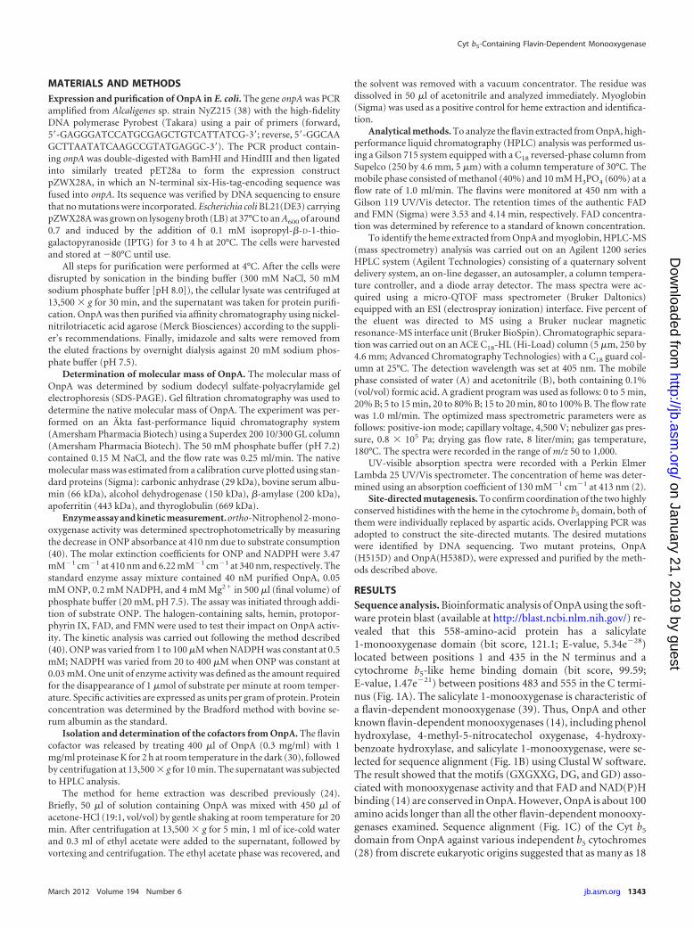
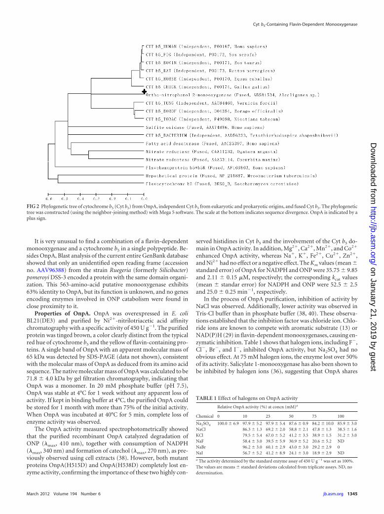
RESULTSSequence analysis. Bioinformatic analysis of OnpA using the soft-ware protein blast (available at http://blast.ncbi.nlm.nih.gov/) re-vealed that this 558-amino-acid protein has a salicylate1-monooxygenase domain (bit score, 121.1; E-value, 5.34e�28)located between positions 1 and 435 in the N terminus and acytochrome b5-like heme binding domain (bit score, 99.59;E-value, 1.47e�21) between positions 483 and 555 in the C termi-nus (Fig. 1A). The salicylate 1-monooxygenase is characteristic ofa flavin-dependent monooxygenase (39). Thus, OnpA and otherknown flavin-dependent monooxygenases (14), including phenolhydroxylase, 4-methyl-5-nitrocatechol oxygenase, 4-hydroxy-benzoate hydroxylase, and salicylate 1-monooxygenase, were se-lected for sequence alignment (Fig. 1B) using Clustal W software.The result showed that the motifs (GXGXXG, DG, and GD) asso-ciated with monooxygenase activity and that FAD and NAD(P)Hbinding (14) are conserved in OnpA. However, OnpA is about 100amino acids longer than all the other flavin-dependent monooxy-genases examined. Sequence alignment (Fig. 1C) of the Cyt b5
domain from OnpA against various independent b5 cytochromes(28) from discrete eukaryotic origins suggested that as many as 18
Cyt b5-Containing Flavin-Dependent Monooxygenase
March 2012 Volume 194 Number 6 jb.asm.org 1343
on January 21, 2019 by guesthttp://jb.asm
.org/D
ownloaded from
amino acid residues were conserved. Most importantly, the twohighly conserved histidines which form the fifth and sixth ligandsof the heme iron (22, 25, 28) were found in OnpA (H515 andH538). However, the carboxyl-terminal membrane anchor do-main (11, 12, 28) was not observed in OnpA or in a bacterial Cyt b5
of unknown function (21, 28). Interestingly, the Cyt b5 in OnpAhas moderate identity (40 to 50%) to the listed eukaryotic Cyt b5
but a lower identity (37%) to the bacterial Cyt b5. A phylogenetictree (Fig. 2) for Cyt b5 from different sources was constructed withMega 5.0 (32) using the neighbor-joining method. This shows thatthe Cyt b5 in OnpA has a closer phylogenetic relationship to inde-pendent eukaryotic Cyt b5 than those originating from bacteria orother fusion Cyt b5, implying that the Cyt b5 domain of onpA is ofeukaryotic origin.
FIG 1 (A) Outline of OnpA primary structure. (B) Multiple-sequence alignment of flavin-dependent monooxygenases and the N terminus of OnpA. (C)Multiple-sequence alignment of b5 cytochromes and the C terminus of OnpA. GenBank accession numbers are provided on the right. OnpA is available underaccession no. ABS81534. The selected flavin-dependent monooxygenases (14) include phenol hydroxylases (P31020), 4-methyl-5-nitrocatechol oxygenase(AAC44479), 4-hydroxybenzoate hydroxylases (AAA73519 and P00438), salicylate monooxygenase (BAA61829 and Q53552). The conserved motifs GXGXXG,DG, and GD are boxed and labeled. The sequences of b5 cytochromes obtained from mouse (P56395), tobacco (P49098), housefly (P49096), yeast (P40312),chicken (P00174), rat (P00173), pig (P00172), bovine (P00171), horse (P00170), rabbit (P00169), human (P00167), and borage (O04354) were all collected froma published review (28). Gray shading indicates conserved residues, and asterisks indicate two histidines coordinating with the heme.
Xiao et al.
1344 jb.asm.org Journal of Bacteriology
on January 21, 2019 by guesthttp://jb.asm
.org/D
ownloaded from
It is very unusual to find a combination of a flavin-dependentmonooxygenase and a cytochrome b5 in a single polypeptide. Be-sides OnpA, Blast analysis of the current entire GenBank databaseshowed that only an unidentified open reading frame (accessionno. AAV96388) from the strain Ruegeria (formerly Silicibacter)pomeroyi DSS-3 encoded a protein with the same domain organi-zation. This 563-amino-acid putative monooxygenase exhibits63% identity to OnpA, but its function is unknown, and no genesencoding enzymes involved in ONP catabolism were found inclose proximity to it.
Properties of OnpA. OnpA was overexpressed in E. coliBL21(DE3) and purified by Ni2�-nitrilotriacetic acid affinitychromatography with a specific activity of 450 U g�1. The purifiedprotein was tinged brown, a color clearly distinct from the typicalred hue of cytochrome b5 and the yellow of flavin-containing pro-teins. A single band of OnpA with an apparent molecular mass of65 kDa was detected by SDS-PAGE (data not shown), consistentwith the molecular mass of OnpA as deduced from its amino acidsequence. The native molecular mass of OnpA was calculated to be71.8 � 4.0 kDa by gel filtration chromatography, indicating thatOnpA was a monomer. In 20 mM phosphate buffer (pH 7.5),OnpA was stable at 4°C for 1 week without any apparent loss ofactivity. If kept in binding buffer at 4°C, the purified OnpA couldbe stored for 1 month with more than 75% of the initial activity.When OnpA was incubated at 40°C for 5 min, complete loss ofenzyme activity was observed.
The OnpA activity measured spectrophotometrically showedthat the purified recombinant OnpA catalyzed degradation ofONP (�max, 410 nm), together with consumption of NADPH(�max, 340 nm) and formation of catechol (�max, 270 nm), as pre-viously observed using cell extracts (38). However, both mutantproteins OnpA(H515D) and OnpA(H538D) completely lost en-zyme activity, confirming the importance of these two highly con-
served histidines in Cyt b5 and the involvement of the Cyt b5 do-main in OnpA activity. In addition, Mg2�, Ca2�, Mn2�, and Co2�
enhanced OnpA activity, whereas Na�, K�, Fe2�, Cu2�, Zn2�,and Ni2� had no effect or a negative effect. The Km values (mean �standard error) of OnpA for NADPH and ONP were 35.75 � 9.85and 2.11 � 0.15 �M, respectively; the corresponding kcat values(mean � standar error) for NADPH and ONP were 52.5 � 2.5and 25.0 � 0.25 min�1, respectively.
In the process of OnpA purification, inhibition of activity byNaCl was observed. Additionally, lower activity was observed inTris-Cl buffer than in phosphate buffer (38, 40). These observa-tions established that the inhibition factor was chloride ion. Chlo-ride ions are known to compete with aromatic substrate (13) orNAD(P)H (29) in flavin-dependent monooxygenases, causing en-zymatic inhibition. Table 1 shows that halogen ions, including F�,Cl�, Br�, and I�, inhibited OnpA activity, but Na2SO4 had noobvious effect. At 75 mM halogen ions, the enzyme lost over 50%of its activity. Salicylate 1-monooxygenase has also been shown tobe inhibited by halogen ions (36), suggesting that OnpA shares
FIG 2 Phylogenetic tree of cytochrome b5 (Cyt b5) from OnpA, independent Cyt b5 from eukaryotic and prokaryotic origins, and fused Cyt b5. The phylogenetictree was constructed (using the neighbor-joining method) with Mega 5 software. The scale at the bottom indicates sequence divergence. OnpA is indicated by aplus sign.
TABLE 1 Effect of halogens on OnpA activity
Chemical
Relative OnpA activity (%) at concn (mM)a
0 10 25 50 75 100
Na2SO4 100.0 � 6.9 97.9 � 5.2 97.9 � 5.4 87.6 � 0.9 84.2 � 10.0 85.9 � 3.0NaCl 86.3 � 1.3 69.2 � 2.0 58.8 � 2.1 47.8 � 1.3 38.5 � 1.6KCl 79.5 � 5.4 67.0 � 5.2 41.2 � 3.5 38.9 � 1.5 31.2 � 3.0NaF 58.4 � 3.0 39.5 � 5.9 30.9 � 5.2 20.6 � 5.2 NDNaBr 96.2 � 3.0 60.1 � 2.9 43.0 � 3.0 29.2 � 2.9 0NaI 56.7 � 5.2 41.2 � 8.9 24.1 � 3.0 18.9 � 2.9 ND
a The activity determined by the standard enzyme assay of 450 U g�1 was set as 100%.The values are means � standard deviations calculated from triplicate assays. ND, nodetermination.
Cyt b5-Containing Flavin-Dependent Monooxygenase
March 2012 Volume 194 Number 6 jb.asm.org 1345
on January 21, 2019 by guesthttp://jb.asm
.org/D
ownloaded from
some characteristics with salicylate 1-monooxygenase. In addi-tion, 75 mM nitrate and nitrite also inhibited OnpA activity by50% and 25%, respectively.
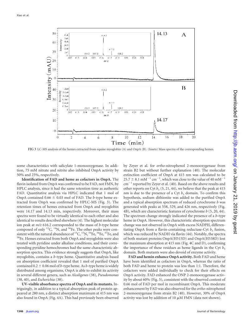
Identification of FAD and heme as cofactors in OnpA. Theflavin isolated from OnpA was confirmed to be FAD, not FMN, byHPLC analysis, since it had the same retention time as authenticFAD. Quantitative analysis via HPLC indicated that 1 mol ofOnpA contained 0.66 � 0.03 mol of FAD. The b-type heme ex-tracted from OnpA was confirmed by HPLC-MS (Fig. 3). Theretention times of hemes extracted from OnpA and myoglobinwere 14.17 and 14.13 min, respectively. Moreover, their massspectra were found to be virtually identical to each other and alsoidentical to results described elsewhere (4). The highest molecularion peak at m/z 616.2 corresponded to the mass of b-type hemecomposed of only 12C, 14N, and 56Fe. The other peaks were con-sistent with the natural abundance of 13C, 15N, 54Fe, 56Fe, 57Fe, and58Fe. Hemes extracted from both OnpA and myoglobin were alsotreated with pyridine under alkaline conditions, and their corre-sponding pyridine hemochromes had the same characteristic ab-sorption spectra. This evidence strongly suggests that OnpA, likemyoglobin, contains a b-type heme. Quantitative analysis basedon absorption coefficient revealed that 1 mol of purified OnpAcontained 0.2 � 0.06 mol of b-type heme. As b-type heme is widelydistributed among organisms, OnpA is able to exhibit its activityin several different genera, such as Alcaligenes (38), Pseudomonas(38, 40), and Escherichia (38).
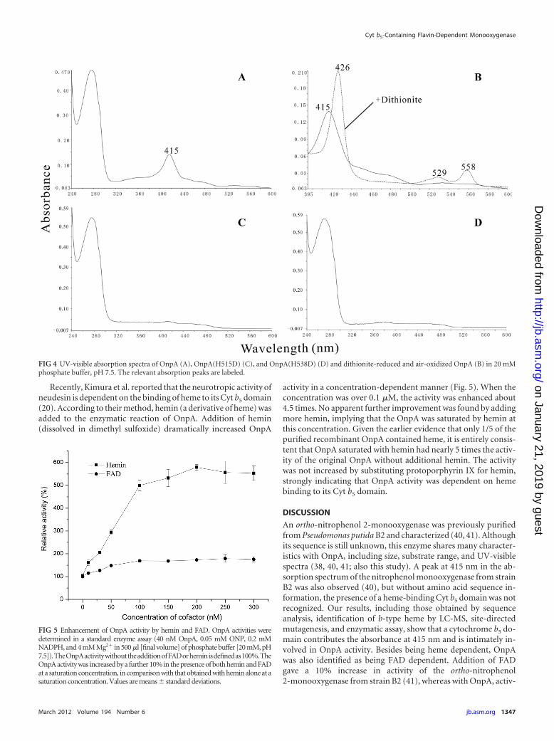
UV-visible absorbance spectra of OnpA and its mutants. In-triguingly, in addition to a typical absorption peak of protein ap-peared at 280 nm, a distinct absorption maximum at 415 nm wasalso found in OnpA (Fig. 4A). This had previously been observed
by Zeyer et al. for ortho-nitrophenol 2-monooxygenase fromstrain B2 but without further explanation (40). The molecularextinction coefficient of OnpA at 415 nm was calculated to be25.7 � 8.1 mM�1 cm�1, which was close to the value of 40 mM�1
cm�1 reported by Zeyer et al. (40). Based on the above results andother reports on Cyt b5 (5, 21, 44), we believe that the peak at 415nm is due to the presence of a Cyt b5 domain. To confirm thishypothesis, sodium dithionite was added to the purified OnpAand a typical absorption spectrum of reduced cytochrome b wasgenerated with peaks at 558, 529, and 426 nm, respectively (Fig.4B), which are characteristic features of cytochrome b (5, 20, 44).The spectrum change strongly indicated the presence of a b-typeheme in OnpA. However, this characteristic absorption spectrumchange was not observed in OnpA with excess NADPH, differen-tiating OnpA from a flavin-containing reductase-Cyt b5 fusion,which was reduced by NADH via flavin (44). Notably, the spectraof both mutant proteins OnpA(H515D) and OnpA(H538D) lostthe maximum absorption at 415 nm (Fig. 4C and D), confirmingthe importance of these residues as heme ligands in the Cyt b5
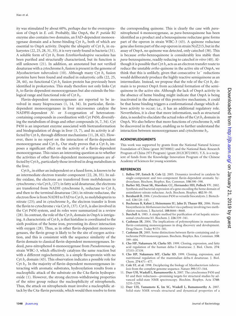
domain. Both mutants were also devoid of enzyme activity.FAD and hemin enhance OnpA activity. Both FAD and heme
have been identified as cofactors in OnpA, whereas the ratio ofboth FAD and heme to protein was less than 1:1. Therefore, thecofactors were added individually to check for their effects onOnpA activity. FAD enhanced the ONP 2-monooxygenase activ-ity by about 60% (Fig. 5), consistent with the observed content of0.66 mol of FAD per mol in recombinant OnpA. This moderateenhancement by FAD was also observed for the ortho-nitrophenol2-monooxygenase from strain B2 (40). However, 30% of OnpAactivity was lost by addition of 10 �M FMN (data not shown).
FIG 3 LC-MS analysis of the hemes extracted from equine myoglobin (A) and OnpA (B). (Insets) Mass spectra of the corresponding hemes.
Xiao et al.
1346 jb.asm.org Journal of Bacteriology
on January 21, 2019 by guesthttp://jb.asm
.org/D
ownloaded from
Recently, Kimura et al. reported that the neurotropic activity ofneudesin is dependent on the binding of heme to its Cyt b5 domain(20). According to their method, hemin (a derivative of heme) wasadded to the enzymatic reaction of OnpA. Addition of hemin(dissolved in dimethyl sulfoxide) dramatically increased OnpA
activity in a concentration-dependent manner (Fig. 5). When theconcentration was over 0.1 �M, the activity was enhanced about4.5 times. No apparent further improvement was found by addingmore hemin, implying that the OnpA was saturated by hemin atthis concentration. Given the earlier evidence that only 1/5 of thepurified recombinant OnpA contained heme, it is entirely consis-tent that OnpA saturated with hemin had nearly 5 times the activ-ity of the original OnpA without additional hemin. The activitywas not increased by substituting protoporphyrin IX for hemin,strongly indicating that OnpA activity was dependent on hemebinding to its Cyt b5 domain.
DISCUSSION
An ortho-nitrophenol 2-monooxygenase was previously purifiedfrom Pseudomonas putida B2 and characterized (40, 41). Althoughits sequence is still unknown, this enzyme shares many character-istics with OnpA, including size, substrate range, and UV-visiblespectra (38, 40, 41; also this study). A peak at 415 nm in the ab-sorption spectrum of the nitrophenol monooxygenase from strainB2 was also observed (40), but without amino acid sequence in-formation, the presence of a heme-binding Cyt b5 domain was notrecognized. Our results, including those obtained by sequenceanalysis, identification of b-type heme by LC-MS, site-directedmutagenesis, and enzymatic assay, show that a cytochrome b5 do-main contributes the absorbance at 415 nm and is intimately in-volved in OnpA activity. Besides being heme dependent, OnpAwas also identified as being FAD dependent. Addition of FADgave a 10% increase in activity of the ortho-nitrophenol2-monooxygenase from strain B2 (41), whereas with OnpA, activ-
FIG 4 UV-visible absorption spectra of OnpA (A), OnpA(H515D) (C), and OnpA(H538D) (D) and dithionite-reduced and air-oxidized OnpA (B) in 20 mMphosphate buffer, pH 7.5. The relevant absorption peaks are labeled.
FIG 5 Enhancement of OnpA activity by hemin and FAD. OnpA activities weredetermined in a standard enzyme assay (40 nM OnpA, 0.05 mM ONP, 0.2 mMNADPH, and 4 mM Mg2� in 500 �l [final volume] of phosphate buffer [20 mM, pH7.5]).TheOnpAactivitywithouttheadditionofFADorheminisdefinedas100%.TheOnpA activity was increased by a further 10% in the presence of both hemin and FADat a saturation concentration, in comparison with that obtained with hemin alone at asaturation concentration. Values are means � standard deviations.
Cyt b5-Containing Flavin-Dependent Monooxygenase
March 2012 Volume 194 Number 6 jb.asm.org 1347
on January 21, 2019 by guesthttp://jb.asm
.org/D
ownloaded from
ity was stimulated by about 60%, perhaps due to the overexpres-sion of OnpA in E. coli. Probably, like OnpA, the P. putida B2enzyme also contains two domains, an FAD-dependent monoox-ygenase domain and a heme-binding Cyt b5, both of which areessential to OnpA activity. Despite the ubiquity of Cyt b5 in eu-karyotes (22, 25, 28, 31, 35), it is very rarely found in bacteria (31).A soluble form of Cyt b5 from Ectothiorhodospira vacuolata hasbeen purified and structurally characterized, but its function isstill unknown (21). In addition, an annotated but not verifieddesaturase with a cytochrome b5 fusion is present in the genome ofMycobacterium tuberculosis (10). Although many Cyt b5 fusionproteins have been found and studied in eukaryotic cells (22, 25,28, 44), no functional Cyt b5 fusion protein has previously beenidentified in prokaryotes. This study therefore not only links Cytb5 to flavin-dependent monooxygenases but also extends the bio-logical range and functional role of Cyt b5.
Flavin-dependent monooxygenases are reported to be in-volved in many bioprocesses (1, 14, 34). In particular, flavin-dependent monooxygenases in liver microsomes catalyze theNADPH-dependent N- or S-oxygenation of heteroatom-containing compounds in coordination with Cyt P450, diversify-ing the metabolism of drugs and other compounds (6, 7, 34). CytP450 is an important enzyme associated with biotransformationand biodegradation of drugs in liver (3, 7), and its activity is af-fected by Cyt b5 through different mechanisms (11, 28, 42). How-ever, there is no report on the interaction of flavin-dependentmonooxygenase and Cyt b5. Our study proves that a Cyt b5 im-poses a significant effect on the activity of a flavin-dependentmonooxygenase. This raises an interesting question as to whetherthe activities of other flavin-dependent monooxygenases are af-fected by Cyt b5, particularly those involved in drug metabolism inthe liver.
Cyt b5, in either an independent or a fused form, is known to bean intermediate electron transfer component (22, 28, 35). In sul-fite oxidase, the electrons are transferred from molybdenum tocytochrome c via Cyt b5 (27); in fatty acid desaturase, the electronsare transferred from NADH cytochrome b5 reductase to Cyt b5
and then to the terminal desaturase (26); in nitrate reductase, theelectron flow is from NADH to FAD to Cyt b5 to molybdopterin tonitrate (23); and in cytochrome b2, the electron transfer is fromthe flavin to cytochrome c via Cyt b5 (37). Cyt b5 is also involved inthe Cyt P450 system, and its roles were summarized in a review(28). In contrast, the role of the Cyt b5 domain in OnpA is intrigu-ing. A characteristic of Cyt b5 is that histidine is coordinated to thesixth position of the heme iron, preventing its direct interactionwith oxygen (28). Thus, as in other flavin-dependent monooxy-genases, the flavin group is likely to be the site of oxygen activa-tion, and this is consistent with the sequence similarity of theflavin domain to classical flavin-dependent monooxygenases. In-deed, para-nitrophenol 4-monooxygenase from Pseudomonas sp.strain WBC-3, which effectively catalyzes the same reaction butwith a different regiochemistry, is a simple flavoprotein with noCyt b5 domain (43). This observation indicates a possible role forCyt b5. In the majority of flavin-dependent monooxygenases in-teracting with aromatic substrates, hydroxylation results from anucleophilic attack of the substrate on the C4a-flavin hydroper-oxide (1). However, the strong electron-withdrawing propertiesof the nitro group reduce the nucleophilicity of nitrophenols.Thus, the attack on nitrophenols must involve a nucleophilic at-tack by the C4a-flavin peroxide, which would then resolve to form
the corresponding quinone. This is clearly the case with para-nitrophenol 4-monooxygenase, as para-benzoquinone has beenidentified as a product and a benzoquinone reductase gene formspart of the operon in strain WBC-3 (43). A quinone reductasegene also forms part of the onp operon in strain NyZ215, but in theassay of OnpA, no quinone was detected, only catechol (38). Thisis because ortho-benzoquinone is considerably less stable thanpara-benzoquinone, readily reducing to catechol in vitro (40). Al-though it is possible that Cyt b5 acts as an electron transfer route toreduce the unstable ortho-quinone in the active site of OnpA, wethink that this is unlikely, given that consecutive 1e� reductionswould deliberately produce the highly reactive semiquinone as anintermediate. Instead, we propose that the role of the Cyt b5 do-main is to protect OnpA from accidental formation of the semi-quinone in the active site. Although the lack of OnpA activity inthe absence of heme binding might indicate that OnpA is beinginactivated in the absence of this protective mechanism, it couldbe that heme binding triggers a conformational change which al-lows activity to occur; i.e., it has an additional regulatory role.Nevertheless, it is clear that more information, such as structuraldata, is needed to elucidate the actual roles of the Cyt b5 domain inOnpA. We also believe that more functions of cytochrome b5 willbe discovered in the future, enabling us to further understand theinteraction between monooxygenases and cytochrome b5.
ACKNOWLEDGMENTS
This work was supported by grants from the National Natural ScienceFoundation of China (grant 30730002) and the National Basic ResearchProgram of China (973 Program) (grant 2012CB721003). Y.X. is a recip-ient of funds from the Knowledge Innovation Program of the ChineseAcademy of Sciences for young scientists.
REFERENCES1. Ballou DP, Entsch B, Cole LJ. 2005. Dynamics involved in catalysis by
single-component and two-component flavin-dependent aromatic hy-droxylases. Biochem. Biophys. Res. Commun. 338:590 –598.
2. Barber MJ, Desai SK, Marohnic CC, Hernandez HH, Pollock VV. 2002.Synthesis and bacterial expression of a gene encoding the heme domain ofassimilatory nitrate reductase. Arch. Biochem. Biophys. 402:38 –50.
3. Bernhardt R. 2006. Cytochromes P450 as versatile biocatalysts. J. Biotech-nol. 124:128 –145.
4. Buchenau B, Kahnt J, Heinemann IU, Jahn D, Thauer RK. 2006. Hemebiosynthesis in Methanosarcina barkeri via a pathway involving two meth-ylation reactions. J. Bacteriol. 188:8666 – 8668.
5. Burchell A. 1985. A simple method for purification of rat hepatic micro-somal cytochrome b5. Biochem. J. 226:339 –341.
6. Cashman JR. 2004. The implications of polymorphisms in mammalianflavin-containing monooxygenases in drug discovery and development.Drug Discov. Today 9:574 –581.
7. Cashman JR. 2005. Some distinctions between flavin-containing and cy-tochrome P450 monooxygenases. Biochem. Biophys. Res. Commun. 338:599 – 604.
8. Cho HP, Nakamura M, Clarke SD. 1999. Cloning, expression, and fattyacid regulation of the human delta-5 desaturase. J. Biol. Chem. 274:37335–37339.
9. Cho HP, Nakamura MT, Clarke SD. 1999. Cloning, expression, andnutritional regulation of the mammalian delta-6 desaturase. J. Biol.Chem. 274:471– 477.
10. Cole ST, et al. 1998. Deciphering the biology of Mycobacterium tubercu-losis from the complete genome sequence. Nature 393:537–544.
11. Durr UH, Waskell L, Ramamoorthy A. 2007. The cytochromes P450 andb5 and their reductases—promising targets for structural studies by ad-vanced solid-state NMR spectroscopy. Biochim. Biophys. Acta 1768:3235–3259.
12. Durr UH, Yamamoto K, Im SC, Waskell L, Ramamoorthy A. 2007.Solid-state NMR reveals structural and dynamical properties of a
Xiao et al.
1348 jb.asm.org Journal of Bacteriology
on January 21, 2019 by guesthttp://jb.asm
.org/D
ownloaded from
membrane-anchored electron-carrier protein, cytochrome b5. J. Am.Chem. Soc. 129:6670 – 6671.
13. Eppink MH, Boeren SA, Vervoort J, van Berkel WJ. 1997. Purificationand properties of 4-hydroxybenzoate 1-hydroxylase (decarboxylating), anovel flavin adenine dinucleotide-dependent monooxygenase from Can-dida parapsilosis CBS604. J. Bacteriol. 179:6680 – 6687.
14. Eppink MH, Schreuder HA, Van Berkel WJ. 1997. Identification of anovel conserved sequence motif in flavoprotein hydroxylases with a puta-tive dual function in FAD/NAD(P)H binding. Protein Sci. 6:2454 –2458.
15. Garrett RM, Bellissimo DB, Rajagopalan KV. 1995. Molecular cloning ofhuman liver sulfite oxidase. Biochim. Biophys. Acta 1262:147–149.
16. Graham SE, Peterson JA. 1999. How similar are P450s and what can theirdifferences teach us? Arch. Biochem. Biophys. 369:24 –29.
17. Hongsthong A, et al. 2006. Revealing the complementation of ferredoxinby cytochrome b 5 in the Spirulina-�6-desaturation reaction byN-terminal fusion and co-expression of the fungal-cytochrome b 5 do-main and Spirulina-�6-acyl-lipid desaturase. Appl. Microbiol. Biotech-nol. 72:1192–1201.
18. Hyde GE, Crawford NM, Campbell WH. 1991. The sequence of squashNADH:nitrate reductase and its relationship to the sequences of otherflavoprotein oxidoreductases. A family of flavoprotein pyridine nucleo-tide cytochrome reductases. J. Biol. Chem. 266:23542–23547.
19. Kay CJ, Lippay EW. 1992. Mutation of the heme-binding crevice offlavocytochrome b2 from Saccharomyces cerevisiae: altered heme potentialand absence of redox cooperativity between heme and FMN centers. Bio-chemistry 31:11376 –11382.
20. Kimura I, et al. 2008. Neurotrophic activity of neudesin, a novel extra-cellular heme-binding protein, is dependent on the binding of heme to itscytochrome b5-like heme/steroid-binding domain. J. Biol. Chem. 283:4323– 4331.
21. Kostanjevecki V, et al. 1999. Structure and characterization of Ectothi-orhodospira vacuolata cytochrome b558, a prokaryotic homologue of cyto-chrome b5. J. Biol. Chem. 274:35614 –35620.
22. Lederer F. 1994. The cytochrome b5-fold: an adaptable module.Biochimie 76:674 – 692.
23. Lu G, Lindqvist Y, Schneider G, Dwivedi U, Campbell W. 1995. Struc-tural studies on corn nitrate reductase: refined structure of the cyto-chrome b reductase fragment at 2.5 A, its ADP complex and an active-sitemutant and modeling of the cytochrome b domain. J. Mol. Biol. 248:931–948.
24. Lubben M, Morand K. 1994. Novel prenylated hemes as cofactors ofcytochrome oxidases. Archaea have modified hemes A and O. J. Biol.Chem. 269:21473–21479.
25. Napier JA, Sayanova O, Stobart AK, Shewry PR. 1997. A new class ofcytochrome b5 fusion proteins. Biochem. J. 328(Pt. 2):717–718.
26. Rioux V, Pedrono F, Legrand P. 2011. Regulation of mammalian desatu-rases by myristic acid: N-terminal myristoylation and other modulations.Biochim. Biophys. Acta 1811:1– 8.
27. Rudolph MJ, Johnson JL, Rajagopalan KV, Kisker C. 2003. The 1.2 Åstructure of the human sulfite oxidase cytochrome b5 domain. Acta Crys-tallogr. D Biol. Crystallogr. 59:1183–1191.
28. Schenkman JB, Jansson I. 2003. The many roles of cytochrome b5. Phar-macol. Ther. 97:139 –152.
29. Seibold B, et al. 1996. 4-Hydroxybenzoate hydroxylase from Pseudomo-nas sp. CBS3. Purification, characterization, gene cloning, sequence anal-ysis and assignment of structural features determining the coenzyme spec-ificity. Eur. J. Biochem. 239:469 – 478.
30. Somerville CC, Nishino SF, Spain JC. 1995. Purification and character-ization of nitrobenzene nitroreductase from Pseudomonas pseudoalcali-genes JS45. J. Bacteriol. 177:3837–3842.
31. Sperling P, Ternes P, Zank TK, Heinz E. 2003. The evolution of desatu-rases. Prostaglandins Leukot. Essent. Fatty Acids 68:73–95.
32. Tamura K, et al. 2011. MEGA5: molecular evolutionary genetics analysisusing maximum likelihood, evolutionary distance, and maximum parsi-mony methods. Mol. Biol. Evol. 28:2731–2739.
33. Torres Pazmino DE, Winkler M, Glieder A, Fraaije MW. 2010. Mono-oxygenases as biocatalysts: classification, mechanistic aspects and biotech-nological applications. J. Biotechnol. 146:9 –24.
34. van Berkel WJ, Kamerbeek NM, Fraaije MW. 2006. Flavoprotein mono-oxygenases, a diverse class of oxidative biocatalysts. J. Biotechnol. 124:670 – 689.
35. Vergeres G, Waskell L. 1995. Cytochrome b5, its functions, structure andmembrane topology. Biochimie 77:604 – 620.
36. White-Stevens RH, Kamin H. 1972. Studies of a flavoprotein, salicylatehydroxylase. I. Preparation, properties, and the uncoupling of oxygenreduction from hydroxylation. J. Biol. Chem. 247:2358 –2370.
37. Xia ZX, et al. 1987. Three-dimensional structure of flavocytochrome b2from baker’s yeast at 3.0-A resolution. Proc. Natl. Acad. Sci. U. S. A.84:2629 –2633.
38. Xiao Y, Zhang JJ, Liu H, Zhou NY. 2007. Molecular characterization ofa novel ortho-nitrophenol catabolic gene cluster in Alcaligenes sp. strainNyZ215. J. Bacteriol. 189:6587– 6593.
39. Yamamoto S, Katagiri M, Maeno H, Hayaishi O. 1965. Salicylate hy-droxylase, a monooxygenase requiring flavin adenine dinucleotide. I. Pu-rification and general properties. J. Biol. Chem. 240:3408 –3413.
40. Zeyer J, Kocher HP. 1988. Purification and characterization of a bacterialnitrophenol oxygenase which converts ortho-nitrophenol to catechol andnitrite. J. Bacteriol. 170:1789 –1794.
41. Zeyer J, Kocher HP, Timmis KN. 1986. Influence of para-substituents onthe oxidative metabolism of o-nitrophenols by Pseudomonas putida B2.Appl. Environ. Microbiol. 52:334 –339.
42. Zhang H, Myshkin E, Waskell L. 2005. Role of cytochrome b5 in catalysisby cytochrome P450 2B4. Biochem. Biophys. Res. Commun. 338:499 –506.
43. Zhang JJ, Liu H, Xiao Y, Zhang XE, Zhou NY. 2009. Identification andcharacterization of catabolic para-nitrophenol 4-monooxygenase andpara-benzoquinone reductase from Pseudomonas sp. strain WBC-3. J.Bacteriol. 191:2703–2710.
44. Zhu H, Qiu H, Yoon HW, Huang S, Bunn HF. 1999. Identification of acytochrome b-type NAD(P)H oxidoreductase ubiquitously expressed inhuman cells. Proc. Natl. Acad. Sci. U. S. A. 96:14742–14747.
Cyt b5-Containing Flavin-Dependent Monooxygenase
March 2012 Volume 194 Number 6 jb.asm.org 1349
on January 21, 2019 by guesthttp://jb.asm
.org/D
ownloaded from


![by Joseph Bruchac illustrated by Teresa Flavin · illustrated by Teresa Flavin by Joseph Bruchac illustrated by Teresa Flavin Pushing Up)&(the Sky FjZhi^dc d[ i]Z LZZ` =dl Yd eZdeaZ](https://img.pdfslide.us/doc/110x75/5f8e7f4dad596368cb63a9b9/by-joseph-bruchac-illustrated-by-teresa-flavin-illustrated-by-teresa-flavin-by-joseph.jpg)