Embed Size (px)
Citation preview
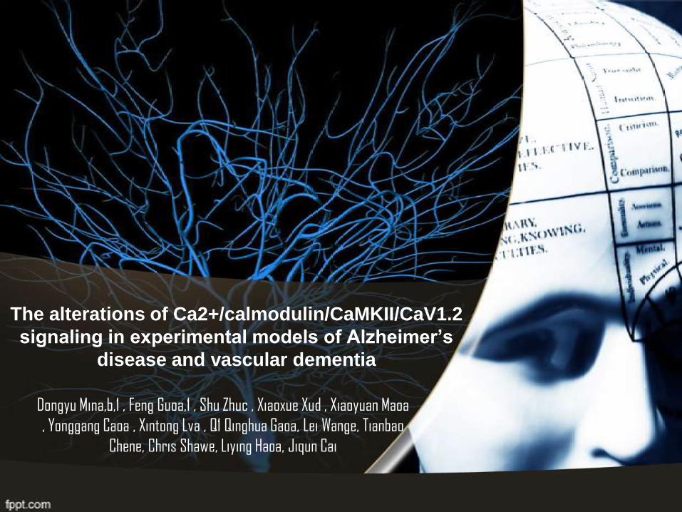
The alterations of Ca2+/calmodulin/CaMKII/CaV1.2
signaling in experimental models of Alzheimer’s
disease and vascular dementia
Dongyu Mina,b,1 , Feng Guoa,1 , Shu Zhuc , Xiaoxue Xud , Xiaoyuan Maoa
, Yonggang Caoa , Xintong Lva , Q1 Qinghua Gaoa, Lei Wange, Tianbao
Chene, Chris Shawe, Liying Haoa, Jiqun Cai
Molecular Biology
III Semester
2013
Manuela Jiménez Obando
Juan Sebastián Marín Cárdenas
INTRODUCTION
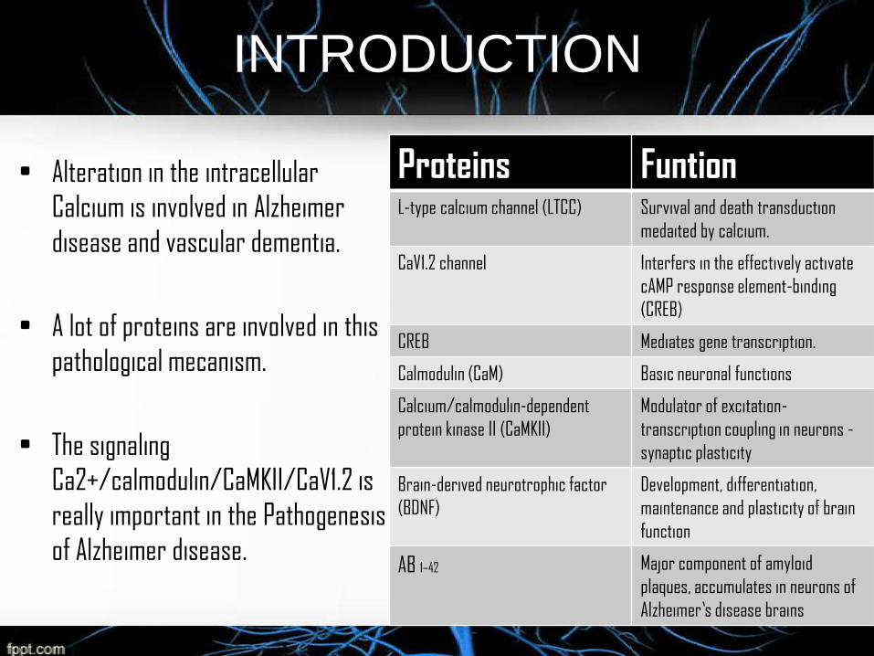
• Alteration in the intracellular
Calcium is involved in Alzheimer
disease and vascular dementia.
• A lot of proteins are involved in this
pathological mecanism.
• The signaling
Ca2+/calmodulin/CaMKII/CaV1.2 is
really important in the Pathogenesis
of Alzheimer disease.
Proteins FuntionL-type calcium channel (LTCC) Survival and death transduction
medaited by calcium.
CaV1.2 channel Interfers in the effectively activate
cAMP response element-binding
(CREB)
CREB Mediates gene transcription.
Calmodulin (CaM) Basic neuronal functions
Calcium/calmodulin-dependent
protein kinase II (CaMKII)
Modulator of excitation-
transcription coupling in neurons -
synaptic plasticity
Brain-derived neurotrophic factor
(BDNF)
Development, differentiation,
maintenance and plasticity of brain
function
AB 1–42 Major component of amyloid
plaques, accumulates in neurons of
Alzheimer’s disease brains
INTRODUCTION
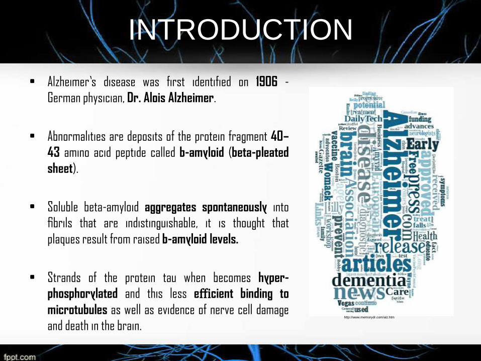
• Alzheimer’s disease was first identified on 1906 -
German physician, Dr. Alois Alzheimer.
• Abnormalities are deposits of the protein fragment 40–
43 amino acid peptide called b-amyloid (beta-pleated
sheet).
• Soluble beta-amyloid aggregates spontaneously into
fibrils that are indistinguishable, it is thought that
plaques result from raised b-amyloid levels.
• Strands of the protein tau when becomes hyper-
phosphorylated and this less efficient binding to
microtubules as well as evidence of nerve cell damage
and death in the brain.http://www.memorydr.com/alz.htm
INTRODUCTION
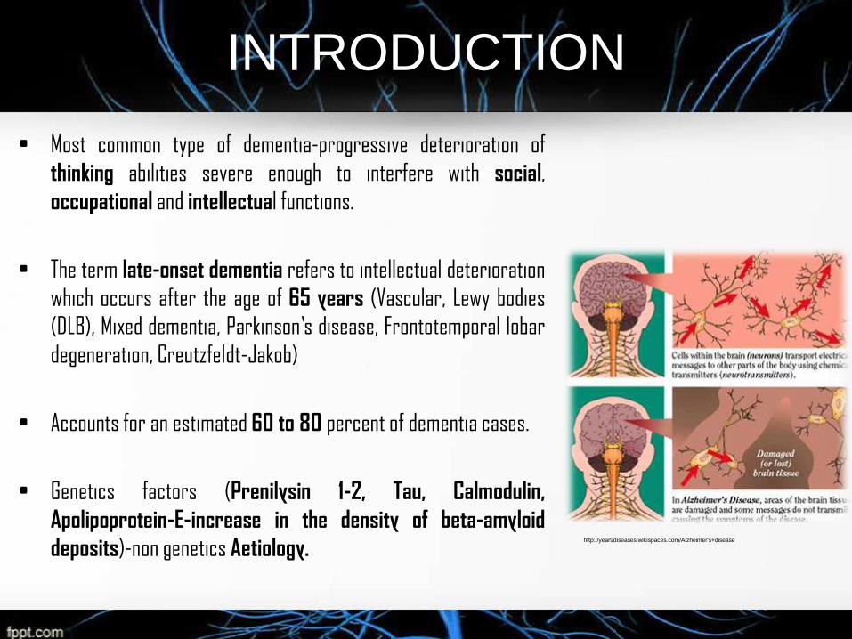
• Most common type of dementia-progressive deterioration of
thinking abilities severe enough to interfere with social,
occupational and intellectual functions.
• The term late-onset dementia refers to intellectual deterioration
which occurs after the age of 65 years (Vascular, Lewy bodies
(DLB), Mixed dementia, Parkinson’s disease, Frontotemporal lobar
degeneration, Creutzfeldt-Jakob)
• Accounts for an estimated 60 to 80 percent of dementia cases.
• Genetics factors (Prenilysin 1-2, Tau, Calmodulin,
Apolipoprotein-E-increase in the density of beta-amyloid
deposits)-non genetics Aetiology.http://year9diseases.wikispaces.com/Alzheimer's+disease
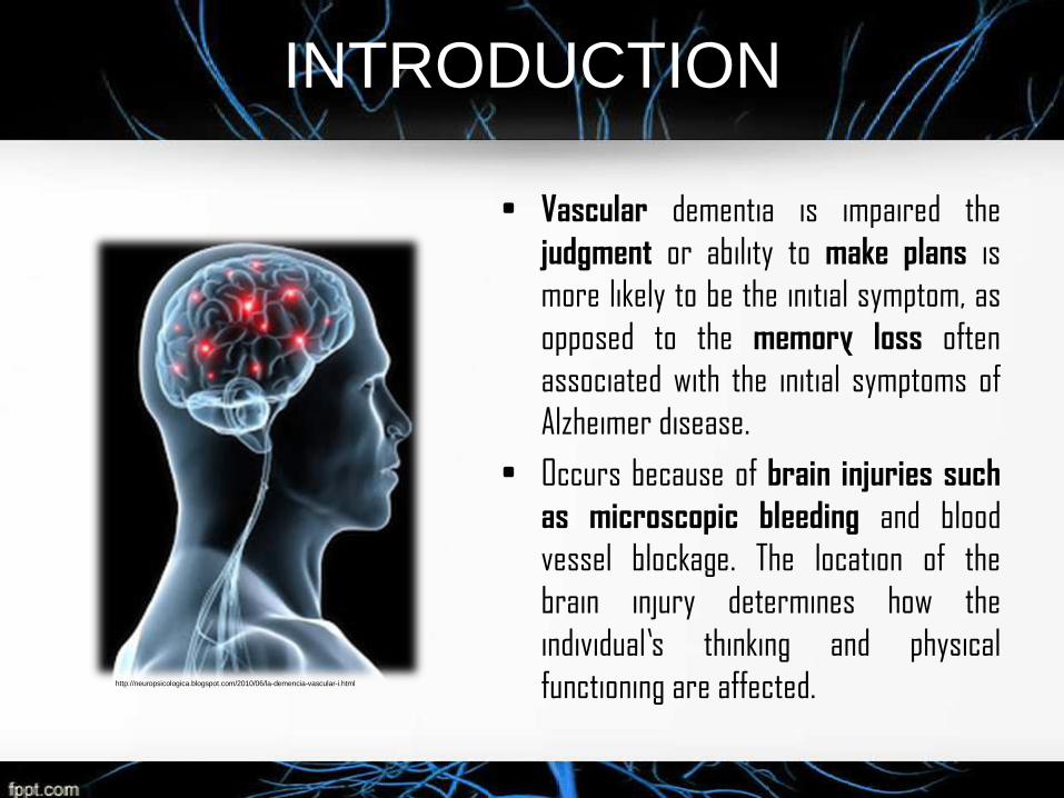
INTRODUCTION
• Vascular dementia is impaired the
judgment or ability to make plans is
more likely to be the initial symptom, as
opposed to the memory loss often
associated with the initial symptoms of
Alzheimer disease.
• Occurs because of brain injuries such
as microscopic bleeding and blood
vessel blockage. The location of the
brain injury determines how the
individual’s thinking and physical
functioning are affected.http://neuropsicologica.blogspot.com/2010/06/la-demencia-vascular-i.html
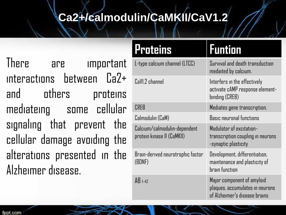
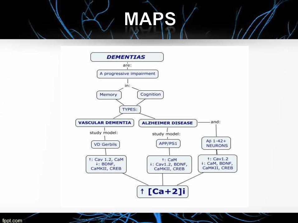
Ca2+/calmodulin/CaMKII/CaV1.2
There are important
interactions between Ca2+
and others proteins
mediateing some cellular
signaling that prevent the
cellular damage avoiding the
alterations presented in the
Alzheimer disease.
Proteins FuntionL-type calcium channel (LTCC) Survival and death transduction
medaited by calcium.
CaV1.2 channel Interfers in the effectively
activate cAMP response element-
binding (CREB)
CREB Mediates gene transcription.
Calmodulin (CaM) Basic neuronal functions
Calcium/calmodulin-dependent
protein kinase II (CaMKII)
Modulator of excitation-
transcription coupling in neurons
-synaptic plasticity
Brain-derived neurotrophic factor
(BDNF)
Development, differentiation,
maintenance and plasticity of
brain function
AB 1–42 Major component of amyloid
plaques, accumulates in neurons
of Alzheimer’s disease brains
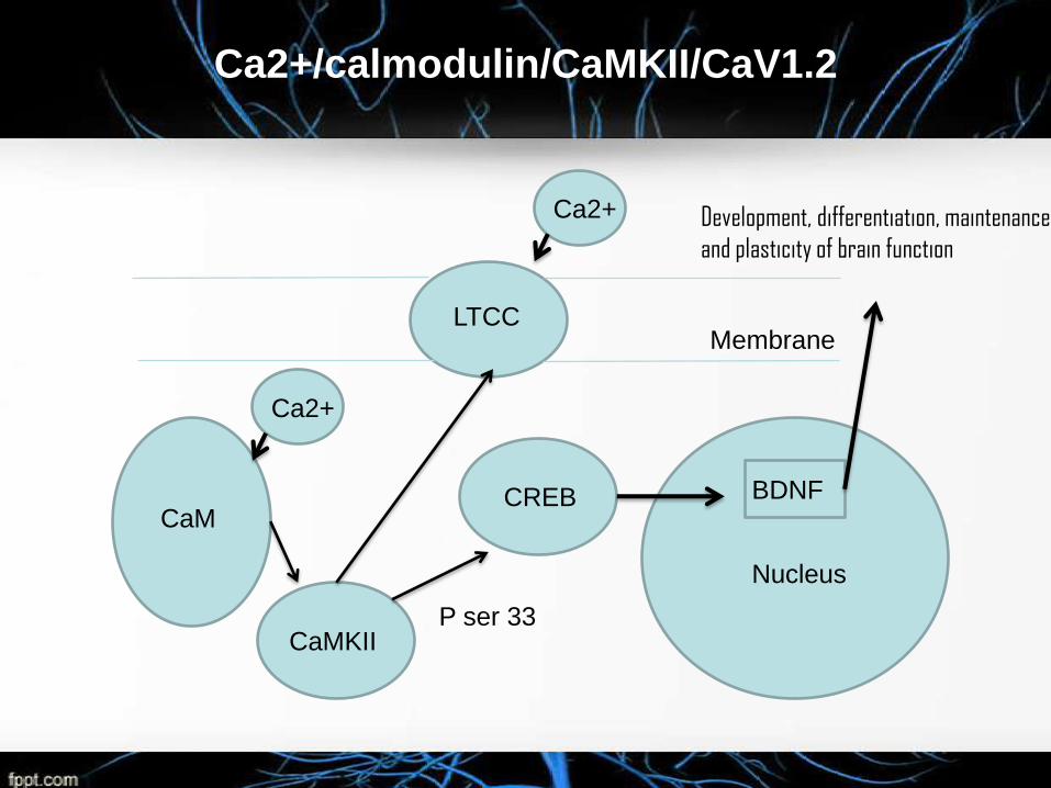
Ca2+/calmodulin/CaMKII/CaV1.2
LTCC
Ca2+
CREBCaM
Ca2+
CaMKII
Nucleus
BDNF
Membrane
Development, differentiation, maintenance
and plasticity of brain function
P ser 33
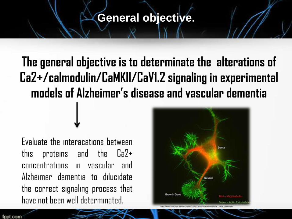
General objective.
The general objective is to determinate the alterations of
Ca2+/calmodulin/CaMKII/CaV1.2 signaling in experimental
models of Alzheimer’s disease and vascular dementia
Evaluate the interacations between
this proteins and the Ca2+
concentrations in vascular and
Alzheimer dementia to dilucidate
the correct signaling process that
have not been well determinated.http://www.elmundo.es/elmundosalud/2008/02/06/neurociencia/1202303660.html
Materiales y metodos.
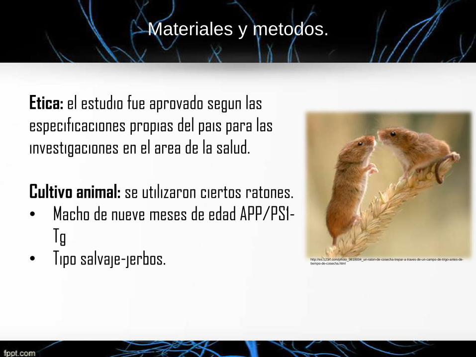
Etica: el estudio fue aprovado segun las
especificaciones propias del pais para las
investigaciones en el area de la salud.
Cultivo animal: se utilizaron ciertos ratones.
• Macho de nueve meses de edad APP/PS1-
Tg
• Tipo salvaje-jerbos. http://es.123rf.com/photo_9818004_un-raton-de-cosecha-trepar-a-traves-de-un-campo-de-trigo-antes-de-
tiempo-de-cosecha.html
Materiales y métodos.
Induccion de isquemia global: los ratones se anestesiaron
anteriormente, luego se indujo la isquemia utilizando pinzas arteriales, a
los 10 minutos, se retiraron las pinzas para reestablecer el flujo. El
raton de caracter control se hizo lo mismo pero son compresión
carotidea.
• Con esto se pretendio inducir la isquemia cerebral y así
evaluar los parámentros de la investigación con el modelo de
demencia vascular.
Materiales y métodos.
http://sosbiologiacelularytisular.blogspot.com/2010/09/biologia-celular-cuerpos-de-nissl.html
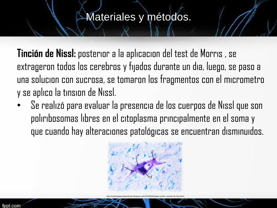
Tinción de Nissl: posterior a la aplicacion del test de Morris , se
extrageron todos los cerebros y fijados durante un dia, luego, se paso a
una solucion con sucrosa, se tomaron los fragmentos con el micrometro
y se aplico la tinsion de Nissl.
• Se realizó para evaluar la presencia de los cuerpos de Nissl que son
poliribosomas libres en el citoplasma principalmente en el soma y
que cuando hay alteraciones patológicas se encuentran disminuidos.
Materiales y métodos.
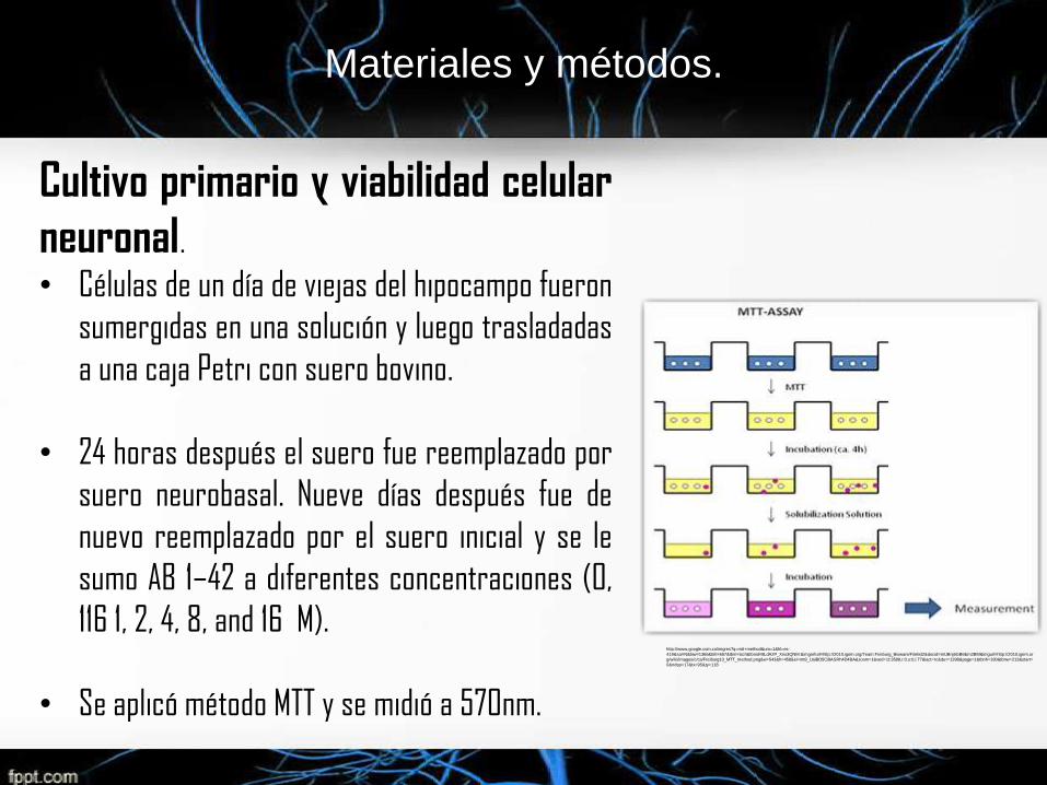
Cultivo primario y viabilidad celular
neuronal.• Células de un día de viejas del hipocampo fueron
sumergidas en una solución y luego trasladadas
a una caja Petri con suero bovino.
• 24 horas después el suero fue reemplazado por
suero neurobasal. Nueve días después fue de
nuevo reemplazado por el suero inicial y se le
sumo AB 1–42 a diferentes concentraciones (0,
116 1, 2, 4, 8, and 16 M).
• Se aplicó método MTT y se midió a 570nm.
http://www.google.com.co/imgres?q=mtt+method&um=1&hl=es-
419&sa=N&biw=1366&bih=667&tbm=isch&tbnid=8LdA2P_Xno3QNM:&imgrefurl=http://2010.igem.org/Team:Freiburg_Bioware/Filelist2&docid=mUBnj6GB6Em2BM&imgurl=http://2010.igem.or
g/wiki/images/c/ca/Freiburg10_MTT_method.png&w=543&h=458&ei=mt8_UaiBD5Ci8ASRh4D4BA&zoom=1&ved=1t:3588,r:0,s:0,i:77&iact=rc&dur=1398&page=1&tbnh=180&tbnw=213&start=
0&ndsp=17&tx=95&ty=118
Materiales y métodos.
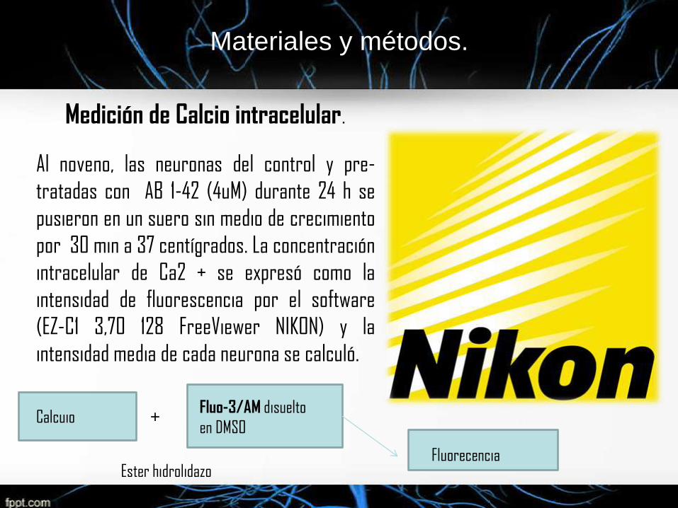
Medición de Calcio intracelular.
Al noveno, las neuronas del control y pre-
tratadas con AB 1-42 (4uM) durante 24 h se
pusieron en un suero sin medio de crecimiento
por 30 min a 37 centígrados. La concentración
intracelular de Ca2 + se expresó como la
intensidad de fluorescencia por el software
(EZ-C1 3,70 128 FreeViewer NIKON) y la
intensidad media de cada neurona se calculó.
+CalcuioFluo-3/AM disuelto
en DMSO
Ester hidrolidazoFluorecencia
Materiales y métodos.
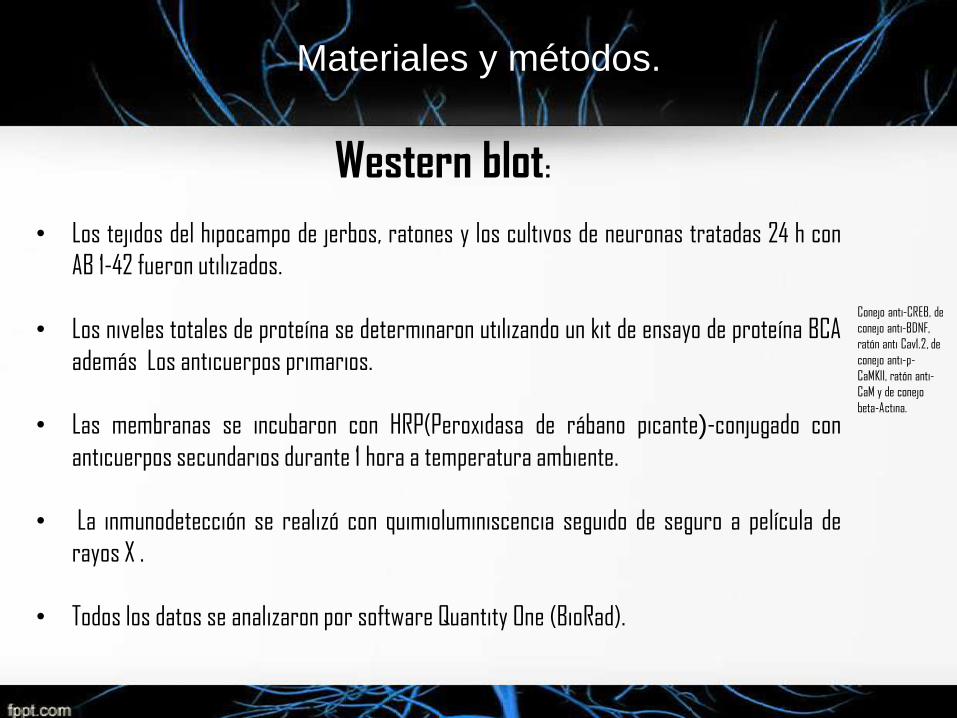
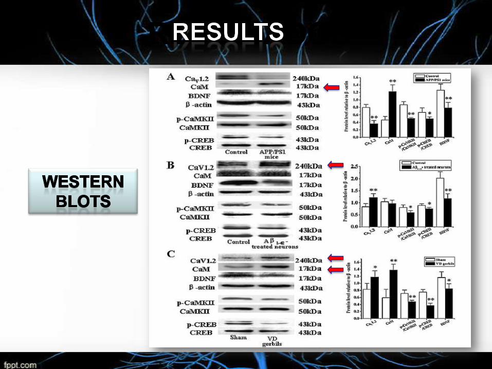
Western blot:
• Los tejidos del hipocampo de jerbos, ratones y los cultivos de neuronas tratadas 24 h con
AB 1-42 fueron utilizados.
• Los niveles totales de proteína se determinaron utilizando un kit de ensayo de proteína BCA
además Los anticuerpos primarios.
• Las membranas se incubaron con HRP(Peroxidasa de rábano picante)-conjugado con
anticuerpos secundarios durante 1 hora a temperatura ambiente.
• La inmunodetección se realizó con quimioluminiscencia seguido de seguro a película de
rayos X .
• Todos los datos se analizaron por software Quantity One (BioRad).
Conejo anti-CREB, de
conejo anti-BDNF,
ratón anti Cav1.2, de
conejo anti-p-
CaMKII, ratón anti-
CaM y de conejo
beta-Actina.
Materiales y métodos.
Materiales y métodos.
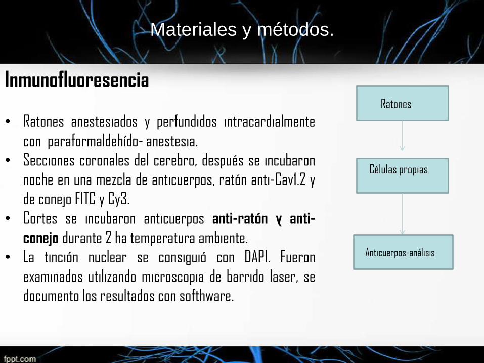
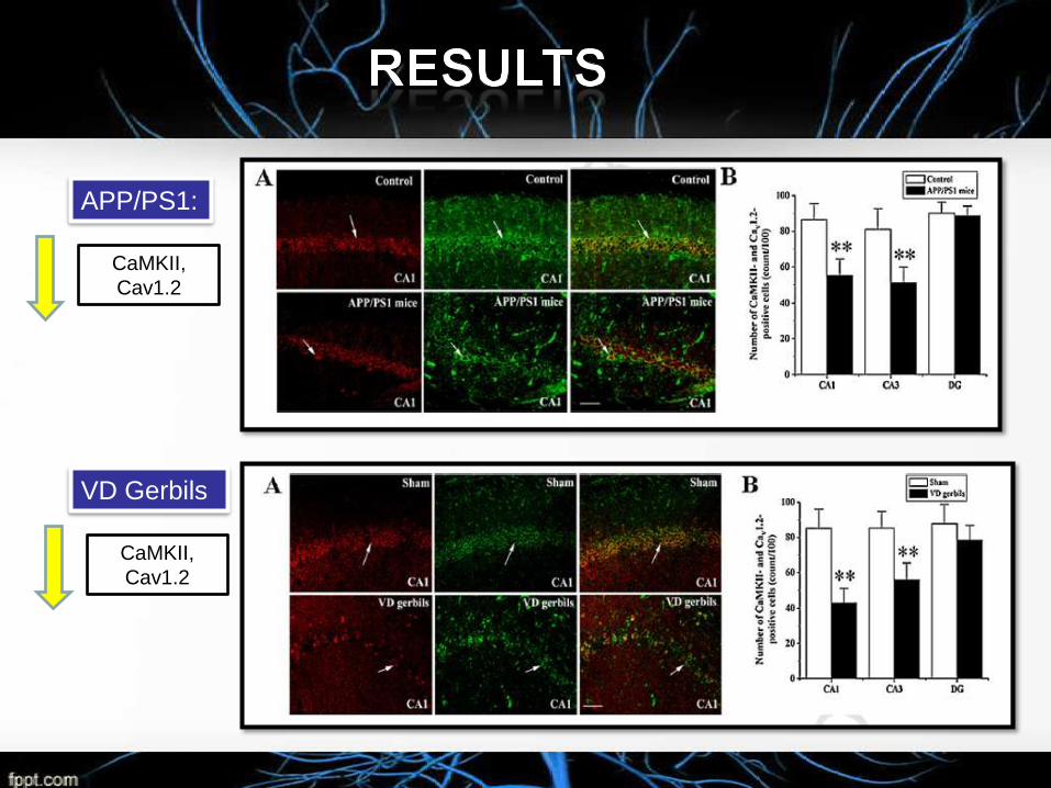
Inmunofluoresencia
• Ratones anestesiados y perfundidos intracardialmente
con paraformaldehído- anestesia.
• Secciones coronales del cerebro, después se incubaron
noche en una mezcla de anticuerpos, ratón anti-Cav1.2 y
de conejo FITC y Cy3.
• Cortes se incubaron anticuerpos anti-ratón y anti-
conejo durante 2 ha temperatura ambiente.
• La tinción nuclear se consiguió con DAPI. Fueron
examinados utilizando microscopia de barrido laser, se
documento los resultados con softhware.
Ratones
Células propias
Anticuerpos-análisis
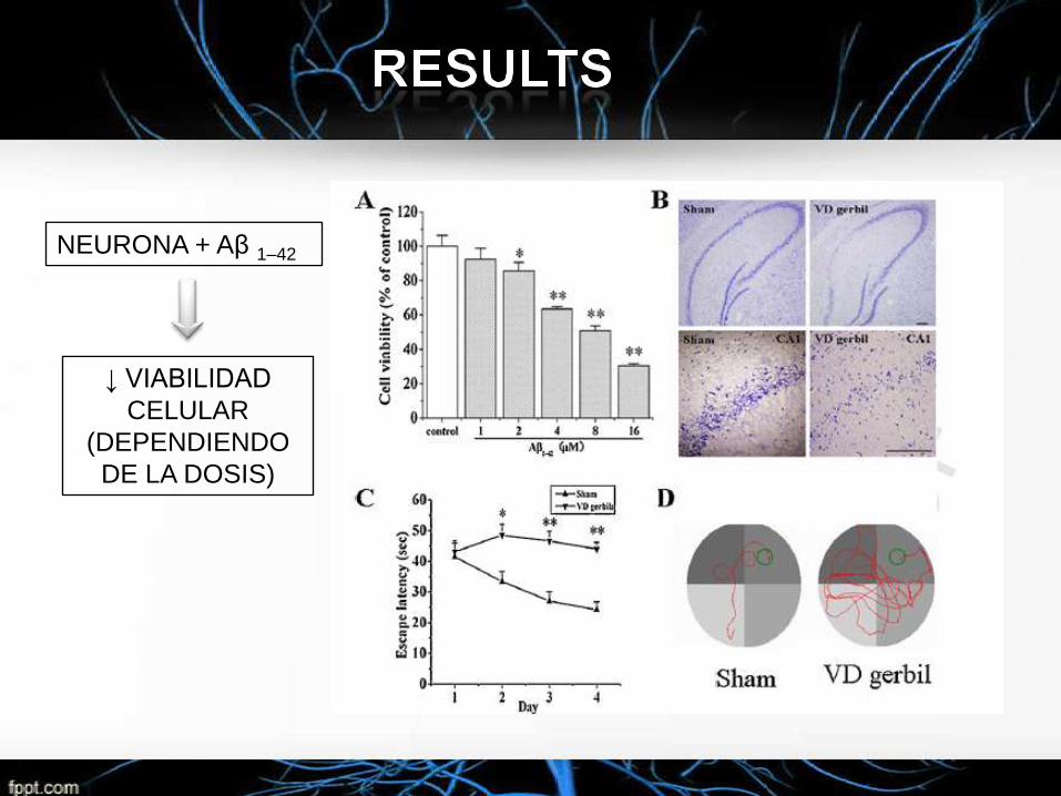
NEURONA + Aβ 1–42
↓ VIABILIDAD
CELULAR
(DEPENDIENDO
DE LA DOSIS)
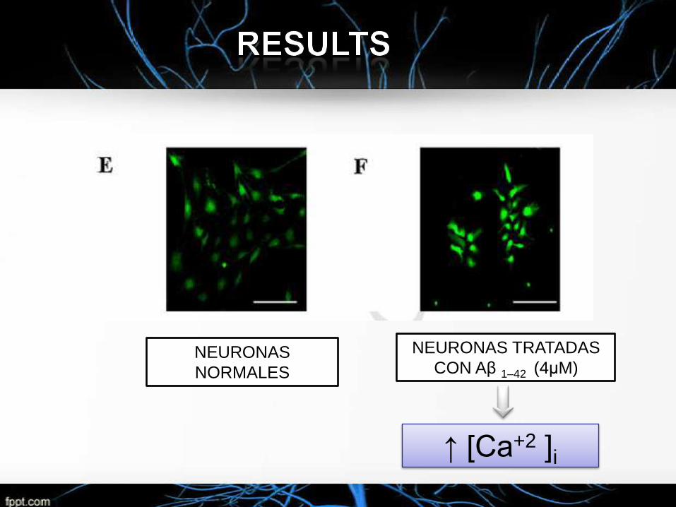
NEURONAS
NORMALES
NEURONAS TRATADAS
CON Aβ 1–42 (4μM)
↑ [Ca+2 ]i
CaMKII,
Cav1.2
APP/PS1:
VD Gerbils
CaMKII,
Cav1.2
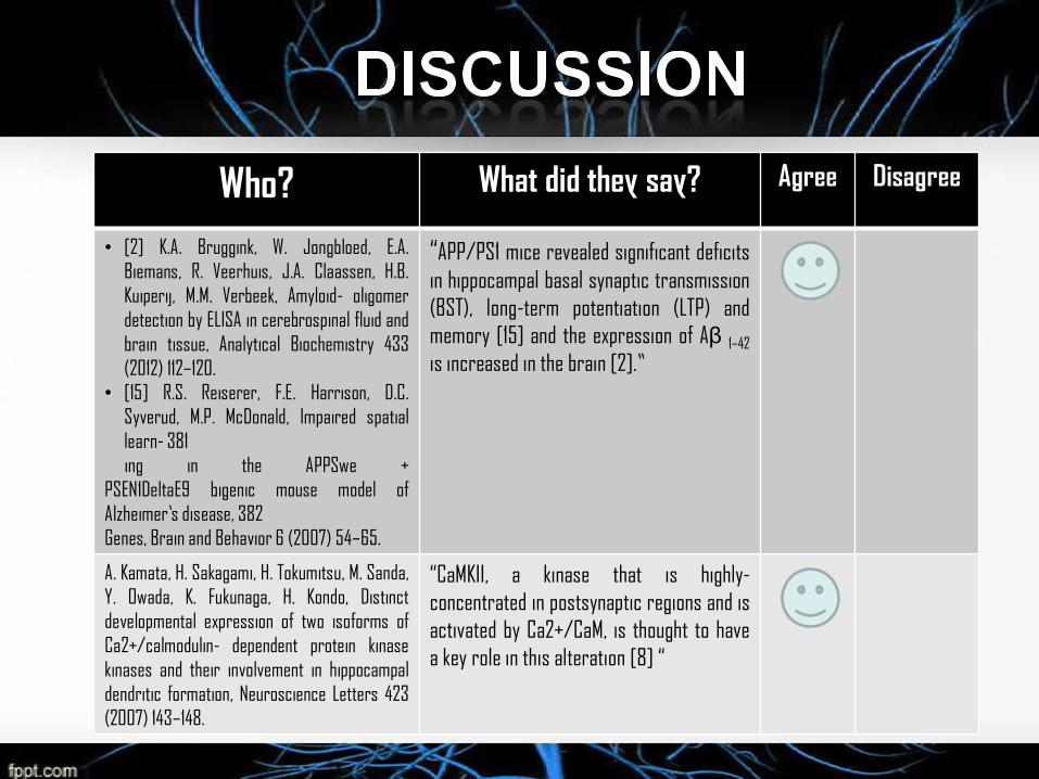
Who? What did they say? Agree Disagree
• [2] K.A. Bruggink, W. Jongbloed, E.A.
Biemans, R. Veerhuis, J.A. Claassen, H.B.
Kuiperij, M.M. Verbeek, Amyloid- oligomer
detection by ELISA in cerebrospinal fluid and
brain tissue, Analytical Biochemistry 433
(2012) 112–120.
• [15] R.S. Reiserer, F.E. Harrison, D.C.
Syverud, M.P. McDonald, Impaired spatial
learn- 381
ing in the APPSwe +
PSEN1DeltaE9 bigenic mouse model of
Alzheimer’s disease, 382
Genes, Brain and Behavior 6 (2007) 54–65.
“APP/PS1 mice revealed significant deficits
in hippocampal basal synaptic transmission
(BST), long-term potentiation (LTP) and
memory [15] and the expression of Aβ 1–42
is increased in the brain [2].”
A. Kamata, H. Sakagami, H. Tokumitsu, M. Sanda,
Y. Owada, K. Fukunaga, H. Kondo, Distinct
developmental expression of two isoforms of
Ca2+/calmodulin- dependent protein kinase
kinases and their involvement in hippocampal
dendritic formation, Neuroscience Letters 423
(2007) 143–148.
“CaMKII, a kinase that is highly-
concentrated in postsynaptic regions and is
activated by Ca2+/CaM, is thought to have
a key role in this alteration [8] “
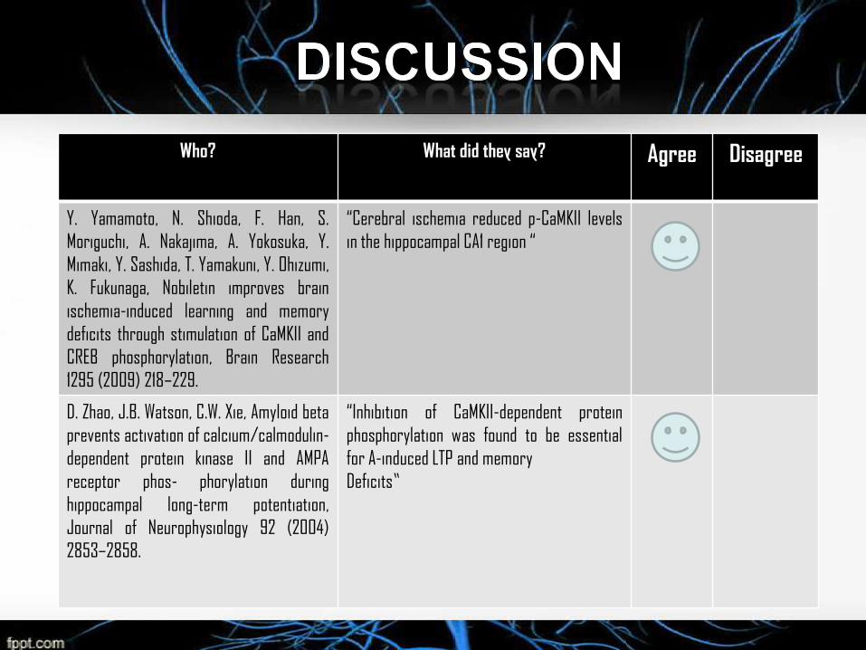
Who? What did they say? Agree Disagree
Y. Yamamoto, N. Shioda, F. Han, S.
Moriguchi, A. Nakajima, A. Yokosuka, Y.
Mimaki, Y. Sashida, T. Yamakuni, Y. Ohizumi,
K. Fukunaga, Nobiletin improves brain
ischemia-induced learning and memory
deficits through stimulation of CaMKII and
CREB phosphorylation, Brain Research
1295 (2009) 218–229.
“Cerebral ischemia reduced p-CaMKII levels
in the hippocampal CA1 region “
D. Zhao, J.B. Watson, C.W. Xie, Amyloid beta
prevents activation of calcium/calmodulin-
dependent protein kinase II and AMPA
receptor phos- phorylation during
hippocampal long-term potentiation,
Journal of Neurophysiology 92 (2004)
2853–2858.
“Inhibition of CaMKII-dependent protein
phosphorylation was found to be essential
for A-induced LTP and memory
Deficits”
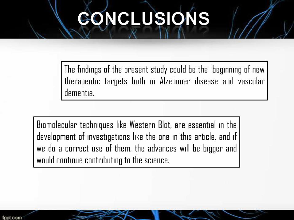
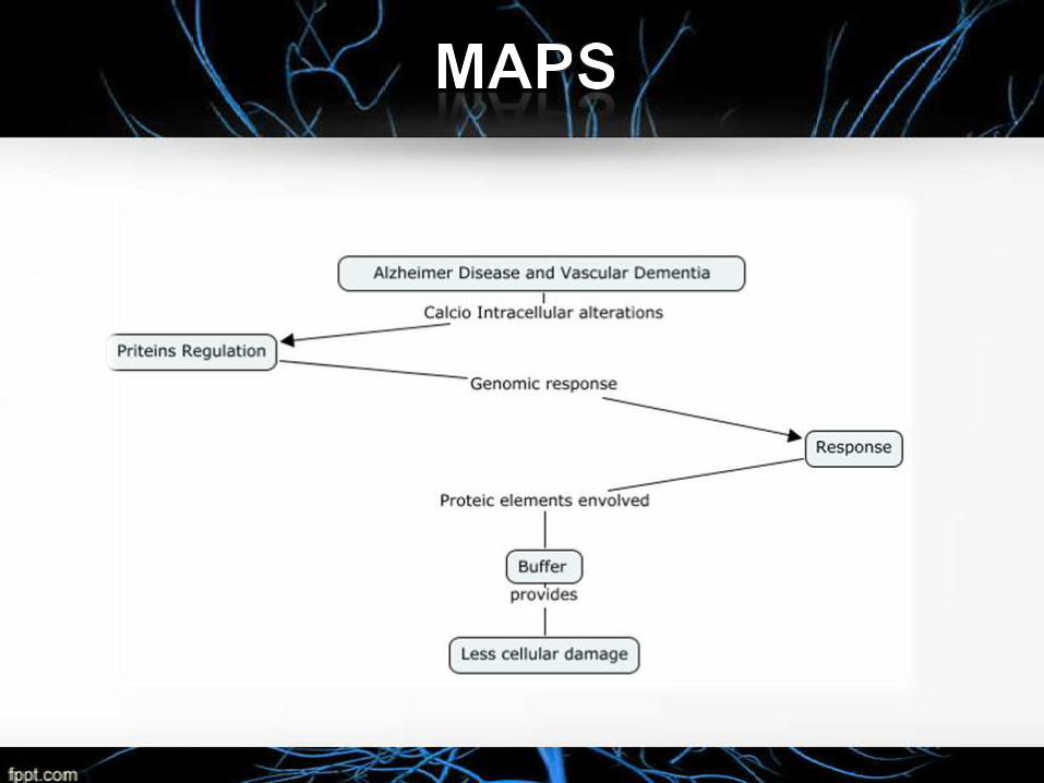
The findings of the present study could be the beginning of new
therapeutic targets both in Alzehimer disease and vascular
dementia.
Biomolecular techniques like Western Blot, are essential in the
development of investigations like the one in this article, and if
we do a correct use of them, the advances will be bigger and
would continue contributing to the science.
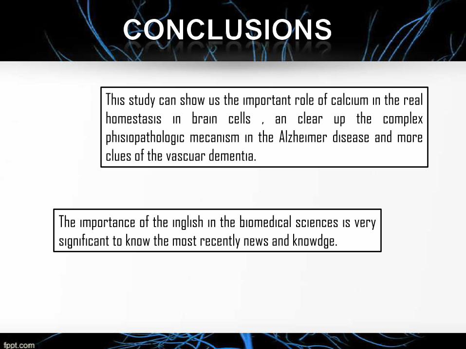
This study can show us the important role of calcium in the real
homestasis in brain cells , an clear up the complex
phisiopathologic mecanism in the Alzheimer disease and more
clues of the vascuar dementia.
The importance of the inglish in the biomedical sciences is very
significant to know the most recently news and knowdge.
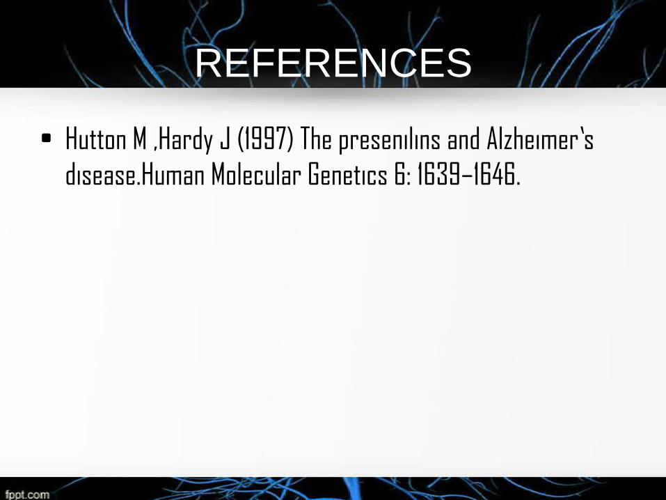
REFERENCES
• Hutton M ,Hardy J (1997) The presenilins and Alzheimer’s
disease.Human Molecular Genetics 6: 1639–1646.


![Seminario molecular[1] listo](https://img.pdfslide.us/doc/110x75/5594b2091a28ab16648b46d8/seminario-molecular1-listo.jpg)