Embed Size (px)
Citation preview
CONEUR-527; NO OF PAGES 8
Self-localization and the entorhinal–hippocampal systemKathryn J Jeffery
Self-localization requires that information from several sensory
modalities and knowledge domains be integrated in order to
identify an environment and determine current location and
heading. This integration occurs by the convergence of highly
processed sensory information onto neural systems in
entorhinal cortex and hippocampus. Entorhinal neurons
combine angular and linear self-motion information to generate
an oriented metric signal that is then ‘attached’ to each
environment using information about landmarks and context.
Neurons in hippocampus use this signal to determine the
animal’s unique position within a particular environment.
Elucidating this process illuminates not only spatial processing
but also, more generally, how the brain builds knowledge
representations from inputs carrying heterogeneous sensory
and semantic content.
Addresses
Institute of Behavioural Neuroscience, Division of Psychology and
Language Science, University College London, 26 Bedford Way,
London WC1H OAP, UK
Corresponding author: Jeffery, Kathryn J ([email protected])
Current Opinion in Neurobiology 2008, 17:1–8
This review comes from a themed issue on
Neurobiology of behaviour
Edited by Edvard Moser and Barry Dickson
0959-4388/$ – see front matter
# 2007 Elsevier Ltd. All rights reserved.
DOI 10.1016/j.conb.2007.11.008
Navigation is a complex cognitive faculty involving many
interacting processes and using information from, to a
greater or lesser extent, all sensory modalities. It requires
an organism to determine its position and orientation
(self-localization), its desired position (goal determi-
nation), and finally, how to get from one place to the
other (route planning).
The problem of self-localization is currently under scru-
tiny because of recent findings concerning the involve-
ment of the hippocampus and surrounding structures.
This article reviews evidence that spatial representation
in the hippocampus arises from convergence of highly
elaborated but disparate inputs onto common neural
matrices, in particular, the entorhinal grid cell system
and the hippocampal place cell system, which then per-
form structure-specific transformations on the infor-
mation. Understanding this system illuminates not only
www.sciencedirect.com
Please cite this article in press as: Jeffery KJ, Self-localization and the entorhinal–hippocampal sy
spatial processing but also the more general problem of
how the brain builds ‘knowledge structures’ from sensory
inputs.
Self-localization and the entorhinal–hippocampal systemBehavioral evidence indicates that when an animal loca-
lizes itself in a familiar environment it uses several
learned cues including the geometry of the environment
[1�], landmarks [2], and contextual cues [3�], which it
combines with information about its own recent move-
ments (path integration [4]). Neurobiological studies
spanning several decades have consistently indicated that
a central structure for self-localization is the hippo-
campus, whose principal neurons, the place cells, are
selectively active in specific regions of the environment
known as ‘place fields’ [5] (Figure 1A). Place cells collec-
tively provide a representation of position [6], which
could be used for navigation. A long-standing question
concerns how place cells determine where to fire, and
recent findings in entorhinal cortex, one synapse
upstream of the place cells, shed light on this issue.
Place cells receive highly processed multimodal sensory
information, and these inputs are functionally segregated
[7]. For example, at least in rodents, proximal environ-
mental boundaries primarily specify the location of place
fields [8,9�], whereas landmarks have a more prominent
role in directional orientation [10,11], particularly if they
are more distant [12], and contextual inputs may select
which representation activates in a particular environ-
ment [13]. Deformation of environments causes a partial
but concordant deformation of the place field array [8],
suggesting that the boundaries supply place cells with a
linear distance metric, telling the cells, in effect, how far
the animal is from (some of) them [14]. A complementary
angular metric, conveying directional orientation, is pro-
cessed by the head direction system, a network of limbic
structures whose neurons, the head direction cells, show
directionally specific firing [15��] (Figure 1C). There is
much current interest in determining how these inputs
reach the hippocampus, how they are integrated, and how
they are updated as the animal moves around.
Origin of angular and linear informationThe question of how angular and linear metric infor-
mation is integrated by place cells has recently been
greatly advanced by the discovery that neurons directly
afferent to the place cells, in the dorso-medial region of
the entorhinal cortex (EC), have both angular and linear
metric properties [16��]. These so-called ‘grid cells’
(Figure 1B) exhibit multiple, evenly spaced place fields
Current Opinion in Neurobiology 2008, 17:1–8
stem, Curr Opin Neurobiol (2008), doi:10.1016/j.conb.2007.11.008
2 Neurobiology of behaviour
CONEUR-527; NO OF PAGES 8
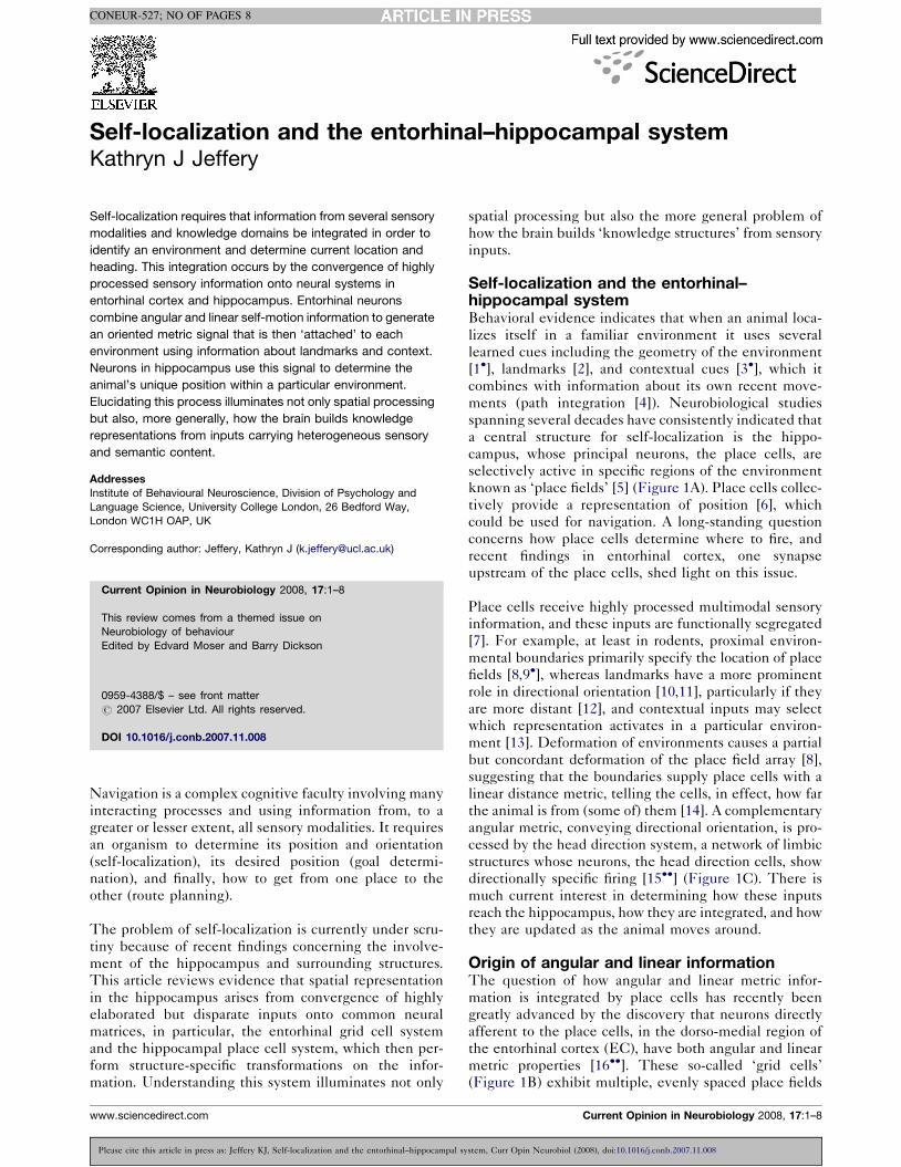
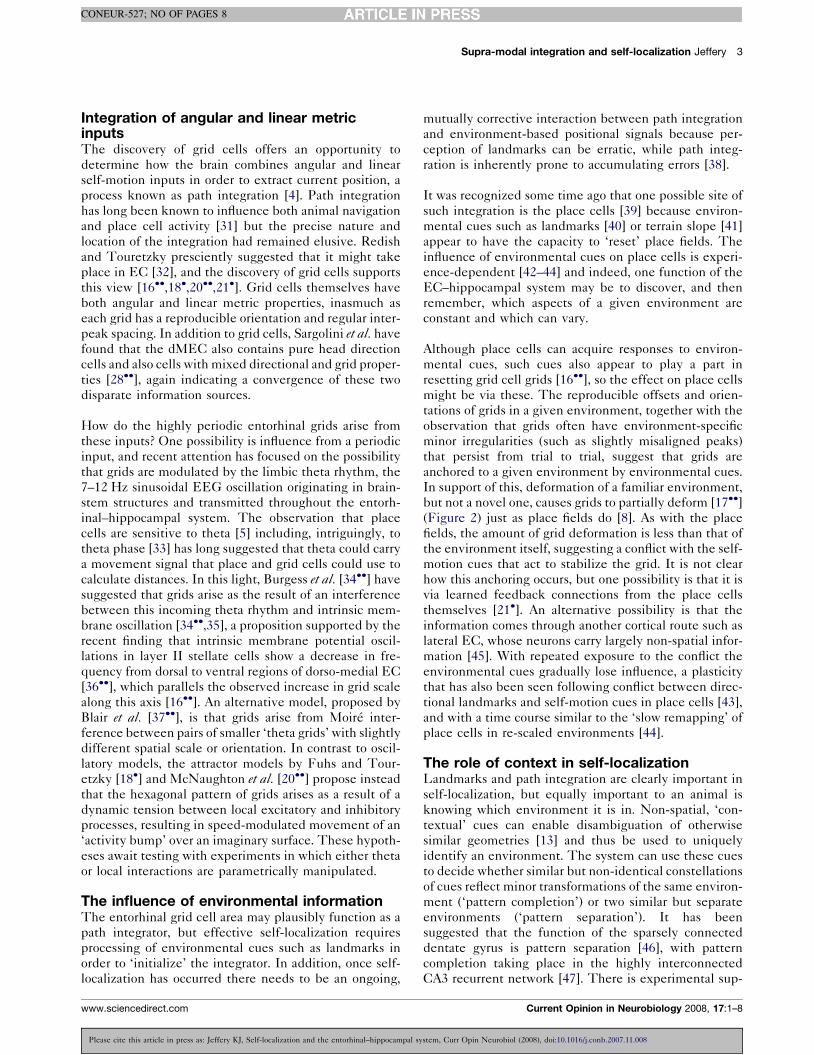
Figure 1
(adapted from reference [56]) Three kinds of spatial-signal-carrying neurons in the rodent limbic system. (A) A classical place cell. The black lines
show the path of a rat as it foraged in a 60 cm square box, and the red squares show action potentials from a single hippocampal neuron,
superimposed on the position of the rat. A typical place cell will, as shown here, concentrate most of its activity in one part of a small
environment (the ‘place field’). (B) Grid cells from dorsal (upper panel) and ventral (lower panel) dorso-medial EC, in the same data format as (A).
Data shown as reported in reference [16��]. The figure shows the hexagonal array of firing fields typical of grid cells. Note the change in inter-
peak spacing between dorsal and ventral regions. (C) Data from a typical head direction cell, showing that firing rate (y axis) increases
dramatically when the rat’s head is within a restricted directional orientation (x axis). Figure kindly supplied by Jeffrey Taube.
that spread across the environment in an apparently
limitless array, producing a striking hexagonal (or triangu-
lar) pattern strongly reminiscent of graph paper. Grid
scales increase from dorsal to ventral areas or dorso-
medial EC, and the grids from a given animal are ran-
domly ‘offset’ but seem to have similar orientation ([17��];Supplementary Figure 4). The even spacing of the grid
nodes could plausibly enable metric computations in
other neurons, such as place cells [16��]. How such
computations are implemented is still a matter of specu-
lation, but the most obvious method is by summation of
grids of different scales (as, for example, from different
dorso-ventral layers of EC), which would in theory pro-
duce widely scattered hot spots of activity with a sparsity
comparable to that of place fields [18�,19,20��,21�].
The remarkable metric properties of grid cell grids shift
back one synapse the question of where this information
comes from. How does a grid cell combine angular and
linear information to determine where to lay down its
peaks? That the peaks occur at the same spacing for a
given neuron even across different environments [16��]suggests an intrinsic (i.e., environment-independent)
metric, which must come from processing of angular
and linear self-motion information.
The route into EC for angular self-motion inputs, via the
head direction (HD) system, has now been worked out
in some detail [15��] and involves a circuit from the
vestibular nuclei through the dorsal tegmental nucleus,
Current Opinion in Neurobiology 2008, 17:1–8
Please cite this article in press as: Jeffery KJ, Self-localization and the entorhinal–hippocampal sy
where neurons sensitive to angular velocity are found
[22,23], and thence via the lateral mammillary nucleus,
anterodorsal thalamus, and post-subiculum to the EC
[15��]. Information concerning landmarks, and perhaps
optic flow, may come from higher cortical regions such as
retrosplenial cortex. Integration of static environmental
inputs with self-motion information is thought to take
place amongst the HD cells themselves, perhaps
mediated by ‘attractor’ processes [24,25] in which the
cells collectively form a stable representation of direc-
tion that is updated in accordance with incoming infor-
mation on angular velocity and abruptly moved (or
‘reset’) by landmark information. Observations that
HD cells always seem to fire coherently, even when
sensory cues are dissociated [26�], support the attractor
view.
The route for linear self-motion integration into the
entorhinal–hippocampal system is far less well estab-
lished than for angular. A weak correlation has been found
between locomotor speed and firing rate for place cells
[27] as well as head direction and grid cells [28��]. A single
recording session of a probable axon from a highly speed-
correlated cell has been reported [29], suggesting the
existence of cells that encode speed directly. The speed
correlate of place cells depends on the integrity of peri-
rhinal cortex [30], so this may be one route for such
information into the hippocampal system. As with angular
motion, it is likely that the vestibular component of linear
motion is routed via brainstem structures.
www.sciencedirect.com
stem, Curr Opin Neurobiol (2008), doi:10.1016/j.conb.2007.11.008
Supra-modal integration and self-localization Jeffery 3
CONEUR-527; NO OF PAGES 8
Integration of angular and linear metricinputsThe discovery of grid cells offers an opportunity to
determine how the brain combines angular and linear
self-motion inputs in order to extract current position, a
process known as path integration [4]. Path integration
has long been known to influence both animal navigation
and place cell activity [31] but the precise nature and
location of the integration had remained elusive. Redish
and Touretzky presciently suggested that it might take
place in EC [32], and the discovery of grid cells supports
this view [16��,18�,20��,21�]. Grid cells themselves have
both angular and linear metric properties, inasmuch as
each grid has a reproducible orientation and regular inter-
peak spacing. In addition to grid cells, Sargolini et al. have
found that the dMEC also contains pure head direction
cells and also cells with mixed directional and grid proper-
ties [28��], again indicating a convergence of these two
disparate information sources.
How do the highly periodic entorhinal grids arise from
these inputs? One possibility is influence from a periodic
input, and recent attention has focused on the possibility
that grids are modulated by the limbic theta rhythm, the
7–12 Hz sinusoidal EEG oscillation originating in brain-
stem structures and transmitted throughout the entorh-
inal–hippocampal system. The observation that place
cells are sensitive to theta [5] including, intriguingly, to
theta phase [33] has long suggested that theta could carry
a movement signal that place and grid cells could use to
calculate distances. In this light, Burgess et al. [34��] have
suggested that grids arise as the result of an interference
between this incoming theta rhythm and intrinsic mem-
brane oscillation [34��,35], a proposition supported by the
recent finding that intrinsic membrane potential oscil-
lations in layer II stellate cells show a decrease in fre-
quency from dorsal to ventral regions of dorso-medial EC
[36��], which parallels the observed increase in grid scale
along this axis [16��]. An alternative model, proposed by
Blair et al. [37��], is that grids arise from Moire inter-
ference between pairs of smaller ‘theta grids’ with slightly
different spatial scale or orientation. In contrast to oscil-
latory models, the attractor models by Fuhs and Tour-
etzky [18�] and McNaughton et al. [20��] propose instead
that the hexagonal pattern of grids arises as a result of a
dynamic tension between local excitatory and inhibitory
processes, resulting in speed-modulated movement of an
‘activity bump’ over an imaginary surface. These hypoth-
eses await testing with experiments in which either theta
or local interactions are parametrically manipulated.
The influence of environmental informationThe entorhinal grid cell area may plausibly function as a
path integrator, but effective self-localization requires
processing of environmental cues such as landmarks in
order to ‘initialize’ the integrator. In addition, once self-
localization has occurred there needs to be an ongoing,
www.sciencedirect.com
Please cite this article in press as: Jeffery KJ, Self-localization and the entorhinal–hippocampal sy
mutually corrective interaction between path integration
and environment-based positional signals because per-
ception of landmarks can be erratic, while path integ-
ration is inherently prone to accumulating errors [38].
It was recognized some time ago that one possible site of
such integration is the place cells [39] because environ-
mental cues such as landmarks [40] or terrain slope [41]
appear to have the capacity to ‘reset’ place fields. The
influence of environmental cues on place cells is experi-
ence-dependent [42–44] and indeed, one function of the
EC–hippocampal system may be to discover, and then
remember, which aspects of a given environment are
constant and which can vary.
Although place cells can acquire responses to environ-
mental cues, such cues also appear to play a part in
resetting grid cell grids [16��], so the effect on place cells
might be via these. The reproducible offsets and orien-
tations of grids in a given environment, together with the
observation that grids often have environment-specific
minor irregularities (such as slightly misaligned peaks)
that persist from trial to trial, suggest that grids are
anchored to a given environment by environmental cues.
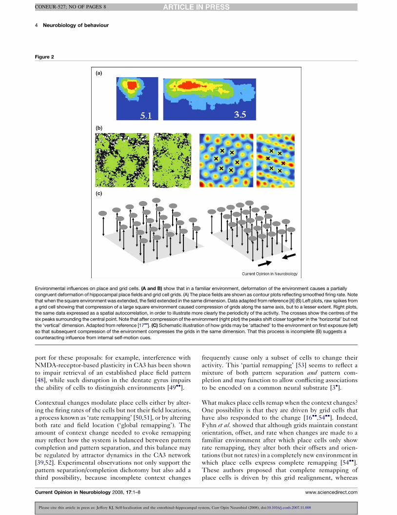
In support of this, deformation of a familiar environment,
but not a novel one, causes grids to partially deform [17��](Figure 2) just as place fields do [8]. As with the place
fields, the amount of grid deformation is less than that of
the environment itself, suggesting a conflict with the self-
motion cues that act to stabilize the grid. It is not clear
how this anchoring occurs, but one possibility is that it is
via learned feedback connections from the place cells
themselves [21�]. An alternative possibility is that the
information comes through another cortical route such as
lateral EC, whose neurons carry largely non-spatial infor-
mation [45]. With repeated exposure to the conflict the
environmental cues gradually lose influence, a plasticity
that has also been seen following conflict between direc-
tional landmarks and self-motion cues in place cells [43],
and with a time course similar to the ‘slow remapping’ of
place cells in re-scaled environments [44].
The role of context in self-localizationLandmarks and path integration are clearly important in
self-localization, but equally important to an animal is
knowing which environment it is in. Non-spatial, ‘con-
textual’ cues can enable disambiguation of otherwise
similar geometries [13] and thus be used to uniquely
identify an environment. The system can use these cues
to decide whether similar but non-identical constellations
of cues reflect minor transformations of the same environ-
ment (‘pattern completion’) or two similar but separate
environments (‘pattern separation’). It has been
suggested that the function of the sparsely connected
dentate gyrus is pattern separation [46], with pattern
completion taking place in the highly interconnected
CA3 recurrent network [47]. There is experimental sup-
Current Opinion in Neurobiology 2008, 17:1–8
stem, Curr Opin Neurobiol (2008), doi:10.1016/j.conb.2007.11.008
4 Neurobiology of behaviour
CONEUR-527; NO OF PAGES 8
Figure 2
Environmental influences on place and grid cells. (A and B) show that in a familiar environment, deformation of the environment causes a partially
congruent deformation of hippocampal place fields and grid cell grids. (A) The place fields are shown as contour plots reflecting smoothed firing rate. Note
that when the square environment was extended, the field extended in the same dimension. Data adapted from reference [8] (B) Left plots, raw spikes from
a grid cell showing that compression of a large square environment caused compression of grids along the same axis, but to a lesser extent. Right plots,
the same data expressed as a spatial autocorrelation, in order to illustrate more clearly the periodicity of the activity. The crosses show the centres of the
six peaks surrounding the central point. Note that after compression of the environment (right plot) the peaks shift closer together in the ‘horizontal’ but not
the ‘vertical’ dimension. Adapted from reference [17��]. (C) Schematic illustration of how grids may be ‘attached’ to the environment on first exposure (left)
so that subsequent compression of the environment compresses the grids in the same dimension. That this process is incomplete (B) suggests a
counteracting influence from internal self-motion cues.
port for these proposals: for example, interference with
NMDA-receptor-based plasticity in CA3 has been shown
to impair retrieval of an established place field pattern
[48], while such disruption in the dentate gyrus impairs
the ability of cells to distinguish environments [49��].
Contextual changes modulate place cells either by alter-
ing the firing rates of the cells but not their field locations,
a process known as ‘rate remapping’ [50,51], or by altering
both rate and field location (‘global remapping’). The
amount of context change needed to evoke remapping
may reflect how the system is balanced between pattern
completion and pattern separation, and this balance may
be regulated by attractor dynamics in the CA3 network
[39,52]. Experimental observations not only support the
pattern separation/completion dichotomy but also add a
third possibility, because incomplete context changes
Current Opinion in Neurobiology 2008, 17:1–8
Please cite this article in press as: Jeffery KJ, Self-localization and the entorhinal–hippocampal sy
frequently cause only a subset of cells to change their
activity. This ‘partial remapping’ [53] seems to reflect a
mixture of both pattern separation and pattern com-
pletion and may function to allow conflicting associations
to be encoded on a common neural substrate [3�].
What makes place cells remap when the context changes?
One possibility is that they are driven by grid cells that
have also responded to the change [16��,54��]. Indeed,
Fyhn et al. showed that although grids maintain constant
orientation, offset, and rate when changes are made to a
familiar environment after which place cells only show
rate remapping, they alter both their offsets and orien-
tations (but not rates) in a completely new environment in
which place cells express complete remapping [54��].These authors proposed that complete remapping of
place cells is driven by this grid realignment, whereas
www.sciencedirect.com
stem, Curr Opin Neurobiol (2008), doi:10.1016/j.conb.2007.11.008
Supra-modal integration and self-localization Jeffery 5
CONEUR-527; NO OF PAGES 8
rate remapping occurs in the absence of grid shift and has
some other cause. However, to explain why place fields
do not merely offset by the same amount as the grids, it is
necessary to assume either that the grids offset by differ-
ent amounts or that not all grids have the same orientation
in a given animal, both propositions for which there is, as
yet, scant evidence. Partial place cell remapping is not
explained by such a scheme either, unless it is assumed
that not all grids undergo realignment.
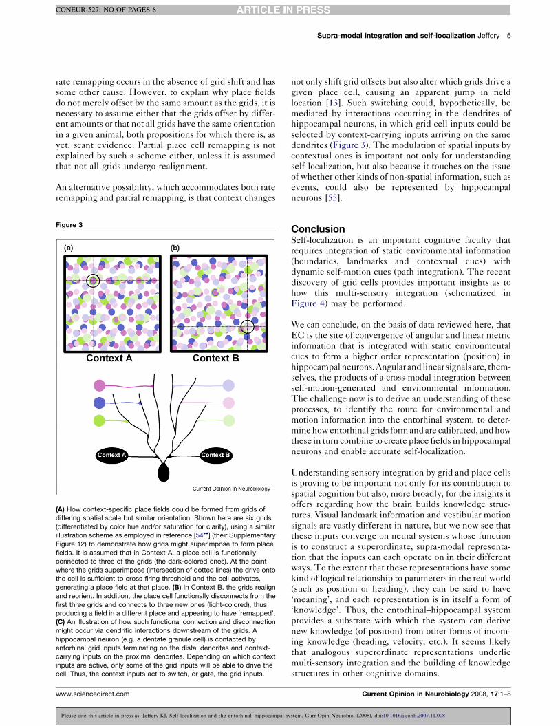
An alternative possibility, which accommodates both rate
remapping and partial remapping, is that context changes
Figure 3
(A) How context-specific place fields could be formed from grids of
differing spatial scale but similar orientation. Shown here are six grids
(differentiated by color hue and/or saturation for clarity), using a similar
illustration scheme as employed in reference [54��] (their Supplementary
Figure 12) to demonstrate how grids might superimpose to form place
fields. It is assumed that in Context A, a place cell is functionally
connected to three of the grids (the dark-colored ones). At the point
where the grids superimpose (intersection of dotted lines) the drive onto
the cell is sufficient to cross firing threshold and the cell activates,
generating a place field at that place. (B) In Context B, the grids realign
and reorient. In addition, the place cell functionally disconnects from the
first three grids and connects to three new ones (light-colored), thus
producing a field in a different place and appearing to have ‘remapped’.
(C) An illustration of how such functional connection and disconnection
might occur via dendritic interactions downstream of the grids. A
hippocampal neuron (e.g. a dentate granule cell) is contacted by
entorhinal grid inputs terminating on the distal dendrites and context-
carrying inputs on the proximal dendrites. Depending on which context
inputs are active, only some of the grid inputs will be able to drive the
cell. Thus, the context inputs act to switch, or gate, the grid inputs.
www.sciencedirect.com
Please cite this article in press as: Jeffery KJ, Self-localization and the entorhinal–hippocampal sy
not only shift grid offsets but also alter which grids drive a
given place cell, causing an apparent jump in field
location [13]. Such switching could, hypothetically, be
mediated by interactions occurring in the dendrites of
hippocampal neurons, in which grid cell inputs could be
selected by context-carrying inputs arriving on the same
dendrites (Figure 3). The modulation of spatial inputs by
contextual ones is important not only for understanding
self-localization, but also because it touches on the issue
of whether other kinds of non-spatial information, such as
events, could also be represented by hippocampal
neurons [55].
ConclusionSelf-localization is an important cognitive faculty that
requires integration of static environmental information
(boundaries, landmarks and contextual cues) with
dynamic self-motion cues (path integration). The recent
discovery of grid cells provides important insights as to
how this multi-sensory integration (schematized in
Figure 4) may be performed.
We can conclude, on the basis of data reviewed here, that
EC is the site of convergence of angular and linear metric
information that is integrated with static environmental
cues to form a higher order representation (position) in
hippocampal neurons. Angular and linear signals are, them-
selves, the products of a cross-modal integration between
self-motion-generated and environmental information.
The challenge now is to derive an understanding of these
processes, to identify the route for environmental and
motion information into the entorhinal system, to deter-
mine how entorhinal grids form and are calibrated, and how
these in turn combine to create place fields in hippocampal
neurons and enable accurate self-localization.
Understanding sensory integration by grid and place cells
is proving to be important not only for its contribution to
spatial cognition but also, more broadly, for the insights it
offers regarding how the brain builds knowledge struc-
tures. Visual landmark information and vestibular motion
signals are vastly different in nature, but we now see that
these inputs converge on neural systems whose function
is to construct a superordinate, supra-modal representa-
tion that the inputs can each operate on in their different
ways. To the extent that these representations have some
kind of logical relationship to parameters in the real world
(such as position or heading), they can be said to have
‘meaning’, and each representation is in itself a form of
‘knowledge’. Thus, the entorhinal–hippocampal system
provides a substrate with which the system can derive
new knowledge (of position) from other forms of incom-
ing knowledge (heading, velocity, etc.). It seems likely
that analogous superordinate representations underlie
multi-sensory integration and the building of knowledge
structures in other cognitive domains.
Current Opinion in Neurobiology 2008, 17:1–8
stem, Curr Opin Neurobiol (2008), doi:10.1016/j.conb.2007.11.008

6 Neurobiology of behaviour
CONEUR-527; NO OF PAGES 8
Figure 4
Schematic illustration of the hypothetical functional connectivity of the entorhinal–hippocampal place system. Salient elements of the system are
indicated by the letter labels. Contextual cues (A) control which grid cells (B) are connected to which place cells, thus generating (by summation)
place fields (C). Local landmarks (D) then anchor the grids, perhaps via feedback from the place cells. Within the angular metric domain, distant
landmarks, together with angular self-motion cues (E) set and update the head direction system (F), which in turn orients the grid cell grids.
Similarly in the linear domain, linear self-motion cues (G) are integrated to form a velocity/distance (H) signal that enables updating of grid cell activity.
This in turn will update place cell activity and enable appropriate ongoing associations to the landmark array.
AcknowledgementsThe work was supported by a grant from the UK Biotechnology andBiological Sciences Research Council. The author would like to thankCaswell Barry, Neil Burgess and Jim Donnett for commenting on themanuscript.
References and recommended readingPapers of particular interest have been highlighted as:
� of special interest�� of outstanding interest
1.�
Cheng K, Newcombe NS: Is there a geometric module forspatial orientation? Squaring theory and evidence. PsychonBull Rev 2005, 12:1-23.
This is a good review of the current status long-standing debate con-cerning the relative roles of geometry vs. landmarks in guiding animalnavigation.
2. Collett TS, Cartwright BA, Smith BA: Landmark learning andvisuo-spatial memories in gerbils. J Comp Physiol [A] 1986,158:835-851.
3.�
Anderson M, Killing S, Morris C, O’Donoghue A, Onyiagha D,Stevenson R, Verriotis M, Jeffery K: Behavioral correlates of thepopulation coding of spatial context. Hippocampus 2006,16:730-742.
Current Opinion in Neurobiology 2008, 17:1–8
Please cite this article in press as: Jeffery KJ, Self-localization and the entorhinal–hippocampal sy
This study used naturalistic methods to study behavior under environ-mental conditions associated with partial place cell remapping. Findingswere consistent with the proposition that the different but overlappingplace cell representations of the same environment can be flexiblyassociated with different, sometimes conflicting behaviors.
4. Etienne AS, Jeffery KJ: Path integration in mammals.Hippocampus 2004, 14:180-192.
5. O’Keefe J, Dostrovsky J: The hippocampus as a spatial map.Preliminary evidence from unit activity in the freely moving rat.Brain Res 1971, 34:171-175.
6. O’Keefe J, Nadel L: The Hippocampus as a Cognitive Map.Oxford: Clarendon Press; 1978.
7. Jeffery KJ: Integration of the sensory inputs to place cells:what, where, why, and how? Hippocampus 2007, 17:775-785.
8. O’Keefe J, Burgess N: Geometric determinants of the placefields of hippocampal neurons. Nature 1996, 381:425-428.
9.�
Siegel JJ, Neunuebel JP, Knierim JJ: Dominance of the proximalcoordinate frame in determining the locations of hippocampalplace cell activity during navigation. J Neurophysiol 2007.
This study examined the relative effects of proximal vs. distal envir-onmental cues by translating a recording box within a room andrecording CA3 and CA1 place fields at either an early or a late phaseof training on a place preference task. Early in training the cellsshowed ambiguous responses to the reference frame disjunction,
www.sciencedirect.com
stem, Curr Opin Neurobiol (2008), doi:10.1016/j.conb.2007.11.008
Supra-modal integration and self-localization Jeffery 7
CONEUR-527; NO OF PAGES 8
but later in training they remained anchored either to the room or (morecommonly) the platform.
10. Jeffery KJ, Donnett JG, Burgess N, O’Keefe JM: Directionalcontrol of hippocampal place fields. Exp Brain Res 1997,117:131-142.
11. Knierim JJ, Rao G: Distal landmarks and hippocampal placecells: effects of relative translation versus rotation.Hippocampus 2003, 13:604-617.
12. Zugaro MB, Berthoz A, Wiener SI: Background, but notforeground, spatial cues are taken as references for headdirection responses by rat anterodorsal thalamus neurons.J Neurosci 2001, 21:RC154.
13. Jeffery KJ, Anderson MI, Hayman R, Chakraborty S: A proposedarchitecture for the neural representation of spatial context.Neurosci Biobehav Rev 2004, 28:201-218.
14. Barry C, Lever C, Hayman R, Hartley T, Burton S, O’Keefe J,Jeffery KJ, Burgess N: The boundary vector cell model of placecell firing and spatial memory. Rev Neurosci 2006, 17:71-97.
15.��
Taube JS: The head direction signal: origins and sensory-motor integration. Annu Rev Neurosci 2007, 30:181-207.
This is a very comprehensive but readable review of the head directioncell literature.
16.��
Hafting T, Fyhn M, Molden S, Moser MB, Moser EI:Microstructure of a spatial map in the entorhinal cortex. Nature2005, 801-806.
This paper presents the first report of entorhinal grid cells and representsa seminal advance in our understanding of the entorhinal–hippocampalplace system.
17.��
Barry C, Hayman R, Burgess N, Jeffery KJ: Experience-dependent rescaling of entorhinal grids. Nat Neurosci 2007,10:682-684.
This study shows that in a familiar environment, grid scale is notentirely dependent on an intrinsic (path-integration-based) metric, butalso receives an input from learned environment cues such as theboundaries of the environment. When a familiar two-dimensionalenvironment was slightly stretched or compressed in one or bothdimensions, grids showed a partial re-scaling in the same dimension.This re-scaling did not transfer to a novel environment. It lessenedwith repeated exposures, suggesting a slow adaptation, of a timescalesimilar to ‘slow remapping’ reported in hippocampal place fields[44].
18.�
Fuhs MC, Touretzky DS: A spin glass model of pathintegration in rat medial entorhinal cortex. J Neurosci 2006,26:4266-4276.
This paper is the first attractor model of entorhinal grid formation andproposes that grids arise from local interactions among entorhinal neu-rons.
19. Solstad T, Moser EI, Einevoll GT: From grid cells to place cells: amathematical model. Hippocampus 2006, 16:1026-1031.
20.��
McNaughton BL, Battaglia FP, Jensen O, Moser EI, Moser MB:Path integration and the neural basis of the ‘cognitive map’.Nat Rev Neurosci 2006, 7:663-678.
This is a review of the discovery of grid cells and its implications for pathintegration hypotheses. A model is presented in which grid cell scale ismodulated by theta frequency, with grids themselves resulting fromasymmetric interactions between connected entorhinal neurons, andsummation of grids of different scales generating the unique patternsof place cell activity seen in hippocampus.
21.�
O’Keefe J, Burgess N: Dual phase and rate coding inhippocampal place cells: theoretical significance andrelationship to entorhinal grid cells. Hippocampus 2005,15:853-866.
This paper is a review of theta and its relationship to hippocampal placecell activity and was one of the first to propose feedback connectionsfrom place cells as a way of anchoring grids to the external environment.
22. Bassett JP, Taube JS: Neural correlates for angular headvelocity in the rat dorsal tegmental nucleus. J Neurosci 2001,21:5740-5751.
23. Sharp PE, Tinkelman A, Cho J: Angular velocity and headdirection signals recorded from the dorsal tegmental nucleusof gudden in the rat: implications for path integration in thehead direction cell circuit. Behav Neurosci 2001, 115:571-588.
www.sciencedirect.com
Please cite this article in press as: Jeffery KJ, Self-localization and the entorhinal–hippocampal sy
24. Skaggs WE, Knierim JJ, Kudrimoti HS, McNaughton BL: A modelof the neural basis of the rat’s sense of direction. Adv Neural InfProcess Syst 1995, 7:173-180.
25. Zhang K: Representation of spatial orientation by the intrinsicdynamics of the head-direction cell ensemble: a theory.J Neurosci 1996, 16:2112-2126.
26.�
Yoganarasimha D, Yu X, Knierim JJ: Head direction cellrepresentations maintain internal coherence duringconflicting proximal and distal cue rotations: comparison withhippocampal place cells. J Neurosci 2006, 26:622-631.
This study explores the effects on head direction cells and place cellswhen proximal vs. distal environmental cues are dissociated. Consistentwith previous findings, the head direction cells always remain in registerwith each other and tend to be mostly influenced by the distal cue set.Interestingly, place cells did not always remain coherent and sometimesdissociated from the head direction cells.
27. McNaughton BL, Barnes CA, O’Keefe J: The contributions ofposition, direction, and velocity to single unit activity in thehippocampus of freely moving rats. Exp Brain Res 1983,52:41-49.
28.��
Sargolini F, Fyhn M, Hafting T, McNaughton BL, Witter MP,Moser MB, Moser EI: Conjunctive representation of position,direction, and velocity in entorhinal cortex. Science 2006,312:758-762.
This paper reports results of a comprehensive analysis of firing correlatesof neurons in dMEC and finds that neurons in the deeper layers (III–VI)show conjunctive properties, with some neurons having pure head-direction properties and many showing directionally modulated gridactivity. All of the neurons were modulated by running speed.
29. O’Keefe J, Burgess N, Donnett JG, Jeffery KJ, Maguire EA: Placecells, navigational accuracy, and the human hippocampus.Philos Trans R Soc Lond B Biol Sci 1998, 353:1333-1340.
30. Muir GM, Bilkey DK: Theta- and movement velocity-relatedfiring of hippocampal neurons is disrupted by lesions centeredon the perirhinal cortex. Hippocampus 2003, 13:93-108.
31. McNaughton BL, Barnes CA, Gerrard JL, Gothard K, Jung MW,Knierim JJ, Kudrimoti H, Qin Y, Skaggs WE, Suster M, Weaver KL:Deciphering the hippocampal polyglot: the hippocampus as apath integration system. J Exp Biol 1996, 199:173-185.
32. Redish AD, Touretzky DS: Cognitive maps beyond thehippocampus. Hippocampus 1997, 7:15-35.
33. O’Keefe J, Recce ML: Phase relationship betweenhippocampal place units and the EEG theta rhythm.Hippocampus 1993, 3:317-330.
34.��
Burgess N, Barry C, O’Keefe J: An oscillatory interferencemodel of grid cell firing. Hippocampus 2007, 17:801-812.
This paper proposes a model of entorhinal grid generation based oninterference between an extrinsically derived membrane potential oscil-lation (due to theta) and an intrinsic membrane potential oscillation, suchas that reported in entorhinal neurons by Giocomo et al. [36��].
35. Hasselmo ME, Giocomo LM, Zilli EA: Grid cell firing may arisefrom interference of theta frequency membrane potentialoscillations in single neurons. Hippocampus 2007.
36.��
Giocomo LM, Zilli EA, Fransen E, Hasselmo ME: Temporalfrequency of subthreshold oscillations scales with entorhinalgrid cell field spacing. Science 2007, 315:1719-1722.
This study shows that subthreshold membrane oscillations in entorhinalneurons are slightly faster in more doral regions and slightly slower moreventrally. Since grid scale also increases dorso-ventrally, this findingsupports oscillatory models of grid generation such as those based oninterferences between theta frequency and local membrane oscillations.
37.��
Blair HT, Welday AC, Zhang K: Scale-invariant memoryrepresentations emerge from moire interference between gridfields that produce theta oscillations: a computational model.J Neurosci 2007, 27:3211-3229.
This paper proposes that entorhinal grids arise from Moire interferencebetween smaller scale grids and makes the intriguing suggestion that thesize invariance properties of such grids could have a more general role inmemory representation.
38. Etienne AS, Maurer R, Seguinot V: Path integration in mammalsand its interaction with visual landmarks. J Exp Biol 1996,199:201-209.
Current Opinion in Neurobiology 2008, 17:1–8
stem, Curr Opin Neurobiol (2008), doi:10.1016/j.conb.2007.11.008
8 Neurobiology of behaviour
CONEUR-527; NO OF PAGES 8
39. Samsonovich A, McNaughton BL: Path integration and cognitivemapping in a continuous attractor neural network model.J Neurosci 1997, 17:5900-5920.
40. Gothard KM, Skaggs WE, McNaughton BL: Dynamics ofmismatch correction in the hippocampal ensemble code forspace: interaction between path integration andenvironmental cues. J Neurosci 1996, 16:8027-8040.
41. Jeffery KJ, Anand RL, Anderson MI: A role for terrain slope inorienting hippocampal place fields. Exp Brain Res 2006,169:218-225.
42. Bostock E, Muller RU, Kubie JL: Experience-dependentmodifications of hippocampal place cell firing. Hippocampus1991, 1:193-205.
43. Jeffery KJ, O’Keefe J: Learned interaction of visual andidiothetic cues in the control of place field orientation. ExpBrain Res 1999, 127:151-161.
44. Lever C, Wills T, Cacucci F, Burgess N, O’Keefe J: Long-termplasticity in hippocampal place-cell representation ofenvironmental geometry. Nature 2002, 416:90-94.
45. Hargreaves EL, Rao G, Lee I, Knierim JJ: Major dissociationbetween medial and lateral entorhinal input to dorsalhippocampus. Science 2005, 308:1792-1794.
46. Leutgeb JK, Leutgeb S, Moser MB, Moser EI: Pattern separationin the dentate gyrus and CA3 of the hippocampus. Science2007, 315:961-966.
47. O’Reilly RC, McClelland JL: Hippocampal conjunctiveencoding, storage, and recall: avoiding a trade-off.Hippocampus 1994, 4:661-682.
48. Nakazawa K, Quirk MC, Chitwood RA, Watanabe M, Yeckel MF,Sun LD, Kato A, Carr CA, Johnston D, Wilson MA, Tonegawa S:Requirement for hippocampal CA3 NMDA receptors inassociative memory recall. Science 2002, 297:211-218.
49.��
McHugh TJ, Jones MW, Quinn JJ, Balthasar N, Coppari R,Elmquist JK, Lowell BB, Fanselow MS, Wilson MA, Tonegawa S:Dentate gyrus NMDA receptors mediate rapid pattern
Current Opinion in Neurobiology 2008, 17:1–8
Please cite this article in press as: Jeffery KJ, Self-localization and the entorhinal–hippocampal sy
separation in the hippocampal network. Science 2007,317:94-99.
The study examined the effects of context change in mutant mice withimpaired dentate gyrus NMDA receptor function. Recordings from CA3indicated a reduced responsiveness of these neurons to transfer from onecontext to another.
50. Hayman RM, Chakraborty S, Anderson MI, Jeffery KJ: Context-specific acquisition of location discrimination by hippocampalplace cells. Eur J Neurosci 2003, 18:2825-2834.
51. Leutgeb S, Leutgeb JK, Barnes CA, Moser EI, McNaughton BL,Moser MB: Independent codes for spatial and episodicmemory in hippocampal neuronal ensembles. Science 2005,309:619-623.
52. Wills TJ, Lever C, Cacucci F, Burgess N, O’Keefe J: Attractordynamics in the hippocampal representation of the localenvironment. Science 2005, 308:873-876.
53. Anderson MI, Jeffery KJ: Heterogeneous modulation ofplace cell firing by changes in context. J Neurosci 2003,23:8827-8835.
54.��
Fyhn M, Hafting T, Treves A, Moser MB, Moser EI: Hippocampalremapping and grid realignment in entorhinal cortex. Nature2007, 446:190-194.
This study compares the behavior of place and grid cells in novelenvironments vs. altered familiar environments and finds that condi-tions that cause place cell activity to alter completely (‘global remap-ping’) are associated with alteration of position and orientation ofentorhinal grids, whereas conditions that cause place cells to altertheir firing rates alone (‘rate remapping’) are accompanied by nochange in entorhinal grids. A hypothesis is proposed in which summa-tion of grids of different scales and orientations causes place fields, and‘grid realignment’ cause the place of summation to alter, with acorresponding change in place fields.
55. Eichenbaum H, Dudchenko P, Wood E, Shapiro M, Tanila H: Thehippocampus, memory, and place cells: is it spatial memory ora memory space? Neuron 1999, 23:209-226.
56. Jeffery KJ, Burgess N: A metric for the cognitive map—found atlast? Trends Cogn Sci 2006, 10:1-3.
www.sciencedirect.com
stem, Curr Opin Neurobiol (2008), doi:10.1016/j.conb.2007.11.008