Embed Size (px)
Citation preview
This file is part of the following reference:
Rutkowski, Rachael (2005) Genetic and cellular analysis of the novel cell proliferation regulator, deflated, in Drosophila. PhD thesis, James Cook
University.
Access to this file is available from:
http://eprints.jcu.edu.au/17563
120
Chapter 5- deflated interacts genetically with known cell proliferation and cell signalling mutants
5.1 Introduction
As described in the previous chapter, deflated is expressed in proliferating and
endoreplicating cells and deflated mutant phenotypes are consistent with a defect in the
regulation of cell proliferation, including the inability to progress normally through the
cell cycle. The conserved protein motifs identified in DEFLATED and the mild wing
and bristle defects observed in the deflated transheterozgous adults suggest that deflated
may have a role in signalling and that its effects on the cell cycle may be indirect. The
aims of the experiments described in this chapter were to further explore deflated’s role
in cell proliferation and cell signalling by examining genetic interactions with other
known cell proliferation and cell signalling mutants. A genetic approach was taken for
two reasons. One, genetic studies allow for the identification of genetically related
genes, particularly those that act in the same biological pathway, and can reveal how
biological pathways relate to one and other. Two, the type of role that a gene product
plays within these pathways can also be determined, such as whether it acts as an
activator or an inhibitor.
This chapter examines the genetic interactions between overexpressed deflated and
other cell proliferation and signalling genes. Two well characterised developmental
systems were studied, the eye and the wing. Both have been extensively used to
understand the developmental control of cell proliferation and cell signalling. The eye
was used to further explore the role deflated plays in regulating S-phase and the wing to
identify genes that interact genetically with deflated.
121
The adult eye comprises approximately 800 ommatidia arrayed in an orderly fashion
and whose development is well understood (Dickson and Hafen, 1993; Wolff and
Ready, 1993). Each ommatidia is made of eight photoreceptor cells and the accessory
cone, pigment, and bristle cells. The eye arises from an epithelial sheet of cells known
as the eye-antennal imaginal disc. The differentiation of ommatidial cells commences
during late third instar larval stage in the wake of the morphogenetic furrow, a
constriction of cells that moves from the posterior of the eye disc to the anterior. As
cells enter the morphogenetic furrow they become arrested in G1. Upon leaving the
furrow some cells have become committed to differentiate. Those that have not enter a
synchronous S-phase followed by a synchronous M-phase (known as the second mitotic
wave) required to generate enough cells for subsequent differentiation. Perturbations to
this strict developmentally controlled cell proliferation and differentiation result in a
disruption of the ommatidial array, resulting in a disordered or rough eye phenotype.
Therefore, this system allows for easy observation to perturbations of both cell
proliferation and differentiation.
The adult wing comprises six longitudinal veins and two cross veins with intervein
regions in between. The adult wing arises from the wing imaginal disc, which forms
from a small group of 20-40 cells in the embryo that proliferate during the larval stages
to form a disc of characteristic size and shape (reviewed in Milan, 1998). Proliferation
in the wing disc occurs in an asynchronous pattern under the control of the
Decapentaplegic (Dpp) and Wingless (Wg) pathways. Unlike the eye disc, perturbation
of cell proliferation in wing discs is generally compensated for via apoptosis and cell
proliferation arrest in response to excess cells or increased cell proliferation rates in
122
response to insufficient cells. Consequently, the resulting adult wing often shows little
or no morphological changes (Milan, 1998), with most morphological defects observed
in adult wings arising from defects in cell fate specification and differentiation as a
consequence of defective cell signalling cascades.
Wing differentiation is under the control of many signalling pathways, including Notch,
Epidermal Growth Factor Receptor (EGFR)/Ras, Dpp and Hedgehog/Wingless. Cells
receive anterior-posterior information from Dpp signalling cues and dorso-ventral cues
from Wingless /Hedgehog signalling (Klein, 2001). The formation of the wing veins
occurs through expression of specific transcription factors to specify intervein and
provein region and to provide vein identity (de Celis, 2003). This patterning is
reinforced by the activation of EGFR and Notch signalling in the proveins. EGFR
signalling is thought to activate Notch expression that then signals to cells at the
provein-intervein border. Dpp signalling in the proveins results in the differentiation of
these cells into veins. Therefore, disruption to signalling pathways often manifests as
disruption of wing patterning or wing vein differentiation.
5.2 Overexpression of deflated can suppress overexpression of Cyclin E but not of
Cyclin A or E2f and Dp
5.2.1 Overexpressed deflated modifies overexpressed Cyclin E
The overexpression of Cyclin E, Cyclin A, and the co-expression of E2f and Dp have
been reported to induce ectopic S-phases (Asano et al., 1996; Dienemann and Sprenger,
2004; Du et al., 1996; Duronio et al., 1996; Knoblich et al., 1994; Richardson et al.,
1995). When these genes are overexpressed within and posterior to the morphogenetic
furrow of the eye imaginal disc (by GMR-Gal4) caused a rough eye phenotype (Figure
123
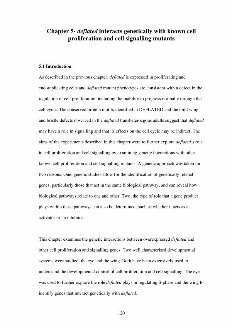
5.1A, B, and C). The three rough eye phenotypes varied slightly from each other,
although all displayed a reduction in ventral tissue and a disrupted ommatidial array.
However, the eyes from P[GMR-Gal4], P[UAS-CycA] (Figure 5.1 B) and P[GMR-
Gal4]; P[UAS-E2f1], P[UAS-DP] (Figure 5.1A) flies had a greater number of bristles
than wild type eyes (Figure 5.1 D). P[GMR-Gal4]; P[UAS-E2f1], P[UAS-DP] eyes
also showed a reproducible furrow running roughly dorso-ventrally, which was not
observed in the other two genotypes. P[GMR-Gal4]; P[UAS-CycE] eyes, on the other
hand, displayed fused ommatidia and were lacking bristles (Figure 5.1 C).
Eye phenotypes resulting from Cyclin E or E2f and Dp overexpression have been
previously shown to be modifiable by mutations in other cell cycle regulators (Lane et
al., 2000; Staehling-Hampton et al., 1999). To determine whether deflated has a role in
S-phase, deflated overexpression in these three genetic backgrounds was investigated.
When one copy of the deflated cDNA was overexpressed using GMR-Gal4 (P[GMR-
Gal4]; P[UAS-defl]AV3) it had no effect on the phenotype of P[GMR-Gal4]; P[UAS-
E2f1], P[UAS-DP] eyes (Figure 5.1 E). These eyes still lacked cells in the ventral
region, the vertical furrow was still present and the eye still contained extra bristles.
deflated overexpression also failed to suppress the Cyclin A overexpression phenotype
(Figure 5.1 F). The eye remained disorganised and contained a similar level of extra
bristles. deflated overexpression, however, did suppress the Cyclin E overexpression
phenotype. The ommatidia were more orderly arrayed, there were fewer, if any, fused
ommatidia and many more ommatidia contained bristles (Figure 5.1 G). The
overexpression of deflated by GMR-Gal4 in an otherwise wild type background
(P[GMR-Gal4]; P[UAS-defl]AV3) had no effect on the ommatidial array or bristle
number of the adult eye (Figure 5.1 H).
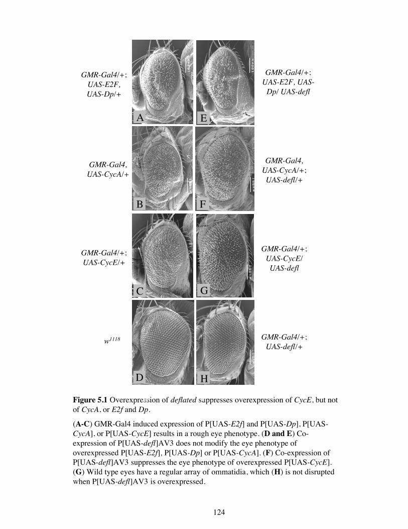
GMR-Gal4/+;UAS-E2F,UAS-Dp/+
GMR-Gal4/+;UAS-E2F, UAS-
Dp/ UAS-defl
GMR-Gal4,UAS-CycA/+
GMR-Gal4,UAS-CycA/+;UAS-defl/+
GMR-Gal4/+;UAS-CycE/+
GMR-Gal4/+;UAS-CycE/UAS-defl
Figure 5.1 Overexpression of deflated suppresses overexpression of CycE, but notof CycA, or E2f and Dp.
(A-C) GMR-Gal4 induced expression of P[UAS-E2f] and P[UAS-Dp], P[UAS-CycA], or P[UAS-CycE] results in a rough eye phenotype. (D and E) Co-expression of P[UAS-defl]AV3 does not modify the eye phenotype ofoverexpressed P[UAS-E2f], P[UAS-Dp] or P[UAS-CycA]. (F) Co-expression ofP[UAS-defl]AV3 suppresses the eye phenotype of overexpressed P[UAS-CycE].(G) Wild type eyes have a regular array of ommatidia, which (H) is not disruptedwhen P[UAS-defl]AV3 is overexpressed.
w1118 GMR-Gal4/+;UAS-defl/+
A D
B E
C F
G H
A
B
C
D
E
F
G
H
124
125
5.2.2 deflated overexpression attenuates the ectopic S-phases induced by Cyclin E
overexpression
In order to understand how deflated could suppress Cyclin E overexpression but not
Cyclin A or E2f and Dp, the effect of deflated on S-phase entry in these eye discs was
examined. To visualise S-phases, the eye-antennal imaginal discs from third instar
larvae were dissected and labelled with BrdU for 30 min (Section 2.24). The
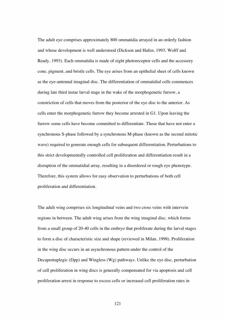
synchronous S-phase posterior to the morphogenetic furrow is clearly seen in the wild
type eye-antennal imaginal disc (Figure 5.2 A and A′). In the Cyclin E overexpressed
eye disc the width of the synchronous S-phase band was increased, indicating that
ectopic S-phases have occurred (Figure 5.2 B and B′). When deflated and Cyclin E are
co-overexpressed, this band of ectopic S-phases was reduced in both size and intensity.
Therefore deflated can act to negatively regulate S-phase in these circumstances (Figure
5.2 C and C′). It is interesting to note that the overexpression of deflated does not affect
the normal band of S-phase cells that are closest to the morphogenetic furrow. When
deflated is overexpressed in an otherwise wild type background, the pattern of S-phases
in the third instar eye imaginal disc is the same as the wild type (Figure 5.2 D and D′).
Together these data indicate that deflated can act to negatively regulate S-phase, but
only when normal cell cycle regulation is perturbed or within cells that should be
differentiating rather than proliferating.
To begin to understand why deflated overexpression could modify Cyclin E but not
Cyclin A overexpression, third instar eye discs from larvae overexpressing Cyclin A
were labelled with BrdU. Previous reports of Cyclin A overexpression using sev-Gal4
showed that ectopic S-phases occurred in cells posterior to the morphogenetic furrow
(Dienemann and Sprenger, 2004). However, when GMR-Gal4 was used to overexpress
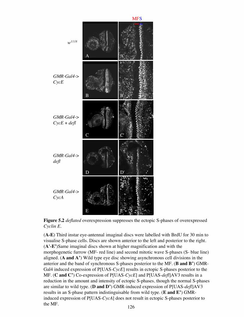
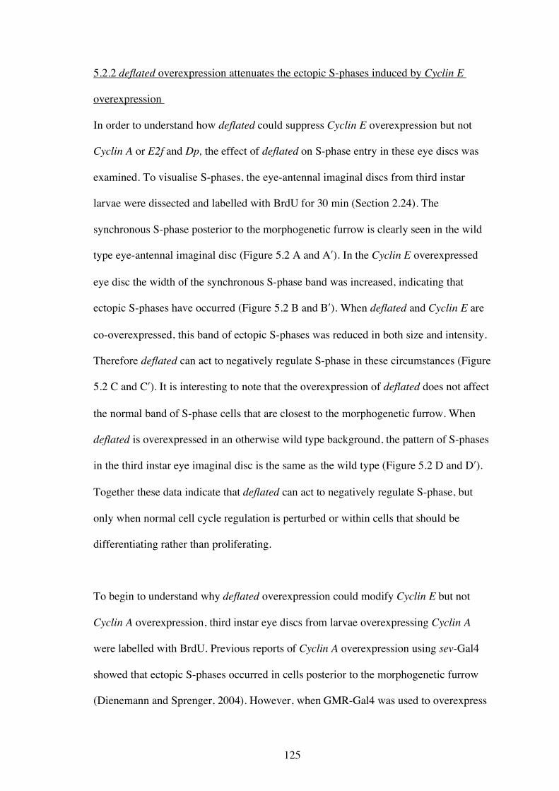
w1118
GMR-Gal4->CycE
GMR-Gal4->CycE + defl
GMR-Gal4->defl
GMR-Gal4->CycA
Figure 5.2 deflated overexpression suppresses the ectopic S-phases of overexpressedCyclin E.
(A-E) Third instar eye-antennal imaginal discs were labelled with BrdU for 30 min tovisualise S-phase cells. Discs are shown anterior to the left and posterior to the right.(A'-E’)Same imaginal discs shown at higher magnification and with themorphogenetic furrow (MF- red line) and second mitotic wave S-phases (S- blue line)aligned. (A and A’) Wild type eye disc showing asynchronous cell divisions in theanterior and the band of synchronous S-phases posterior to the MF. (B and B’) GMR-Gal4 induced expression of P[UAS-CycE] results in ectopic S-phases posterior to theMF. (C and C’) Co-expression of P[UAS-CycE] and P[UAS-defl]AV3 results in areduction in the amount and intensity of ectopic S-phases, though the normal S-phasesare similar to wild type. (D and D’) GMR-induced expression of P[UAS-defl]AV3results in an S-phase pattern indistinguisable from wild type. (E and E’) GMR-induced expression of P[UAS-CycA] does not result in ectopic S-phases posterior tothe MF.
A
B
C
D
E
MFS
A'
B'
C'
D'
E'
126
127
Cyclin A, ectopic S-phases were not observed posterior to the morphogenetic furrow
(Figure 5.2 E and E′). Both GMR- and sev-Gal4 drive expression in differentiating
cells, but as GMR-Gal4 also induces expression in the morphogenetic furrow, it is
possible that these cells, or those just posterior, are less sensitive to increased Cyclin A
levels. The S-phase pattern in eye imaginal discs overexpressing E2f and Dp were not
examined as it well established that they affect more than just S-phase entry (see
discussion) and the cellular effects were not pursued further since deflated did not
modify the overexpression phenotype.
5.2.3 Halving the wild type dose of deflated does not dominantly modify overexpressed
Cyclin E
To try to understand the genetic relationship between Cyclin E and deflated further, the
effects of heterozygosity for deflated on the Cyclin E overexpression phenotype was
investigated. If the relationship between deflated overexpression and reduction of
deflated is a simple reciprocal relationship then it would be expected that halving the
gene dose of deflated would enhance the Cyclin E eye phenotype. All four deflated
alleles (deflZ, deflL, deflP, and deflΔ) were introduced into the P[GMR-Gal4]; P[UAS-
CycE] background and all failed to dominantly modify the eye phenotype (data not
shown). It is possible that no modification was observed, despite halving the genetic
dose of wild type deflated, because there was still sufficient DEFLATED protein to
provide wild type function. Alternatively, the overexpression of Cyclin E results in an
over abundance of protein such that its normal regulation is saturated, masking any
effects.
128
This alternative is given support by the finding that halving the genetic dose of dpp
(dpphr92 or dpp10638) does not modify CycE overexpression (data not shown) despite
previous reports of suppression of the hypomorphic CycEJP allele by a number of dpp
alleles (Horsfield et al., 1998).
5.3 Identification of genes that genetically interact with deflated
5.3.1 Generation of an overexpression phenotype
To try to further understand the role of deflated in regulating S- and M-phase entry,
genetic interactions with other cell proliferation regulators were tested. If deflated has a
general role in regulating S- and/or M-phase then a number of loci controlling these
processes would be expected to modify a deflated phenotype. If deflated is primarily a
positive or negative regulator of cell proliferation then this should be reflected in the
nature of the interactions. However, if deflated has a complex role, as hinted by the data
already obtained, then the interactions may not provide straightforward conclusions.
To test candidate cell proliferation regulators for a genetic interaction with deflated, a
wing overexpression phenotype was used. The recessive phenotypes (lethality, mild
wing and bristle, and chorion phenotypes) described in chapter 3 were pleiotrophic and
therefore poorly suited to assessing genetic interactions. Overexpression of the cDNA
in a tissue specific manner was chosen since it will affect only one part of the fly and
make observed genetic interactions easier to interpret. This type of approach has been
successfully employed in genetic screens for modifiers of S-phase regulators such as
Cyclin E (Lane et al., 2000) and E2f/Dp (Staehling-Hampton et al., 1999).
129
To generate a suitable phenotype, the deflated cDNA was overexpressed using a
number of Gal4 driver lines and the effects observed. Discernable phenotypes could
only be detected when two copies of the deflated cDNA (P[UAS-defl]AV3; P[UAS-
defl]Z1) were overexpressed and the flies were raised at 29°C (Gal4 is more active at
higher temperatures). This was unexpected since deflated is only required at low levels
throughout development (Section 4.7.2) and implies that extra deflated can be tolerated.
Interestingly, even under these conditions only a few of the Gal4 drivers could induce
an overexpression phenotype with the wing being the most sensitive to overexpression.
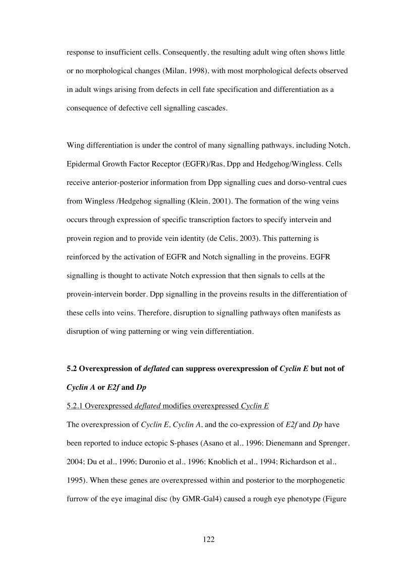
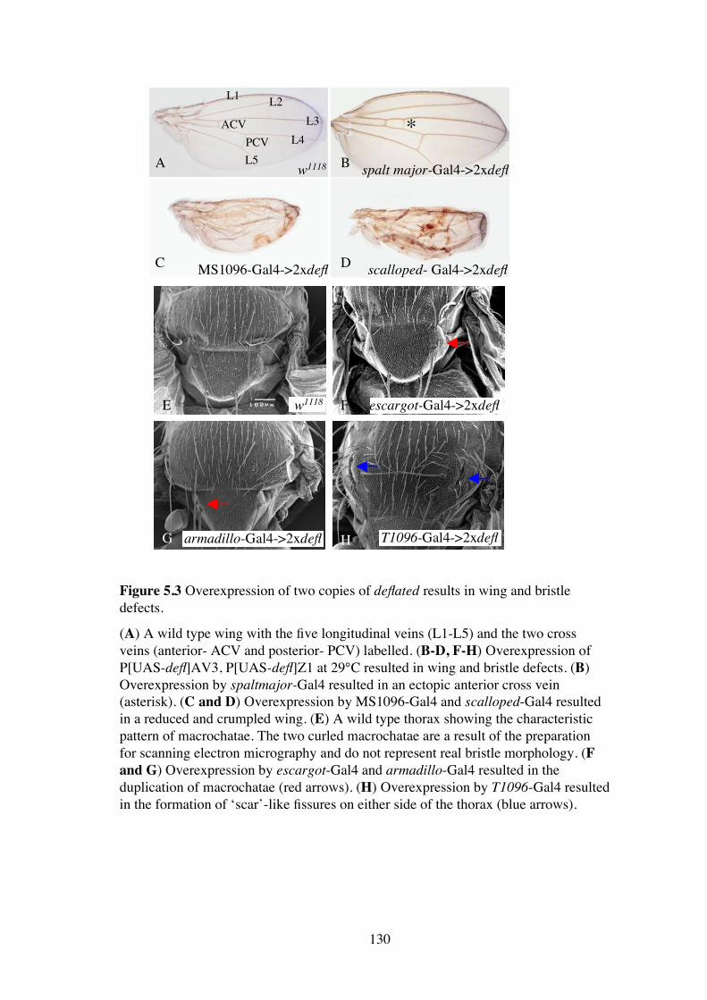
Overexpression of two copies of the deflated cDNA by the Gal4 wing drivers, spalt
major-Gal4 (Figure 5.3 B), MS1096-Gal4 (Figure 5.3 C), and scalloped-Gal4 (Figure
5.3 D), resulted in wings that had an extra posterior cross vein (asterisk, Figure 5.3 B)
or a reduced and crumpled wing (Figure 5.3 C and D). Low level, ubiquitous Gal4
drivers such as armadillo-Gal4 and escargot-Gal4 resulted in a mild phenotype in
which the machrochaetae on the notum, particularly the scutellum, were frequently
duplicated (Figure 5.3 F and G). In addition, an unusual phenotype was observed with
the driver T1096-Gal4. The notum contained “scar” like fissures on either side (Figure
5.3 H). The overexpression caused by the Gal4 driver MS1096 was chosen to test
genetic interactions, as its effect was moderate so modification (enhancement or
suppression) should be easily scored.
5.3.2 deflated interacts genetically with Irregular facets
In the course of these overexpression experiments, it was noted that overexpressed
deflated could suppress the Irregular facets (If) rough eye phenotype (Figure 5.4 A, B
and C). This observation arose because the scalloped-Gal4 driver line contains the If
A B
C D
E F
G H
*L1 L2
L3L4
L5
ACVPCV
w1118 spalt major-Gal4->2xdefl
MS1096-Gal4->2xdefl scalloped- Gal4->2xdefl
w1118 escargot-Gal4->2xdefl
armadillo-Gal4->2xdefl T1096-Gal4->2xdefl
Figure 5.3 Overexpression of two copies of deflated results in wing and bristledefects.
(A) A wild type wing with the five longitudinal veins (L1-L5) and the two crossveins (anterior- ACV and posterior- PCV) labelled. (B-D, F-H) Overexpression ofP[UAS-defl]AV3, P[UAS-defl]Z1 at 29°C resulted in wing and bristle defects. (B)Overexpression by spaltmajor-Gal4 resulted in an ectopic anterior cross vein(asterisk). (C and D) Overexpression by MS1096-Gal4 and scalloped-Gal4 resultedin a reduced and crumpled wing. (E) A wild type thorax showing the characteristicpattern of macrochatae. The two curled macrochatae are a result of the preparationfor scanning electron micrography and do not represent real bristle morphology. (Fand G) Overexpression by escargot-Gal4 and armadillo-Gal4 resulted in theduplication of macrochatae (red arrows). (H) Overexpression by T1096-Gal4 resultedin the formation of ‘scar’-like fissures on either side of the thorax (blue arrows).
130
131
allele on the second chromosome and, in scoring these flies, a difference between If
flies with and without the deflated cDNA was noted. While scalloped-Gal4 is
predominantly a wing driver, expression of a UAS-GFP construct by this driver
showed some fluorescence in third instar eye imaginal discs (data not shown), which
implies that deflated is overexpressed to a small degree in these eyes when driven by
scalloped-Gal4. If is a gain of function allele of the transcription factor Krüppel
(Carrera et al., 1998). This allele causes misexpression of Krüppel in the eye, which
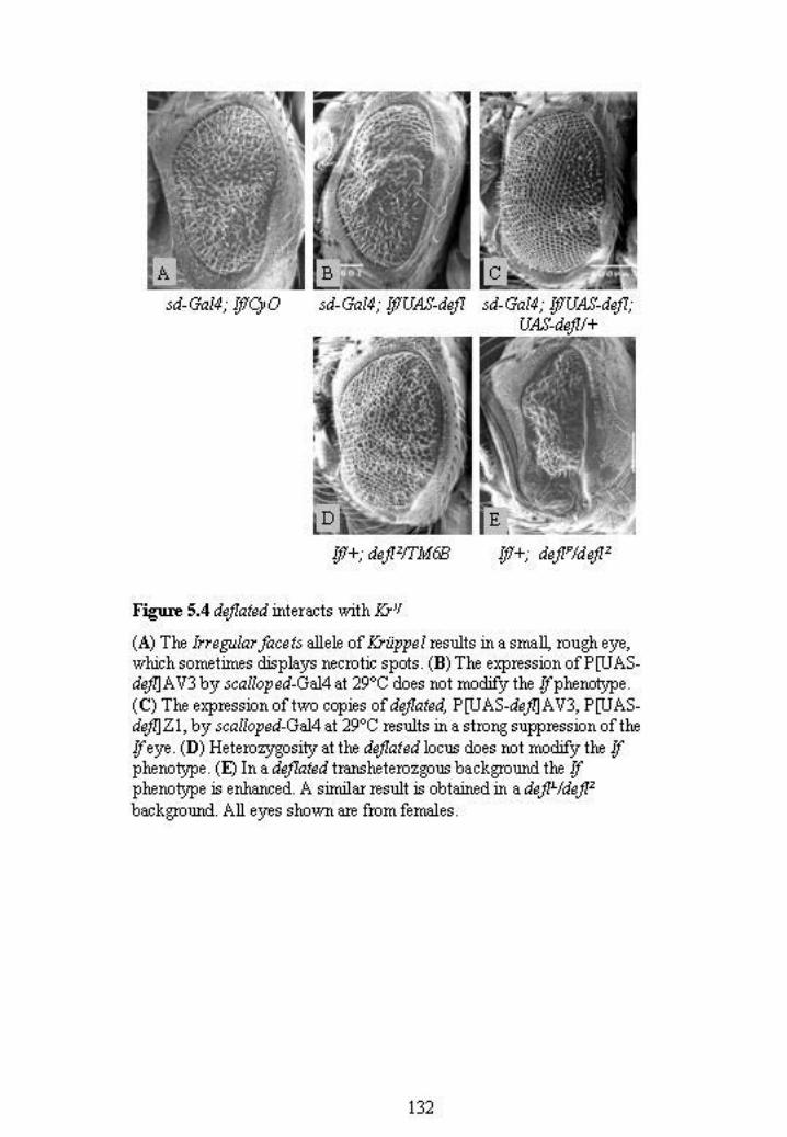
results in a reduced eye with fused ommatidia and necrotic spots (Figure 5.4 A). This
phenotype can vary between individuals and is more severe in males. Although the
expression of one copy of deflated did not modify the If phenotype (Figure 5.4 B), the
expression of two copies (P[UAS-defl]AV3; P[UAS-defl]Z1) resulted in strong
suppression (Figure 5.4 C).
To further understand the genetic relationships between deflated and Krüppel, the effect
of loss of wild type deflated function on If was examined. The If phenotype in deflated
heterozygotes looked similar to milder If eyes and deflated was therefore considered to
not be a dominant modifier (Figure 5.4 D). However, the If eye phenotype was strongly
enhanced in deflP/deflZ (Figure 5.4 E) and deflL/deflZ (data not shown)
transheterozygotes. These observations support an antagonistic role for deflated in
Krüppel function as loss of wild type DEFLATED protein caused a worsening of the If
gain of function phenotype.
5.3.3 Identification of candidate dominant modifiers of the deflated overexpression
wing phenotype
To test candidate genetic interactors, a recombined second chromosome containing two
133
copies of the defl cDNA (P[UAS-defl]BB1, P[UAS-defl]BA2) was generated and
balanced over a CyO(Act-GFP) balancer (to aid in genotyping larvae) and driven by the
MS1096-Gal4 driver, which is on chromosome X. This combination of deflated
transgenes was used to make the testing easier due to both transgenes being on the same
chromosome. The initial overexpression phenotypes were generated in flies that
contained one second chromosome copy and one third chromosome copy (P[UAS-
defl]AV3, P[UAS-defl]Z1). The wing phenotype was less severe in flies containing the
two second chromosome transgenes than one on II, one on III combination. This
observation indicates that the wing phenotype was dose dependent and likely to be
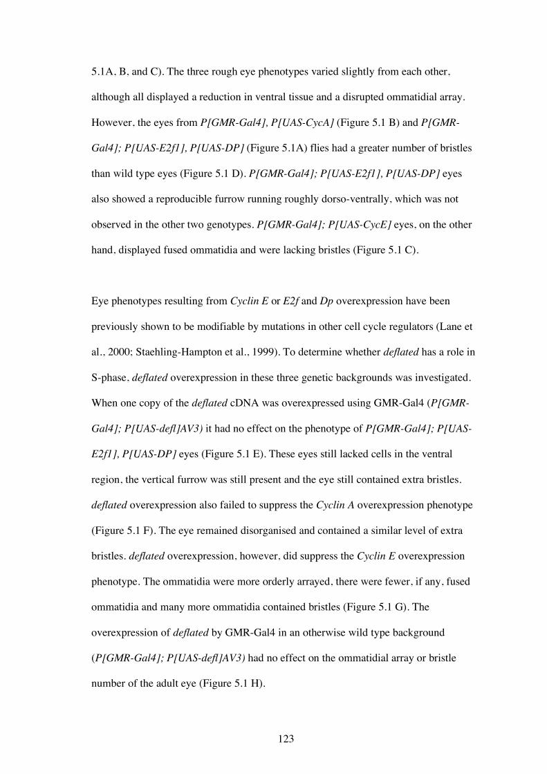
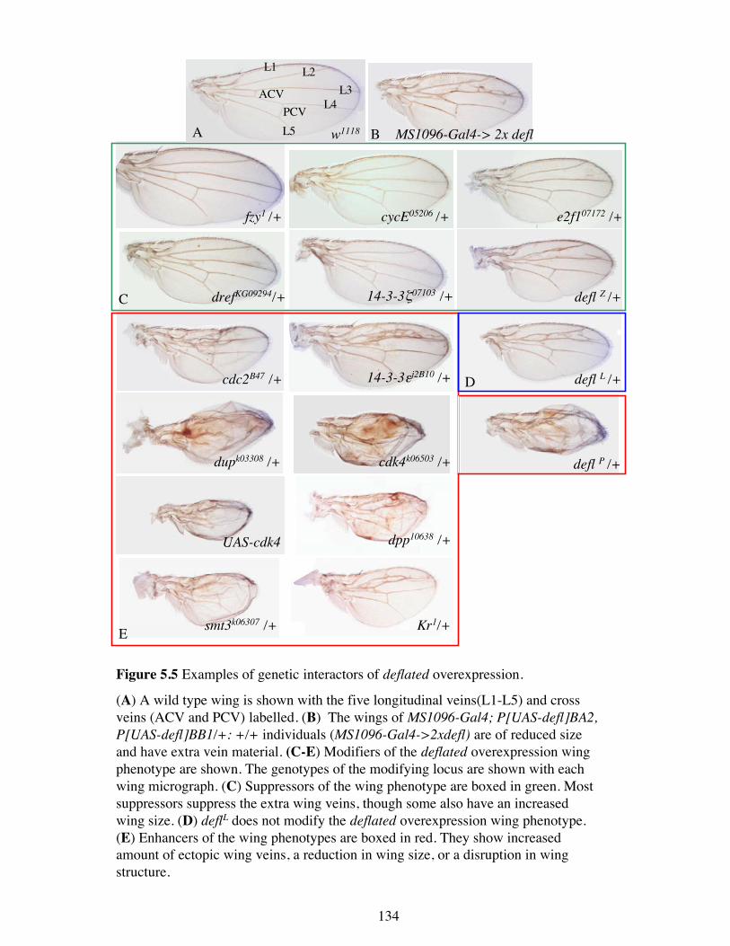
modifiable. The overexpression of P[UAS-defl]BB1, P[UAS-defl]BA2 resulted in a
smaller wing than wild type with ectopic wing vein material anterior to L3 and a slight
reduction in the intervein region between L3 and L4 around the anterior cross vein
(Figure 5.5 B).
Known cell proliferation genes, cell signalling genes, and other genes that were
identified as potential deflated interactors were tested for their ability to dominantly
modify the deflated overexpression wing phenotype. This type of test measures the
ability of an allele to modify the deflated wing phenotype as a heterozygote, which
otherwise shows no phenotype in a wild type background. Modification indicates that
the gene tested acts in a similar biological pathway to deflated. This is because the wing
has become sensitised by the deflated overexpression to changes in the levels of
proteins that act in similar biological pathways. Many of the alleles tested were loss of
function alleles, however some genes were tested for the ability to modify while
overexpressed. Many of the candidate genes tested were able to modify the wing
phenotype (Table 5.1). The strength of the modification (either enhancement or
MS1096-Gal4-> 2x defl
14-3-3εj2B10 /+
14-3-3ζ07103 /+drefKG09294/+
e2f107172 /+
cdc2B47 /+
cdk4k06503 /+
cycE05206 /+
dpp10638 /+
dupk03308 /+
fzy1 /+
Kr1/+
defl L /+
defl Z /+
defl P /+
smt3k06307 /+
UAS-cdk4
A
L1
L5
ACVPCV
w1118 B
C
E
D
Figure 5.5 Examples of genetic interactors of deflated overexpression.
(A) A wild type wing is shown with the five longitudinal veins(L1-L5) and crossveins (ACV and PCV) labelled. (B) The wings of MS1096-Gal4; P[UAS-defl]BA2,P[UAS-defl]BB1/+: +/+ individuals (MS1096-Gal4->2xdefl) are of reduced sizeand have extra vein material. (C-E) Modifiers of the deflated overexpression wingphenotype are shown. The genotypes of the modifying locus are shown with eachwing micrograph. (C) Suppressors of the wing phenotype are boxed in green. Mostsuppressors suppress the extra wing veins, though some also have an increasedwing size. (D) deflL does not modify the deflated overexpression wing phenotype.(E) Enhancers of the wing phenotypes are boxed in red. They show increasedamount of ectopic wing veins, a reduction in wing size, or a disruption in wingstructure.
L4L3
L2
134
135
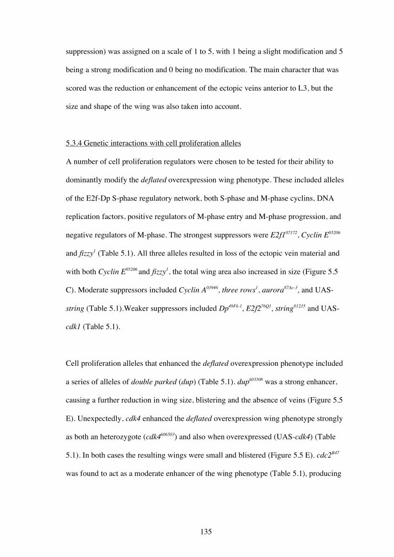
suppression) was assigned on a scale of 1 to 5, with 1 being a slight modification and 5
being a strong modification and 0 being no modification. The main character that was
scored was the reduction or enhancement of the ectopic veins anterior to L3, but the
size and shape of the wing was also taken into account.
5.3.4 Genetic interactions with cell proliferation alleles
A number of cell proliferation regulators were chosen to be tested for their ability to
dominantly modify the deflated overexpression wing phenotype. These included alleles
of the E2f-Dp S-phase regulatory network, both S-phase and M-phase cyclins, DNA
replication factors, positive regulators of M-phase entry and M-phase progression, and
negative regulators of M-phase. The strongest suppressors were E2f107172, Cyclin E05206
and fizzy1 (Table 5.1). All three alleles resulted in loss of the ectopic vein material and
with both Cyclin E05206 and fizzy1, the total wing area also increased in size (Figure 5.5
C). Moderate suppressors included Cyclin A03946, three rows1, aurora87Ac-3, and UAS-
string (Table 5.1).Weaker suppressors included Dp49Fk-1, E2f276Q1, string01235 and UAS-
cdk1 (Table 5.1).
Cell proliferation alleles that enhanced the deflated overexpression phenotype included
a series of alleles of double parked (dup) (Table 5.1). dupk03308 was a strong enhancer,
causing a further reduction in wing size, blistering and the absence of veins (Figure 5.5
E). Unexpectedly, cdk4 enhanced the deflated overexpression wing phenotype strongly
as both an heterozygote (cdk4k06503) and also when overexpressed (UAS-cdk4) (Table
5.1). In both cases the resulting wings were small and blistered (Figure 5.5 E). cdc2B47
was found to act as a moderate enhancer of the wing phenotype (Table 5.1), producing
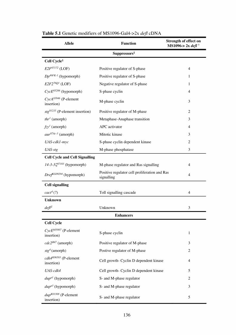
3UnknowndeflZ
Unknown
5S- and M-phase regulatordupK03308 (P-element
insertion)
3S- and M-phase regulatordupa3 (hypomorph)
2S- and M-phase regulatordupa1 (hypomorph)
5Cell growth- Cyclin D dependent kinaseUAS-cdk4
4Cell growth- Cyclin D dependent kinasecdk4K06503 (P-element
insertion)
2Postive regulator of M-phasestg4 (amorph)
3Positive regulator of M-phasecdc2B47 (amorph)
1S-phase cyclinCycEk05007 (P-element
insertion)
Cell Cycle
Enhancers
4Positive regulator cell proliferation and Ras
signallingDrefKG09294 (hypomorph)
Suppressors§
3M-phase phosphataseUAS-stg
2S-phase cyclin dependent kinaseUAS-cdk1-myc
3Mitotic kinaseaur87Ac-3 (amorph)
4APC activatorfzy1 (amorph)
4S-phase cyclinCycE05206 (hypomorph)
3M-phase cyclinCycA03946 (P-element
insertion)
Cell signalling
4Toll signalling cascadecact4 (?)
4M-phase regulator and Ras signalling14-3-3!07103 (hypomorph)
Cell Cycle and Cell Signalling
3Metaphase-Anaphase transitionthr1 (amorph)
2Positive regulator of M-phasestg01235 (P-element insertion)
1Negative regulator of S-phaseE2F276Q1 (LOF)
1Positive regulator of S-phaseDp49FK-1 (hypomorph)
4Positive regulator of S-phaseE2F07172 (LOF)
Cell Cycle§
Strength of effect onMS1096-> 2x defl †FunctionAllele
Table 5.1 Genetic modifiers of MS1096-Gal4->2x defl cDNA
136
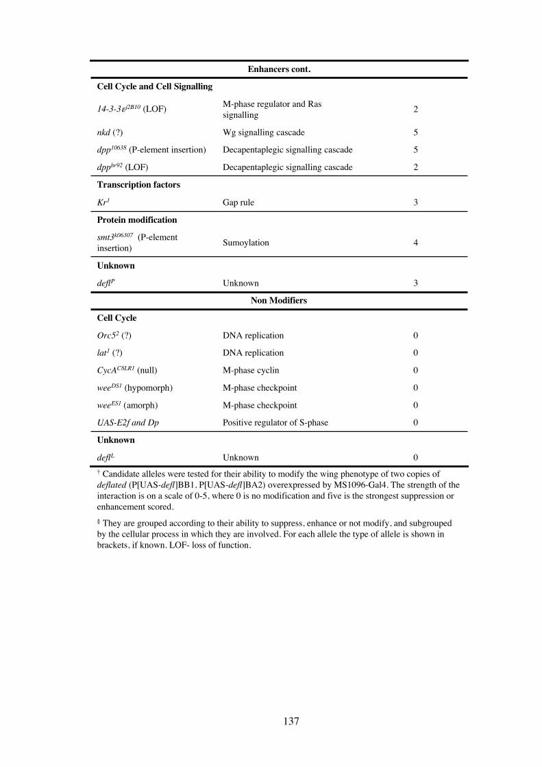
2M-phase regulator and Ras
signalling14-3-3"j2B10 (LOF)
Cell Cycle and Cell Signalling
0UnknowndeflL
Unknown
0Positive regulator of S-phaseUAS-E2f and Dp
0M-phase checkpointweeES1 (amorph)
0M-phase checkpointweeDS1 (hypomorph)
0M-phase cyclinCycAC8LR1 (null)
0DNA replicationlat1 (?)
0DNA replicationOrc52 (?)
Cell Cycle
Non Modifiers
3UnknowndeflP
Unknown
Protein modification
4Sumoylationsmt3k06307 (P-element
insertion)
3Gap ruleKr1
Transcription factors
2Decapentaplegic signalling cascadedpphr92 (LOF)
5Decapentaplegic signalling cascadedpp10638 (P-element insertion)
5Wg signalling cascadenkd (?)
Enhancers cont.
† Candidate alleles were tested for their ability to modify the wing phenotype of two copies of
deflated (P[UAS-defl]BB1, P[UAS-defl]BA2) overexpressed by MS1096-Gal4. The strength of the
interaction is on a scale of 0-5, where 0 is no modification and five is the strongest suppression or
enhancement scored.
§ They are grouped according to their ability to suppress, enhance or not modify, and subgrouped
by the cellular process in which they are involved. For each allele the type of allele is shown in
brackets, if known. LOF- loss of function.
137

138
smaller wings that contained small blisters (Figure5.5 E). Weak enhancers of deflated
overexpression included string4 and Cyclin Ek05007 (Table 5.1).
Alleles of genes that were found to not modify the deflated overexpression wing
phenotype included those involved in DNA replication (Orc52 and lat1), the M-phase
checkpoint (weeDS1 and weeES1), Cyclin AC8LR1, and co-overexpression of E2f and Dp
(Table 5.1). This last result is consistent with the findings observed in the eye, where
overexpressed deflated could not modify overexpressed E2f and Dp.
Three of the genes tested for genetic interaction with deflated are involved in regulating
the cell cycle as well as having roles in regulating the Ras signalling pathway. These
are the 14-3-3 isoforms ε and ζ and the transcription factor DREF. 14-3-3 proteins have
established roles in the regulation of M-phase entry (Sections 1.1.3 and 1.1.4) as well as
regulating Raf, MEK and MAPK activation (Section 1.2.1). The two different 14-3-3
isoforms differed in their ability to modify the deflated overexpression wing phenotype.
14-3-3εJ2B10 was a weak enhancer with the resulting wings containing an increase in
ectopic vein material (Figure 5.5 E). On the other hand, 14-3-3ζ07103 was a strong
suppressor and the resulting wings displayed no ectopic vein material (Figure 5.5 C).
The transcription factor DREF was originally identified as required for the transcription
of DNA replication and S-phase genes such as Cyclin A and E2f1 (Ohno et al., 1996;
Sawado et al., 1998), but it also transcriptionally activates members of the Ras
signalling pathway (Ryu et al., 1997; Yoshida et al., 2004). The DrefKG09294 allele was a
strong suppressor of the deflated wing overexpression phenotype, resulting in wings
lacking the ectopic vein material (Figure 5.5 C).
139
5.3.5 deflated interacts genetically with various cell signalling pathway components
The L5 wing vein defect observed in transheterozygous adult deflated escapees
suggested that deflated may affect signalling cascades involved in wing vein
differentiation (Section 4.7.5). The deflated overexpression wing phenotype (Figure 5.5
B) supports this conclusion. The formation of ectopic vein material anterior to L3
indicates that, when overexpressed, deflated interferes with normal differentiation,
possibly through perturbation of cell signalling pathways. To explore this possibility, a
number of alleles of genes encoding components of various signalling cascades were
tested for their ability to dominantly modify the deflated wing overexpression
phenotype.
Only one allele tested was able to suppress the wing phenotype. This was the Toll
pathway component, cactus4 (Table 5.1). The two dpp alleles tested, dpphr92 and
dpp10638, enhanced the phenotype (Table 5.1), with dpphr92 was weakly enhancing and
dpp10638 strongly enhancing. The resulting wings from the dpp10638 cross were reduced in
size and blistered, with some extra wing vein material (Figure 5.5 E). Naked cuticle
(nkd), a member of the Wingless signalling cascade, was also a strong enhancer (Table
5.1). Taken together, these data show that deflated can interact genetically with a
number of cell signalling components.
5.3.6 deflated also interacts genetically with Krüppel in the wing
Since deflated antagonises the effects of Krüppel misexpression in the eye (Figure 5.4),
it was assessed whether Krüppel could also dominantly modify the deflated
overexpression wing phenotype. Kr1 was able to moderately enhance the deflated wing
phenotype (Table 5.1). The resulting wings were reduced in size and the proximal-distal
140
axis was shortened. There was a further decrease in the intervein region between L2
and L3 and the presence of some ectopic vein material (Figure 5.5 E). This result
further supports an antagonistic role between deflated and Krüppel, as reducing
Krüppel activity enhances the deflated overexpression phenotype.
5.3.7 deflated interacts with smt3, which encodes SUMO
An allele of smt3, which encodes the ubiquitin-like protein SUMO, was tested to assess
whether the conserved putative SUMO-modification site (a.a. 872-875; Figure 4. 2) in
DEFLATED may be functional. Unlike ubiquitin, the conjugation of SUMO to proteins
does not target them for degradation by the proteosome, but is thought to protect them
from degradation by preventing ubiquitination (Shih et al., 2001). Halving the dose of
wild type SUMO (smt3k06307) resulted in enhancement of the deflated overexpression
phenotype (Table 5.1). The resulting wing was reduced in size and contained blisters
(Figure 5.5 E). This finding indicates that DEFLATED may be directly modified by
SUMO conjugation with the modification serving to negatively regulate DEFLATED
function. Alternatively, SUMO may regulate another protein that interacts with
DEFLATED.
5.3.8 Genetic interaction between the deflated alleles
The deflated alleles generated in Chapter 3 were tested for their ability to suppress the
deflated overexpression wing phenotype. If the phenotype was due to excess but normal
function of deflated, then it would be expected that reducing the dose of wild type
deflated by mutation would suppress the wing phenotype. Unexpectedly, only deflZ
could suppress the wing overexpression phenotype. Wings from deflZ heterozygotes
resulted in a near absence of the ectopic wing vein material (Figure 5.5 C). deflL failed
141
to modify the phenotype and deflP enhanced the deflated wing overexpression
phenotype (Table 5.1). In deflL heterozygote wings, the amount and positioning of the
ectopic veins remained unchanged (Figure 5.5 D). However, wings from deflP
heterozygotes were smaller and blistered and the veins were not easily observed (Figure
5.5 E). Taken together, these data indicate that the genetic relationships between the
deflated alleles and its overexpression are not straightforward, indicating perhaps that
not all of the deflated alleles are hypomorphic in nature.
5.4 Discussion
In an attempt to further define the cellular role(s) of deflated, a series of genetic studies
were performed. It should be noted that in interpreting these data it was assumed that
the interacting loci were indeed the candidates tested even though there is the
possibility that other unknown loci on the chromosome may be mediating the
interaction. The genetic interactions observed with overexpressed deflated support a
role for deflated in regulating the cell cycle. In particular, interactions with Cyclin E
suggested that deflated has a negative role in regulating S-phase in the differentiating
part of the eye imaginal disc. These genetic interaction data also support a possible role
for deflated in cell signalling, a finding given further weight by the nature of the
phenotypes observed when deflated is overexpressed. Duplication of macrochaetae and
disruption of normal wing vein specification and formation are very suggestive of
perturbation of signalling pathways. Surprisingly, these interactions show that deflated
is a likely antagonist of Krüppel, a finding that could not be predicted by the putative
protein domains or expression patterns.
142
5.4.1 deflated’s role in regulating S-phase
Overexpression of deflated was not able to suppress the rough eye phenotype of Cyclin
A (Figure 5.1 F). Unlike previous reports (Dienemann and Sprenger, 2004), Cyclin A
was found to not induce ectopic S-phases posterior to the morphogenetic furrow (Figure
5.2 E and E′), which indicates that the rough eye phenotype in Figure 5.1 B is not due
to an increase in cells entering S-phase. Whatever the cause of the rough eye
phenotype, it is unlikely that deflated is involved as deflated failed to modify the Cyclin
A overexpression rough eye phenotype. This study overexpressed Cyclin A using GMR-
Gal4 (which drives expression in the morphogenetic furrow and all the posterior cells),
whereas the previous study used sev-Gal4 (which is expressed in a subset of the
photoreceptor cells). It is possible that cells in the morphogenetic furrow or just
posterior are not sensitive to increases in Cyclin A levels and are therefore not induced
into an ectopic S-phase. Cells further away from the furrow, such as those in which sev-
Gal4 is expressed may be more sensitive and able therefore to undergo an ectopic S–
phase. A likely explanation for the proposed increased resistance to Cyclin A levels in
the morphogenetic furrow is the expression of roughex in the furrow, which is a known
inhibitor of Cyclin A and is one of the gene products required for the G1 arrest (Section
1.3.3 ; Avedisov et al., 2000; Thomas et al., 1994).
Overexpressed deflated was also not able to modify the effects of co-expression of E2f1
and Dp in the eye. Since the co-overexpression of E2f and Dp is known to induce
cellular processes such as apoptosis as well as ectopic S-phases (Asano et al., 1996; Du
et al., 1996), the rough eye phenotype is likely to be caused by the additive or
synergistic effects of these different cellular processes. As deflated overexpression
failed to modify this eye phenotype, it is likely that deflated does not play a role in all
143
of the same cellular processes as E2f and Dp. Alternatively, if deflated is a
transcriptional target of E2F (Section 4.7.2), then it would already be upregulated when
E2f and Dp are overexpressed and further overexpression may not have an affect. The
finding that overexpression of E2f and Dp in the wing did not modify the deflated wing
overexpression phenotype (Table 5.1) argues against the second alternative because if
deflated was a transcriptional target then increasing its expression through
overexpression of E2f and Dp would result in enhancement of the phenotype. In
conclusion, deflated overexpression and E2f and Dp overexpression do not interact
genetically, indicating that even though deflated mutants phenocopy E2f and Dp
mutants (4.7.3), the proteins are likely to act in separate cellular pathways.
The finding that deflated overexpression suppressed the ectopic S-phases induced by
Cyclin E overexpression suggests that deflated may act a negative regulator of S-phase.
This finding seems to contradict the observations of Chapter 4, in which cells from deflP
homozygous brains were less likely to be in S-phase at any given time, which indicates
that deflated may be required positively for S-phase. However, brains from deflL
homozygous individuals showed a slight increase of cells in S-phase, therefore
supporting a conclusion that deflated is a negative regulator of S-phase. Since deflated
overexpression only had an effect on the ectopic S-phases that occurred in cells that
should be differentiating in the eye (Figure 5.2 C and C′), it is possible that deflated
may play different roles depending on the requirements of the cell. In cells that should
be proliferating, such as the brain, deflated is required for S-phase, but in cells that
should be differenting, deflated in required to inhibit S-phase. In the wing the genetic
relationships between deflated and Cyclin E further obscure deflated’s role in regulating
S-phase. Two alleles of Cyclin E were tested, Cyclin E05206 strongly suppressed the
144
deflated wing overexpression phenotype, whereas Cyclin Ek05007 weakly enhanced the
phenotype. These allelic differences indicate that even within one tissue deflated seems
to have opposing roles in regulating S-phase.
In order to understand deflated’s interaction with Cyclin E further, it was assessed
whether decreasing the genetic dose of wild type deflated could modify the rough eye
phenotype. It was found that none of the four deflated alleles modified the rough eye
phenotype. Alleles of dpp, which is a known genetic interactor of Cyclin E, also failed
to modify. It is possible that overexpressed Cyclin E may result in too much Cyclin E
protein for modifications to be observed. This notion is supported by the finding that
many more interactors were identified when a Cyclin E hypomorphic allele, Cyclin EJP,
was used (Brumby et al., 2004) rather than Cyclin E overexpression (Lane et al., 2000).
The dpp interaction was observed using the Cyclin EJP allele (Horsfield et al., 1998).
Since this allele is sensitive to modifiers, it would be interesting to see if the deflated
alleles could dominantly modify this allele. Brumby et al. (2004) did find that a
deficiency (Df(3L)AC1) covering the deflated region (67A2-67D7-13) enhanced the
Cyclin EJP phenotype. If deflated is a negative regulator of S-phase, as the
overexpression interactions suggest (Figure 5.2), then it is unlikely that deflated is the
causative locus in the deficiency. However, if deflated is a positive regulator of
S-phase, as the analysis of brains from deflP/deflP second instar larvae seems to suggest
(Section 4.7.6), then it is possible that deflated is the causative locus identified by
Brumby et al. (2004).
145
5.4.2 deflated may be involved in linking DNA synthesis with M-phase entry
What is the role of deflated in regulating S-phase? At this stage the data is
contradictory. Many cell cycle regulators interact genetically with deflated when it is
overexpressed in the wing (Table 5.1) but interpreting these interactions has proved
difficult. As already discussed, the interactions with Cyclin E and E2f and Dp in the
developing eye and the requirements for deflated in second instar larval brains suggest
opposing roles for deflated in S-phase regulation. However, from the genetic
interactions observed between deflated overexpression and various cell cycle
regulators, it appears that deflated may be involved in linking DNA synthesis and its
completion with M-phase entry. Five distinct sets of data are consistent with this
interpretation.
Firstly, a number of positive regulators of cell cycle progression, E2f1, fzy, thr, and aur
suppressed the deflated overexpression wing phenotype. This indicates that the wing
phenotype is in part due to an increase in cell proliferation. Therefore, from these data,
it appears that overexpressed deflated causes an increase in cell cycle progression,
which can be reduced by decreasing the levels of these positive regulators.
Secondly, two genes for which the observed interactions were consistent were cdc2
(cdk1) and dup, which both have roles in ensuring that DNA synthesis must occur
before mitosis can begin. Halving the dose of cdc2 resulted in suppression of the wing
phenotype and overexpression of cdk1 resulted in enhancement. Taken together these
data suggest that the overexpression of deflated either results in the upregulation of
cdk1 or acts in a pathway that can be affected by cdk1 activity. All three alleles of dup
that were tested enhanced the deflated overexpression wing phenotype. dup encodes the
146
cdt1 homologue, which is a pre-replication complex component (Thomer et al., 2004).
Homozygous dup mutants are unable to replicate their DNA in postblastoderm cycles
yet enter mitosis with unreplicated DNA (Whittaker et al., 2000). As mitosis and DNA
synthesis are uncoupled in dup mutants, and dup can enhance the deflated
overexpression phenotype, it is possible that overexpressed deflated may also cause an
uncoupling of mitosis and DNA synthesis. If this is the case then an explanation can be
provided for the finding that halving the dose of cdc2 suppresses the deflated
overexpression phenotype, while increasing the cdc2 enhances the deflated
overexpression phenotype. Prevention of M-phase entry by decreasing cdc2 results in
fewer cells overexpressing deflated entering M-phase with unreplicated or partially
replicated DNA and the severity of the phenotype is thereby reduced.
Thirdly, the notion that overexpressed deflated uncouples DNA synthesis with M-phase
entry fits with the finding that wee alleles fail to modify the overexpression phenotype.
The M-phase checkpoint is thought to be triggered by the presence of unreplicated
DNA. The checkpoint is not triggered if DNA synthesis is not initated, as in dup
mutants, and therefore M-phase occurs (Whittaker et al., 2000). Therefore, changes in
wee gene dosage would have no effect. If DNA replication does not occur when
deflated is overexpressed then the observation that lat and Orc5 also fail to modify the
overexpression phenotype also makes sense.
Fourthly, the genetic interactions with the two 14-3-3 isoforms could be due to the roles
that these proteins play in regulating M-phase, their roles in regulating Ras signalling
(see Chapter 6), or to some other cellular process. In Drosophila 14-3-3ε is required to
time mitosis and effects a mitotic delay following irradiation in post-blastoderm
147
embryos by keeping cdk1 in check, whereas 14-3-3ζ is required for correct
chromosome segregation in syncytial embryos (Su et al., 2001). Halving the dose of
14–3-3ε enhanced the deflated overexpression phenotype, which indicates that if 14-3-3
and deflated interact in regulating M-phase then overexpression of deflated is likely to
result in an increase in M-phase entry. This interpretation is consistent with the
interpretations of the dup and cdc2 interactions (see above). Halving the 14-3-3ζ dose
resulted in the suppression of the deflated overexpression phenotype and this finding is
also consistent with the interpretation that deflated overexpression may induce
inappropriate M-phase entry. Reducing the amount of 14-3-3ζ may prevent or slow
down APC/C activation, affecting the timing of chromosome segregation and mitotic
exit, thereby counteracting the effects of premature M-phase entry.
Fifthly, the finding that a Dref allele can dominantly suppress the deflated
overexpression phenotype also supports an interpretation that deflated overexpression
causes premature M-phase. DREF is required for the transcription of many cell cycle
genes including those required for M-phase entry (Hyun et al., 2005). When DREF
activity is reduced the transcription of these M-phase regulators is also reduced and
therefore M-phase entry would take longer. However, since DREF induces the
transcription of many cell cycle genes the interaction observed may reflect a different
role for deflated.
In contrast the evidence discussed above, the finding that heterozygosity and
overexpression of the M-phase regulator string (Section 1.3.3) do not consistently
modify the deflated overexpression phenotype clouds the argument that deflated
overexpression causing premature M-phase entry. stg01235 was observed to be a weak
148
suppressor, whereas stg4 caused weak enhancement, and overexpression of string
resulted in moderate suppression. At this point is is difficult to reconcile these findings
with the postulated role of deflated in regulating M-phase entry once S-phase is
completed.
The genetic interactions observed between deflated and cdk4 suggest that deflated may
affect the regulation of cellular growth and M-phase entry. However, as cdk4 was found
to enhance the deflated overexpression phenotype when the former was both
heterozygotic or overexpressed, it is likely that deflated has an indirect effect on
cellular growth and the role cdk4 plays in M-phase. It is possible that when cdk4 is
overexpressed alone it could result in a small and crumpled wing and therefore not
really be enhancing the deflated overexpression phenotype. However, previous studies
have shown that the overexpression of cdk4 without co-overexpression of Cyclin D in
the wing does not result in a phenotype (Datar et al., 2000). Therefore the severe
phenotype observed must be due to either enhancement of deflated overexpression or
an additive effect of overexpression of the two genes. Since CyclinD/Cdk4 can also
promote G2-M progression (Datar et al., 2000; Neufeld et al., 1998), it is possible that
the genetic interactions observed may reflect a potential role for deflated in the
regulation of M-phase entry. Thus, the finding that cdk4 enhances when either
heterozygous or when overexpressed may reflect both of these roles.
Taken as a whole, these genetic interaction data do support a case for deflated being
involved in linking DNA replication with M-phase entry. This model for deflated
function is also consistent with the findings of cell cycle progression in second instar
larval brains. In brains from homozygous deflL and deflP individuals, cells were less
149
likely to be in M-phase, indicating that deflated is required for M-phase entry (Section
4.7.6). To test this hypothesis, further analyses of the genetic interactions that focus on
DNA replication and M-phase entry would be required.
5.4.3 The deflated overexpression phenotype suggests a role for deflated in cell
signalling
Many cells and tissues are able to tolerate deflated overexpression with no obvious
developmental perturbations. Two copies of the cDNA transgene were required before
phenotypes were observed and even then only the wing and the notum showed
abnormalities (Figure 5.3). The wing phenotypes observed included extra veins, a
reduction in the wing size, or a reduction in the intervein region. These phenotypes may
well be due to perturbation of cell proliferation, however they are equally reminiscent
of cell signalling phenotypes, particularly of member of Ras and Dpp pathways
(Ralston and Blair, 2005).
The conclusion that deflated overexpression may perturb signalling pathways is further
supported by the duplication of mainly scutellar macrochaetae when deflated is
overexpressed. Macrochaetae are sensory organs and their specification and formation
are under the control of a number of signalling pathways, including Notch, EGFR/Ras,
Dpp, and Gbb (Culi et al., 2001; Tomoyasu et al., 1998; Wharton et al., 1999). The mild
macrochaetae phenotype observed when deflated is overexpressed is most like that seen
in glass bottom boat (gbb) mutants or when there is a slight reduction in EGFR
signalling (Clifford and Schubach, 1989; Diaz-Benjumea and Garcia-Bellido, 1990;
Wharton et al., 1999). Simarily to the deflated overexpression phenotype,
extramacrochaetae mutants also display extra macrochaetae over the notum, due to its
150
role in regulating responsiveness to the proneural signals of achaete and scute (Cubas
and Modolell, 1992). Its activity is regulated by both EGFR and Notch signalling. emc
mutants also show ectopic wing veins and emc acts antagonistically with Ras during
wing vein differentiation (Baonza and Garcia-Bellido, 1999). Since deflated and emc
have similar phenotypes it is possible that they provide similar function or act in similar
pathways. Both ectopic wing vein formation and macrochaetae duplication observed
when deflated is overexpressed strongly supports a role for deflated in regulating cell
signalling.
To test the role of deflated in cell signalling, cactus from the Toll signalling pathway,
dpp from the Decapentapelgic (TGFβ) pathway and nkd from the Wingless pathway
were tested for their ability to modify the deflated overexpression phenotype.
cactus was found to dominantly suppress the deflated wing phenotype (Table 5.1). Toll
signalling is involved in dorsal/ventral patterning of the egg and embryo and in innate
immunity (Hoffmann, 2003). CACTUS (an IκB homologue) binds DORSAL (an NF-
κB homologue) and prevents it from entering the nucleus in the absence of signal
(Kidd, 1992). Upon signalling, CACTUS becomes phosphorylated, ubiquitinated, and
degraded by the proteosome. DORSAL is also phosphorylated and is able to enter the
nucleus and transcriptionally activate ventral genes and suppress dorsal genes (Drier et
al., 1999; Whalen and Steward, 1993). There is no evidence to suggest that the Toll
pathway plays a role in wing development. However, CACTUS and DORSAL play
Toll-independent roles, and seemingly cooperative roles, in muscle and neuromuscular
junction development and function in larva and adults (Bolatto et al., 2003; Cantera et
151
al., 1999), so it is possible that the suppression of with deflated overexpression in the
wing reflects an unknown function of cactus.
In mammals, NF-κB is activated in response to apoptotic signals. It acts in a protective
capacity in response to tumour necrosis factor receptor, but can induce a pro-apoptotic
response to p53 (Ryan et al., 2000). Other pathways can also activate NF-κB, including
a Raf/MAPK-dependent but Ras-independent pathway (Ghoda et al., 1997; Schouten et
al., 1997). While it is not known if DORSAL is involved in apoptotic pathways in
Drosophila, it is formally possible that it may and the genetic interaction observed
between cactus and deflated reflects this role. While it is difficult to know how deflated
and cactus interact at this point, it can be concluded that deflated overexpression may
result in increased CACTUS levels or may affect some other process that is ameliorated
by reduced CACTUS levels.
Dpp signalling is required for proper wing vein formation, with the cross veins being
most sensitive to Dpp loss. The deflated wing overexpression phenotype is similar to
increased Dpp signalling (Conley et al., 2000) but it more closely phenocopies ectopic
gbb driven by ptc-Gal4 (Khalsa et al., 1998). The protein encoded by gbb, like Dpp, is a
BMP ligand and the relative abundance of the two is important for proper wing
development, as demonstrated by the fact that ectopic dpp can counteract ectopic gbb
(Khalsa et al., 1998). Therefore, the finding that dpp alleles dominantly enhance the
deflated phenotype (Table 5.1 and Figure 5.5 E) is difficult to interpret if Dpp
signalling is considered on its own. However, if the defect is an increase in Gbb
signalling, then loss of dpp would exacerbate the phenotype as the balance between
Dpp and Gbb activities would be further disrupted.
152
This postulated disruption of relative Dpp and Gbb levels could also explain the
interaction with naked cuticle (Table 5.1). naked cuticle is an antagonist of the
Wingless signalling pathway (Zeng et al., 2001) that can function to repress Dpp
signalling in establishing dorso-ventral patterning (Johnston and Shubiger, 1996).
Therefore halving the dose of naked cuticle will increase Wingless signalling, which
would cause a reduction in Dpp signalling. Together these data support a conclusion
that overexpressing deflated may perturb Dpp signalling either directly or indirectly.
5.4.4 deflated is an antagonist of Krüppel
The finding that deflated interacts antagonistically with Krüppel was unexpected. In
Drosophila, Krüppel has been most studied in its role as a gap gene during
embryogenesis (Pankratz and Jackle, 1993). It is required to specify segments during
embryogenesis by repressing the expression of other gap and pair-rule genes. In
mammals there are 16 Krüppel-like factors (Suske et al., 2005). Interestingly, some of
them have opposing roles in regulating cell proliferation (Ghaleb et al., 2005). KLF4
acts as a negative regulator of cell proliferation, in part through the p53 pathway (Yoon
et al., 2003), whereas KLF5 is a positive regulator of cell proliferation and mediates the
oncogenic potential of Ras (Nandan et al., 2004). KLF2 represses the transcription of
WEE1 in response to DNA damage, thereby preventing M-phase entry (Wang et al.,
2005) and it also upregulates the cdk inhibitor p21 (Wu and Lingrel, 2004). KLF6 can
also prevent cell proliferation by inhibiting Cyclin D1 (Benzeno et al., 2004). Unlike
the mammalian proteins, there is no experimental evidence to suggest that Drosophila
genes Krüppel, Krüppel-homologue 1 or Krüppel-homologue 2 have a role in the
regulation of cell proliferation but shuch a role cannot be excluded.
153
The ability of overexpressed deflated to suppress the eye phenotype of If, the loss of
wild type deflated to enhance the phenotype, and the ability of Krüppel to dominantly
enhance overexpressed deflated, when taken together, implies an antagonistic
relationship between the two gene products. It is possible that Krüppel antagonises
deflated but, as the loss of deflated in the transheterozygotes does not on its own result
in a rough eye phenotype, the If phenotype could not be due to reduced deflated
function. Therefore, it is more likely that deflated antagonises Krüppel. This
antagonism could be upstream of Krüppel, directed at Krüppel itself, or by activating
Krüppel targets (Figure 5.6). It is unlikely that it is directed at Krüppel targets for two
reasons. Firstly, it is expected that there are many Krüppel targets. Secondly, the targets
are likely to differ between the eye and the wing. Since deflated has the same effects on
Krüppel acitivity in two different tissues, it is unlikely to regulate all Krüppel targets.
Alternatively, deflated could be upstream of Krüppel, for example affecting its
transcription or translation, or it could directly affect Krüppel as an inhibitor by
affecting its cellular localisation or its turnover. Further studies would be required to
determine how deflated antagonises Krüppel and how this relates to the functional
capacity of deflated. While it is possible that there is an unknown function for Krüppel
in cell proliferation or cell signalling regulation, it is more likely that deflated affects
Krüppel’s known roles.
Krüppel
deflated
?
?
downstream targets
?
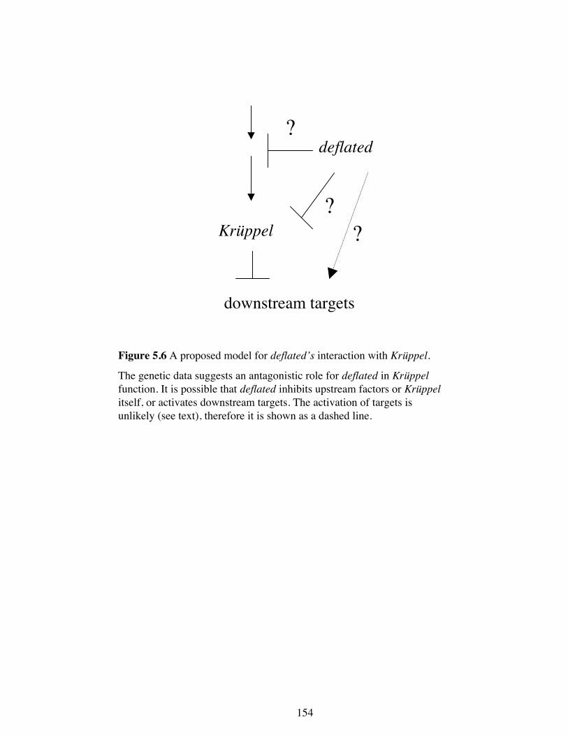
Figure 5.6 A proposed model for deflated’s interaction with Krüppel.
The genetic data suggests an antagonistic role for deflated in Krüppelfunction. It is possible that deflated inhibits upstream factors or Krüppelitself, or activates downstream targets. The activation of targets isunlikely (see text), therefore it is shown as a dashed line.
154
155
In further support of an antagonistic role for deflated in Krüppel function is the finding
that loss of deflated phenocopies a downstream target of Krüppel, ken and barbie (ken),
which is repressed by Krüppel activity (Matyash et al., 2004). Mutations in ken result in
the absence of external genitalia and the posterior abdominal segment, which is similar
to what is seen in the deflated transheterozygotes (Figure 4.4). It is possible that the
lack of genitalia in the deflated mutants is due to loss of inhibition of Krüppel, which
would result in less KEN activity.
5.4.5 Genetic interaction studies provide information on how DEFLATED may be
regulated
The findings that DEFLATED contains a putative 14-3-3 binding site and a putative
SUMO modification site (Figure 4.2) and that overexpressed deflated interacts
genetically with both 14-3-3 isoforms and smt3 (Figure 5.5) indicate that DEFLATED
may be directly regulated by these proteins. Furthermore, the interactions between three
of the deflated alleles and deflated overexpression also provide some insight on how
DEFLATED may be regulated.
The different abilities of the different deflated alleles to modify the overexpression of
deflated allows for the speculation upon which regions of DEFLATED may mediate its
own regulation. Many cells seem to tolerate excess deflated so if it is assumed that the
overexpression phenotype in the wing is due to an inability of these cells to tolerate the
excess deflated by not correctly regulating its normal activity then the genetic
interactions may identify a region of negative regulation. Examination of the different
alleles’ abilities to modify the overexpression phenotype and the protein products that
these alleles are predicted to produce should aid in identifying this regulatory region.
156
deflZ was the only deflated allele that suppressed the overexpression wing phenotype.
This is surprising because it is predicted to be the least truncated of the mutant proteins
(Figure 5.7), only lacking 15 amino acids from the C-terminus and it might therefore be
expected to provide near-wild type function. As this is was observed to not be the case
it suggests that deflZ protein behaves neither like the exogenous full length protein nor
the endogenous wild type protein. Instead, the apparent activity of deflated is reduced,
which results in an almost wild type wing. How this allele produces a seemingly
antimorphic phenotype is currently unknown.
In contrast to deflZ, deflL was observed to not dominantly modify the deflated wing
overexpression phenotype. This suggests that the predicted truncated protein encoded
by deflL is able to act like the wild type protein in this assay. deflP does not reduce
deflated function nor does it produce any more in this assay. As the DEFLL protein
appears to be regulated as though it were wild type endogenous protein it is likely
therefore to contain the necessary regulatory region(s).
Surprisingly, deflP was found to enhance the deflated wing overexpression phenotype.
While is it formally possible that this surprising result may be due to an uncharacterised
lesion not linked to the deflated locus, assuming that it is due to the deflP suggests that
the DEFLP product behaves in a similar manner to the overexpressed protein. One
explanation for this is that DEFLP is lacking the necessary regulatory region(s) that act
to hold the wild type protein in check. When these regulatory mechanisms are swamped
by excess wild type protein, DEFLATED is no longer correctly regulated. When
DEFLP is also present there is even more protein being misregulated and the phenotype
1001
986
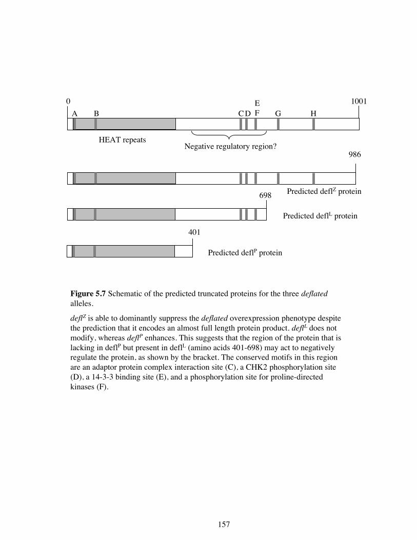
Figure 5.7 Schematic of the predicted truncated proteins for the three deflatedalleles.
deflZ is able to dominantly suppress the deflated overexpression phenotype despite
the prediction that it encodes an almost full length protein product. deflL does not
modify, whereas deflP enhances. This suggests that the region of the protein that is
lacking in deflP but present in defl
L (amino acids 401-698) may act to negatively
regulate the protein, as shown by the bracket. The conserved motifs in this region
are an adaptor protein complex interaction site (C), a CHK2 phosphorylation site
(D), a 14-3-3 binding site (E), and a phosphorylation site for proline-directed
kinases (F).
0
HEAT repeats
A B C D
E
F G H
Predicted deflZ protein
Predicted deflL protein
Predicted deflP protein
Negative regulatory region?
401
698
157
158
is worsened. Therefore, these regulatory regions are likely to lie in the region of the
protein that is present in DEFLL (because it is still properly regulated) but are absent in
DEFLP (because it is not properly regulated). Examination of the predicted protein
products shows that this region comprises the amino acids 401-698 (Figure 5.7).
Therefore, this region is likely to comprise a regulatory region. This region contains a
number of conserved putative protein motifs; one of the adaptor complex binding sites,
a Chk2 phosphorylation site, a 14-3-3 binding site, and a phosphorylation site for
proline-directed kinases. Any of these sites, either separately or in combination, could
serve to negatively regulate DEFLATED. Alternatively, the negative regulation could
be due to another as yet unidentified motif.
The finding that deflated overexpression can be modified by halving the amount of
14-3-3 proteins indicates that the putative 14-3-3 binding site at amino acids 650-655
may be a genuine binding site that mediates this negative regulatory effect.
Interestingly, it appears that the 14-3-3ε protein may act to negatively regulate
DEFLATED function as 14-3-3ε enhanced the deflated overexpression phenotype
when heterozygous. It is possible that DEFLATED could also be positively regulated
by 14-3-3ζ at this site, since halving its dose was able to suppress the excess deflated
activity. If the 14-3-3 site is important (and sufficient) for the regulation of
DEFLATED activity, then mutation at this site in an otherwise wild type protein should
phenocopy the effects of deflated overexpression and the phenotype of the deflP allele.
While not recognised as being within the postulated regulatory region of amino acids
401-698, it is possible that DEFLATED may also be regulated by SUMO modification.
SUMO modification generally acts to activate its targets. However, the genetic data
159
supports an interpretation that DEFLATED would be negatively regulated by
sumoylation, as reducing the amount of SUMO resulted in a enhancement of the
deflated overexpression phenotype. In Drosophila, SUMO and its conjugation and
deconjugation machinery are localised to the nucleus in older embryos and S2 cells
(Shih et al., 2001; Smith et al., 2004). As a result nearly all SUMO-conjugated proteins
are nuclear. Recently it has been reported that sumoylation acts at the plasma
membrane to regulate ion channel function (Rajan et al., 2005). Therefore, if
DEFLATED is a SUMO target it is likely that it would localise to the nucleus but it is
also possible that it would localise to the plasma membrane. Alternatively, the SUMO
modification site may not be functional and the genetic interaction observed between
smt3 and deflated is caused by a reduction in the activation of some negative regulator
of DEFLATED, causing the worsening of the phenotype observed. To differentiate
between these alternatives biochemical analysis of DEFLATED will be required to test
the involvement of SUMO conjugation in its regulation.
5.4.6 Conclusions about deflated function
The conclusions about deflated function that can be drawn from the genetic interactions
described in this chapter are: (i) it interacts with Cyclin E and can act as a negative
regulator of S-phase; (ii) it may be involved in linking DNA replication completion
with M-phase entry; (iii) it appears to be a negative effector of Dpp signalling; (iv) it is
an antagonist of Krüppel; (v) it may contain a negative regulatory region between
amino acids 401 and 698, which may involve 14-3-3 binding at amino acids 650-655;
and (vi) it may be modified by SUMO. Obviously these tests were not exhaustive,
particularly in regards to examining the role of deflated in various signalling pathways.
The genes and pathways examined were chosen in part because the alleles were readily

160
available in the lab or from the Bloomington Stock Centre and the results obtained
suggest further genetic analysis is required to fully understand the role of deflated in
these pathways. Apart from further genetic analysis, in depth cell biology and
biochemical studies will be required to determine the mechanisms of deflated function
and regulation.
161
Chapter 6- deflated is involved in Ras signalling
6.1 Introduction
Control of the cell cycle is coordinated with growth and development in multicellular
organisms through signalling pathways. Some signalling pathways result in
upregulation of cell proliferation, while some result in downregulation (Section 1.2).
Therefore these signalling pathways play important roles during development and
tumourogenesis. One pathway that has received much attention is the RTK/Ras
signalling pathway since it plays important roles in differentiation and cell proliferation
(Section 1.2.2). Disruption of this pathway can result in perturbation of cell
differentiation as observed in Drosophila adult wings and eyes (de Celis, 2003; Simon
et al., 1991), or it can result in the loss of cell proliferation regulation as observed in
over 30% of human cancers (Chang et al., 2003). Therefore an understanding of this
pathway and of its regulation will aid in understanding how cell proliferation and
differentiation are coordinately controlled.
Some of the phenotypes displayed by deflated mutants and when deflated is
overexpressed suggests that deflated may play a role in regulating cell signalling. These
phenotypes include the loss of some of the L5 vein in the deflated transheterozgyote
wings (Section 4.7.5) and the formation of extra vein material and the duplication of
macrochaetae when deflated is overexpressed (Section 5.4.3). Furthermore, deflated
interacts genetically with components of three different signalling cascades, Toll, Dpp,
and Wg. Since deflated affects both cell proliferation and cell signalling, it is pertinent
to know whether the role of deflated is primarily one of cell proliferation or cell
signalling.
sev-RasV12/+ sev-RasV12/+; deflP/+ sev-RasV12/+; deflΔ/+
w1118 sev-RasV12/+; deflZ/+ sev-RasV12/+; deflL/+
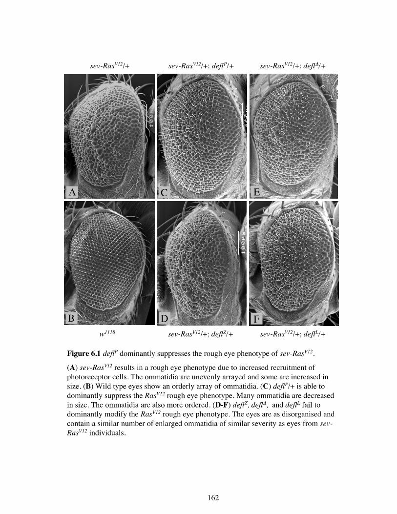
Figure 6.1 deflP dominantly suppresses the rough eye phenotype of sev-RasV12.
(A) sev-RasV12 results in a rough eye phenotype due to increased recruitment ofphotoreceptor cells. The ommatidia are unevenly arrayed and some are increased insize. (B) Wild type eyes show an orderly array of ommatidia. (C) deflP/+ is able todominantly suppress the RasV12 rough eye phenotype. Many ommatidia are decreasedin size. The ommatidia are also more ordered. (D-F) deflZ, deflΔ, and deflL fail todominantly modify the RasV12 rough eye phenotype. The eyes are as disorganised andcontain a similar number of enlarged ommatidia of similar severity as eyes from sev-RasV12 individuals.
A C E
B D F
162
163
To further examine the role(s) of deflated in cell signalling, a Drosophila tumour model
of activated Ras (RasV12) was used. RasV12 is a constitutive allele that is unable to
hydrolyse GTP, thus it remains in an active state. When RasV12 is expressed in the
developing eye under the sevenless promoter (sev-RasV12), a rough eye phenotype with
disorganised and blistered-looking ommatidia results due to cone cells adopting an R7
photoreceptor fate (Fortini et al., 1992). The RasV12 model has been used extensively to
identify many of the members of the Ras signalling cascade through genetic
modification of the rough eye phenotype (Chang and Rubin, 1997; Karim et al., 1996).
Since in this model the only cells that are affected are post-mitotic and the outcome is
differentiation, it was hoped that the function of deflated in signalling could be further
investigated in a context where cell proliferation does not play a role.
6.2 Genetic interactions between Ras signalling components and deflated
6.2.1 deflated interacts genetically with RasV12
To test the effects of altered deflated expression on the RasV12 eye phenotype, the four
different deflated alleles (deflZ, deflL, deflP and deflΔ) were crossed into the RasV12
background and their heterozygous effects on the Ras phenotype were assessed. deflP
was able to suppress the rough eye phenotype producing ommatidia that were less
disorganised and most were of normal size (Figure 6.1 B). Unexpectedly, deflL, deflZ,
and deflΔ failed to modify the sev-RasV12 rough eye phenotype (Figure 6.1 D-F). The
resulting eyes were equally disorganised and contained as many enlarged ommatidia as
the sev-RasV12 eye (Figure 6.1A). This result suggested that deflated may act
downstream from activated Ras or in a parallel pathway that affects the Ras signalling
outcome. Since the null allele of deflated (deflΔ) does not suppress the Ras phenotype
sev-RasV12/+ sev-RasV12/+;deflP/deflZ
sev-RasV12/+;deflL/deflZ
No adultseclosed
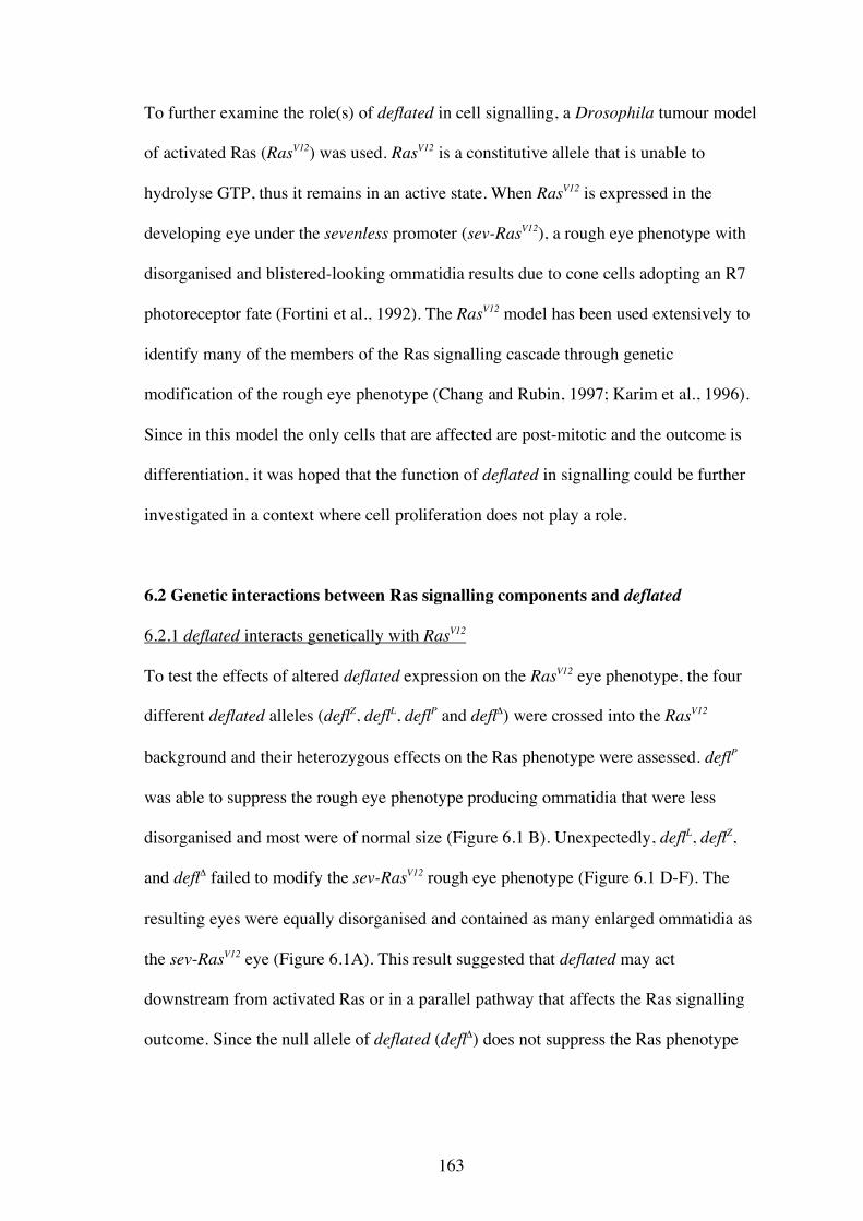
Figure 6.2 The phenotype of sev-RasV12 is enhanced in a deflatedtransheterozygous background.
(A) sev-RasV12 results in a rough eye phenotype. (B) The expression of sev-RasV12
in a deflP/deflZ transheterozygous background results in an enhancement of therough eye phenotype. (C) sev-RasV12/+; deflL/deflZ flies fail to eclose,presumably due to strong enhancement of Ras signalling leading to lethality.
A B C
164
w1118/+; +/+;
sev-RasV12/+w1118
/+; sev-Gal4/+;
sev-RasV12/UAS-deflw1118
/+; +/+;
sev-RasV12/UAS-defl
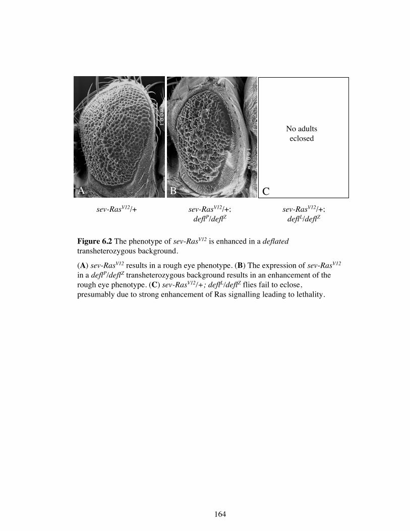
Figure 6.3 Overexpression of deflated suppresses the sev-RasV12 eye
phenotype.
(A) sev-RasV12 results in a rough eye phenotype. (B) Expression of P[UAS-defl]AV3 by sev-Gal4 or (C) leaky expression of P[UAS-defl]AV3 in the
absence of Gal4 suppresses the rough eye phenotype in a dose-sensitive
manner.
A B C
165
166
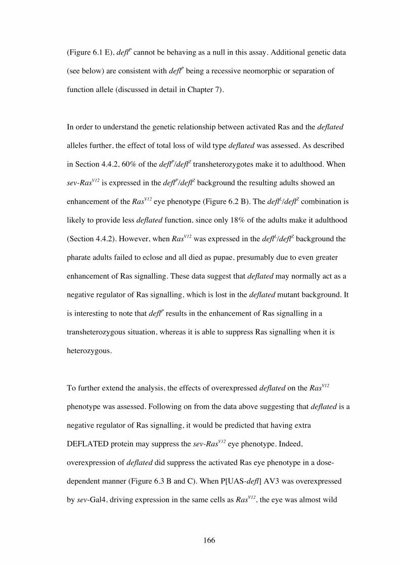
(Figure 6.1 E), deflP cannot be behaving as a null in this assay. Additional genetic data
(see below) are consistent with deflP being a recessive neomorphic or separation of
function allele (discussed in detail in Chapter 7).
In order to understand the genetic relationship between activated Ras and the deflated
alleles further, the effect of total loss of wild type deflated was assessed. As described
in Section 4.4.2, 60% of the deflP/deflZ transheterozygotes make it to adulthood. When
sev-RasV12 is expressed in the deflP/deflZ background the resulting adults showed an
enhancement of the RasV12 eye phenotype (Figure 6.2 B). The deflL/deflZ combination is
likely to provide less deflated function, since only 18% of the adults make it adulthood
(Section 4.4.2). However, when RasV12 was expressed in the deflL/deflZ background the
pharate adults failed to eclose and all died as pupae, presumably due to even greater
enhancement of Ras signalling. These data suggest that deflated may normally act as a
negative regulator of Ras signalling, which is lost in the deflated mutant background. It
is interesting to note that deflP results in the enhancement of Ras signalling in a
transheterozygous situation, whereas it is able to suppress Ras signalling when it is
heterozygous.
To further extend the analysis, the effects of overexpressed deflated on the RasV12
phenotype was assessed. Following on from the data above suggesting that deflated is a
negative regulator of Ras signalling, it would be predicted that having extra
DEFLATED protein may suppress the sev-RasV12 eye phenotype. Indeed,
overexpression of deflated did suppress the activated Ras eye phenotype in a dose-
dependent manner (Figure 6.3 B and C). When P[UAS-defl] AV3 was overexpressed
by sev-Gal4, driving expression in the same cells as RasV12, the eye was almost wild
A B
C D
sev-Gal4, rlSEM/ +; +/+ sev-Gal4, rlSEM/+; deflP/+
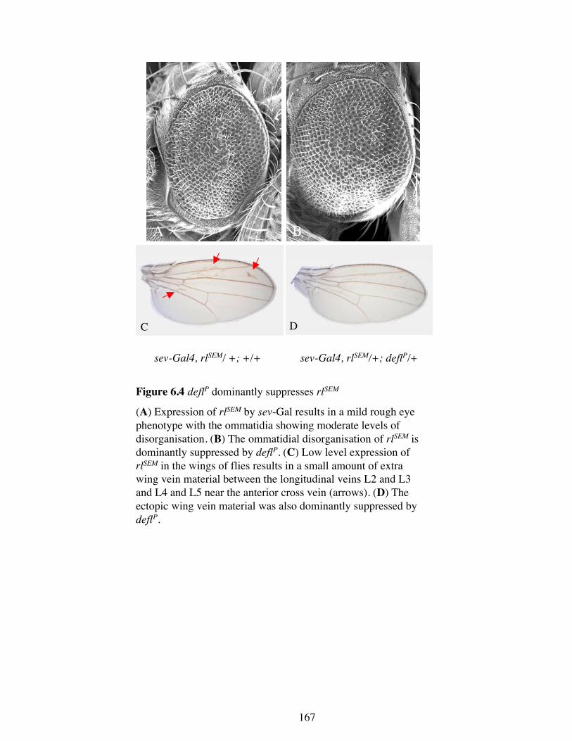
Figure 6.4 deflP dominantly suppresses rlSEM
(A) Expression of rlSEM by sev-Gal results in a mild rough eyephenotype with the ommatidia showing moderate levels ofdisorganisation. (B) The ommatidial disorganisation of rlSEM isdominantly suppressed by deflP. (C) Low level expression ofrlSEM in the wings of flies results in a small amount of extrawing vein material between the longitudinal veins L2 and L3and L4 and L5 near the anterior cross vein (arrows). (D) Theectopic wing vein material was also dominantly suppressed bydeflP.
167
168
type and shows very mild ommatidial disorganisation (Figure 6.3 B). Leaky expression
of the P[UAS-defl] AV3 transgene in the absence of a Gal4 driver also suppressed the
rough eyes, though to a lesser extent (Figure 6.3 C). These data implicate deflated as a
negative regulator of Ras signalling.
6.2.2 deflP dominantly suppresses the MAPK allele rlSEM
If deflated acts as a negative regulator of Ras signalling then it be predicted to also
interact with other Ras signalling cascade components such as MAPK (Section 1.2). To
test this, deflP was crossed into a rlSEM background. rlSEM is a hypermorphic allele of
rolled, the gene that encodes MAPK. This allele results in both a mild eye and wing
phenotype. Increased Ras signalling in the eyes, caused by rlSEM, results in enhanced
recruitment of R7 cells, resulting in a rough eye phenotype (Figure 6.4 A). Increased
Ras signalling in the wing results in ectopic vein formation between L2 and L3 and
around the anterior cross vein (Figure 6.4 C). deflP was found to be able to suppress
both the eye and wing phenotypes of the rlSEM allele. The eyes were more organised
(Figure 6.4 B) and the wings no longer contained the ectopic wing veins (Figure 6.4 D).
Since the rlSEM allele is still capable of responding to upstream signals, the suppression
observed does not allow deflated to be placed either upstream or downstream of
MAPK. However, the ability of deflP to suppress the rlSEM phenotypes shows that the
effects of deflated are not restricted to activated Ras and that deflated can consistently
modify the Ras signalling pathway.
6.3 Overexpression of deflated affects the activation of MAPK
Since deflated can affect Ras signalling it was possible that the ectopic vein material
observed in wings overexpressing deflated (Figure 5.3) was due to perturbation of Ras
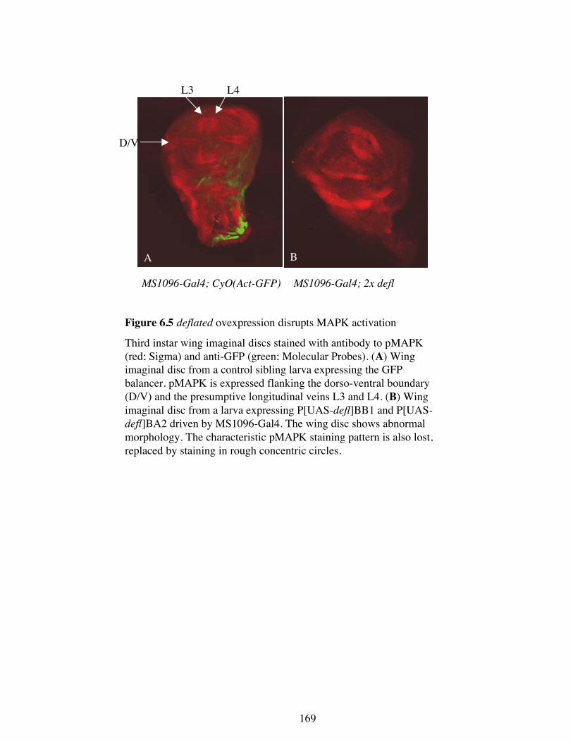
A B
MS1096-Gal4; CyO(Act-GFP) MS1096-Gal4; 2x defl
Figure 6.5 deflated ovexpression disrupts MAPK activation
Third instar wing imaginal discs stained with antibody to pMAPK(red; Sigma) and anti-GFP (green; Molecular Probes). (A) Wingimaginal disc from a control sibling larva expressing the GFPbalancer. pMAPK is expressed flanking the dorso-ventral boundary(D/V) and the presumptive longitudinal veins L3 and L4. (B) Wingimaginal disc from a larva expressing P[UAS-defl]BB1 and P[UAS-
defl]BA2 driven by MS1096-Gal4. The wing disc shows abnormalmorphology. The characteristic pMAPK staining pattern is also lost,replaced by staining in rough concentric circles.
D/V
L3 L4
169
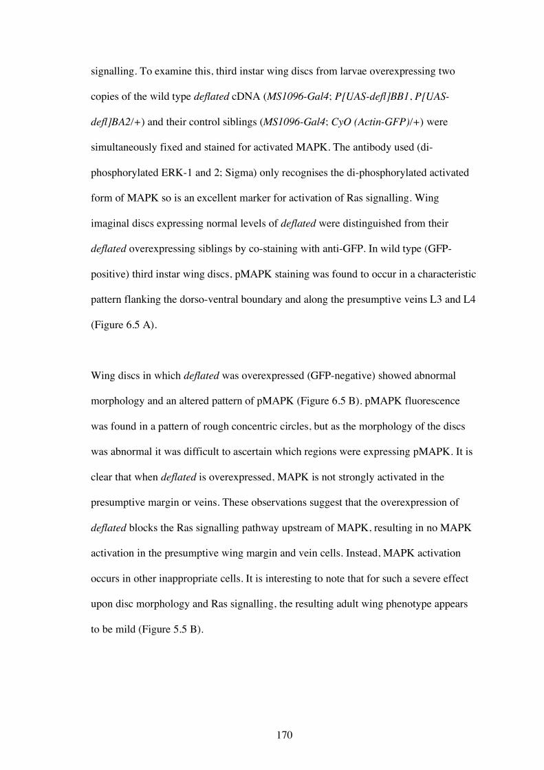
170
signalling. To examine this, third instar wing discs from larvae overexpressing two
copies of the wild type deflated cDNA (MS1096-Gal4; P[UAS-defl]BB1, P[UAS-
defl]BA2/+) and their control siblings (MS1096-Gal4; CyO (Actin-GFP)/+) were
simultaneously fixed and stained for activated MAPK. The antibody used (di-
phosphorylated ERK-1 and 2; Sigma) only recognises the di-phosphorylated activated
form of MAPK so is an excellent marker for activation of Ras signalling. Wing
imaginal discs expressing normal levels of deflated were distinguished from their
deflated overexpressing siblings by co-staining with anti-GFP. In wild type (GFP-
positive) third instar wing discs, pMAPK staining was found to occur in a characteristic
pattern flanking the dorso-ventral boundary and along the presumptive veins L3 and L4
(Figure 6.5 A).
Wing discs in which deflated was overexpressed (GFP-negative) showed abnormal
morphology and an altered pattern of pMAPK (Figure 6.5 B). pMAPK fluorescence
was found in a pattern of rough concentric circles, but as the morphology of the discs
was abnormal it was difficult to ascertain which regions were expressing pMAPK. It is
clear that when deflated is overexpressed, MAPK is not strongly activated in the
presumptive margin or veins. These observations suggest that the overexpression of
deflated blocks the Ras signalling pathway upstream of MAPK, resulting in no MAPK
activation in the presumptive wing margin and vein cells. Instead, MAPK activation
occurs in other inappropriate cells. It is interesting to note that for such a severe effect
upon disc morphology and Ras signalling, the resulting adult wing phenotype appears
to be mild (Figure 5.5 B).
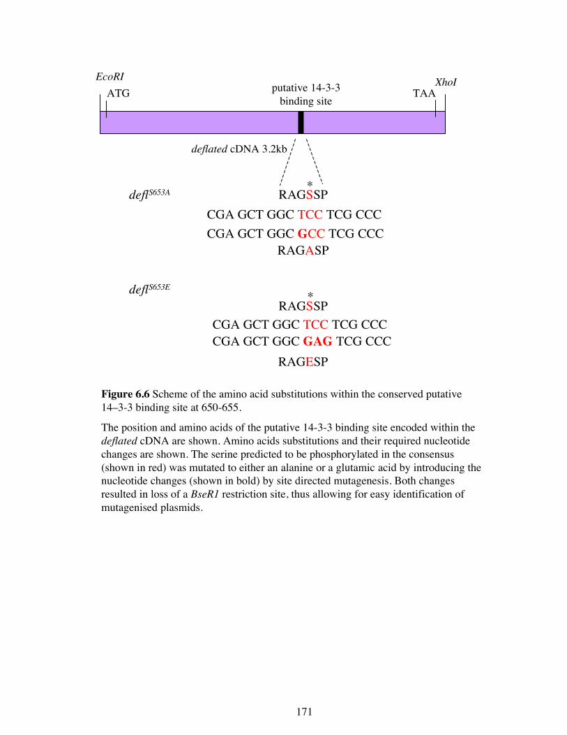
Figure 6.6 Scheme of the amino acid substitutions within the conserved putative14–3-3 binding site at 650-655.
The position and amino acids of the putative 14-3-3 binding site encoded within thedeflated cDNA are shown. Amino acids substitutions and their required nucleotidechanges are shown. The serine predicted to be phosphorylated in the consensus(shown in red) was mutated to either an alanine or a glutamic acid by introducing thenucleotide changes (shown in bold) by site directed mutagenesis. Both changesresulted in loss of a BseR1 restriction site, thus allowing for easy identification ofmutagenised plasmids.
deflated cDNA 3.2kb
EcoRI XhoIATG TAAputative 14-3-3
binding site
RAGSSP*
CGA GCT GGC TCC TCG CCCCGA GCT GGC GCC TCG CCC
RAGASP
deflS653A
RAGSSPCGA GCT GGC TCC TCG CCCCGA GCT GGC GAG TCG CCC
RAGESP
deflS653E *
171
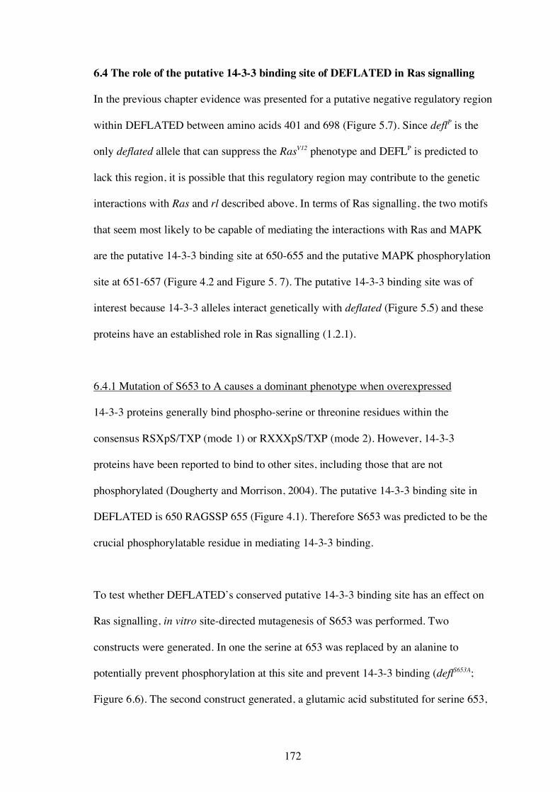
172
6.4 The role of the putative 14-3-3 binding site of DEFLATED in Ras signalling
In the previous chapter evidence was presented for a putative negative regulatory region
within DEFLATED between amino acids 401 and 698 (Figure 5.7). Since deflP is the
only deflated allele that can suppress the RasV12 phenotype and DEFLP is predicted to
lack this region, it is possible that this regulatory region may contribute to the genetic
interactions with Ras and rl described above. In terms of Ras signalling, the two motifs
that seem most likely to be capable of mediating the interactions with Ras and MAPK
are the putative 14-3-3 binding site at 650-655 and the putative MAPK phosphorylation
site at 651-657 (Figure 4.2 and Figure 5. 7). The putative 14-3-3 binding site was of
interest because 14-3-3 alleles interact genetically with deflated (Figure 5.5) and these
proteins have an established role in Ras signalling (1.2.1).
6.4.1 Mutation of S653 to A causes a dominant phenotype when overexpressed
14-3-3 proteins generally bind phospho-serine or threonine residues within the
consensus RSXpS/TXP (mode 1) or RXXXpS/TXP (mode 2). However, 14-3-3
proteins have been reported to bind to other sites, including those that are not
phosphorylated (Dougherty and Morrison, 2004). The putative 14-3-3 binding site in
DEFLATED is 650 RAGSSP 655 (Figure 4.1). Therefore S653 was predicted to be the
crucial phosphorylatable residue in mediating 14-3-3 binding.
To test whether DEFLATED’s conserved putative 14-3-3 binding site has an effect on
Ras signalling, in vitro site-directed mutagenesis of S653 was performed. Two
constructs were generated. In one the serine at 653 was replaced by an alanine to
potentially prevent phosphorylation at this site and prevent 14-3-3 binding (deflS653A;
Figure 6.6). The second construct generated, a glutamic acid substituted for serine 653,
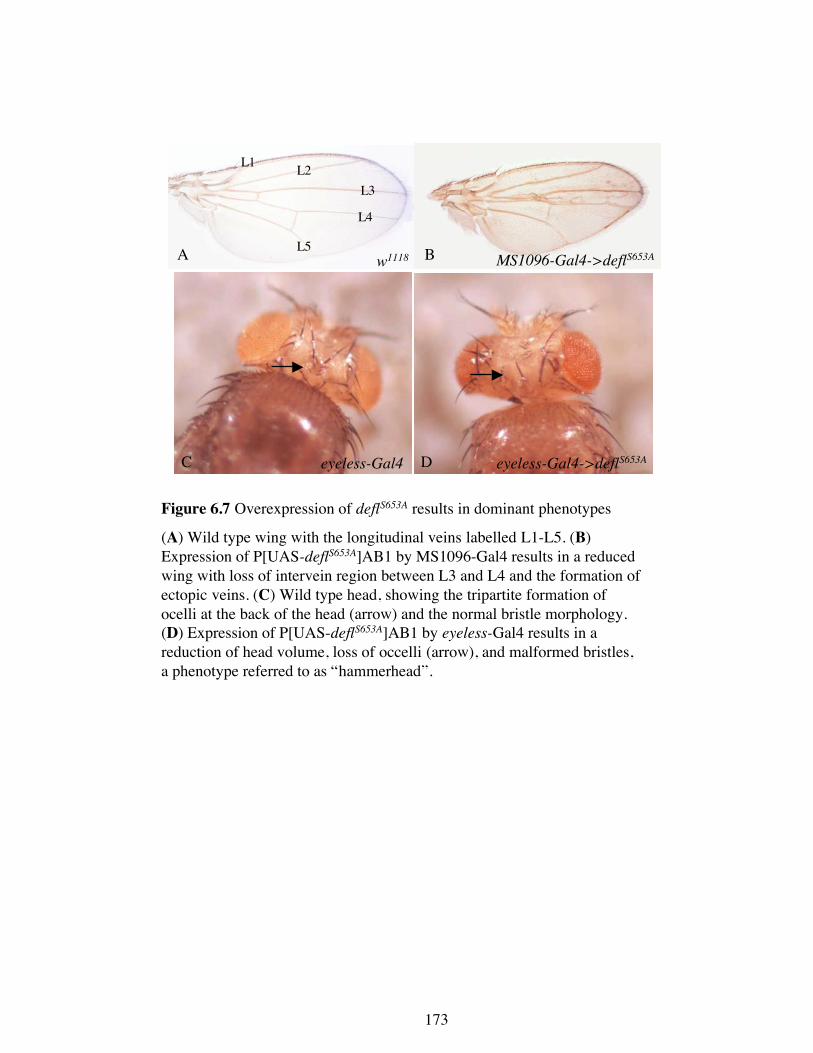
C D
A Bw1118 MS1096-Gal4->deflS653A
eyeless-Gal4 eyeless-Gal4->deflS653A
Figure 6.7 Overexpression of deflS653A results in dominant phenotypes
(A) Wild type wing with the longitudinal veins labelled L1-L5. (B)
Expression of P[UAS-deflS653A]AB1 by MS1096-Gal4 results in a reduced
wing with loss of intervein region between L3 and L4 and the formation of
ectopic veins. (C) Wild type head, showing the tripartite formation of
ocelli at the back of the head (arrow) and the normal bristle morphology.
(D) Expression of P[UAS-deflS653A]AB1 by eyeless-Gal4 results in a
reduction of head volume, loss of occelli (arrow), and malformed bristles,
a phenotype referred to as “hammerhead”.
L1L2
L3
L4
L5
173
174
which would potentially mimic constitutive phosphorylation (deflS653E; Figure 6.6).
These mutations were made via site-directed mutagenesis and cloned into the Gal4
responsive pUAST vector (Section 2.16.3). Transgenic flies containing both of these
constructs were generated. Thirteen independent transgenic lines of P[UAS-deflS653A]
and five independent transgenic lines of P[UAS-deflS653E] were generated.
To characterise the effects of deflS653A and deflS653E expression compared to wild type
expression, a series of Gal4 drivers were used. Most tissues were tolerant of excess wild
type DEFLATED since two copies were required for a phenotype to be readily
observed (Section 5.3.1). In general, tissues were more sensitive to deflS653A than to wild
type deflated. One copy of P[UAS-deflS653A] expressed at 29°C resulted in phenotypes in
the wing, head, and thoracic bristles in at least two of the seven independent lines
tested. When P[UAS-deflS653A]AR1 or AB1 were used to overexpress deflS653A in the
wing using MS1096-Gal4, extra wing veins, a reduction in intervein regions, and a
slight curl was observed (Figure 6.7 B). Overexpression of AR1 or AB1 by eyeless-
Gal4 resulted in an abnormal ‘hammerhead’ phenotype, which was more severe in
males. This phenotype was characterised by a marked decrease in head volume at the
back of the head, a lack of occelli, and a disruption and thickening of the macrochaetae
(Figure 6.7 D). When P[UAS-deflS653A]AB1, T2, or AJ1 were overexpressed by nos-
Gal4, thoracic bristles were frequently duplicated (data not shown), similar to what was
observed when two copies of wild type deflated was overexpressed (Figure 5.3). No
perturbations in eye development was observed when deflS653A was overexpressed using
GMR-Gal4 or sev-Gal4 and no other Gal4 driver lines were tested. These indicate
deflS653A behaves as a dominant allele since overexpression caused phenotypes that were
either more severe or not observed when wild type deflated was overexpressed. These
175
data also show that S653 is an important residue in normal deflated function and is
likely to be required for regulation of DEFLATED.
In contrast to the effects of deflS653A, overexpression of one copy of deflS653E using nos-
Gal4, eyeless-Gal4, sevenless- Gal4, or GMR-Gal4 29ºC did not produce any abnormal
phenotypes (three independent lines tested). P[UAS-deflS653E]H2 did show duplication
of thoracic macrochaetae, but as this was the only line that produced this phenotype, it
is difficult to ascertain whether the effect is general or due to transgene position effects.
These data imply that deflS653E is likely to act as a recessive allele and that the S to E
substitution mimicking phosphorylation at this site has little or no effect on endogenous
DEFLATED protein function.
6.4.2 Mutation of S653 to A or E produces a protein that cannot provide the necessary
function throughout development
To test whether the deflS653A and deflS653E encoded proteins could provide full wild type
function, transgenes carrying these mutant alleles were assessed for their ability to
rescue the second instar lethality of deflL/deflL and deflP/deflP individuals. Since there
was no need to drive expression of the deflated cDNA transgenes (Section 4.5), two
deflS653A and one deflS653E second chromosome lines were crossed into the homozygous
deflL and deflP backgrounds in the absence of a Gal4 driver. All three transgenes failed
to rescue the second instar lethality of homozygous deflL or deflP individuals. Therefore,
mutating the serine at position 653 to either an alanine or a glutamic acid results in a
protein that seems to be unable to complement deflP/deflP or deflL/deflL lethality. Taken
together, these data show that the S653 is a crucial residue for normal DEFLATED
function.
176
6.5 The subcellular localisation of DEFLATED::GFP
6.5.1 Generation and analysis of flies expressing DEFLATED::GFP
To examine DEFLATED’s subcellular localisation, a construct was generated in which
the Green Fluorescent Protein (GFP) coding sequence was fused in frame to the C-
terminus of wild type deflated cDNA (Section 2.16.2). This gene fusion was cloned into
both pUAST and pUASP to allow for high levels of expression in both somatic and
germline tissues, respectively. Nine independent transgenic lines of pUAST-defl::GFP
and five independent lines of pUASP-defl::GFP were established. These lines were
characterised to ensure that they retained the same function as the untagged wild type
transgenes. Each transgene was first overexpressed using various Gal4 driver lines at
29°C. In all instances phenotypes that were observed were of the same type and
severity as when untagged deflated cDNA was overexpressed. These transgenes were
then crossed into homozygous deflΔ and deflP backgrounds to assess their ability to
rescue the second instar homozygous lethality of these alleles. All transgenes tested
were capable of rescue. Two independent pUAST-defl::GFP lines (AF2 and C2) were
found to fully rescue deflL/deflL, so that a healthy stock without the TM6B balancer
could be established. One pUASP-defl::GFP line (BI1) could rescue deflΔ/deflΔ to the
pupal stage, lethality at which stage would most likely be due to homozygosity of the
nbs deletion (Section 4.5). These data show that tagging the wild type DEFLATED
protein C-terminally with GFP results in a fusion protein that seems not to differ from
the wild type protein in function.
0 1 2 3
4 5 6 7
8 9 10 11
Figure 6.8 Subcellular localisation of DEFLATED::GFP in embryos.
(A) DEFL::GFP, expressed by nanos-Gal4::VP16 induced expression ofP[UAS-defl::GFP]AH1, localises throughout the cell, with some cellsshowing strong nuclear localisation. (A′) A higher magnification of the regionboxed in A. (B) During mitosis DEFL::GFP localises to the mitotic spindleand to the midbody after the metaphase to anaphase transition. DEFL::GFPwas not found localised to chromosomes, seen as the black shadow at themetaphase equator (6 min timepoint). The images are a series of time lapseimages. The number in the bottom right hand corner indicates the number ofminutes after the first image was captured. (C) Higher magnification showingDEFL::GFP localisation to mitotic spindles but not to chromosome and to thespindle midbody in late anaphase.
dorsalventral
A stage 15-16 embryo expressing nanos Gal4-> defl::GFP
Syncytial embryo expressingnanos Gal4-> defl::GFP
A
B
C
A′
6′ 8′C
177
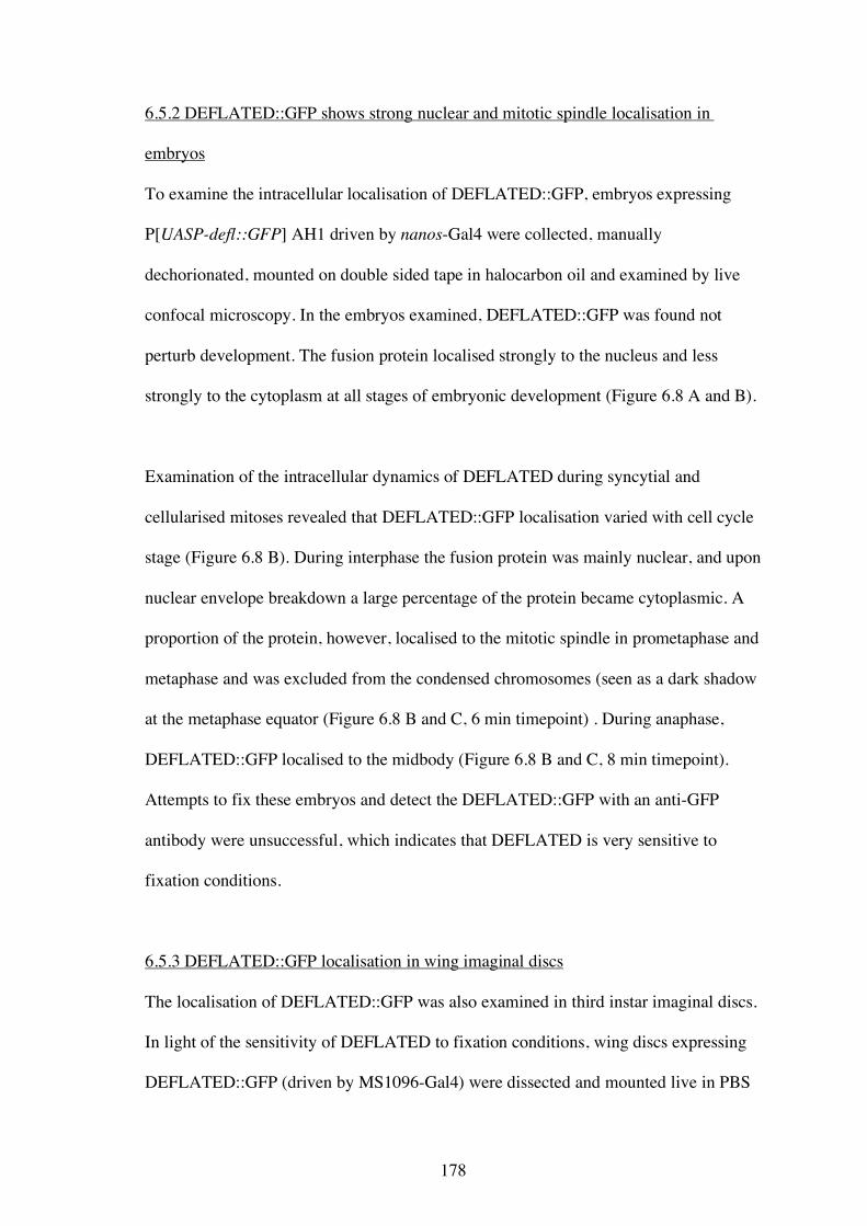
178
6.5.2 DEFLATED::GFP shows strong nuclear and mitotic spindle localisation in
embryos
To examine the intracellular localisation of DEFLATED::GFP, embryos expressing
P[UASP-defl::GFP] AH1 driven by nanos-Gal4 were collected, manually
dechorionated, mounted on double sided tape in halocarbon oil and examined by live
confocal microscopy. In the embryos examined, DEFLATED::GFP was found not
perturb development. The fusion protein localised strongly to the nucleus and less
strongly to the cytoplasm at all stages of embryonic development (Figure 6.8 A and B).
Examination of the intracellular dynamics of DEFLATED during syncytial and
cellularised mitoses revealed that DEFLATED::GFP localisation varied with cell cycle
stage (Figure 6.8 B). During interphase the fusion protein was mainly nuclear, and upon
nuclear envelope breakdown a large percentage of the protein became cytoplasmic. A
proportion of the protein, however, localised to the mitotic spindle in prometaphase and
metaphase and was excluded from the condensed chromosomes (seen as a dark shadow
at the metaphase equator (Figure 6.8 B and C, 6 min timepoint) . During anaphase,
DEFLATED::GFP localised to the midbody (Figure 6.8 B and C, 8 min timepoint).
Attempts to fix these embryos and detect the DEFLATED::GFP with an anti-GFP
antibody were unsuccessful, which indicates that DEFLATED is very sensitive to
fixation conditions.
6.5.3 DEFLATED::GFP localisation in wing imaginal discs
The localisation of DEFLATED::GFP was also examined in third instar imaginal discs.
In light of the sensitivity of DEFLATED to fixation conditions, wing discs expressing
DEFLATED::GFP (driven by MS1096-Gal4) were dissected and mounted live in PBS
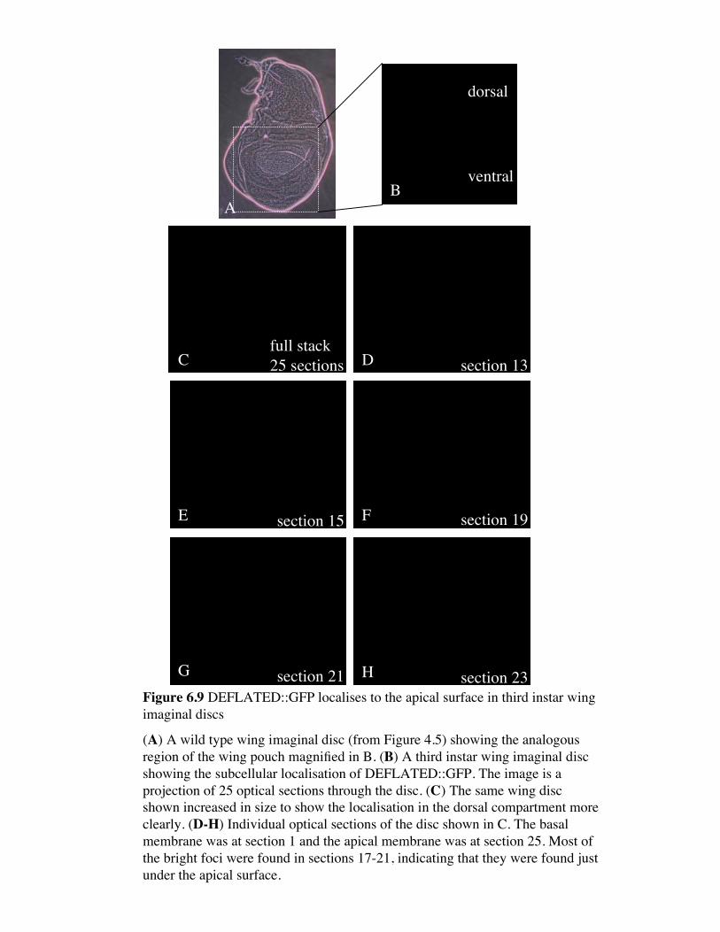
AB
Cfull stack25 sections section 13
section 15
section 21
section 19
section 23
dorsal
ventral
D
E
G
F
HFigure 6.9 DEFLATED::GFP localises to the apical surface in third instar wingimaginal discs
(A) A wild type wing imaginal disc (from Figure 4.5) showing the analogousregion of the wing pouch magnified in B. (B) A third instar wing imaginal discshowing the subcellular localisation of DEFLATED::GFP. The image is aprojection of 25 optical sections through the disc. (C) The same wing discshown increased in size to show the localisation in the dorsal compartment moreclearly. (D-H) Individual optical sections of the disc shown in C. The basalmembrane was at section 1 and the apical membrane was at section 25. Most ofthe bright foci were found in sections 17-21, indicating that they were found justunder the apical surface.
179
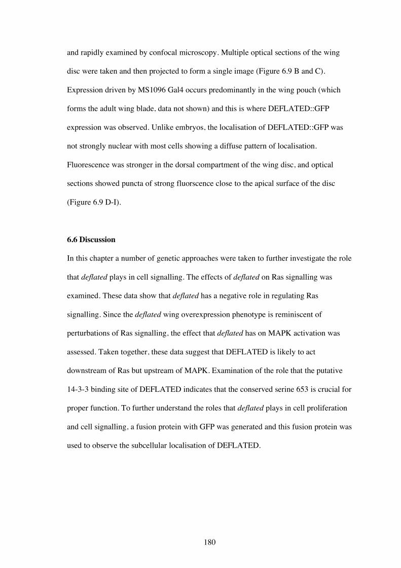
180
and rapidly examined by confocal microscopy. Multiple optical sections of the wing
disc were taken and then projected to form a single image (Figure 6.9 B and C).
Expression driven by MS1096 Gal4 occurs predominantly in the wing pouch (which
forms the adult wing blade, data not shown) and this is where DEFLATED::GFP
expression was observed. Unlike embryos, the localisation of DEFLATED::GFP was
not strongly nuclear with most cells showing a diffuse pattern of localisation.
Fluorescence was stronger in the dorsal compartment of the wing disc, and optical
sections showed puncta of strong fluorscence close to the apical surface of the disc
(Figure 6.9 D-I).
6.6 Discussion
In this chapter a number of genetic approaches were taken to further investigate the role
that deflated plays in cell signalling. The effects of deflated on Ras signalling was
examined. These data show that deflated has a negative role in regulating Ras
signalling. Since the deflated wing overexpression phenotype is reminiscent of
perturbations of Ras signalling, the effect that deflated has on MAPK activation was
assessed. Taken together, these data suggest that DEFLATED is likely to act
downstream of Ras but upstream of MAPK. Examination of the role that the putative
14-3-3 binding site of DEFLATED indicates that the conserved serine 653 is crucial for
proper function. To further understand the roles that deflated plays in cell proliferation
and cell signalling, a fusion protein with GFP was generated and this fusion protein was
used to observe the subcellular localisation of DEFLATED.

181
6.6.1 The role of deflated in Ras signalling
The genetic interactions observed between deflated, RasV12 and rolledSEM demonstrate
that deflated is involved in the Ras signalling pathway. The data obtained was unable to
distinguish between the possibility that DEFLATED is a component of the pathway, or
has a role in another cellular process that affects the outcome of Ras signalling. The
data are consistent with DEFLATED acting as a negative regulator of Ras signalling
since the RasV12 phenotype was enhanced in deflP/deflZ transheterozygotes (Figure 6.2)
and suppressed by deflated overexpression (Figure 6.3). While these data are consistent
with a role as a negative regulator, they do not categorically show that DEFLATED is a
negative regulator acting in the Ras pathway. To address this, mitotic clones of deflΔ
would be required to properly assess the role of DEFLATED as a negative regulator.
These clones would contain no DEFLATED protein and therefore Ras signalling could
be examined in these cells. If DEFLATED is normally a negative regulator of Ras
signalling then it would be expected that Ras signalling would be enhanced in these
clones. If DEFLATED does not normally act upon the Ras pathway then Ras signalling
should be unaffected.
In the absence of data obtained from mitotic clones it is still likely that DEFLATED is
required to negatively regulate Ras signalling. The findings that both deflP and deflated
overexpression suppress Ras signalling, indicates that Ras signalling is negatively
regulated under these circumstances. While the expression and stability of DEFLP still
requires confirmation, the genetic data indicate that the protein encoded by deflP and the
exogoneous DEFLATED protein appear to behave as neomorphs, separation of
function, or dominant negative proteins. Assuming that the genetic interaction observed
is not due to another unlinked lesion on the deflP chromosome, it is unlikely that deflP
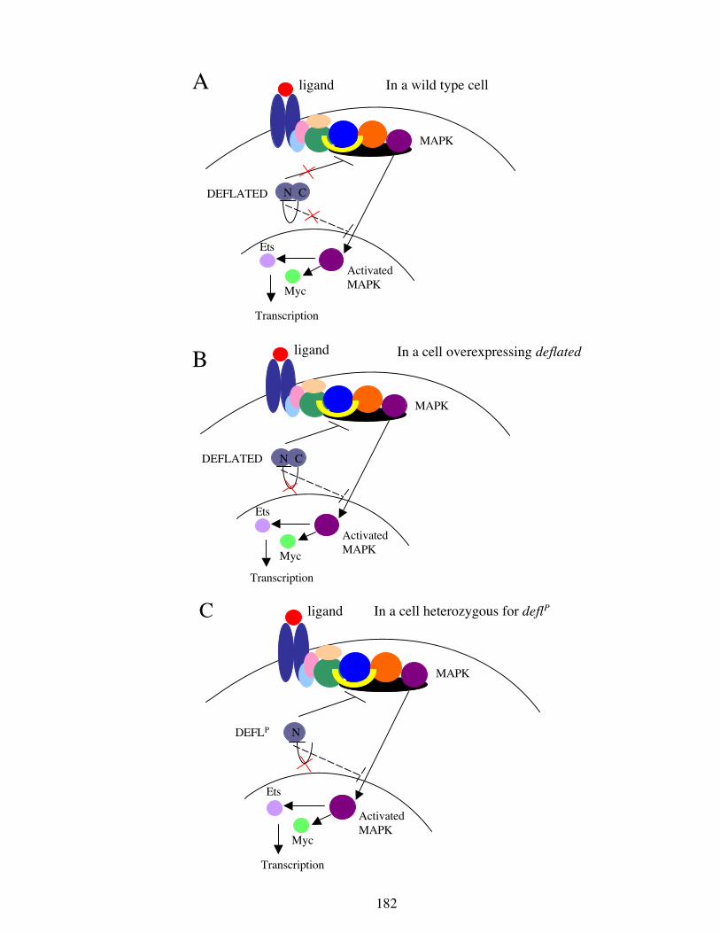
In a cell overexpressing deflated
Transcription
Ets
Myc
ligand
DEFLATED
In a wild type cell
Transcription
Ets
Myc
ligand
DEFLATED
N C
N C
A
B
MAPK
ActivatedMAPK
MAPK
ActivatedMAPK
Ets
Myc
ligand
DEFLP
In a cell heterozygous for deflP
N
Transcription
C
ActivatedMAPK
MAPK
182
Figure 6.10 A hypothetical model for DEFLATED function in Ras signalling
(A) In a wild type cell Ras signalling is not affected by DEFLATED, due tothe regulatory region in the C-terminus (a.a. 401-968) negatively regulatingthe N-terminus. (B) When deflated is overexpressed the excess DEFLATED isno longer negatively regulated and the protein can suppress Ras signalling ateither the plasma membrane (or endosome) and prevent the subsequentactivation of MAPK or by preventing activated MAPK translocating into thenucleus. This second possibility is less likely for reasons explained in the textand therefore is represented by the dashed line. (C) When DEFLP protein ispresent it cannot be negatively regulated as it lacks the C-terminus. Thistruncated protein therefore is capable suppressing Ras signalling. See Figure1.6 for an explanation of the proteins involved in the Ras signalling cascade.
183
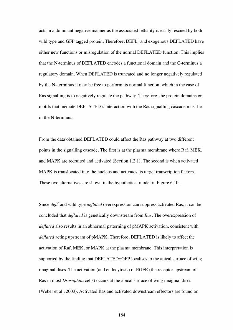
184
acts in a dominant negative manner as the associated lethality is easily rescued by both
wild type and GFP tagged protein. Therefore, DEFLP and exogenous DEFLATED have
either new functions or misregulation of the normal DEFLATED function. This implies
that the N-terminus of DEFLATED encodes a functional domain and the C-terminus a
regulatory domain. When DEFLATED is truncated and no longer negatively regulated
by the N–terminus it may be free to perform its normal function, which in the case of
Ras signalling is to negatively regulate the pathway. Therefore, the protein domains or
motifs that mediate DEFLATED’s interaction with the Ras signalling cascade must lie
in the N-terminus.
From the data obtained DEFLATED could affect the Ras pathway at two different
points in the signalling cascade. The first is at the plasma membrane where Raf, MEK,
and MAPK are recruited and activated (Section 1.2.1). The second is when activated
MAPK is translocated into the nucleus and activates its target transcription factors.
These two alternatives are shown in the hypothetical model in Figure 6.10.
Since deflP and wild type deflated overexpression can suppress activated Ras, it can be
concluded that deflated is genetically downstream from Ras. The overexpression of
deflated also results in an abnormal patterning of pMAPK activation, consistent with
deflated acting upstream of pMAPK. Therefore, DEFLATED is likely to affect the
activation of Raf, MEK, or MAPK at the plasma membrane. This interpretation is
supported by the finding that DEFLATED::GFP localises to the apical surface of wing
imaginal discs. The activation (and endocytosis) of EGFR (the receptor upstream of
Ras in most Drosophila cells) occurs at the apical surface of wing imaginal discs
(Weber et al., 2003). Activated Ras and activated downstream effectors are found on
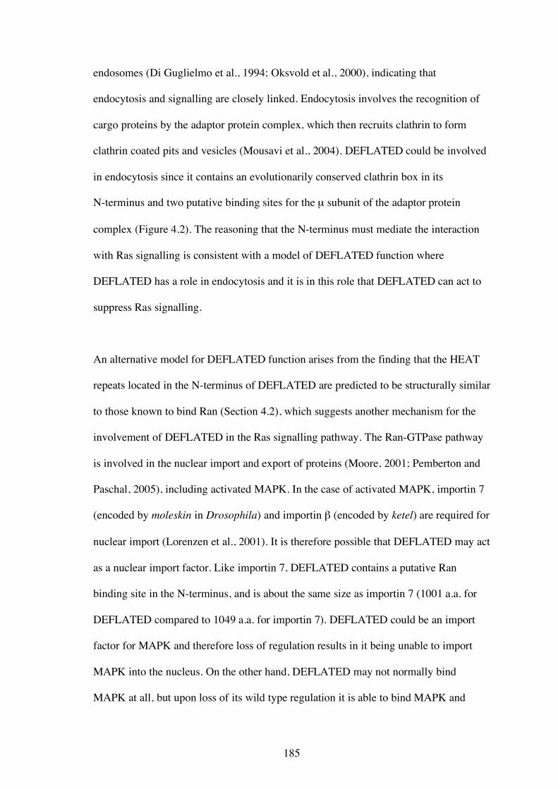
185
endosomes (Di Guglielmo et al., 1994; Oksvold et al., 2000), indicating that
endocytosis and signalling are closely linked. Endocytosis involves the recognition of
cargo proteins by the adaptor protein complex, which then recruits clathrin to form
clathrin coated pits and vesicles (Mousavi et al., 2004). DEFLATED could be involved
in endocytosis since it contains an evolutionarily conserved clathrin box in its
N-terminus and two putative binding sites for the µ subunit of the adaptor protein
complex (Figure 4.2). The reasoning that the N-terminus must mediate the interaction
with Ras signalling is consistent with a model of DEFLATED function where
DEFLATED has a role in endocytosis and it is in this role that DEFLATED can act to
suppress Ras signalling.
An alternative model for DEFLATED function arises from the finding that the HEAT
repeats located in the N-terminus of DEFLATED are predicted to be structurally similar
to those known to bind Ran (Section 4.2), which suggests another mechanism for the
involvement of DEFLATED in the Ras signalling pathway. The Ran-GTPase pathway
is involved in the nuclear import and export of proteins (Moore, 2001; Pemberton and
Paschal, 2005), including activated MAPK. In the case of activated MAPK, importin 7
(encoded by moleskin in Drosophila) and importin β (encoded by ketel) are required for
nuclear import (Lorenzen et al., 2001). It is therefore possible that DEFLATED may act
as a nuclear import factor. Like importin 7, DEFLATED contains a putative Ran
binding site in the N-terminus, and is about the same size as importin 7 (1001 a.a. for
DEFLATED compared to 1049 a.a. for importin 7). DEFLATED could be an import
factor for MAPK and therefore loss of regulation results in it being unable to import
MAPK into the nucleus. On the other hand, DEFLATED may not normally bind
MAPK at all, but upon loss of its wild type regulation it is able to bind MAPK and
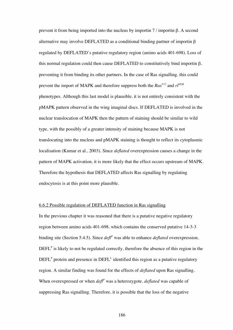
186
prevent it from being imported into the nucleus by importin 7 / importin β. A second
alternative may involve DEFLATED as a conditional binding partner of importin β
regulated by DEFLATED’s putative regulatory region (amino acids 401-698). Loss of
this normal regulation could then cause DEFLATED to constitutively bind importin β,
preventing it from binding its other partners. In the case of Ras signalling, this could
prevent the import of MAPK and therefore suppress both the RasV12 and rlSEM
phenotypes. Although this last model is plausible, it is not entirely consistent with the
pMAPK pattern observed in the wing imaginal discs. If DEFLATED is involved in the
nuclear translocation of MAPK then the pattern of staining should be similar to wild
type, with the possibly of a greater intensity of staining because MAPK is not
translocating into the nucleus and pMAPK staining is thought to reflect its cytoplasmic
localisation (Kumar et al., 2003). Since deflated overexpression causes a change in the
pattern of MAPK activation, it is more likely that the effect occurs upstream of MAPK.
Therefore the hypothesis that DEFLATED affects Ras signalling by regulating
endocytosis is at this point more plausible.
6.6.2 Possible regulation of DEFLATED function in Ras signalling
In the previous chapter it was reasoned that there is a putative negative regulatory
region between amino acids 401-698, which contains the conserved putative 14-3-3
binding site (Section 5.4.5). Since deflP was able to enhance deflated overexpression,
DEFLP is likely to not be regulated correctly, therefore the absence of this region in the
DEFLP protein and presence in DEFLL identified this region as a putative regulatory
region. A similar finding was found for the effects of deflated upon Ras signalling.
When overexpressed or when deflP was a heterozygote, deflated was capable of
suppressing Ras signalling. Therefore, it is possible that the loss of the negative
187
regulatory region at 401-698 or saturation of the regulatory mechanisms that act on this
region causes the suppression of the Ras signalling pathway.
There are three pieces of data that suggest that the regulation of DEFLATED in this
region may be mediated by 14-3-3 binding. The first is that deflated overexpression is
dominantly modified by both 14-3-3 isoforms (Figure 5.5). While this modification
may indicate a role for DEFLATED in M-phase entry (Section 5.4.2), it may
alternatively reflect the role of DEFLATED in Ras signalling. Secondly, it is likely that
DEFLATED affects the Ras signalling pathway at the plasma membrane or on the
endosomes where Raf, MEK and MAPK are activated. This is also where 14-3-3
proteins are known to act in Ras signalling (Section 1.2.1). So it is possible that
DEFLATED is a target of 14-3-3 regulation at the plasma membrane. Thirdly, genetic
interactions show that 14-3-3 proteins act positively in Ras signalling. If 14-3-3 binds
DEFLATED to keep it inactive (and therefore prevent it from inhibiting Ras) then this
scenario is consistent with the positive roles of 14-3-3 proteins.
While it is unknown whether DEFLATED can physically bind 14-3-3, the predicted
phosphorylated serine (S653) predicted to mediate 14-3-3 binding has is likely to be
essential for DEFLATED function. Since expression of deflS653A acted dominantly while
deflS653E did not, it is possible that phosphorylation at this site is required for negative
regulation of DEFLATED function. If this site is phosphorylated then it is likely to bind
14-3-3 proteins. This raises two questions. The first is what is the identity of the kinase
that phosphorylates DEFLATED at S653? This site was not recognised as a known
phosphorylation site of any currently characterised kinase as identified by protein motif
predictions. The second question is how would binding of 14-3-3 at this site negatively
188
regulate DEFLATED function? Generally 14-3-3 proteins form dimers that regulate
their targets’ function by forming either intra- or inter- molecular interactions. Three
additional putative 14-3-3 binding sites were identified in DEFLATED but as they did
not show strong conservation amongst DEFLATED homologues they were not tested
by site-directed mutagenesis. It is possible that one or more of these sites are functional
and therefore DEFLATED could be regulated intra-molecularly. If only the 650-655
14-3-3 binding site is functional, then DEFLATED may be regulated by 14-3-3
bringing it into contact with a second protein. While most known cases of 14-3-3
binding involves 14-3-3 dimers, there is at least one case in which a protein is regulated
by a 14-3-3 monomer. The protein SLOB is bound by a 14-3-3 monomer and when
bound is able to negatively regulate the calcium-dependent potassium channel,
SLOWPOKE (Zhou et al., 2003). Therefore it is feasible that DEFLATED binds one
14-3-3 protein and this is sufficient to negatively regulate its function.
6.6.3 DEFLATED at the mitotic spindle
The findings that DEFLATED::GFP localises to nuclei and to the mitotic spindle
support a direct role for DEFLATED in cell proliferation. Its localisation to the mitotic
spindle may involve the potential binding of Ran or clathrin, two proteins that also
localise to mitotic spindles and have essential roles in its formation and function.
The Ran pathway functions by generating a gradient of high Ran-GTP in the nucleus
and high Ran-GDP in the cytoplasm. It would be expected that this gradient would be
disrupted during mitosis when the nuclear envelope breaks down. However, the
gradient in maintained by the localisation of RCC1 on the chromatin during mitosis
(Carazo-Salas et al., 1999), thereby generating a high concentration of Ran-GTP in the
189
vicinity of the chromosomes. A high concentration of Ran-GTP is sufficient to nucleate
microtubules (Carazo-Salas et al., 1999; Wilde et al., 2001) and it is thought that the
generation of a high concentration of Ran-GTP at the chromatin is important for proper
spindle formation and function. In accordance with this model, various components of
the Ran pathway are localised along mitotic spindles. In mammalian cells, RanGAP1
and RanBP2 localise to both spindles and microtubule associated kinetochores, with
higher localisation at the kinetochores (Joseph et al., 2004; Joseph et al., 2002; Matunis
et al., 1996). In C. elegans, Ran localises along the length of the chromosomes at points
of microtubule attachment (Bamba et al., 2002). In Drosophila, Ran (in GTP bound
form) localises around, but not directly on, the spindles. RCC1 localises to the
chromatin and RanGAP is occasionally found along the outside edge of microtubules
(Trieselmann and Wilde, 2002). Therefore, the findings that DEFLATED::GFP
localises to the spindle and is a potential Ran binding protein suggests that DEFLATED
may form a complex with Ran or may be involved in the Ran mediated regulation of
spindle formation and function.
Aside from its well known role in endocytosis, clathrin also localises to the mitotic
spindle (Okamoto et al., 2000; Royle et al., 2005) and its localisation is dependent on
the N-terminus of the heavy chain, which is the region that binds proteins that contain a
clathrin box. The localisation of clathrin occurs as a triskelion and is important for
normal spindle formation. In cells lacking the clathrin heavy chain, chromosomes do
not congress to the metaphase plate correctly and, as a result, the cells arrest due to
activation of the spindle checkpoint. As DEFLATED contains a well conserved clathrin
box, it is a good candidate for recruiting clathrin to the mitotic spindle. It is possible
that DEFLATED may bind both Ran and clathrin, acting as a bridge between them.
190
The localisation of DEFLATED::GFP to nuclei and to spindles during mitotis provides
further support for a role in regulating cell proliferation. Although this localisation may
be an artifact of overexpression, since deflated mRNA was not detected in syncytial or
cellularised embryos, post cellularised cells also revealed a nuclear localisation. As the
attempts to fix DEFLATED::GFP embryos were unsuccessful (Section 6.5.4), it could
not be confirmed that DEFLATED::GFP localised to spindles (by co-staining with anti-
tubulin) in older embryos, where it is known that deflated is expressed. However, the
localisation of DEFLATED::GFP to spindles does appear to reflect real DEFLATED
localisation for a number of reasons. Firstly, DEFLATED is likely to bind Ran and/or
clathrin, two proteins that localise to mitotic spindles and have roles in regulating
spindle function (Royle et al., 2005; Triselmann and Wilde, 2002). Secondly, the
overexpression of deflated::GFP does not result in abnormal phenotypes, which
indicates that this localisation does not interfere with normal cellular function.
Although confirmation of a genuine spindle localisation during mitosis will require
DEFLATED antibodies to be raised, the presence of clathrin and Ran binding motifs in
DEFLATED argues strongly that the spindle localisation is not an artefact.
Localisation of DEFLATED to the mitotic spindle supports the genetic findings that
deflated may link DNA synthesis with M-phase entry (Section 5.4.2). Furthermore
spindle localisation indicates that DEFLATED is involved in regulating spindle
formation or function. Since DEFLATED::GFP localises to the midbody in anaphase,
there is also a possibility that DEFLATED has a role in mitotic exit and/or cytokinesis.
DEFLATED may regulate the function of the spindle by physically binding Ran and/or
clathrin.
191
In conclusion, the findings of this chapter support a role for DEFLATED in regulating
Ras signalling, which could explain its effects on cell cycle progression. However, the
finding that DEFLATED localises to mitotic spindles also supports a direct role for
DEFLATED in regulating aspects of M-phase progression. These results support the
genetic findings presented in Chapter 5 that suggest DEFLATED links DNA replication
with M-phase entry.
192
Chapter 7- Towards a model of DEFLATED function
This thesis describes the initial characterisation of the novel Drosophila gene, deflated.
Due to high co-expression with the S-phase genes Ctf4 and Dp, deflated was
hypothesised to be a regulator of cell proliferation. The mRNA expression patterns and
the phenotypes of homozygous, transheterozygous, and rescued deflated– mutant
individuals were consistent with deflated having a role in regulating cell proliferation.
Genetic interaction analysis with cell cycle regulators suggested that the role of deflated
in cell proliferation may be in linking DNA synthesis with entry into M-phase. The
finding that DEFLATED::GFP localises to the mitotic spindle implicates DEFLATED
in spindle formation and function, further supporting this role.
The results presented in the previous chapters demonstrate that deflated interacts
genetically with signalling pathways, including the Ras pathway, and that the wing and
bristle phenotypes produced by deflated overexpression, indicate that deflated may play
a role in regulating cell signalling. This suggests that the effect deflated has on cell
signalling may in part be due this role in cell signalling. In Chapter 6, the effect of
deflated on Ras signalling was examined in some detail. It was shown that DEFLATED
is likely to play a role near the beginning of the Ras signalling cascade, perhaps at the
plasma membrane (Figure 6.10). Since DEFLATED was found to have an
evolutionarily conserved motif known to bind clathrin (Figures 4.1 and 4.2) and
DEFLATED::GFP was observed to have an interphase localisation pattern that could
reflect clathrin coated vesicles or endosomes (Figure 6.9), it is possible that
DEFLATED may function at the points in which signalling pathways intersect with
endocytic pathways. The presence of a putative Ran binding domain (Section 4.2)
193
suggests that DEFLATED may also function in the Ran pathway. This last alternative
may explain deflated’s antagonistic role in Krüppel function. The questions this chapter
addresses are: is it feasible that DEFLATED could function within each of these
pathways? What roles do these pathways have in regulating cell proliferation? Is it
likely that DEFLATED could function separately in all three pathways or can it be a
molecule that acts at the intersection of these three pathways?
7.1 Upon the genetic natures of the deflated alleles
The deflated alleles generated in this study through P-element mutagenesis all resulted
in deletion of the entire coding region or a portion thereof (Figure 3.8). Three of the
alleles, deflP, deflL, and deflZ, are predicted to generate proteins that lack the C-terminus,
while deflΔ produces no protein (Figure 5.7). Throughout this study all alleles have
behaved consistently in each of the assays and therefore conclusions can be drawn
about their natures. deflΔ is certainly a null allele, yet it was not used in many of the
assays as this allele is also null for nbs. Therefore, any data obtained using deflΔ could
be confounded by loss of nbs function, which is expected to have a role in DNA
replication and cell cycle arrest (Section 1.1.4). Recently, the sequencing of a piggyBac
insertion (deflc106100, also known as nbsc106100) shows that this transposon has inserted 49
bp downstream of the deflated start codon (Gene Disruption Project Members, 2005;
Thibault et al., 2004). This indicates that this insertion is likely to be null for deflated,
but how it affects nbs function still needs be determined.
The three different partial deletion alleles, deflL, deflP, and deflZ showed different
genetic interactions and therefore are different types of alleles. deflL consistently
behaved as a hypomorphic allele. In trans to deflZ, it produced a stronger phenotype
194
than deflP, a phenotype similar to deflΔ, and was less easily rescued by cDNA
expression than deflP (Section 4.5). On the basis of these data, deflL appears to be a
stronger allele (retaining less function) than deflP. In constrast, deflP appears to be a
recessive neomorph or a separation of function mutation rather than a hypomorphic
mutation, based on the genetic interactions observed between deflP and activated Ras
and deflP and deflated overexpression in the wing (Sections 5.4.5 and 6.6.2). In both of
these situations, deflP behaves similarly to wild type overexpression. deflZ behaved
consistently as a weak hypomorphic allele, however this allele was not used in many of
the analyses due to the presence of a second mutation elsewhere on the chromosome,
which could potentially confound any observations made (Section 3.4). Finally, the
phenotypes observed from the overexpression of wild type deflated are likely to occur
due to loss of the normal checks on deflated function. This conclusion can be made due
to the fact that high levels of DEFLATED protein are required to induce abnormal
phenotypes. This suggests that the regulators controlling DEFLATED function are
normally in excess of DEFLATED and it is only within the wing imaginal disc that
these putative regulatory mechanisms become rate limiting when deflated is
overexpressed (Section 5.3.1).
7.2 Possible functional domains of DEFLATED
DEFLATED is predicted to have functional domains and motifs indicative of a role in
cell cycle regulation, cell signalling, endocytosis and/or the Ran pathway (Section 4.2).
All of these predictions are based on evolutionarily conservation of primary amino acid
sequence or 3-D structure predictions based on primary amino acid sequence, and
therefore require confirmation within a biochemical context. Within this study, none of
these domains or motifs were fully experimentally tested, however, for some evidence
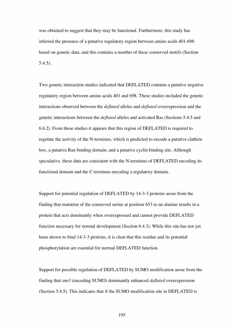
195
was obtained to suggest that they may be functional. Furthermore, this study has
inferred the presence of a putative regulatory region between amino acids 401-698
based on genetic data, and this contains a number of these conserved motifs (Section
5.4.5).
Two genetic interaction studies indicated that DEFLATED contains a putative negative
regulatory region between amino acids 401 and 698. These studies included the genetic
interactions observed between the deflated alleles and deflated overexpression and the
genetic interactions between the deflated alleles and activated Ras (Sections 5.4.5 and
6.6.2). From these studies it appears that this region of DEFLATED is required to
regulate the activity of the N-terminus, which is predicted to encode a putative clathrin
box, a putative Ran binding domain, and a putative cyclin binding site. Although
speculative, these data are consistent with the N-terminus of DEFLATED encoding its
functional domain and the C-terminus encoding a regulatory domain.
Support for potential regulation of DEFLATED by 14-3-3 proteins arose from the
finding that mutation of the conserved serine at position 653 to an alanine results in a
protein that acts dominantly when overexpressed and cannot provide DEFLATED
function necessary for normal development (Section 6.4.3). While this site has not yet
been shown to bind 14-3-3 proteins, it is clear that this residue and its potential
phosphorylation are essential for normal DEFLATED function.
Support for possible regulation of DEFLATED by SUMO modification arose from the
finding that smt3 (encoding SUMO) dominantly enhanced deflated overexpression
(Section 5.4.5). This indicates that if the SUMO modification site in DEFLATED is
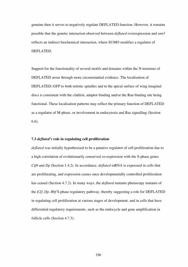
196
genuine then it serves to negatively regulate DEFLATED function. However, it remains
possible that the genetic interaction observed between deflated overexpression and smt3
reflects an indirect biochemical interaction, where SUMO modifies a regulator of
DEFLATED.
Support for the functionality of several motifs and domains within the N-terminus of
DEFLATED arose through more circumstantial evidence. The localisation of
DEFLATED::GFP to both mitotic spindles and to the apical surface of wing imaginal
discs is consistent with the clathrin, adaptor binding and/or the Ran binding site being
functional. These localisation patterns may reflect the primary function of DEFLATED
as a regulator of M-phase, or involvement in endocytosis and Ras signalling (Section
6.6).
7.3 deflated’s role in regulating cell proliferation
deflated was initially hypothesised to be a putative regulator of cell proliferation due to
a high correlation of evolutionarily conserved co-expression with the S-phase genes
Ctf4 and Dp (Section 1.4.2). In accordance, deflated mRNA is expressed in cells that
are proliferating, and expression ceases once developmentally controlled proliferation
has ceased (Section 4.7.2). In many ways, the deflated mutants phenocopy mutants of
the E2f, Dp, Rbf S-phase regulatory pathway, thereby suggesting a role for DEFLATED
in regulating cell proliferation at various stages of development, and in cells that have
differential regulatory requirements, such as the endocycle and gene amplification in
follicle cells (Section 4.7.3).
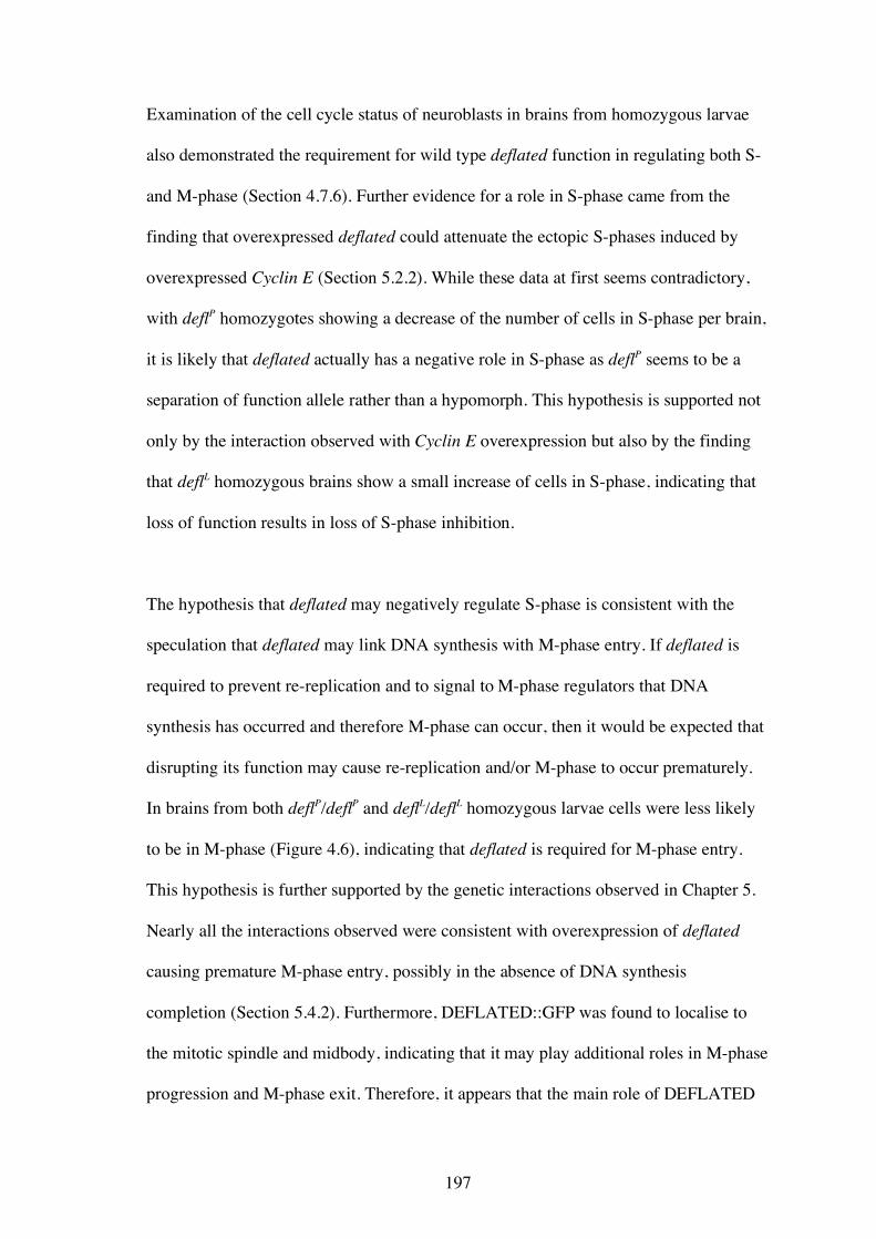
197
Examination of the cell cycle status of neuroblasts in brains from homozygous larvae
also demonstrated the requirement for wild type deflated function in regulating both S-
and M-phase (Section 4.7.6). Further evidence for a role in S-phase came from the
finding that overexpressed deflated could attenuate the ectopic S-phases induced by
overexpressed Cyclin E (Section 5.2.2). While these data at first seems contradictory,
with deflP homozygotes showing a decrease of the number of cells in S-phase per brain,
it is likely that deflated actually has a negative role in S-phase as deflP seems to be a
separation of function allele rather than a hypomorph. This hypothesis is supported not
only by the interaction observed with Cyclin E overexpression but also by the finding
that deflL homozygous brains show a small increase of cells in S-phase, indicating that
loss of function results in loss of S-phase inhibition.
The hypothesis that deflated may negatively regulate S-phase is consistent with the
speculation that deflated may link DNA synthesis with M-phase entry. If deflated is
required to prevent re-replication and to signal to M-phase regulators that DNA
synthesis has occurred and therefore M-phase can occur, then it would be expected that
disrupting its function may cause re-replication and/or M-phase to occur prematurely.
In brains from both deflP/deflP and deflL/deflL homozygous larvae cells were less likely
to be in M-phase (Figure 4.6), indicating that deflated is required for M-phase entry.
This hypothesis is further supported by the genetic interactions observed in Chapter 5.
Nearly all the interactions observed were consistent with overexpression of deflated
causing premature M-phase entry, possibly in the absence of DNA synthesis
completion (Section 5.4.2). Furthermore, DEFLATED::GFP was found to localise to
the mitotic spindle and midbody, indicating that it may play additional roles in M-phase
progression and M-phase exit. Therefore, it appears that the main role of DEFLATED
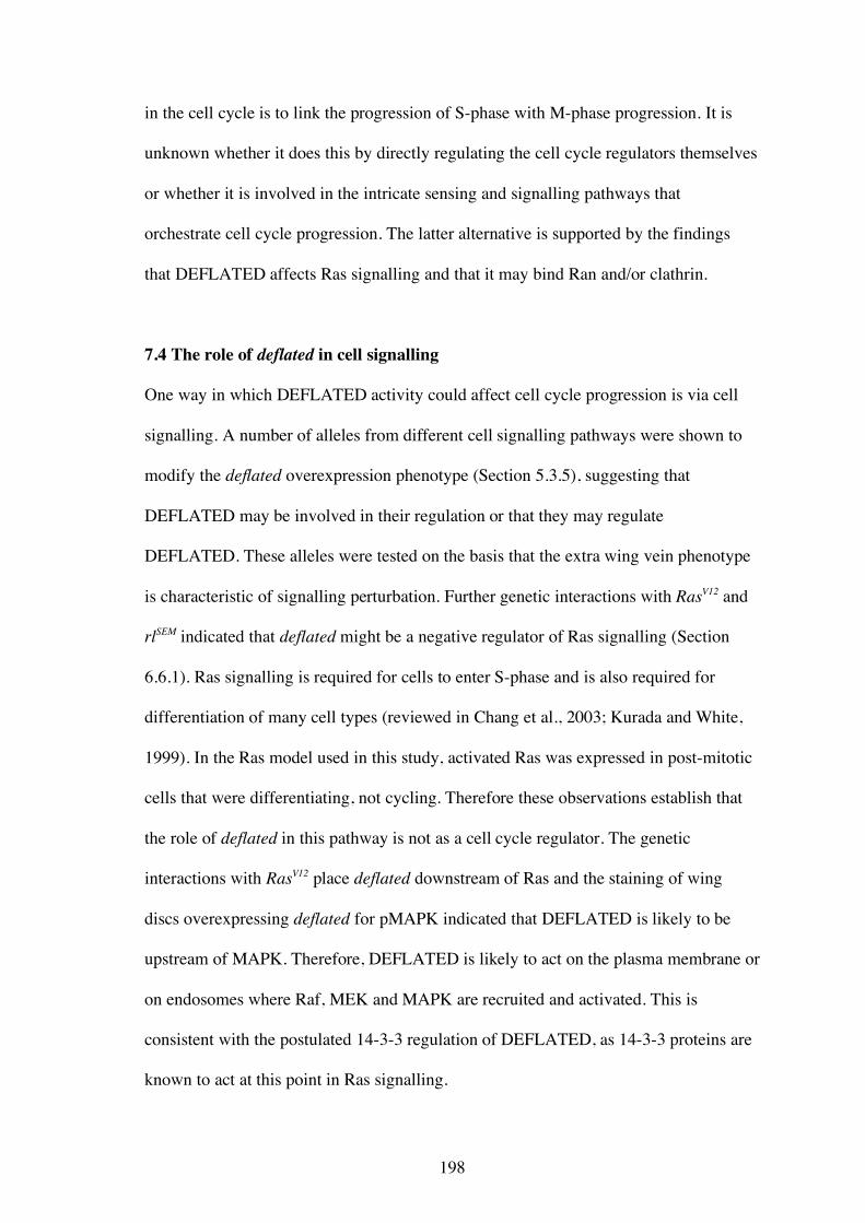
198
in the cell cycle is to link the progression of S-phase with M-phase progression. It is
unknown whether it does this by directly regulating the cell cycle regulators themselves
or whether it is involved in the intricate sensing and signalling pathways that
orchestrate cell cycle progression. The latter alternative is supported by the findings
that DEFLATED affects Ras signalling and that it may bind Ran and/or clathrin.
7.4 The role of deflated in cell signalling
One way in which DEFLATED activity could affect cell cycle progression is via cell
signalling. A number of alleles from different cell signalling pathways were shown to
modify the deflated overexpression phenotype (Section 5.3.5), suggesting that
DEFLATED may be involved in their regulation or that they may regulate
DEFLATED. These alleles were tested on the basis that the extra wing vein phenotype
is characteristic of signalling perturbation. Further genetic interactions with RasV12 and
rlSEM indicated that deflated might be a negative regulator of Ras signalling (Section
6.6.1). Ras signalling is required for cells to enter S-phase and is also required for
differentiation of many cell types (reviewed in Chang et al., 2003; Kurada and White,
1999). In the Ras model used in this study, activated Ras was expressed in post-mitotic
cells that were differentiating, not cycling. Therefore these observations establish that
the role of deflated in this pathway is not as a cell cycle regulator. The genetic
interactions with RasV12 place deflated downstream of Ras and the staining of wing
discs overexpressing deflated for pMAPK indicated that DEFLATED is likely to be
upstream of MAPK. Therefore, DEFLATED is likely to act on the plasma membrane or
on endosomes where Raf, MEK and MAPK are recruited and activated. This is
consistent with the postulated 14-3-3 regulation of DEFLATED, as 14-3-3 proteins are
known to act at this point in Ras signalling.
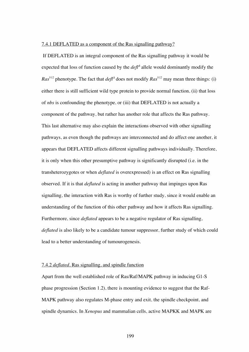
199
7.4.1 DEFLATED as a component of the Ras signalling pathway?
If DEFLATED is an integral component of the Ras signalling pathway it would be
expected that loss of function caused by the deflΔ allele would dominantly modify the
RasV12 phenotype. The fact that deflΔ does not modify RasV12 may mean three things: (i)
either there is still sufficient wild type protein to provide normal function, (ii) that loss
of nbs is confounding the phenotype, or (iii) that DEFLATED is not actually a
component of the pathway, but rather has another role that affects the Ras pathway.
This last alternative may also explain the interactions observed with other signalling
pathways, as even though the pathways are interconnected and do affect one another, it
appears that DEFLATED affects different signalling pathways individually. Therefore,
it is only when this other presumptive pathway is significantly disrupted (i.e. in the
transheterozygotes or when deflated is overexpressed) is an effect on Ras signalling
observed. If it is that deflated is acting in another pathway that impinges upon Ras
signalling, the interaction with Ras is worthy of further study, since it would enable an
understanding of the function of this other pathway and how it affects Ras signalling.
Furthermore, since deflated appears to be a negative regulator of Ras signalling,
deflated is also likely to be a candidate tumour suppressor, further study of which could
lead to a better understanding of tumourogenesis.
7.4.2 deflated, Ras signalling, and spindle function
Apart from the well established role of Ras/Raf/MAPK pathway in inducing G1-S
phase progression (Section 1.2), there is mounting evidence to suggest that the Raf-
MAPK pathway also regulates M-phase entry and exit, the spindle checkpoint, and
spindle dynamics. In Xenopus and mammalian cells, active MAPKK and MAPK are
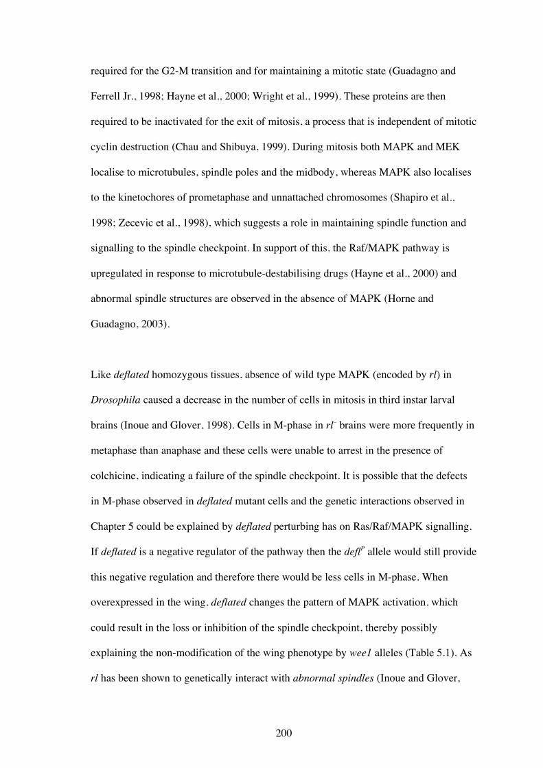
200
required for the G2-M transition and for maintaining a mitotic state (Guadagno and
Ferrell Jr., 1998; Hayne et al., 2000; Wright et al., 1999). These proteins are then
required to be inactivated for the exit of mitosis, a process that is independent of mitotic
cyclin destruction (Chau and Shibuya, 1999). During mitosis both MAPK and MEK
localise to microtubules, spindle poles and the midbody, whereas MAPK also localises
to the kinetochores of prometaphase and unnattached chromosomes (Shapiro et al.,
1998; Zecevic et al., 1998), which suggests a role in maintaining spindle function and
signalling to the spindle checkpoint. In support of this, the Raf/MAPK pathway is
upregulated in response to microtubule-destabilising drugs (Hayne et al., 2000) and
abnormal spindle structures are observed in the absence of MAPK (Horne and
Guadagno, 2003).
Like deflated homozygous tissues, absence of wild type MAPK (encoded by rl) in
Drosophila caused a decrease in the number of cells in mitosis in third instar larval
brains (Inoue and Glover, 1998). Cells in M-phase in rl– brains were more frequently in
metaphase than anaphase and these cells were unable to arrest in the presence of
colchicine, indicating a failure of the spindle checkpoint. It is possible that the defects
in M-phase observed in deflated mutant cells and the genetic interactions observed in
Chapter 5 could be explained by deflated perturbing has on Ras/Raf/MAPK signalling.
If deflated is a negative regulator of the pathway then the deflP allele would still provide
this negative regulation and therefore there would be less cells in M-phase. When
overexpressed in the wing, deflated changes the pattern of MAPK activation, which
could result in the loss or inhibition of the spindle checkpoint, thereby possibly
explaining the non-modification of the wing phenotype by wee1 alleles (Table 5.1). As
rl has been shown to genetically interact with abnormal spindles (Inoue and Glover,
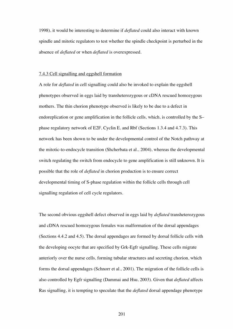
201
1998), it would be interesting to determine if deflated could also interact with known
spindle and mitotic regulators to test whether the spindle checkpoint is perturbed in the
absence of deflated or when deflated is overexpressed.
7.4.3 Cell signalling and eggshell formation
A role for deflated in cell signalling could also be invoked to explain the eggshell
phenotypes observed in eggs laid by transheterozygous or cDNA rescued homozygous
mothers. The thin chorion phenotype observed is likely to be due to a defect in
endoreplication or gene amplification in the follicle cells, which, is controlled by the S–
phase regulatory network of E2F, Cyclin E, and Rbf (Sections 1.3.4 and 4.7.3). This
network has been shown to be under the developmental control of the Notch pathway at
the mitotic-to-endocycle transition (Shcherbata et al., 2004), whereas the developmental
switch regulating the switch from endocycle to gene amplification is still unknown. It is
possible that the role of deflated in chorion production is to ensure correct
developmental timing of S-phase regulation within the follicle cells through cell
signalling regulation of cell cycle regulators.
The second obvious eggshell defect observed in eggs laid by deflated transheterozygous
and cDNA rescued homozygous females was malformation of the dorsal appendages
(Sections 4.4.2 and 4.5). The dorsal appendages are formed by dorsal follicle cells with
the developing oocyte that are specified by Grk-Egfr signalling. These cells migrate
anteriorly over the nurse cells, forming tubular structures and secreting chorion, which
forms the dorsal appendages (Schnorr et al., 2001). The migration of the follicle cells is
also controlled by Egfr signalling (Dammai and Hsu, 2003). Given that deflated affects
Ras signalling, it is tempting to speculate that the deflated dorsal appendage phenotype
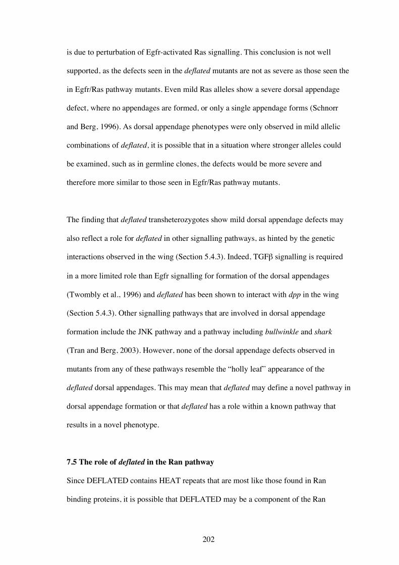
202
is due to perturbation of Egfr-activated Ras signalling. This conclusion is not well
supported, as the defects seen in the deflated mutants are not as severe as those seen the
in Egfr/Ras pathway mutants. Even mild Ras alleles show a severe dorsal appendage
defect, where no appendages are formed, or only a single appendage forms (Schnorr
and Berg, 1996). As dorsal appendage phenotypes were only observed in mild allelic
combinations of deflated, it is possible that in a situation where stronger alleles could
be examined, such as in germline clones, the defects would be more severe and
therefore more similar to those seen in Egfr/Ras pathway mutants.
The finding that deflated transheterozygotes show mild dorsal appendage defects may
also reflect a role for deflated in other signalling pathways, as hinted by the genetic
interactions observed in the wing (Section 5.4.3). Indeed, TGFβ signalling is required
in a more limited role than Egfr signalling for formation of the dorsal appendages
(Twombly et al., 1996) and deflated has been shown to interact with dpp in the wing
(Section 5.4.3). Other signalling pathways that are involved in dorsal appendage
formation include the JNK pathway and a pathway including bullwinkle and shark
(Tran and Berg, 2003). However, none of the dorsal appendage defects observed in
mutants from any of these pathways resemble the “holly leaf” appearance of the
deflated dorsal appendages. This may mean that deflated may define a novel pathway in
dorsal appendage formation or that deflated has a role within a known pathway that
results in a novel phenotype.
7.5 The role of deflated in the Ran pathway
Since DEFLATED contains HEAT repeats that are most like those found in Ran
binding proteins, it is possible that DEFLATED may be a component of the Ran
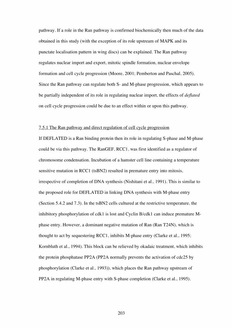
203
pathway. If a role in the Ran pathway is confirmed biochemically then much of the data
obtained in this study (with the exception of its role upstream of MAPK and its
punctate localisation pattern in wing discs) can be explained. The Ran pathway
regulates nuclear import and export, mitotic spindle formation, nuclear envelope
formation and cell cycle progression (Moore, 2001; Pemberton and Paschal, 2005).
Since the Ran pathway can regulate both S- and M-phase progression, which appears to
be partially independent of its role in regulating nuclear import, the effects of deflated
on cell cycle progression could be due to an effect within or upon this pathway.
7.5.1 The Ran pathway and direct regulation of cell cycle progression
If DEFLATED is a Ran binding protein then its role in regulating S-phase and M-phase
could be via this pathway. The RanGEF, RCC1, was first identified as a regulator of
chromosome condensation. Incubation of a hamster cell line containing a temperature
sensitive mutation in RCC1 (tsBN2) resulted in premature entry into mitosis,
irrespective of completion of DNA synthesis (Nishitani et al., 1991). This is similar to
the proposed role for DEFLATED in linking DNA synthesis with M-phase entry
(Section 5.4.2 and 7.3). In the tsBN2 cells cultured at the restrictive temperature, the
inhibitory phosphorylation of cdk1 is lost and Cyclin B/cdk1 can induce premature M-
phase entry. However, a dominant negative mutation of Ran (Ran T24N), which is
thought to act by sequestering RCC1, inhibits M-phase entry (Clarke et al., 1995;
Kornbluth et al., 1994). This block can be relieved by okadaic treatment, which inhibits
the protein phosphatase PP2A (PP2A normally prevents the activation of cdc25 by
phosphorylation (Clarke et al., 1993)), which places the Ran pathway upstream of
PP2A in regulating M-phase entry with S-phase completion (Clarke et al., 1995).
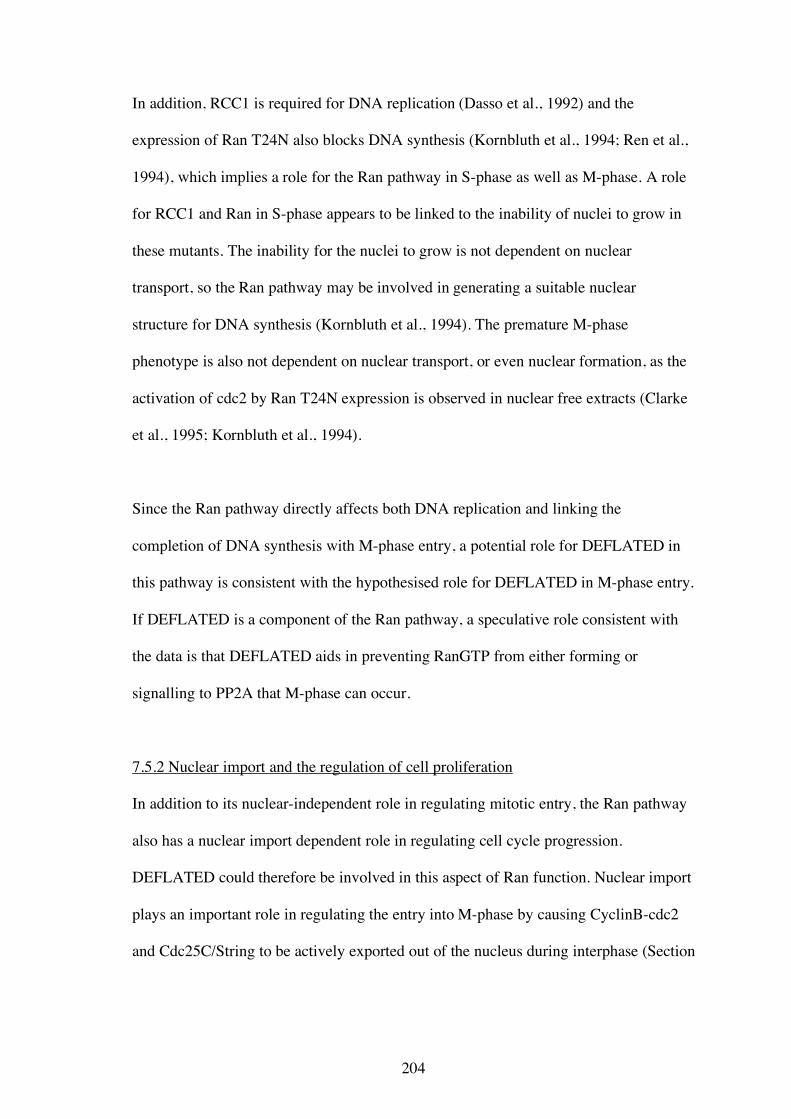
204
In addition, RCC1 is required for DNA replication (Dasso et al., 1992) and the
expression of Ran T24N also blocks DNA synthesis (Kornbluth et al., 1994; Ren et al.,
1994), which implies a role for the Ran pathway in S-phase as well as M-phase. A role
for RCC1 and Ran in S-phase appears to be linked to the inability of nuclei to grow in
these mutants. The inability for the nuclei to grow is not dependent on nuclear
transport, so the Ran pathway may be involved in generating a suitable nuclear
structure for DNA synthesis (Kornbluth et al., 1994). The premature M-phase
phenotype is also not dependent on nuclear transport, or even nuclear formation, as the
activation of cdc2 by Ran T24N expression is observed in nuclear free extracts (Clarke
et al., 1995; Kornbluth et al., 1994).
Since the Ran pathway directly affects both DNA replication and linking the
completion of DNA synthesis with M-phase entry, a potential role for DEFLATED in
this pathway is consistent with the hypothesised role for DEFLATED in M-phase entry.
If DEFLATED is a component of the Ran pathway, a speculative role consistent with
the data is that DEFLATED aids in preventing RanGTP from either forming or
signalling to PP2A that M-phase can occur.
7.5.2 Nuclear import and the regulation of cell proliferation
In addition to its nuclear-independent role in regulating mitotic entry, the Ran pathway
also has a nuclear import dependent role in regulating cell cycle progression.
DEFLATED could therefore be involved in this aspect of Ran function. Nuclear import
plays an important role in regulating the entry into M-phase by causing CyclinB-cdc2
and Cdc25C/String to be actively exported out of the nucleus during interphase (Section
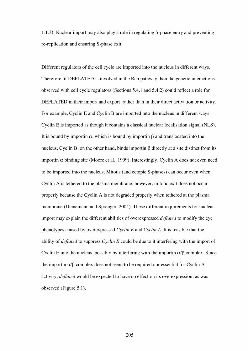
205
1.1.3). Nuclear import may also play a role in regulating S-phase entry and preventing
re-replication and ensuring S-phase exit.
Different regulators of the cell cycle are imported into the nucleus in different ways.
Therefore, if DEFLATED is involved in the Ran pathway then the genetic interactions
observed with cell cycle regulators (Sections 5.4.1 and 5.4.2) could reflect a role for
DEFLATED in their import and export, rather than in their direct activation or activity.
For example, Cyclin E and Cyclin B are imported into the nucleus in different ways.
Cyclin E is imported as though it contains a classical nuclear localisation signal (NLS).
It is bound by importin α, which is bound by importin β and translocated into the
nucleus. Cyclin B, on the other hand, binds importin β directly at a site distinct from its
importin α binding site (Moore et al., 1999). Interestingly, Cyclin A does not even need
to be imported into the nucleus. Mitotis (and ectopic S-phases) can occur even when
Cyclin A is tethered to the plasma membrane, however, mitotic exit does not occur
properly because the Cyclin A is not degraded properly when tethered at the plasma
membrane (Dienemann and Sprenger, 2004). These different requirements for nuclear
import may explain the different abilities of overexpressed deflated to modify the eye
phenotypes caused by overexpressed Cyclin E and Cyclin A. It is feasible that the
ability of deflated to suppress Cyclin E could be due to it interfering with the import of
Cyclin E into the nucleus, possibly by interfering with the importin α/β complex. Since
the importin α/β complex does not seem to be required nor essential for Cyclin A
activity, deflated would be expected to have no effect on its overexpression, as was
observed (Figure 5.1).
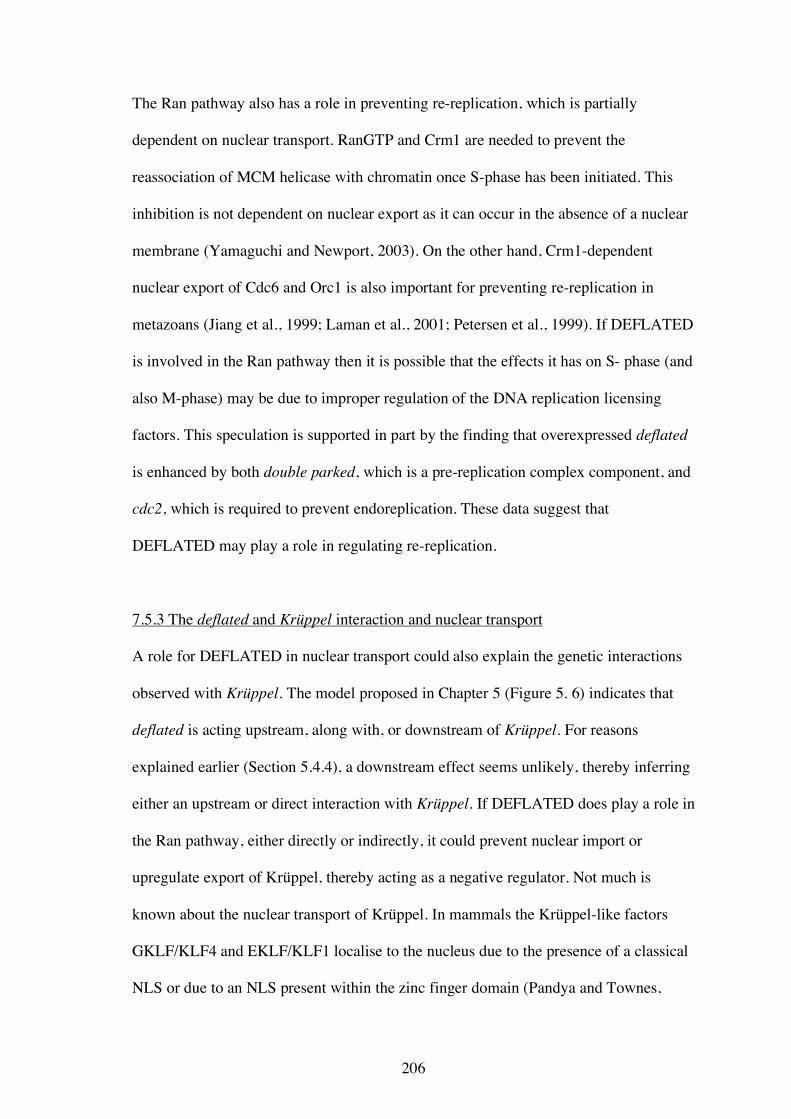
206
The Ran pathway also has a role in preventing re-replication, which is partially
dependent on nuclear transport. RanGTP and Crm1 are needed to prevent the
reassociation of MCM helicase with chromatin once S-phase has been initiated. This
inhibition is not dependent on nuclear export as it can occur in the absence of a nuclear
membrane (Yamaguchi and Newport, 2003). On the other hand, Crm1-dependent
nuclear export of Cdc6 and Orc1 is also important for preventing re-replication in
metazoans (Jiang et al., 1999; Laman et al., 2001; Petersen et al., 1999). If DEFLATED
is involved in the Ran pathway then it is possible that the effects it has on S- phase (and
also M-phase) may be due to improper regulation of the DNA replication licensing
factors. This speculation is supported in part by the finding that overexpressed deflated
is enhanced by both double parked, which is a pre-replication complex component, and
cdc2, which is required to prevent endoreplication. These data suggest that
DEFLATED may play a role in regulating re-replication.
7.5.3 The deflated and Krüppel interaction and nuclear transport
A role for DEFLATED in nuclear transport could also explain the genetic interactions
observed with Krüppel. The model proposed in Chapter 5 (Figure 5. 6) indicates that
deflated is acting upstream, along with, or downstream of Krüppel. For reasons
explained earlier (Section 5.4.4), a downstream effect seems unlikely, thereby inferring
either an upstream or direct interaction with Krüppel. If DEFLATED does play a role in
the Ran pathway, either directly or indirectly, it could prevent nuclear import or
upregulate export of Krüppel, thereby acting as a negative regulator. Not much is
known about the nuclear transport of Krüppel. In mammals the Krüppel-like factors
GKLF/KLF4 and EKLF/KLF1 localise to the nucleus due to the presence of a classical
NLS or due to an NLS present within the zinc finger domain (Pandya and Townes,
207
2002; Quadrini and Bieker, 2002; Shie and Tseng, 2001). EKLF/KLF1 binds both
importin β and importin α in vitro so Krüppel is likely to be translocated via a Ran-
dependent mechanism in vivo (Quadrini and Bieker, 2002).
Although compelling, a potential role for DEFLATED in the Ran pathway can not
readily explain the punctate apical localisation of DEFLATED::GFP nor the finding
that DEFLATED is likely to be upstream of MAPK activation in the Ras signalling
cascade, if all the effects of the Ran pathway occur in the nucleus. However, there is
precedence for a non nuclear role for a Ran binding protein. RanBPM localises to the
plasma membrane and plays a role in signal transduction. RanBPM was first discovered
as a Ran binding protein in a yeast two hybrid assay, even though it does not contain a
classical Ran binding domain as found in importin β (Nakamura et al., 1998). Initially it
was found that RanBPM had a role in regulating microtubule nucleation. However,
subsequent studies have not substantiated this role. Instead there is increasing data in
the literature to suggest that RanBPM may be a scaffold protein integrating signalling
molecules, such as receptor protein-tyrosine kinases and integrins, with their
downstream effectors, such as the Ras/MAPK signalling pathway (Denti et al., 2004;
Wang et al., 2002). Even though the putative Ran-binding domain in DEFLATED was
recognised due to its similarity with domains found in proteins such as importin β, and
is therefore different to RanBPM, it is possible that DEFLATED is a Ran binding
protein that has functions in the nucleus and at the plasma membrane, analogous to
RanBPM.
208
7.6 The role of deflated in endocytosis
Endocytosis is the regulated uptake of molecules at the plasma membrane, and can take
place by four different methods; clathrin-dependent endocytosis, caveolae-mediated
endocytosis, macropinocytosis, and dynamin- and clathrin- independent endocytosis
(reviewed in Mousavi et al., 2004; Seto et al., 2002). The best studied is clathrin-
dependent endocytosis. It has been well documented that clathrin-mediated endocytosis
plays an important role in regulating cell signalling (Seto et al., 2002). What is not so
clear is the precise role endocytosis plays in cell signalling. Initially it was thought that
the internalisation of receptors following their activation by ligand binding was
required to desensitise or down-regulate signalling. While this does seem to occur,
particularly for G protein-coupled receptors (Scott et al., 2002), it is becoming clear that
in some contexts endocytosis may in fact increase signalling, and that there is
substantial cross-talk between endocytic and signalling pathways.
7.6.1. Endocytosis and Ras signalling
Endocytosis plays a role in the regulation of RTK/MAPK, Notch, Wg/Wnt, TGFβ/Dpp,
and Hedgehog signalling pathways. The RTK/Ras/Raf/MAPK pathway is a good
example to illustrate the interactions between signalling and endocytosis since the
mechanisms are reasonably well understood. Moreover, to understand how
DEFLATED may act in either Ras signalling or endocytosis, an examination of the two
processes is required. Proteins involved in endocytosis that are phosphorylated as a
result of RTK activation include clathrin (Wilde et al., 1999), Eps15 (Confalonieri et
al., 2000), and Hrs (Komada and Kitamura, 1995). The phosphorylation of these
proteins is necessary for the internalisation of the receptor and for the signal to be
transduced. In the case of EGFR internalisation, the debate about whether
209
internalisation attenuates or enhances signalling has been addressed. Signalling is
enhanced in situations where recycling back to the membrane is favoured over
degradation, due to the presence of receptor heterodimers rather than homodimers
(Lenferink et al., 1998). Signalling also is enhanced and fine tuned by the formation of
endosomes, in which receptors associate with their targets, since the majority of the
phosphorylated forms of these targets are found in endosomal fractions (Di Guglielmo
et al., 1994; Oksvold et al., 2000). Furthermore, it is possible that internalisation allows
selective activation of targets as it was found that MAPK is not activated when
internalisation was prevented, but other targets were activated as normal (Kranenburg et
al., 1999).
Endosomes form multivesicular bodies (MVBs) where it is thought that receptors are
sorted and either recycled to the cell surface or are sent to the lysosome for degradation.
Two known inhibitors of Ras signalling play a role in the sorting to lysosomes, Cbl and
Hrs. Cbl is a ubiquitin ligase and has been shown to ubiquitinate RTKs (Levkowitz et
al., 1998). Hrs can bind ubiquitin through its ubiquitin-interacting motif (Lloyd et al.,
2002). Both proteins result in the upregulation of receptor degradation, with ubiquitin
possibly acting as the sorting signal. Therefore, while internalisation of the receptor is
needed for signalling to occur, it is in the regulation of the downstream events, such as
how long the receptor and their targets remain on endosomes and whether the receptors
are recycled or sent to lysosomes, that determines whether the overall outcome of
internalisation is one of attenuation or enhancement. It is clear that the cross talk
between cell signalling and endocytosis serves to fine-tune the signalling process, by
allowing regulation to occur at various points, therefore allowing for subtle signalling
outcomes dependent on cell type or developmental stage to take place.
210
7.6.2 Is DEFLATED an endocytic component or a Ras signalling component?
The findings that DEFLATED contains a well conserved clathrin box and adaptor
protein binding sites, is localised to the apical surface in wing discs, displays genetic
interactions with the Ras signalling pathway, and that deflated overexpression prevents
spatially-correct MAPK activation, all support a role for DEFLATED in endocytosis.
DEFLATED might regulate signalling through endocytosis or it may be a protein that is
part of a signalling cascade that is regulated by endocytosis. Genetically, deflated is
downstream of Ras, so it is unlikely that it plays a role in regulating the initial
internalisation of receptor tyrosine kinases. However, since cell signalling may be
increased by concentrating active receptors and their downstream effectors on the
surface of the endosome (Seto et al., 2002) it is possible that DEFLATED plays a role
at the endosome.
If DEFLATED is either involved in regulating the activation of Raf, MEK, or MAPK at
the plasma membrane or on endosomes, or is an endocytic component involved in
creating these endosomes, then DEFLATED should be found in endosomes. The
punctate localisation pattern of DEFLATED::GFP in the wing imaginal discs is similar
to that of endosomes, supporting this speculation. Double labelling with known
endosomal components would be required to confirm that the punctate localisation
pattern is indeed endosomal. Since it appears that DEFLATED is a negative regulator
of Ras signalling it is tempting to speculate on how it may function. If DEFLATED is
an endocytic protein then it may negatively regulate clathrin-coated pit or vesicle
formation or it may promote the disassembly of clathrin. If DEFLATED is endocytosed
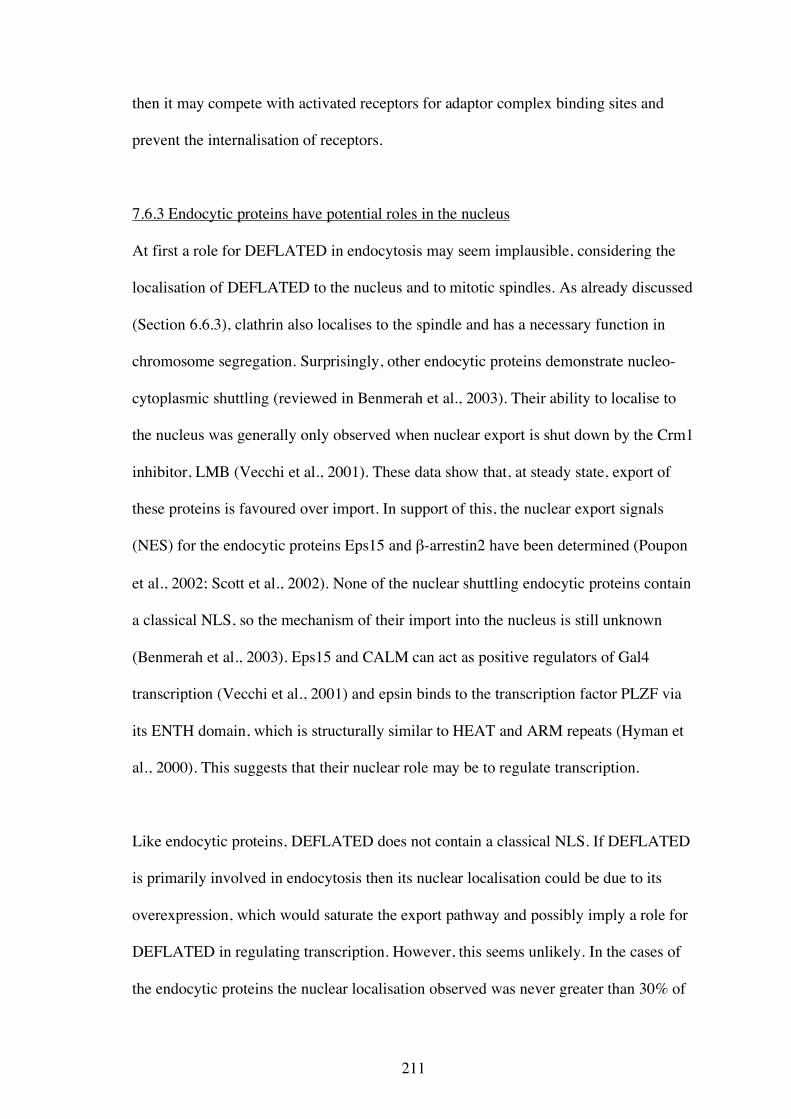
211
then it may compete with activated receptors for adaptor complex binding sites and
prevent the internalisation of receptors.
7.6.3 Endocytic proteins have potential roles in the nucleus
At first a role for DEFLATED in endocytosis may seem implausible, considering the
localisation of DEFLATED to the nucleus and to mitotic spindles. As already discussed
(Section 6.6.3), clathrin also localises to the spindle and has a necessary function in
chromosome segregation. Surprisingly, other endocytic proteins demonstrate nucleo-
cytoplasmic shuttling (reviewed in Benmerah et al., 2003). Their ability to localise to
the nucleus was generally only observed when nuclear export is shut down by the Crm1
inhibitor, LMB (Vecchi et al., 2001). These data show that, at steady state, export of
these proteins is favoured over import. In support of this, the nuclear export signals
(NES) for the endocytic proteins Eps15 and β-arrestin2 have been determined (Poupon
et al., 2002; Scott et al., 2002). None of the nuclear shuttling endocytic proteins contain
a classical NLS, so the mechanism of their import into the nucleus is still unknown
(Benmerah et al., 2003). Eps15 and CALM can act as positive regulators of Gal4
transcription (Vecchi et al., 2001) and epsin binds to the transcription factor PLZF via
its ENTH domain, which is structurally similar to HEAT and ARM repeats (Hyman et
al., 2000). This suggests that their nuclear role may be to regulate transcription.
Like endocytic proteins, DEFLATED does not contain a classical NLS. If DEFLATED
is primarily involved in endocytosis then its nuclear localisation could be due to its
overexpression, which would saturate the export pathway and possibly imply a role for
DEFLATED in regulating transcription. However, this seems unlikely. In the cases of
the endocytic proteins the nuclear localisation observed was never greater than 30% of
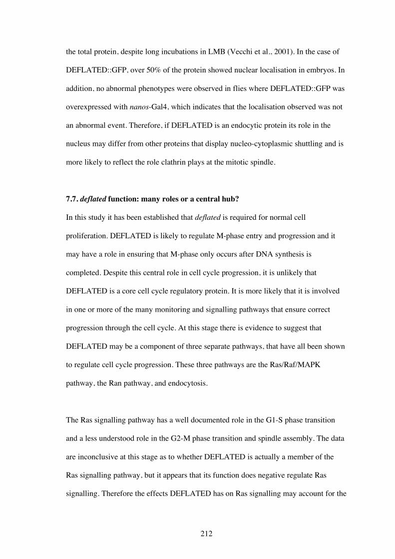
212
the total protein, despite long incubations in LMB (Vecchi et al., 2001). In the case of
DEFLATED::GFP, over 50% of the protein showed nuclear localisation in embryos. In
addition, no abnormal phenotypes were observed in flies where DEFLATED::GFP was
overexpressed with nanos-Gal4, which indicates that the localisation observed was not
an abnormal event. Therefore, if DEFLATED is an endocytic protein its role in the
nucleus may differ from other proteins that display nucleo-cytoplasmic shuttling and is
more likely to reflect the role clathrin plays at the mitotic spindle.
7.7. deflated function: many roles or a central hub?
In this study it has been established that deflated is required for normal cell
proliferation. DEFLATED is likely to regulate M-phase entry and progression and it
may have a role in ensuring that M-phase only occurs after DNA synthesis is
completed. Despite this central role in cell cycle progression, it is unlikely that
DEFLATED is a core cell cycle regulatory protein. It is more likely that it is involved
in one or more of the many monitoring and signalling pathways that ensure correct
progression through the cell cycle. At this stage there is evidence to suggest that
DEFLATED may be a component of three separate pathways, that have all been shown
to regulate cell cycle progression. These three pathways are the Ras/Raf/MAPK
pathway, the Ran pathway, and endocytosis.
The Ras signalling pathway has a well documented role in the G1-S phase transition
and a less understood role in the G2-M phase transition and spindle assembly. The data
are inconclusive at this stage as to whether DEFLATED is actually a member of the
Ras signalling pathway, but it appears that its function does negative regulate Ras
signalling. Therefore the effects DEFLATED has on Ras signalling may account for the
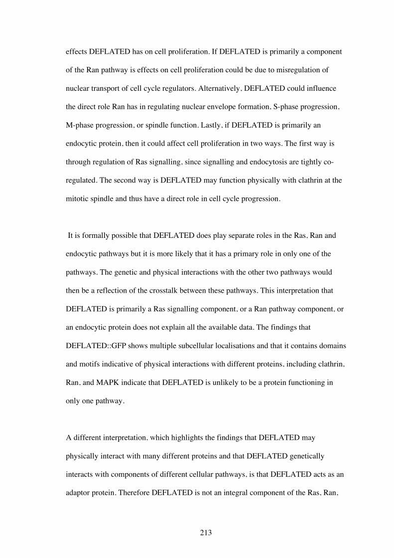
213
effects DEFLATED has on cell proliferation. If DEFLATED is primarily a component
of the Ran pathway is effects on cell proliferation could be due to misregulation of
nuclear transport of cell cycle regulators. Alternatively, DEFLATED could influence
the direct role Ran has in regulating nuclear envelope formation, S-phase progression,
M-phase progression, or spindle function. Lastly, if DEFLATED is primarily an
endocytic protein, then it could affect cell proliferation in two ways. The first way is
through regulation of Ras signalling, since signalling and endocytosis are tightly co-
regulated. The second way is DEFLATED may function physically with clathrin at the
mitotic spindle and thus have a direct role in cell cycle progression.
It is formally possible that DEFLATED does play separate roles in the Ras, Ran and
endocytic pathways but it is more likely that it has a primary role in only one of the
pathways. The genetic and physical interactions with the other two pathways would
then be a reflection of the crosstalk between these pathways. This interpretation that
DEFLATED is primarily a Ras signalling component, or a Ran pathway component, or
an endocytic protein does not explain all the available data. The findings that
DEFLATED::GFP shows multiple subcellular localisations and that it contains domains
and motifs indicative of physical interactions with different proteins, including clathrin,
Ran, and MAPK indicate that DEFLATED is unlikely to be a protein functioning in
only one pathway.
A different interpretation, which highlights the findings that DEFLATED may
physically interact with many different proteins and that DEFLATED genetically
interacts with components of different cellular pathways, is that DEFLATED acts as an
adaptor protein. Therefore DEFLATED is not an integral component of the Ras, Ran,
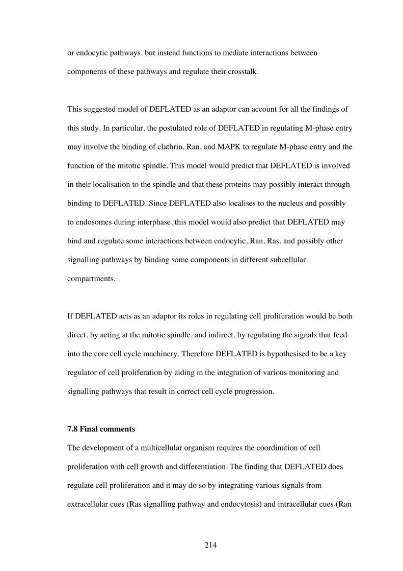
214
or endocytic pathways, but instead functions to mediate interactions between
components of these pathways and regulate their crosstalk.
This suggested model of DEFLATED as an adaptor can account for all the findings of
this study. In particular, the postulated role of DEFLATED in regulating M-phase entry
may involve the binding of clathrin, Ran, and MAPK to regulate M-phase entry and the
function of the mitotic spindle. This model would predict that DEFLATED is involved
in their localisation to the spindle and that these proteins may possibly interact through
binding to DEFLATED. Since DEFLATED also localises to the nucleus and possibly
to endosomes during interphase, this model would also predict that DEFLATED may
bind and regulate some interactions between endocytic, Ran, Ras, and possibly other
signalling pathways by binding some components in different subcellular
compartments.
If DEFLATED acts as an adaptor its roles in regulating cell proliferation would be both
direct, by acting at the mitotic spindle, and indirect, by regulating the signals that feed
into the core cell cycle machinery. Therefore DEFLATED is hypothesised to be a key
regulator of cell proliferation by aiding in the integration of various monitoring and
signalling pathways that result in correct cell cycle progression.
7.8 Final comments
The development of a multicellular organism requires the coordination of cell
proliferation with cell growth and differentiation. The finding that DEFLATED does
regulate cell proliferation and it may do so by integrating various signals from
extracellular cues (Ras signalling pathway and endocytosis) and intracellular cues (Ran
215
pathway and possibly other as of yet unknown pathways) indicates that it plays an
important but complex role.
The genetic observations described in Chapter 5 support a role for DEFLATED in
regulating S- to M-phase progression. This role would require the sensing of the state of
DNA replication and signalling to the M-phase machinery. Since DEFLATED is likely
to be an adaptor protein it is predicted to act by integrating or modulating these signals.
The proper regulation of S- to M-phase progression is crucial to ensure correct
development and to prevent tumourogenesis, so further study of DEFLATED is likely
to increase our understanding of this regulation.
The role of DEFLATED as an adaptor is likely to extend to other signalling pathways,
including those involving Ras and Ran. Therefore, further study of deflated is
warranted to to understand how these pathways are regulated and how they influence
cell proliferation. The further study of deflated will no doubt contain some surprises,
but will also yield insight not only into the primary function of DEFLATED but also
how various cellular processes intersect to facilitate one of those crucial decisions for a
cell- to divide or not divide.