Embed Size (px)
Citation preview
Rheumatic Valvular Heart Disease
Paul J Kovack, DO, FACOI, FACC Cardiologist
Metro Health Hospital

Case
• Ana K is a 45 year old Nepalese woman • She presents with an interpreter hoping to get a refill on a medication she
was prescribed back home for an abnormal heart rhythm she has had for several years
• This is the only medication she is on • It was started when she had a small stroke effecting her speech 15 years
ago. She has no residual problems from the stroke • She does not know what type of abnormal heart rhythm she has • She really has no symptoms, specifically SOB, palpitations,
lightheadedness, chest discomfort, weight gain or swelling • She is tired, but no more than she has been

Physical exam
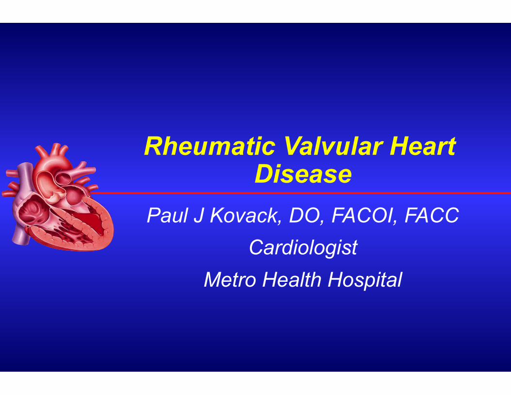
• Irregularly irregular pulse • Diastolic murmur (loud) • No residual neurologic deficits

Grading Heart Murmurs
– graded on a 6 point scale • Grade 1 = very faint, heard intermittently • Grade 2 = quiet but heard immediately • Grade 3 = moderately loud • Grade 4 =very loud with palpable thrill • Grade 5 = very loud and can still hear with edge of stethoscope • Grade 6 = heard with stethoscope partly off the chest • *Note: Thrills are assoc. with murmurs of grades 4 - 6
Ana’s ECG

Echocardiogram

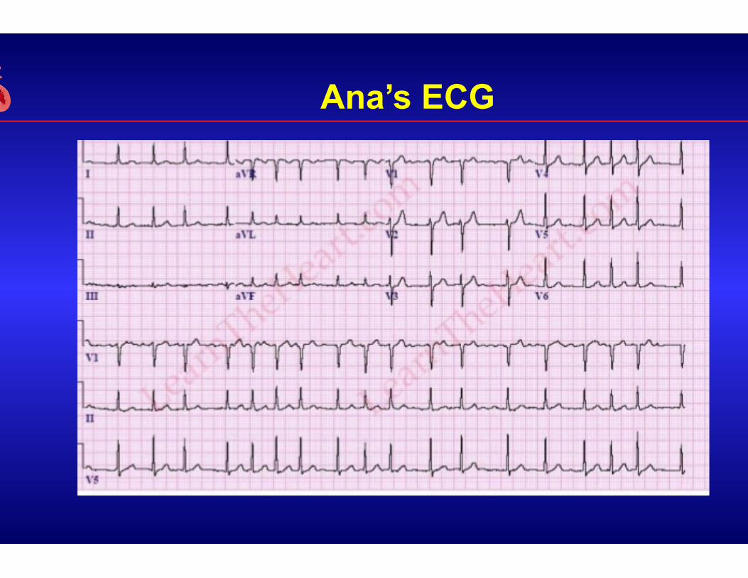
2 D Echocardiogram characteristics
• Thickened and calcified mitral valve leaflets and subvalvular apparatus
• Restricted motion • Diastolic doming of leaflets (hockey stick appearance) • Increased LA size • Fish mouth appearance in short axis view
Domed mitral valve leaflets
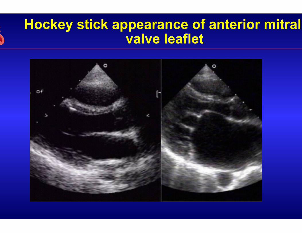
Hockey stick appearance of anterior mitral valve leaflet

Stenotic mitral valve end diastole (fish mouth appearance)
Spotted blue puffer fish

Severity by mean gradient across the mitral valve
Epidemiology of Rheumatic Fever
• ARF caused by group A beta- hemolytic streptococcus effects 20 million people and is the leading cause of cardiovascular death in the first 5 decade of life
• Mean incidence world wide is 19 per 100,000 • US 5 per 100,000
– Resurgence in past few decades • The worst affected areas are sub-Saharan Africa, south-central
Asia and the Pacific where incidence has been reported as high as 1 percent.

Rheumatic Heart Disease
• RF principally involves the heart, joint, CNS, skin and subcutaneous tissues
• Rheumatic Heart Disease refers to the cardiac involvement that develops in 50% of patients with RF
• Can effect the endocardium, myocardium or pericardium • It may later effect the heart valves causing chronic valve
disease leading to CHF and death • There is a latent period of up to several decades before valve
disease becomes severe enough to recognize.
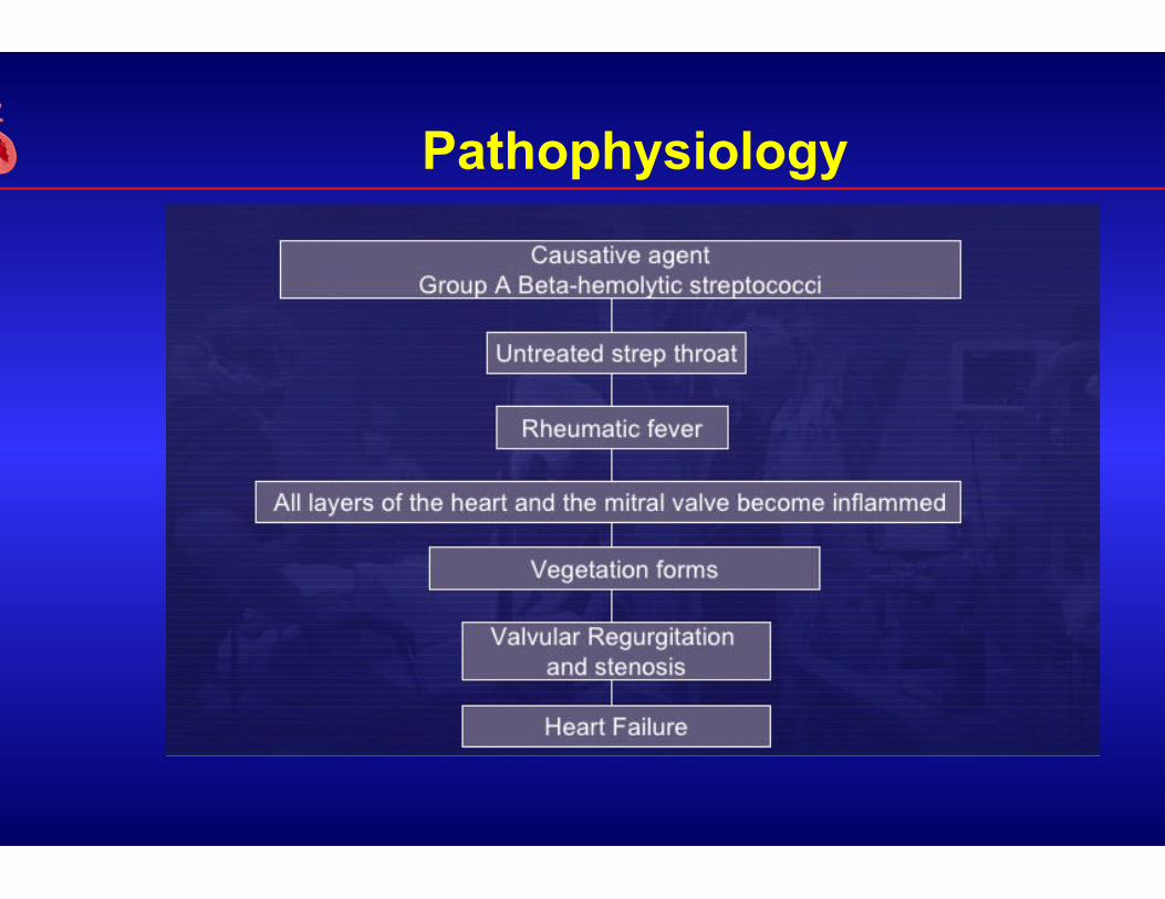
Pathophysiology

Key feature of RF is granuloma formation
1. Exudative and degenerative phase
2. The proliferative phase (Granulomatous period)
3. Scar phase (healed phase\Fibrosis phase\Hardening phase)
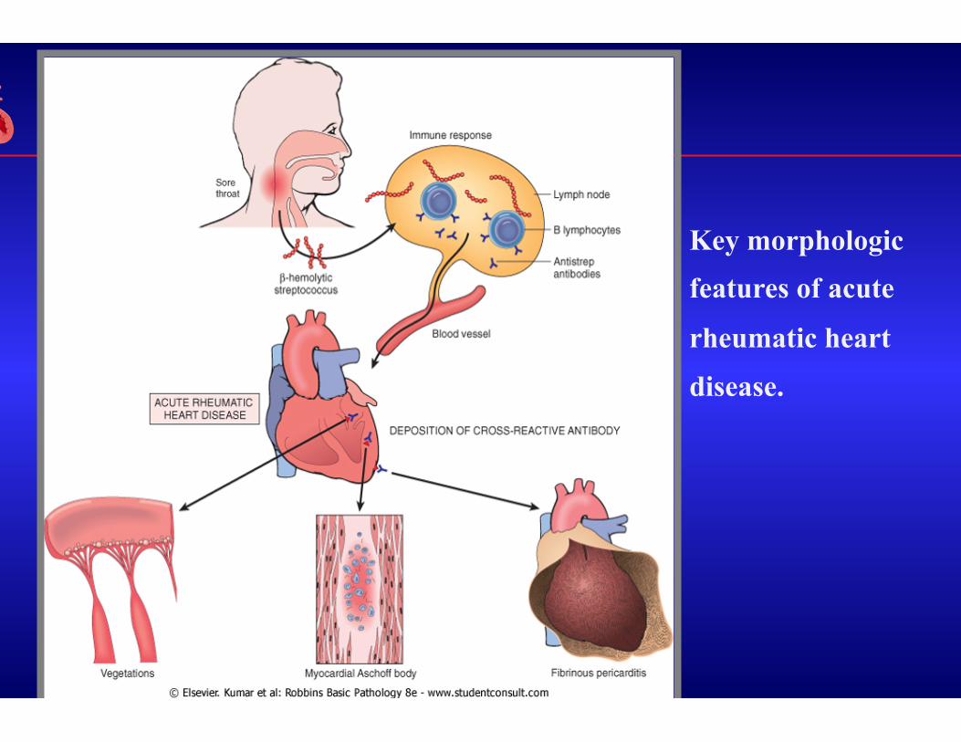
Key morphologic features of acute
rheumatic heart
disease.

Acute rheumatic endocarditis: small (diameter 1- to 2-mm) vegetations along the mitral valve margin, insufficient to cause valvular deformation.
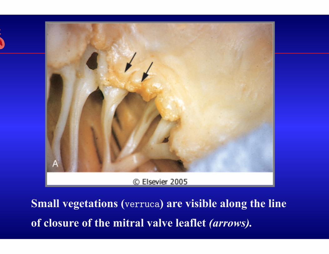
Small vegetations (verruca) are visible along the line
of closure of the mitral valve leaflet (arrows).
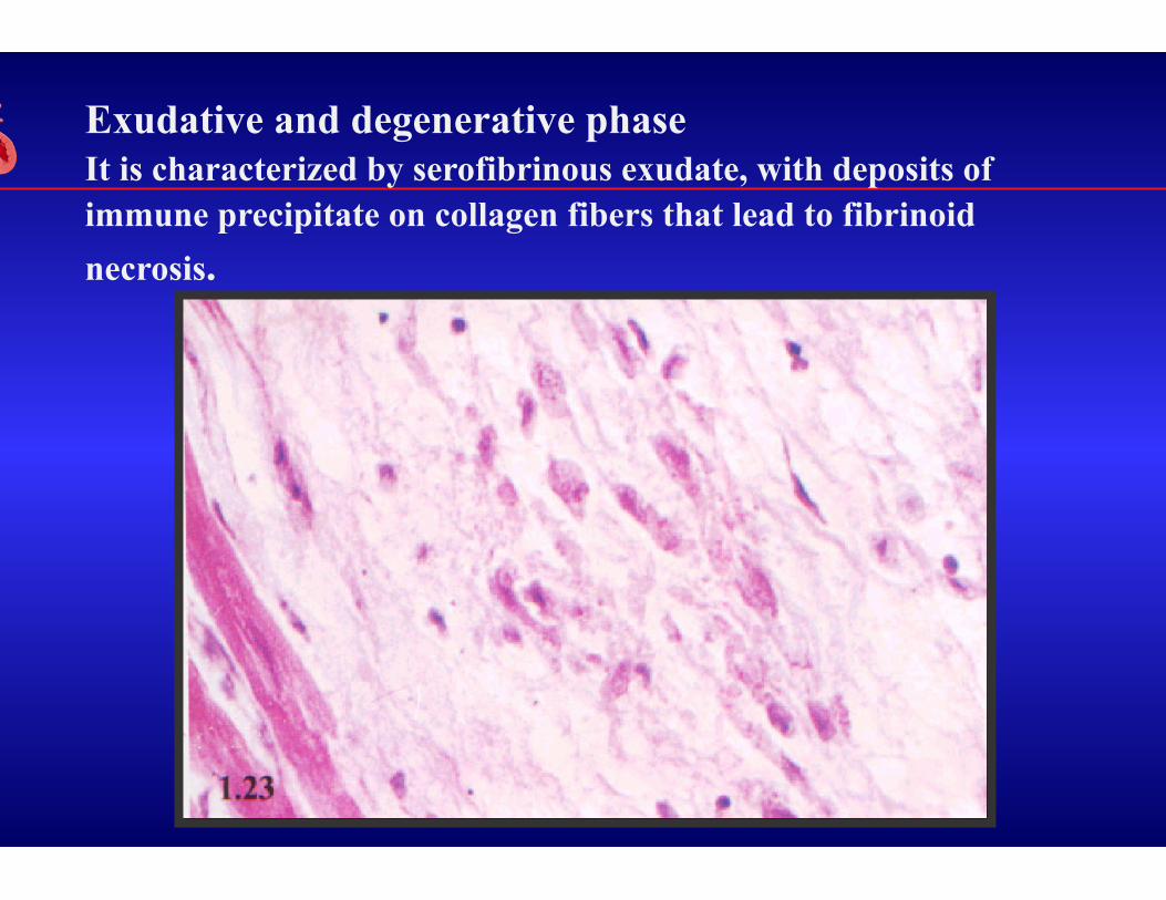
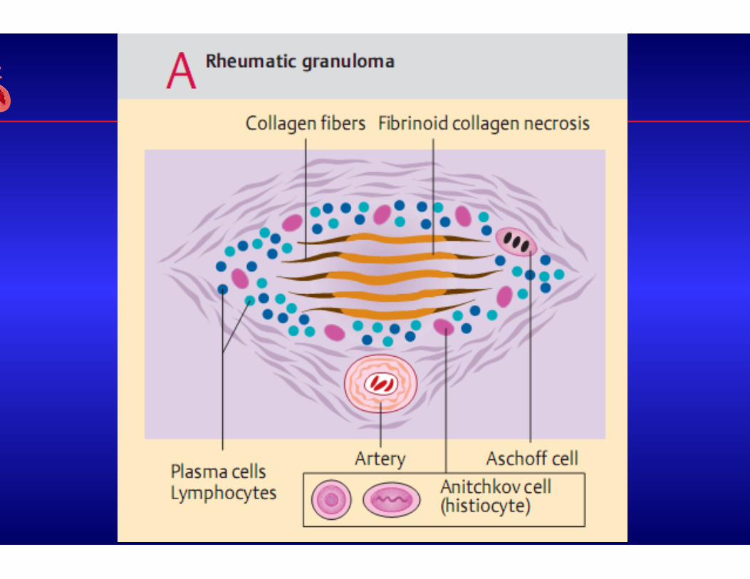
Exudative and degenerative phase It is characterized by serofibrinous exudate, with deposits of immune precipitate on collagen fibers that lead to fibrinoid necrosis.

Serous pericarditis Fibrinous pericarditis

pericardial effusion
Can lead to heart sound far, around the heart boundary expanding, serious cardiac X-ray showed a flask

Adhesive pericarditis is in cardiac surface of patients. From the epicardial surface to the pericardial sac visible fibrinous exudate, which is typical for a fibrinous pericarditis.
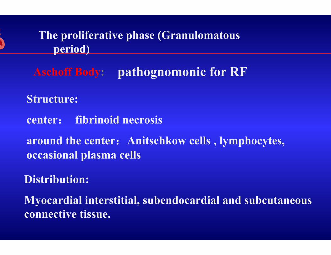
The proliferative phase (Granulomatous period)
Aschoff Body:
Structure:
center: fibrinoid necrosis
around the center:Anitschkow cells , lymphocytes, occasional plasma cells
Distribution:
Myocardial interstitial, subendocardial and subcutaneous connective tissue.
pathognomonic for RF

Anitschkow cells
These distinctive cells have abundant cytoplasm
and central round-to-ovoid nuclei in which the
chromatin is disposed in a central, slender, wavy
ribbon (hence the designation "caterpillar cells“
cross section named Owl 's eye cells).
Some of the larger macrophages become multinucleated to form Aschoff cells(inflammatory giant cells).


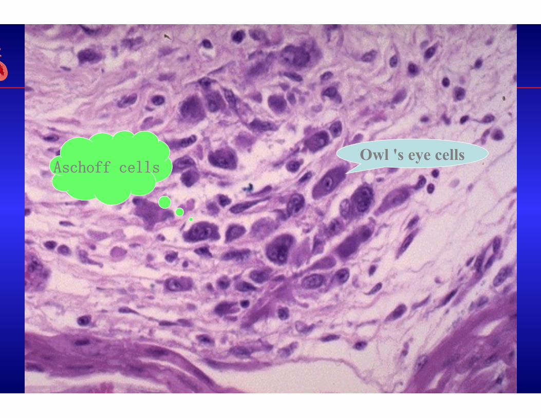
Owl 's eye cells Aschoff cells

Early changes to mitral valve on echocardiogram
Valves effected
• Mitral valve most common • Then aortic valve • Tricuspid and pulmonary valves effected less than 5 % of the
time
Mitral stenosis
• Most common cause is RF (up to 99%) • Become symptomatic is 2nd to 4th decade of life • More women than men (2/3rds) • 25% of patients with chronic RF have pure MS • 40% have combined mitral stenosis and mitral regurgitation
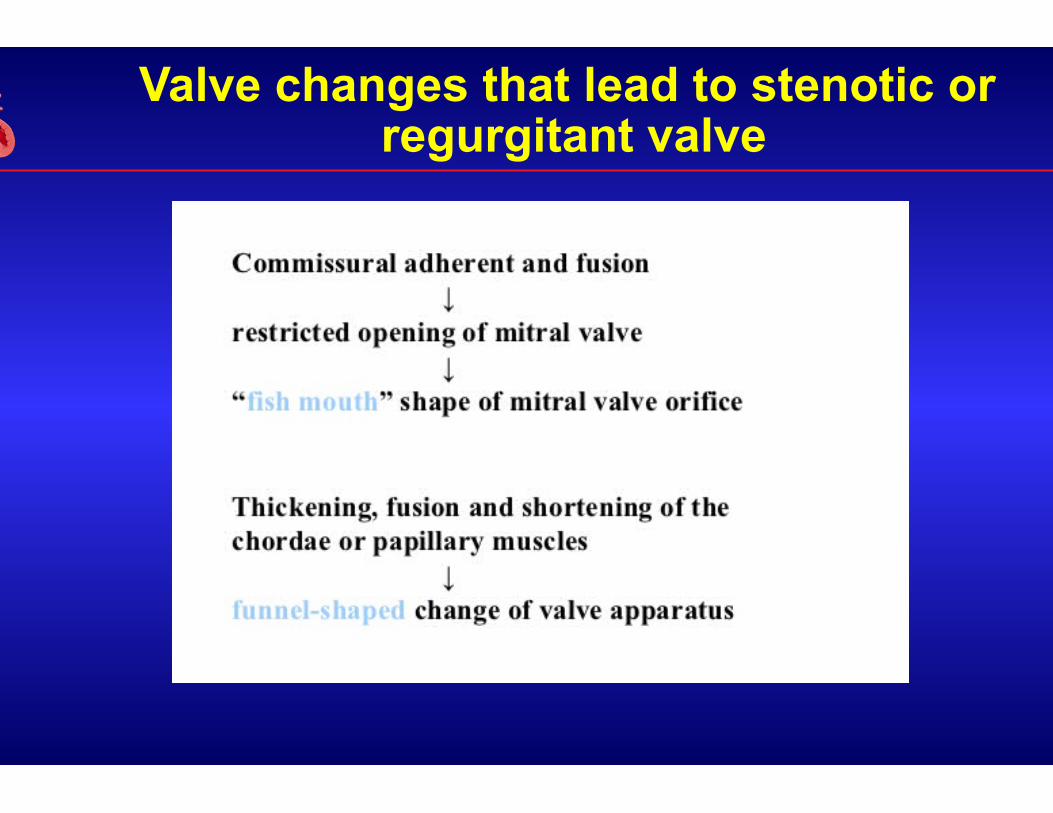
Valve changes that lead to stenotic or regurgitant valve
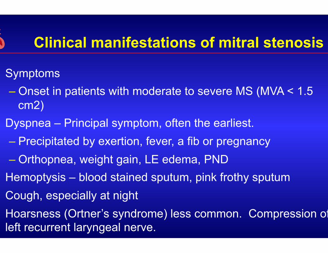
Clinical manifestations of mitral stenosis
• Symptoms – Onset in patients with moderate to severe MS (MVA < 1.5
cm2) • Dyspnea – Principal symptom, often the earliest.
– Precipitated by exertion, fever, a fib or pregnancy – Orthopnea, weight gain, LE edema, PND
• Hemoptysis – blood stained sputum, pink frothy sputum • Cough, especially at night • Hoarsness (Ortner’s syndrome) less common. Compression of
left recurrent laryngeal nerve.
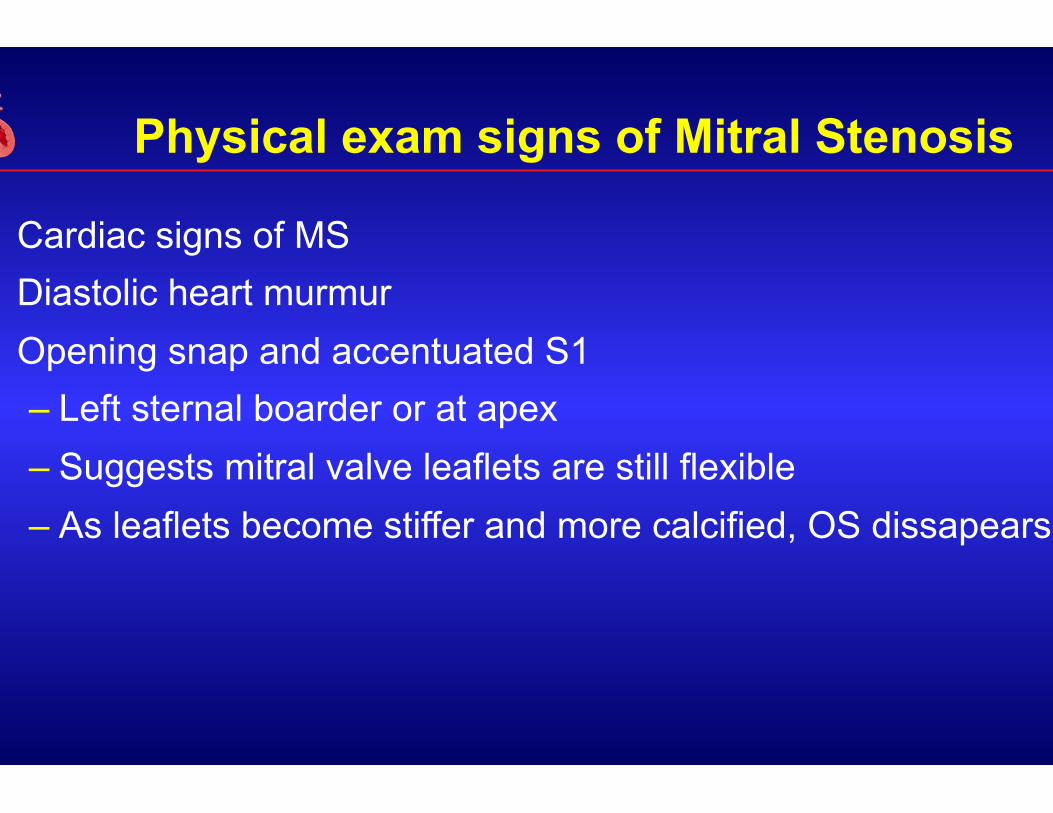
Physical exam signs of Mitral Stenosis
• Cardiac signs of MS • Diastolic heart murmur • Opening snap and accentuated S1
– Left sternal boarder or at apex – Suggests mitral valve leaflets are still flexible – As leaflets become stiffer and more calcified, OS dissapears
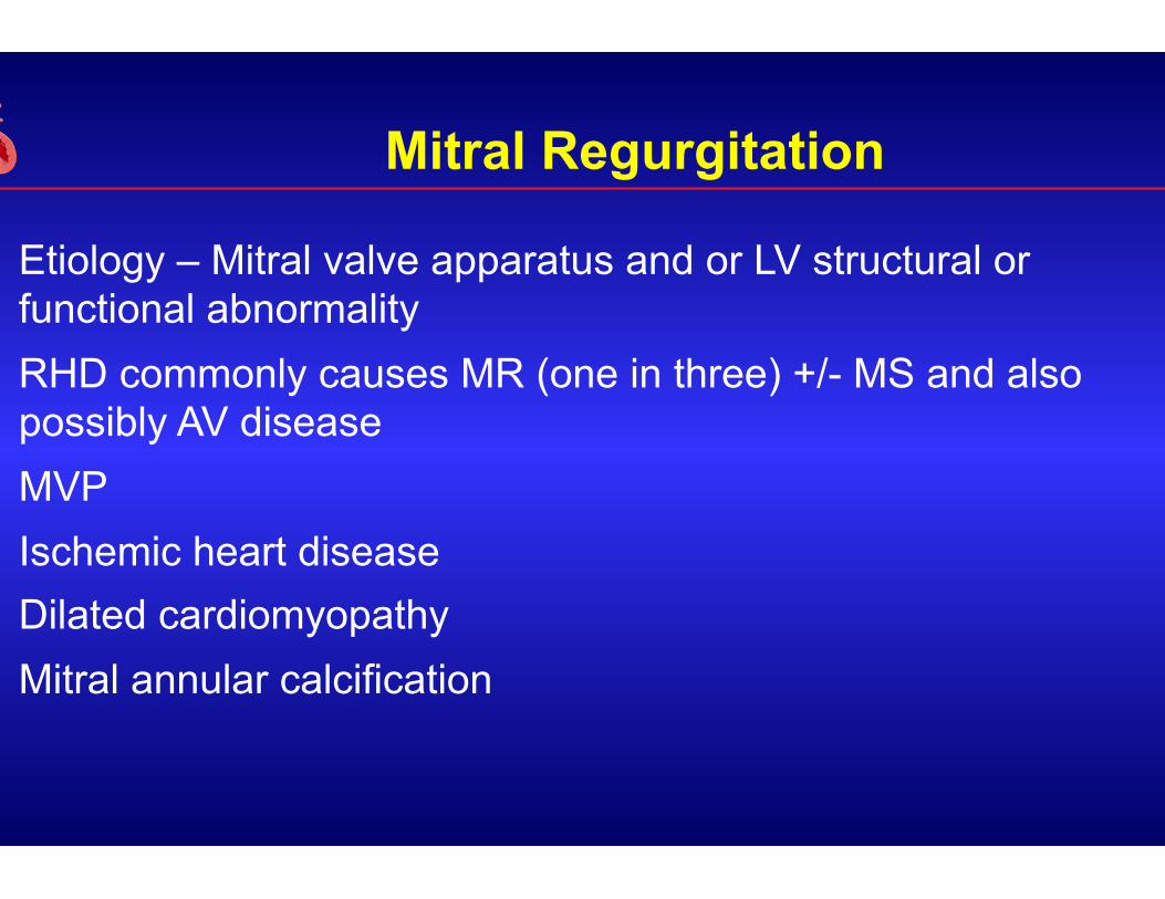
Mitral Regurgitation
• Etiology – Mitral valve apparatus and or LV structural or functional abnormality
• RHD commonly causes MR (one in three) +/- MS and also possibly AV disease
• MVP • Ischemic heart disease • Dilated cardiomyopathy • Mitral annular calcification

Systolic murmur of mitral regurgitation
• RHD – pan systolic, blowing, high-pitched murmur • Loudest at apex (axilla, back) • MVP – mid to late systolic murmur • Papillary muscle dysfunction – variable murmur • Rupture of chordae tendonea - musical

Differential diagnosis of systolic murmur (MI.AS.TI.PS)
• Tricuspid regurgitation – systolic murmur along the left sternal border (Erb’s point), Increased with inspiration.
• VSD – loudest at left sternal border and can be accompanied by a parasternal thrill
• Systolic ejection murmur Aortic Stenosis vs pulmonic stenosis
• Hypertrophic cardiomyopathy

Aortic stenosis
• RHD less common etiology but suspect it when AS is seen with MS and AI
• Degenerative calcific AS. Most common cause of severe AS – Most common in the elderly – Can be associated with mitral annular calcification
• Bicuspid AV.

Physical exam in aortic stenosis
• Systolic ejection murmur – Blowing harsh crescendo decrescendo – Heard best at aortic listening post – Radiates to neck and apex – The more sever the AS, the longer the duration of the
murmur – Although, once LV fails (dilates) and cardiac output
decreases, murmur will deminish
Aortic regurgitation
• RHD is most common cause - 2/3rds originate from RF – Can be associated with AS and MV disease
• Infective endocarditis • Bicuspid AV • Aortic root dilatation
– Marfan syndrome – Aortic dissection – Syphilitic aortitis

Physical examination in AI
• Chronic, severe aortic insufficiency • Peripheral arterial signs due to widened pulse pressure
– Water hammer pulse (early rise and fall) – Pistol shot sounds – loud systolic and diastolic sounds over
femoral artery – Duroziez’s sign – bruit over partially compressed femoral
artery – Quincke’s sign – subunguinal capillary pulsations – de Musset’s sign
Aortic insufficiency murmur • Diastolic murmur • High-pitched, blowing, decrescendo pattern • When due to primary aortic valve disease
– Diastolic murmur heard best along left sternal border – 3rd and 4th intercostal space
• When due to dilation of the ascending aorta – Heard best along right upper sternal border
• Austin Flint murmur – apical mid or late diastolic low-pitched murmur. Common in severe AI. Due to partial closure of MV by the regurgitant aortic jet.

Final thoughts
• Obtaining history of Rheumatic fever as a child – History of repeated throat infections, fevers, missing school – Remember half of those later diagnosed with rheumatic
valvular heart disease have no recollection of having RF as a child
• Atrial fibrillation – Common consequence of mitral valve disease – Due to LA dilatation – Anticoagulate with Coumadin, not one of the new drugs