Embed Size (px)
Citation preview
7/23/2016
1
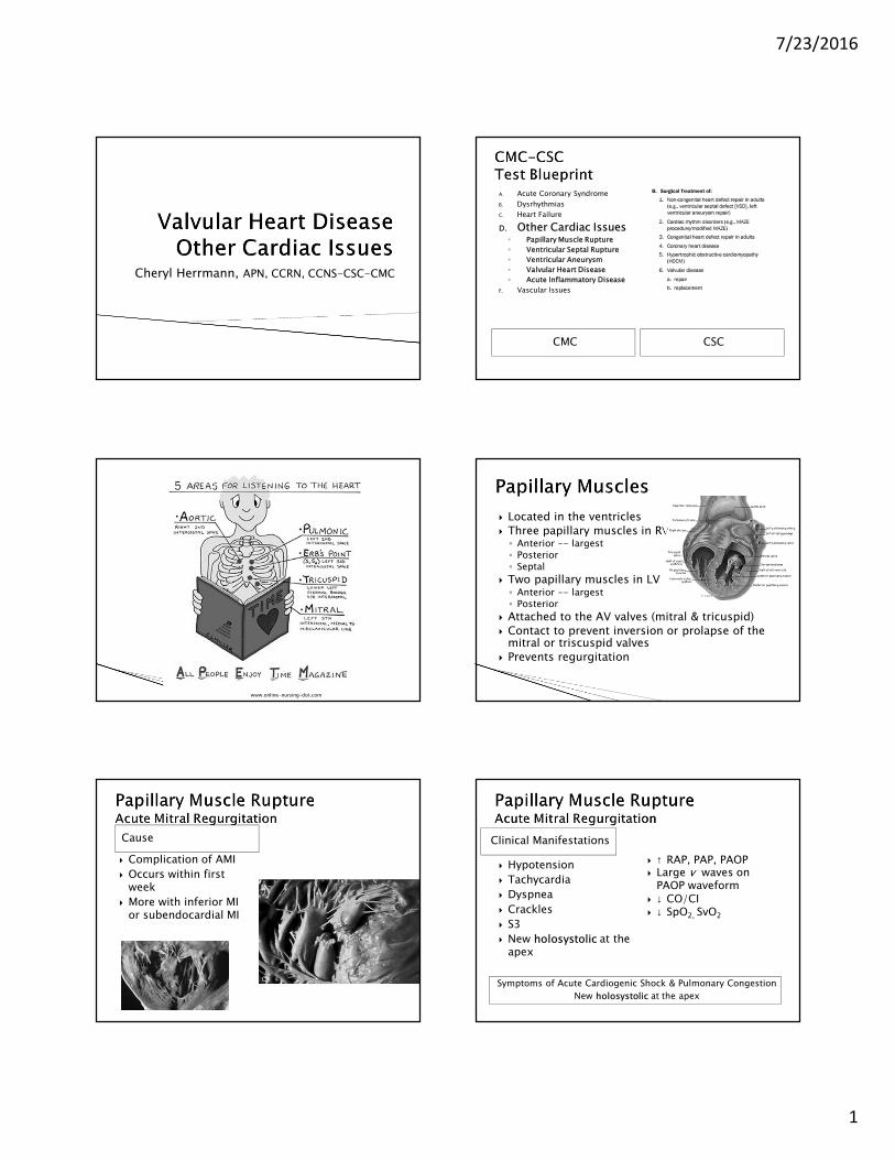
Cheryl Herrmann, APN, CCRN, CCNS-CSC-CMC
CMC CSC
A. Acute Coronary Syndrome
B. Dysrhythmias
C. Heart Failure
D. Other Cardiac Issues◦ Papillary Muscle Rupture
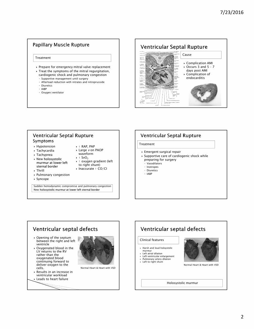
◦ Ventricular Septal Rupture
◦ Ventricular Aneurysm
◦ Valvular Heart Disease
◦ Acute Inflammatory Disease
E. Vascular Issues
www.online-nursing-dot.com
� Located in the ventricles
� Three papillary muscles in RV◦ Anterior -- largest◦ Posterior◦ Septal
� Two papillary muscles in LV◦ Anterior -- largest◦ Posterior
� Attached to the AV valves (mitral & tricuspid)
� Contact to prevent inversion or prolapse of the mitral or triscuspid valves
� Prevents regurgitation
Cause
� Complication of AMI
� Occurs within first week
� More with inferior MI or subendocardial MI
Symptoms of Acute Cardiogenic Shock & Pulmonary Congestion
New holosystolic at the apex
� Hypotension
� Tachycardia
� Dyspnea
� Crackles
� S3
� New holosystolic at the apex
� ↑ RAP, PAP, PAOP� Large v waves on
PAOP waveform� ↓ CO/CI� ↓ SpO2, SvO2
Clinical Manifestations
7/23/2016
2
Treatment
� Prepare for emergency mitral valve replacement
� Treat the symptoms of the mitral regurgitation, cardiogenic shock and pulmonary congestion◦ Supportive management until surgery
◦ Afterload reduction with nitrates and nitroprusside
◦ Diuretics
◦ IABP
◦ Oxygen/ventilator
Cause
� Complication AMI� Occurs 3 and 5 – 7
days post AMI� Complication of
endocarditis
Sudden hemodynamic compromise and pulmonary congestion
New holosystolic murmur at lower left sternal border
� Hypotension
� Tachycardia
� Tachypnea
� New holosystolic murmur at lower left sternal border
� Thrill
� Pulmonary congestion
� Syncope
� ↑ RAP, PAP� Large v on PAOP
waveform� ↑ SvO2
� ↑ oxygen gradient (left to right shunt)
� Inaccurate ↑ CO/CI
Treatment
� Emergent surgical repair
� Supportive care of cardiogenic shock while preparing for surgery◦ Vasodilators
◦ Inotropes
◦ Diuretics
◦ IABP
� Opening of the septum between the right and left ventricle
� Oxygenated blood in the LV returns to the RV rather than the oxygenated blood continuing forward to deliver oxygen to the cells.
� Results in an increase in ventricular workload
� Leads to heart failure
Normal Heart & Heart with VSD
Clinical features
� Harsh and loud holoystolic murmur
� Left atrial dilation� Left ventricular enlargement� Pulmonary artery dilation� Left to right shunt
Normal Heart & Heart with VSD
Holosystolic murmur
7/23/2016
3
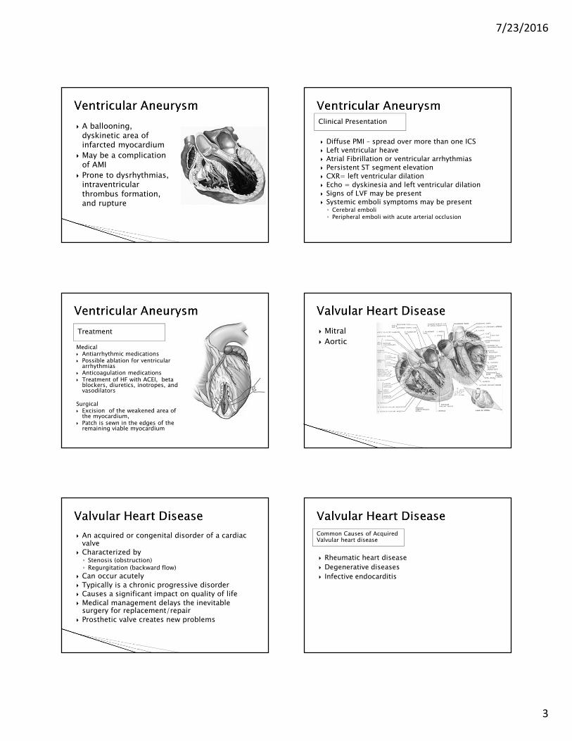
� A ballooning, dyskinetic area of infarcted myocardium
� May be a complication of AMI
� Prone to dysrhythmias, intraventricular thrombus formation, and rupture
Clinical Presentation
� Diffuse PMI – spread over more than one ICS
� Left ventricular heave
� Atrial Fibrillation or ventricular arrhythmias
� Persistent ST segment elevation
� CXR= left ventricular dilation
� Echo = dyskinesia and left ventricular dilation
� Signs of LVF may be present� Systemic emboli symptoms may be present◦ Cerebral emboli
◦ Peripheral emboli with acute arterial occlusion
Treatment
Medical� Antiarrhythmic medications� Possible ablation for ventricular
arrhythmias� Anticoagulation medications� Treatment of HF with ACEI, beta
blockers, diuretics, inotropes, and vasodilators
Surgical� Excision of the weakened area of
the myocardium,� Patch is sewn in the edges of the
remaining viable myocardium
� Mitral
� Aortic
� An acquired or congenital disorder of a cardiac valve
� Characterized by ◦ Stenosis (obstruction)◦ Regurgitation (backward flow)
� Can occur acutely
� Typically is a chronic progressive disorder
� Causes a significant impact on quality of life
� Medical management delays the inevitable surgery for replacement/repair
� Prosthetic valve creates new problems
Common Causes of Acquired Valvular heart disease
� Rheumatic heart disease
� Degenerative diseases
� Infective endocarditis
7/23/2016
4
Clinical Management
� Cardiac compensatory mechanisms can maintain stability for years before symptoms occur.
� Key is early diagnosis to prevent the long-term consequences◦ Pulmonary hypertension
◦ Heart Failure
◦ Atrial fibrillation
◦ Thromboembolism
� Important to understand◦ The structure and function of the valves
◦ The causes and treatments of each disorder
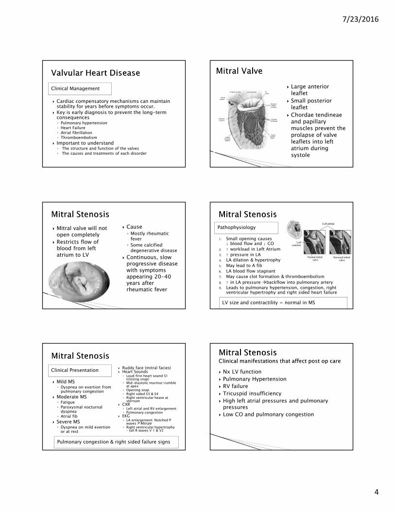
� Large anterior leaflet
� Small posterior leaflet
� Chordae tendineae and papillary muscles prevent the prolapse of valve leaflets into left atrium during systole
� Mitral valve will not open completely
� Restricts flow of blood from left atrium to LV
� Cause◦ Mostly rheumatic
fever
◦ Some calcified degenerative disease
� Continuous, slow progressive disease with symptoms appearing 20-40 years after rheumatic fever
Pathophysiology
1. Small opening causes ↓ blood flow and ↓ CO
2. ↑ workload in Left Atrium
3. ↑ pressure in LA4. LA dilation & hypertrophy
5. May lead to A fib
6. LA blood flow stagnant
7. May cause clot formation & thromboembolism
8. ↑ in LA pressure �backflow into pulmonary artery9. Leads to pulmonary hypertension, congestion, right
ventricular hypertrophy and right sided heart failure
LV size and contractility = normal in MS
Clinical Presentation
Pulmonary congestion & right sided failure signs
� Mild MS◦ Dyspnea on exertion from
pulmonary congestion
� Moderate MS◦ Fatigue
◦ Paroxysmal nocturnal dyspnea
◦ Atrial fib
� Severe MS◦ Dyspnea on mild exertion
or at rest
� Ruddy face (mitral facies)� Heart Sounds◦ Loud first heart sound S1
(closing snap)◦ Mid-diastolic murmur/rumble
at apex◦ Opening snap◦ Right sided S3 & S4◦ Right ventricular heave at
sternum� CXR◦ Left atrial and RV enlargement◦ Pulmonary congestion
� EKG◦ LA enlargement: Notched P
waves P Mitrale◦ Right ventricular hypertrophy
– tall R waves V 1 & V2
� Nx LV function
� Pulmonary Hypertension
� RV failure
� Tricuspid insufficiency
� High left atrial pressures and pulmonary pressures
� Low CO and pulmonary congestion
7/23/2016
5
� Assess for pulmonary hypertension
� Increased PVR leads to RV failure
� Increased CVP = possible RV decompression
� TEE to assess for RV and LV function
� Dobutamine, Milrinone, Norepinephrine to increase
contractility of RV and � PVR
� Fluid administration
� PAD does not reflect LA filling pressures related to pulmonary hypertension – Wedge more accurate
� PA catheter may be placed farther in related to dilated pulmonary arteries
� IABP usually not indicated as no LV dysfunction but RV dysfunction
� Causes◦ Anything that affects any
part of the MV � Mitral annulus
� Valve leaflets
� Chord tendineae
� Papillary muscle
◦ Mitral Valve Prolapse◦ Rheumatic heart disease◦ Infective endocarditis◦ Cardiomyopathy◦ Ischemic heart disease
◦
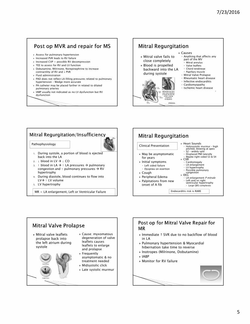
� Mitral valve fails to close completely
� Blood is propelled backward into the LA during systole
Pathophysiology
1. During systole, a portion of blood is ejected back into the LA
2. ↓ blood in LV � ↓ CO
3. ↑ blood in LA � ↑ LA pressures � pulmonary congestion and ↑ pulmonary pressures � RV hypertrophy
4. During diastole, blood continues to flow into LV� ↑ LV volume
5. LV hypertrophy
MR = LA enlargement, Left or Ventricular Failure
Clinical Presentation
� May be asymptomatic for years
� Initial symptoms◦ Left sided failure
◦ Dyspnea on exertion
� Cough
� Peripheral Edema
� Palpitations from new onset of A fib
� Heart Sounds◦ Holosystolic murmur – high
pitched, blowing at apex◦ S2 – widely split◦ Displaced PMI laterally◦ Maybe right sided S3 & S4
� CXR◦ Cardiomegaly◦ LA enlargement◦ LV hypertrophy◦ Possible pulmonary
congestion� EKG◦ LA enlargement P mitrale◦ Left and/or right
ventricular hypertrophy� Large QRS complexes
Endocarditis risk is RARE
� Mitral valve leaflets prolapse back into the left atrium during systole
� Cause: myxomatousdegeneration of valve leaflets causes leaflets to enlarge and prolapse
� Frequently asymptomatic & no treatment needed
� Midsystolic click
� Late systolic murmur
� Immediate � SVR due to no backflow of blood in LA
� Pulmonary hypertension & Myocardial hibernation take time to reverse
� Inotropes (Milrinone, Dobutamine)
� IABP
� Monitor for RV failure
7/23/2016
6
� Repair preserves native valve
� Repair is favored due to disadvantages of prosthetic valves◦ No anticoagulation needed for repair
� Technically more difficult◦ Depends on degree of regurgitation,
◦ Pathophysiology of the regurgitation
◦ LV function,
◦ Ability of surgeon
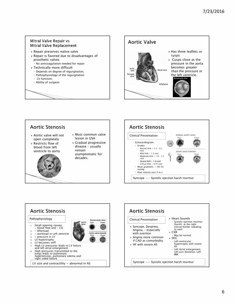
� Has three leaflets or cusps
� Cusps close as the pressure in the aorta becomes greater than the pressure in the left ventricle.
� Aortic valve will not open completely
� Restricts flow of blood from left ventricle to aorta
� Most common valve lesion in USA
� Gradual progressive disease – usually remain asymptomatic for decades.
Clinical Presentation
Syncope --- Systolic ejection harsh murmur
◦ Echocardiogram:
� Grades:
� Normal AVA > 2.5 – 3.5 cm2
� Mild AVA > 1.5 cm2
� Moderate AVA = 1.0 – 1.5 cm2
� Severe AVA < 1.0 cm2
� Critical AVA < 0.75 cm2
� Mean gradients: > 40-45 mmHg
� Peak velocity over 4 m/s
Pathophysiology
1. Small opening causes ↓ blood flow and ↓ CO
2. ↑ Afterload3. ↑ workload in Left ventricle4. ↑ pressure in LV5. LV hypertrophy6. LV becomes stiff7. High LV pressures leads to LV failure
and left atrial enlargement8. High pressures transmitted to the
lungs leads to pulmonary hypertension, pulmonary edema and right sided failure
LV size and contractility = abnormal in AS
Clinical Presentation
Syncope --- Systolic ejection harsh murmur
� Syncope, Dyspnea, Angina, – especially with exertion
� Angina more common if CAD as comorbidity
� HF with severe AS
� Heart Sounds◦ Systolic ejection murmur
(harsh) at the right sternal border radiating to neck
� CXR◦ May be normal
� EKG◦ Left ventricular
hypertrophy with severe AS
◦ Left Atrial enlargement, Left axis deviation, Left BBB
7/23/2016
7
� Inotropes rarely needed ◦ Except in patient with impaired LV function – may
be caused by myocardial stunning
� Avoid ↑ BP in immediate post-op period◦ “thin” aorta in bicuspid valve patients– more likely to rupture
under high pressure
� Maintain AV Synchrony
� Maintain adequate preload� PCWP > 15 mm HG
� Avoid hypovolemia and inotropes
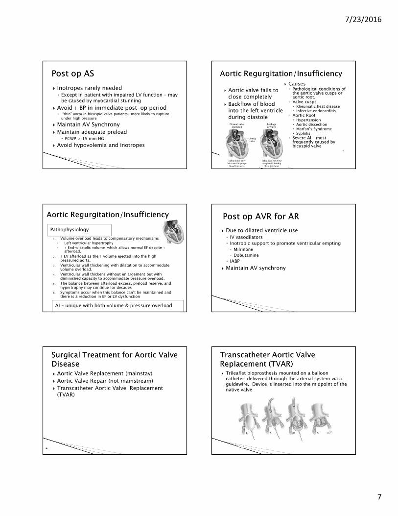
� Causes◦ Pathological conditions of
the aortic valve cusps or aortic root. ◦ Valve cusps� Rheumatic heat disease� Infective endocarditis◦ Aortic Root� Hypertension� Aortic dissection� Marfan’s Syndrome� Syphilis◦ Severe AI – most
frequently caused by bicuspid valve
◦
� Aortic valve fails to close completely
� Backflow of blood into the left ventricle during diastole
Pathophysiology
1. Volume overload leads to compensatory mechanisms◦ Left ventricular hypertrophy
◦ ↑ End-diastolic volume which allows normal EF despite ↑afterload.
2. ↑ LV afterload as the ↑ volume ejected into the high pressured aorta.
3. Ventricular wall thickening with dilatation to accommodate volume overload.
4. Ventricular wall thickens without enlargement but with diminished capacity to accommodate pressure overload.
5. The balance between afterload excess, preload reserve, and hypertrophy may continue for decades
6. Symptoms occur when this balance can’t be maintained and there is a reduction in EF or LV dysfunction
AI – unique with both volume & pressure overload
� Due to dilated ventricle use◦ IV vasodilators
◦ Inotropic support to promote ventricular empting
� Milrinone
� Dobutamine
◦ IABP
� Maintain AV synchrony
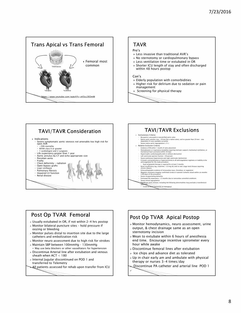
� Aortic Valve Replacement (mainstay)
� Aortic Valve Repair (not mainstream)
� Transcatheter Aortic Valve Replacement (TVAR)
� Trileaflet bioprosthesis mounted on a balloon catheter delivered through the arterial system via a guidewire. Device is inserted into the midpoint of the native valve
7/23/2016
8
� Femoral most common
https://www.youtube.com/watch?v=ztl3cc2EOmM
Pro’s
� Less invasive than traditional AVR’s
� No sternotomy or cardiopulmonary bypass
� Less ventilation time or extubated in OR
� Shorter ICU length of stay and often discharged within 48 hours postop
Con’s� Elderly population with comorbidities
� Higher risk for delirium due to sedation or pain management
� Screening for physical therapy
� Indications◦ Severe symptomatic aortic stenosis not amenable/too high risk for
open AVR� >50% mortality� NYHA class II or greater� 1 cardiologist and 2 surgeons
◦ Life expectancy greater than 1 year◦ Aortic annulus by CT and echo appropriate size◦ Porcelain aorta◦ Frailty◦ Chest deformity - radiation◦ Open bypass grafts◦ Liver cirrhosis◦ Pulmonary fibrosis◦ Impaired LV function◦ Renal disease
� Exclusionary Criteria:
◦ Bicuspid or unicuspid or noncalcified aortic valve
◦ Native aortic annulus size <18 mm (for a native valve), and no greater than 29 mm – size dependent (4 sizes available currently)
◦ Severe native aortic regurgitation (>3+)
� Relative Exclusion Criteria:
◦ Evidence of MI within 1 month of valve placement
◦ Hemodynamic or respiratory instability requiring inotropic support, mechanical ventilation, or mechanical heart assistance within 30 days
◦ Hypertrophic cardiomyopathy with or without obstruction
◦ Left ventricular ejection fraction <20 percent
◦ Severe pulmonary hypertension and right ventricular dysfunction
◦ A known contraindication or hypersensitivity to all anticoagulation regimens or inability to be anticoagulated for the study procedure
� Dual antiplatelet therapy is required for at least 3 months
◦ Renal insufficiency (eg, creatinine >3.0 mg/dL) and/or end-stage renal disease requiring chronic dialysis
◦ Echocardiographic evidence of intracardiac mass, thrombus, or vegetation
◦ Magnetic resonance imaging-confirmed stroke or transient ischemic attack within six months (180 days) of the procedure.
◦ Severe incapacitating dementia
◦ Estimated life expectancy <12 months due to noncardiac comorbid conditions
◦ Severe mitral regurgitation
◦ Significant aortic disease, including the following abnormalities may preclude a transfemoral approach:
� Could be done transaortic or transapical
� Usually extubated in OR, if not within 2-4 hrs postop
� Monitor bilateral puncture sites – hold pressure if oozing or bleeding
� Monitor pulses distal to insertion site due to the large catheters and embolization risk
� Monitor neuro assessment due to high risk for strokes
� Maintain SBP between 100mmHg – 130mmHg • May use beta blockers or other vasodilators for hypertension
� Discontinue Arterial line after extubation and venous sheath when ACT < 180
� Internal Jugular discontinued on POD 1 and transferred to Telemetry
� All patients assessed for rehab upon transfer from ICU
� Monitor hemodynamics, neuro assessment, urine output, & chest drainage same as an open sternotomy incision
� Wean to extubate within 6 hours of anesthesia end time. Encourage incentive spirometer every hour while awake
� Discontinue femoral lines after extubation
� Ice chips and advance diet as tolerated
� Up in chair early am and ambulate with physical therapy or nurses 3-4 times/day
� Discontinue PA catheter and arterial line POD 1
�
7/23/2016
9
� Complete Heart Block due to Aortic Valve edema.
� Hypotension◦ Monitor amount of sedation or vasodilating medications for
cause of hypotension
� Check groin sites for bleeding, lower abdomen for signs of retroperitoneal bleed, check peripheral vascular pulses
� Monitor Labs (Hgb/Ht)
� Vasovagal response
� Stroke
- Assess neuro status with VS’s
� Causes◦ Pathological conditions of
the aortic valve cusps or aortic root. ◦ Valve cusps� Rheumatic heat disease� Infective endocarditis◦ Aortic Root� Hypertension� Aortic dissection� Marfan’s Syndrome� Syphilis◦ Severe AI – most
frequently caused by bicuspid valve
◦
� Aortic valve fails to close completely
� Backflow of blood into the left ventricle during diastole
Pathophysiology
1. Volume overload leads to compensatory mechanisms◦ Left ventricular hypertrophy◦ ↑ End-diastolic volume which allows normal EF despite ↑ afterload.
2. ↑ LV afterload as the ↑ volume ejected into the high pressured aorta.
3. Eccentric hypertrophy (ventricular wall thickening with dilatation) to accommodate volume overload.
4. Modest concentric hypertrophy (ventricular wall thickens without enlargement but with diminished capacity) to accommodate pressure overload.
5. The balance between afterload excess, preload reserve, and hypertrophy may continue for decades
6. Symptoms occur when this balance can’t be maintained and there is a reduction in EF or LV dysfunction
AI – unique with both volume & pressure overload
Clinical Presentation
� May be asymptomatic for years
� Fatigue
� Dyspnea on exertion
� Angina
� Palpitations
� Widen pulse pressure > 50 mmHG
� Heart Sounds◦ Decrescendo diastolic
blowing murmur – best heard in upright position and leaning forward.
� CXR◦ Enlarged heart
◦ Dilation of proximal aorta
� EKG◦ Left ventricular
hypertrophy
Wide pulse pressure – Diastolic blowing murmur
Clinical Presentation Signs
� Austin Flint murmur◦ Decrescendo diastolic blowing murmur
� Hill Sign ◦ Systolic BP in lower extremities at least 20mmHg higher than
arms
� Duroziez sign◦ Systolic and diastolic bruit heard when femoral artery is
compressed by stethoscope
� Corrigan pulse◦ ↑ volume and rate of the rise of the radial pulse when the
wrist is elevated perpendicular to the body of a supine patient
� de Musset sign◦ Bobbing of the head with each systolic beat
https://www.youtube.com/watch?v=C6mTmpP9Lvw
� Rheumatic
� Endocarditis (esp IV drug abuser)
� Functional (most common form): secondary to left sided pathology often accompanied by pulm HTN
� Congenital: AV canal, VSD, Ebsteins, Myxoma
7/23/2016
10
� Primary indication for tricuspid valve repair is severe TR in patients requiring surgery for mitral valve disease
� Tricuspid stenosis: don’t see often
� TR: with left sided lesion, right sided failure, mod to severe TR
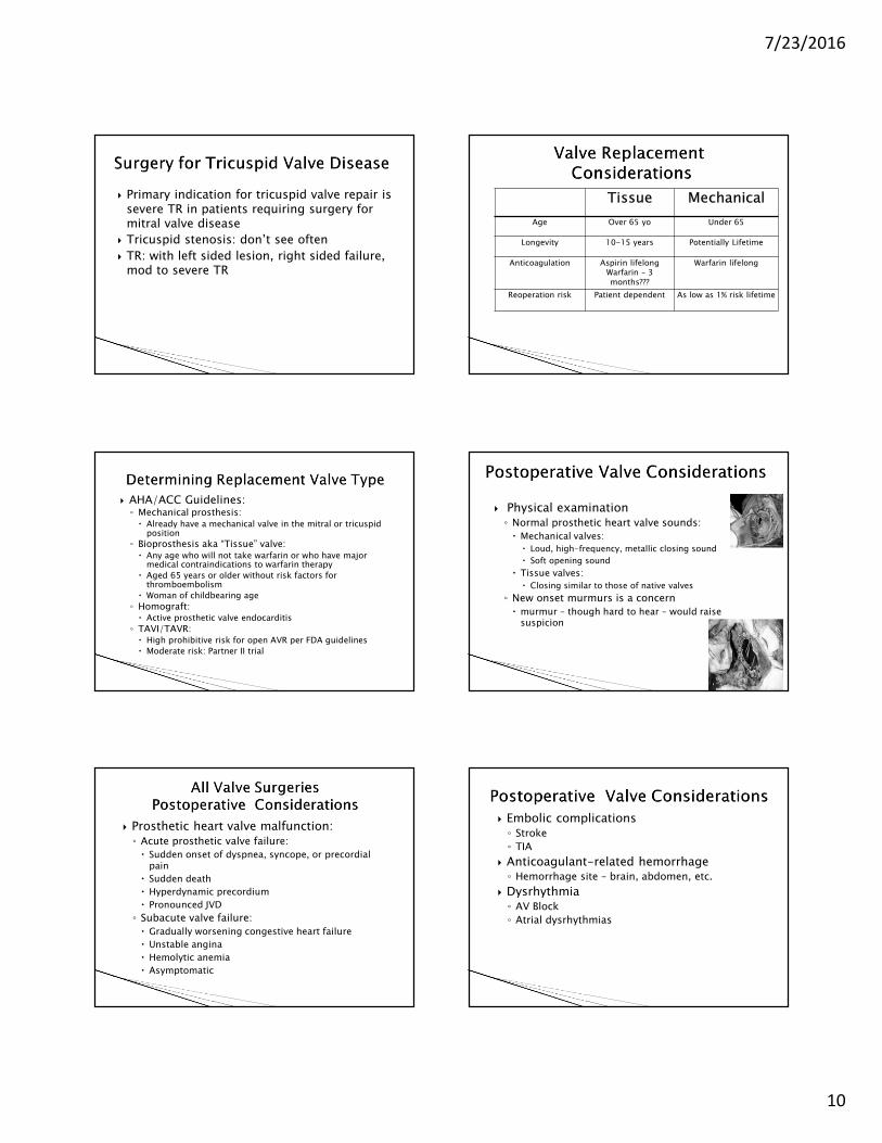
Tissue Mechanical
Age Over 65 yo Under 65
Longevity 10-15 years Potentially Lifetime
Anticoagulation Aspirin lifelongWarfarin – 3 months???
Warfarin lifelong
Reoperation risk Patient dependent As low as 1% risk lifetime
� AHA/ACC Guidelines:◦ Mechanical prosthesis:� Already have a mechanical valve in the mitral or tricuspid
position
◦ Bioprosthesis aka “Tissue” valve:� Any age who will not take warfarin or who have major
medical contraindications to warfarin therapy
� Aged 65 years or older without risk factors for thromboembolism
� Woman of childbearing age
◦ Homograft:� Active prosthetic valve endocarditis
◦ TAVI/TAVR:� High prohibitive risk for open AVR per FDA guidelines
� Moderate risk: Partner II trial
� Physical examination◦ Normal prosthetic heart valve sounds:
� Mechanical valves:
� Loud, high-frequency, metallic closing sound
� Soft opening sound
� Tissue valves:
� Closing similar to those of native valves
◦ New onset murmurs is a concern
� murmur – though hard to hear – would raise suspicion
� Prosthetic heart valve malfunction:◦ Acute prosthetic valve failure:
� Sudden onset of dyspnea, syncope, or precordial pain
� Sudden death
� Hyperdynamic precordium
� Pronounced JVD
◦ Subacute valve failure:
� Gradually worsening congestive heart failure
� Unstable angina
� Hemolytic anemia
� Asymptomatic
� Embolic complications◦ Stroke
◦ TIA
� Anticoagulant-related hemorrhage◦ Hemorrhage site – brain, abdomen, etc.
� Dysrhythmia◦ AV Block
◦ Atrial dysrhythmias
7/23/2016
11
� Blood borne bacterial traveling to the heart and growing on the valve
� Dental or other procedures may provoke bacteremia
� He is discharged on POD # 3
� His post op recovery for the next 8 weeks is unremarkable.
� Went to dentist for dental cleaning six days ago
� A few days ago he began feeling very tired, his pulse was irregular
� Went to ED in rural hospital
� Atrial Fibrillation Rate 140
� Given Lopressor 5 mg IV
� Transferred to referral hospital
� BP 137/80
� HR 98
� RR 18
� T 100.9 Orally
� SpO2 97%
� Clindamycin 600 mg prior to cleaning (Allergic to PCN)
� Started experiencing diarrhea the next two days so took Imodium
� Diarrhea subsided now (three days later)
� Echodense mobile structure on the posterior leaflet suggestive of infective endocarditis close to the annular ring on the posterior leaflet without signs of abscess
7/23/2016
12
� Antibiotic prophylaxis is indicated for the following high-risk cardiac conditions:◦ Prosthetic cardiac valve
◦ History of infective endocarditis
◦ Congenital heart disease (CHD)
◦ Cardiac transplantation recipients with cardiac valvular disease
� For these procedures◦ Dental
◦ Invasive respiratory (bronch)
� Amoxicillin◦ Adult dose: 2 g PO
◦ Pediatric dose: 50 mg/kg PO; not to exceed 2 g/dose
◦ Administer once as a single dose 30-60 min before the procedure.
� Ampicillin, Clindamycin, Cephalexin, Cefazolin, or Ceftriazone◦ May be used if allergic or unable to take oral
◦ See guidelines for specific doses
� Medical therapy is first line treatment
� Surgery indicated for those patients with acute infective valve endocarditis and life-threatening heart failure or cardiogenic shock
1. Aortic Stenosis
2. Aortic Insufficiency
3. Mitral Stenosis
4. Mitral Regurgitation
1. .
2. .
3. Mitral Stenosis
7/23/2016
13
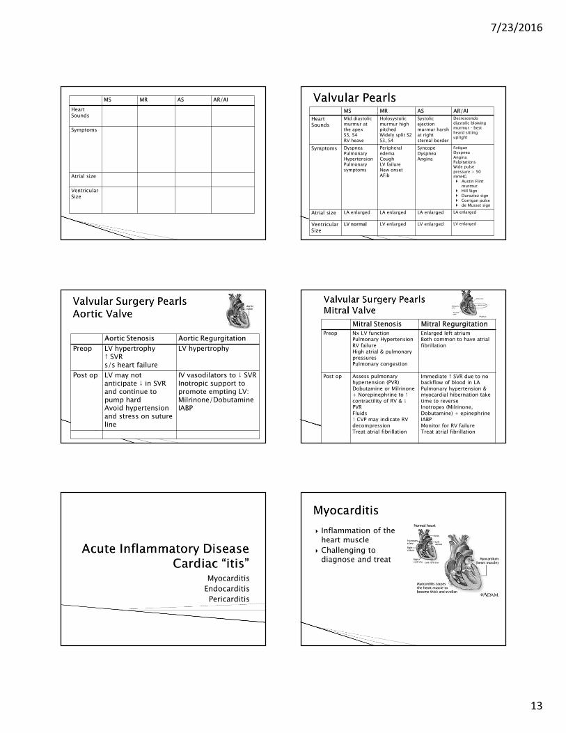
MS MR AS AR/AI
Heart Sounds
Symptoms
Atrial size
Ventricular Size
MS MR AS AR/AI
Heart Sounds
Mid diastolic murmur at the apexS3, S4RV heave
Holosystolicmurmur high pitchedWidely split S2S3, S4
Systolic ejection murmur harsh at right sternal border
Decrescendodiastolic blowing murmur – best heard sitting upright
Symptoms DyspneaPulmonary HypertensionPulmonary symptoms
PeripheraledemaCoughLV failureNew onset AFib
SyncopeDyspneaAngina
FatigueDyspneaAnginaPalpitationsWide pulse pressure > 50 mmHG� Austin Flint
murmur� Hill Sign � Duroziez sign� Corrigan pulse� de Musset sign
Atrial size LA enlarged LA enlarged LA enlarged LA enlarged
Ventricular Size
LV normal LV enlarged LV enlarged LV enlarged
Aortic Stenosis Aortic Regurgitation
Preop LV hypertrophy↑ SVRs/s heart failure
LV hypertrophy
Post op LV may not anticipate ↓ in SVR and continue to pump hardAvoid hypertension and stress on suture line
IV vasodilators to ↓ SVRInotropic support to promote empting LV: Milrinone/DobutamineIABP
Mitral Stenosis Mitral Regurgitation
Preop Nx LV functionPulmonary HypertensionRV failureHigh atrial & pulmonary pressuresPulmonary congestion
Enlarged left atriumBoth common to have atrialfibrillation
Post op Assess pulmonary hypertension (PVR)Dobutamine or Milrinone+ Norepinephrine to ↑ contractility of RV & ↓ PVRFluids↑ CVP may indicate RV decompressionTreat atrial fibrillation
Immediate � SVR due to no backflow of blood in LAPulmonary hypertension & myocardial hibernation take time to reverseInotropes (Milrinone, Dobutamine) + epinephrineIABPMonitor for RV failureTreat atrial fibrillation
Myocarditis
Endocarditis
Pericarditis
� Inflammation of the heart muscle
� Challenging to diagnose and treat
7/23/2016
14
Causes
� Infectious◦ Bacterial
◦ Spirochetal
◦ Fungal
◦ Protozoal
◦ Parasitic
◦ Rickettsial
◦ Viral
� Coxsackievirus
� Human immunodeficiency virus
� Immune Mediated◦ Allergens◦ Alloantigens◦ Autoantigens
� Chagas Disease� Systemic lupus� Sarcoidosis
� Toxic Myocarditis◦ Drugs
� Amphetamines� Anthracyclines� Cocaine� Ethanol
◦ Heavy metals� Copper � Iron � Lead
◦ Physical agents� Electrical shock� Radiation
Phases
Source: Moser & Riegal. Cardiac Nursing 2008
� Acute: Day 0 – 3◦ Viral infection
◦ Myocyte change
◦ Myocyte antigens released
◦ Cytokines released
� Subacute: Day 4 – 14◦ Infiltrating mononuclear
cells◦ Cytokine production◦ T and B lymphocytes
activated◦ Neutralizing antibodies◦ Viral clearance
� Chronic myocarditis: Day 15 – 90◦ Fibrosis◦ Cardiac enlargement◦ Apoptosis
Clinical Presentation
Wide variance in symptoms – minimal symptoms to fulminant Heart Failure
� History of recent flu-like symptoms◦ Fever◦ Arthralgia (joint pain)◦ Fatigue◦ Chest pain
� Labs◦ ↑ Troponin I◦ ↑ CK MB◦ ↑ ERS◦ Leukocytosis◦ Cardiac immunoglobulin
antibody titer 1:40 or greater
� Heart Sounds◦ Distant◦ S3 or S4◦ Pericardial friction rub
� EKG◦ Ventricular Arrhythmias◦ Heart Block◦ Diffuse ST segment changes
and T wave changes� CXR◦ Cardiomegaly◦ Pulmonary congestion◦ Pleural or pericardial effusion
� Endomyocardial biopsy for definitive diagnosis
Treatment
� Supportive management of HF symptoms◦ IABP◦ VAD
� Treat cause◦ Antibiotics◦ Antifungals◦ Antivirals◦ Corticosteroids◦ Withdrawal of offending agent
� Anticoagulation if AF� Early consideration for
transplant if:◦ Progressive HF◦ Giant cell myocarditis
� Observe for pericarditis and tamponade
� Bedrest due to increased risk of myocardial inflammation
� Advise to abstain from exercise for several months
� Close and consitent follow-up every 1 – 3 months
◦ Persistent inflammation will lead to dilated cardiomyopathy
� Inflammation/infection of the endocardium or lining of the heart.
� Usually involves the membrane lining the heart valves
� The invading pathogen typically adheres to the heart valves
� The pathogen creates solid vegetation or masses of bacteria, WBC, platelets, and fibrin that invade and destroy surrounding tissues
� Affects aortic and mitral valves more than tricuspid
Causes
Most common bacterial pathogens are Staphylococcus and Streptococcus and fungal pathogens Aspergillusfumigatus and Candida albicans
� IV drug use
� Body piercing
� Dental procedures
� Poor oral hygiene
� Rheumatic fever
� Invasive procedures
7/23/2016
15
High risk groups
� Valve repair or replacement patients
� Patients with invasive lines◦ Central lines
◦ Pacemakers
◦ Cardiac caths
� Hemodialysis patients
� Congenital or acquired valvular heart disease◦ Septal defects
◦ Bicuspid aortic valve
◦ Mitral valve prolapse
� Immunosuppressed state
Acute infective endocarditis
Subacute endocarditis
� Progresses rapidly
� Often caused by Staphylococcus infection
� Typically occurs with normal valves and causes severe damage to the valves
� Manifests initially as flu-like symptoms and progresses slowly and prolonged course
� Often caused by Streptococcus or gram negative infection
� Typically occurs with already damaged valves
� Outcome is usually good with adequate treatment
Clinical Presentation
� May be nonspecific ◦ Fever◦ Diaphoresis◦ Weight loss◦ Myalgia (muscle pain)◦ Night sweatsor
� Overt complications◦ Embolic stroke◦ Heart failure
� Heart sounds◦ New or changed murmur◦ S3◦ Pericardial friction rub
� Embolic or allergic vasculitis signs
◦ Osler’s nodes: painful nodules on pads of fingers and toes
◦ Janeway lesions: nontender macules on palms and soles
◦ Roth’s spots: round white lesions on the retina
◦ Occur from microembolization of the original vegetation
Diagnosis
� Labs◦ Positive blood cultures
◦ ↑ ERS
◦ Anemia
◦ Leukosytosis
◦ ↑ C-reactive protein
◦ Abnormal urinalysis
◦ ↑ WBC
� Echocardiogram◦ Intracardiac vegetation
◦ Dysfunctional valves
Complications
� Acute Renal Failure
� CVA
� Conduction Abnormalities
� Heart Failure
� Mycotic aneurysm
� Paravalvular abscess, perforation, or fistula
� Pericarditis
� Pulmonary emboli
� Septic arthritis
� Splenic abscess/infarct
� Systemic embolizationSource: Moser & Riegal. Cardiac Nursing 2008
Treatment
� Antimicrobial therapy
� Eradiation of endocardial vegetations
� Long term IV bacterial antibiotics
� Supportive therapy to treat and prevent complications from the disease progression
� If not responsive to medical therapy, surgery may be an option◦ Valve repair/replacement
◦ Optimal time for surgery is when the patient is hemodynamical stable
7/23/2016
16
Prophylaxis
� Prevention is essential in high risk groups � Prophylactic Antibiotics ◦ Dental, oral, upper respiratory tract procedures
� Amoxicillin� Ampicillin� Clindamycin� Azithromycin� Cephalexin� Cefazolin◦ Genitourinary or gastrointestinal procedures
� Amoxicillin� Ampicillin� Vancomycin
� Avoid unnecessary invasive lines� “Don’t Do Drugs!”� Avoid body piercing
� Two days ago noticed abdomen and back were swollen. It was difficult to feel his spine as there was so much fluid
� Unable to walk across the room without becoming SOB
� Breathing worse when lying flat
� Difficulty speaking in full sentence
� Dry cough
� Weight gain of 10 pounds in last week
� BP 140/90
� HR 109
� RR 24, moderately labored
� T 98.7
� SpO2 97% on 3 liters nasal cannula
� Weight 66.2 kg
� Pansystolic murmur 4/6 noted loudest at the left sternal border
� Coarse rales throughout all lung fields
� Recent admission five days ago for hematuria, hyperkalemia, and acute renal insufficiency
� Left ureteral stent placed for sever hydrohephrosis of the left kidney
� Received 2 units of blood at that time
• One pack a day smoker
• Rarely drinks
• Occasional use of marijuana
• Has tried cocaine, Ecstasy, Adderall in the past
• Had several tattoos done a few months ago.
7/23/2016
17
• Procalcitonin 0.14 (normal)
• Urine toxicology (with earlier
admission)
– Positive for methamphetamines and cannabinoids
• DAU-12– Positive for benzodiazepines and
THC (marijuana)
• BNP 5002
� Given Lasix with 1300 ml urine output in ED
� Started on Rocephin and Azithromycin
� Plan for echocardiogram
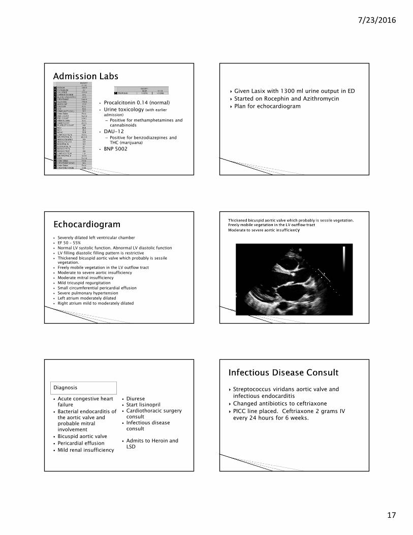
� Severely dilated left ventricular chamber
� EF 50 – 55%
� Normal LV systolic function. Abnormal LV diastolic function
� LV filling diastolic filling pattern is restrictive
� Thickened bicuspid aortic valve which probably is sessile vegetation.
� Freely mobile vegetation in the LV outflow tract
� Moderate to severe aortic insufficiency
� Moderate mitral insufficiency
� Mild tricuspid regurgitation
� Small circumferential pericardial effusion
� Severe pulmonary hypertension
� Left atrium moderately dilated
� Right atrium mild to moderately dilated
Diagnosis
� Acute congestive heart failure
� Bacterial endocarditis of the aortic valve and probable mitral involvement
� Bicuspid aortic valve
� Pericardial effusion
� Mild renal insufficiency
� Diurese� Start lisinopril� Cardiothoracic surgery
consult� Infectious disease
consult
� Admits to Heroin and LSD
� Streptococcus viridans aortic valve and infectious endocarditis
� Changed antibiotics to ceftriaxone
� PICC line placed. Ceftriaxone 2 grams IV every 24 hours for 6 weeks.
7/23/2016
18
� The organisms are most abundant in the mouth, and one member of the group, S. mutans, is the etiologic agent of dental caries. Others may be involved in other mouth or gingival infections.
� If they are introduced into the bloodstream, they have the potential of causing endocarditis, particularly in individuals with damaged heart valves. They are the most common causes of subacute bacterial endocarditis.
� Viridans streptococci have the unique ability to synthesize dextrans from glucose, which allows them to adhere to fibrin-platelet aggregates at damaged heart valves. This mechanism underlies their ability to cause subacute valvular heart disease following their introduction into the bloodstream (e.g., following tooth extraction).
Source: Wikipedia. August 29, 2011
� Aortic Valve Replacement with #23 Trifecta Valve
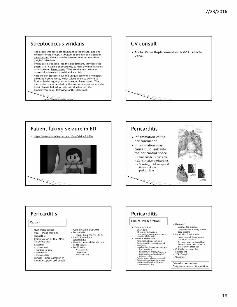
� https://www.youtube.com/watch?v=Q6sRyrB_UMA� Inflammation of the
pericardial sac
� Inflammation may cause fluid leak into the pericardial space◦ Tamponade is possible
◦ Constrictive pericarditis
� Scarring, thickening and fibrous of the pericardium
Causes
� Numerous causes
� Viral – most common
� Idiopathic
� Complication of HIV-AIDS –TB pericarditis
� Bacterial◦ Stab wound
◦ Cardiac surgery
◦ Pneumonia
◦ Endocarditis
� Fungal – more common in immunosuppressed people
� Complication after AMI� Metastasis
◦ Esp in lung cancer (30 %)� Radiation induced
pericarditis� Uremic pericarditis- chronic
renal failure� Medications◦ Procainamide
◦ Hydralazine
◦ INH (isoniazid)
Clinical Presentation
� Can mimic AMI◦ Chest pain◦ ST segment elevation◦ Have patient point to the exact
location of the pain
� Pleuritic chest pain◦ Persistent, sharp, stabbing◦ Aggravated by inspiration and
coughing◦ Localized to the retrosternal and
left pericardium � With deep inspiration the
diaphragm pulls on the inflamed pericardium causing the sharp pain that radiates
◦ Pain is worse when recumbent◦ Pain maybe relieved by sitting
upright and leaning forward� Mohammed’s Sign
� Dyspnea*
◦ Unrelated to exertion
◦ Caused by the inability to take deep breaths
� Pericardial friction rub
◦ Heard best left lower sternal border, 4t or 5th ICS
◦ To heard best, pt should lean forward so the pericardium is closer to the chest wall
� Chills/Fever – may be antecedent
� SOB/Cough� Weakness
Pain when recumbent
Dyspnea unrelated to exertion
7/23/2016
19
Clinical presentation
� Cardiac tamponade as evidenced by◦ True dyspnea and SOB◦ Pulmonary congestion◦ ↓ CO◦ Decreased tissue perfusion◦ Pericardial knock
� High pitched, sharp, early diastolic sound heard best at lower LSB with patient leaning forward
◦ PAP > 50 mmHG
Muffled heart sounds = tamponade
Pericardial knock = constrictive pericarditis
Diagnostics
� 12 Lead EKG – most definitive diagnostic test
� CXR – may have enlarged cardiac silhouette (water bottle heart)
� Echo◦ Possibly Pericardial
effusion
◦ Pericardial thickening with constrictive pericarditis
� LABs◦ Troponin, CK negative
◦ ↑ WBC, Sed rate, C-reactive protein
◦ Positive blood cultues if due to infection
Acute: Pericardial inflammatory response
Chronic:
Dressler syndrome
� Pericarditis occurring 2 – 7 days after AMI
� Related to the inflammation and healing process
� Autoimmune response to myocardial necrosis
� Involves both the pleura and pericardium
� Occurs 2 – 4 weeks to several months post AMI
� Diffuse ST elevation� Scooping upwardly concave ST segment elevation
in almost all leads except AVR� No reciprocal ST depression except in AVR� PR depression
7/23/2016
20
Clinical Management
� Relieve pain◦ NSAIDs◦ Indomethacin & colchicines
– for intolerance to NSAIDs◦ If unresponsive to NSAIDS
� Narcotic analgesia� Short course of
corticosteroids
� Treat cause◦ Antibiotics if bacterial
� If anticoagulants are given, assess for tamponade
� Pericarditis after AMI◦ NO corticosteroids or
anti-inflammatory agents� Can cause rupture of the
infarcted area
� Pericardiocentesis for tamponade
� Pericardial window for recurrent effusion
� Pericardiectomy –constrictive pericarditis
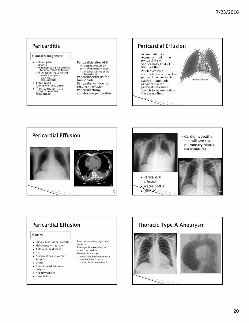
� Accumulation of excessive fluid in the pericardial sac
� Sac normally holds 15 –50 ml of fluid
� When excessive accumulation is slow, the pericardium can stretch.
� Cardiac tamponade occurs when the pericardium cannot stretch to accommodate the excess fluid
117
118
� Pericardial Effusion
� Water bottle
� (Above)
� Cardiomyopathy --- will see the pulmonary hiatus (vasculature)
Causes
� Initial causes of pericarditis
� Malignancy or radiation
� Autoimmune disease
� AMI
� Complications of cardiac surgery
� Drugs
� Chronic renal failure on dialysis
� Hypothyroidism
� Heart failure
� Blunt or penetrating chest trauma
� Retrograde extension of aortic dissection
� Iatrogenic causes
◦ Myocardial perforation with invasive lines (pacers, central lines, angiogram)
7/23/2016
21
Clinical Presentation
� Slow occurring effusions◦ Asymptomatic
◦ Often undetected as the gradual stretching of pericardium doesn’t compromise ventricular filling
� Rapid occurring effusions◦ Accumulation of pericardial
fluid or buildup of a volume of pericardial fluid exceeding the pericardium’s ability to expand◦ Leads to tamponade
◦ Compromises ventricular filling
◦ ↓ Cardiac output◦ 100 – 200 ml of extra fluid
can elevate pericardial pressure from 1 – 5 mmHg to 30 mmHg or greater.
� Compression of the heart due to collection of fluid or blood in the pericardial space
Clinical Presentation
� Beck’s Triad◦ Hypotension
◦ Neck vein distention
◦ Muffled heart sounds
� Hypotension� Low urine output� Rising & equalization of CVP
& PAD� Large a and v on PAOP “M”� Falling SVO2, CO/CI� Widening mediastinum on
CXR� Cool extremities� Tachycardia� Pulses Paradox >10 mmHG
◦ ↓ in systolic BP with inspiration
124
Clinical Management
� Closed Pericardiocentesis (preferred)
� Open Surgical Pericardiocentesis◦ Creation of a pericardial window to drain pericardium
Pericardial EffusionPost pericardial window for pericardial effusion
126
7/23/2016
22
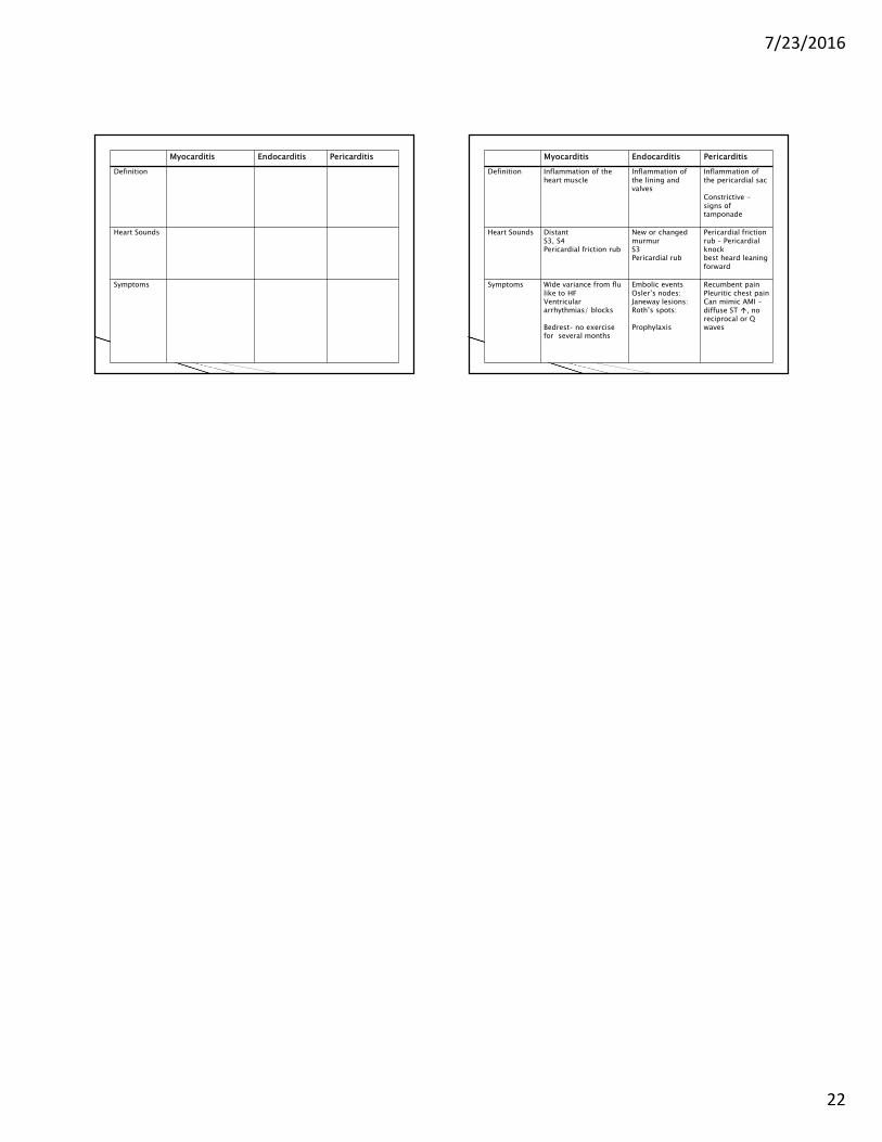
Myocarditis Endocarditis Pericarditis
Definition
Heart Sounds
Symptoms
Myocarditis Endocarditis Pericarditis
Definition Inflammation of the heart muscle
Inflammation ofthe lining and valves
Inflammation of the pericardial sac
Constrictive –signs of tamponade
Heart Sounds DistantS3, S4Pericardial friction rub
New or changed murmurS3Pericardial rub
Pericardial friction rub – Pericardial knockbest heard leaning forward
Symptoms Wide variance from flulike to HFVentricular arrhythmias/ blocks
Bedrest– no exercise for several months
Embolic eventsOsler’s nodes:Janeway lesions:Roth’s spots:
Prophylaxis
Recumbent painPleuritic chest painCan mimic AMI –diffuse ST �, no reciprocal or Q waves