Embed Size (px)
Citation preview
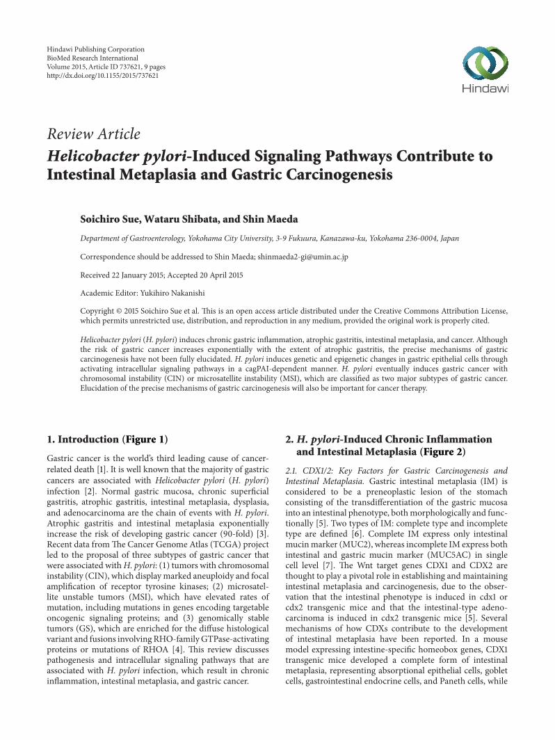
Review ArticleHelicobacter pylori-Induced Signaling Pathways Contribute toIntestinal Metaplasia and Gastric Carcinogenesis
Soichiro Sue, Wataru Shibata, and Shin Maeda
Department of Gastroenterology, Yokohama City University, 3-9 Fukuura, Kanazawa-ku, Yokohama 236-0004, Japan
Correspondence should be addressed to Shin Maeda; [email protected]
Received 22 January 2015; Accepted 20 April 2015
Academic Editor: Yukihiro Nakanishi
Copyright © 2015 Soichiro Sue et al. This is an open access article distributed under the Creative Commons Attribution License,which permits unrestricted use, distribution, and reproduction in any medium, provided the original work is properly cited.
Helicobacter pylori (H. pylori) induces chronic gastric inflammation, atrophic gastritis, intestinal metaplasia, and cancer. Althoughthe risk of gastric cancer increases exponentially with the extent of atrophic gastritis, the precise mechanisms of gastriccarcinogenesis have not been fully elucidated. H. pylori induces genetic and epigenetic changes in gastric epithelial cells throughactivating intracellular signaling pathways in a cagPAI-dependent manner. H. pylori eventually induces gastric cancer withchromosomal instability (CIN) or microsatellite instability (MSI), which are classified as two major subtypes of gastric cancer.Elucidation of the precise mechanisms of gastric carcinogenesis will also be important for cancer therapy.
1. Introduction (Figure 1)
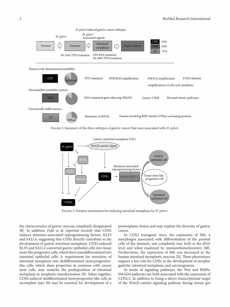
Gastric cancer is the world’s third leading cause of cancer-related death [1]. It is well known that the majority of gastriccancers are associated with Helicobacter pylori (H. pylori)infection [2]. Normal gastric mucosa, chronic superficialgastritis, atrophic gastritis, intestinal metaplasia, dysplasia,and adenocarcinoma are the chain of events with H. pylori.Atrophic gastritis and intestinal metaplasia exponentiallyincrease the risk of developing gastric cancer (90-fold) [3].Recent data fromThe Cancer Genome Atlas (TCGA) projectled to the proposal of three subtypes of gastric cancer thatwere associated withH. pylori: (1) tumors with chromosomalinstability (CIN), which displaymarked aneuploidy and focalamplification of receptor tyrosine kinases; (2) microsatel-lite unstable tumors (MSI), which have elevated rates ofmutation, including mutations in genes encoding targetableoncogenic signaling proteins; and (3) genomically stabletumors (GS), which are enriched for the diffuse histologicalvariant and fusions involving RHO-familyGTPase-activatingproteins or mutations of RHOA [4]. This review discussespathogenesis and intracellular signaling pathways that areassociated with H. pylori infection, which result in chronicinflammation, intestinal metaplasia, and gastric cancer.
2. H. pylori-Induced Chronic Inflammationand Intestinal Metaplasia (Figure 2)
2.1. CDX1/2: Key Factors for Gastric Carcinogenesis andIntestinal Metaplasia. Gastric intestinal metaplasia (IM) isconsidered to be a preneoplastic lesion of the stomachconsisting of the transdifferentiation of the gastric mucosainto an intestinal phenotype, bothmorphologically and func-tionally [5]. Two types of IM: complete type and incompletetype are defined [6]. Complete IM express only intestinalmucinmarker (MUC2), whereas incomplete IM express bothintestinal and gastric mucin marker (MUC5AC) in singlecell level [7]. The Wnt target genes CDX1 and CDX2 arethought to play a pivotal role in establishing and maintainingintestinal metaplasia and carcinogenesis, due to the obser-vation that the intestinal phenotype is induced in cdx1 orcdx2 transgenic mice and that the intestinal-type adeno-carcinoma is induced in cdx2 transgenic mice [5]. Severalmechanisms of how CDXs contribute to the developmentof intestinal metaplasia have been reported. In a mousemodel expressing intestine-specific homeobox genes, CDX1transgenic mice developed a complete form of intestinalmetaplasia, representing absorptional epithelial cells, gobletcells, gastrointestinal endocrine cells, and Paneth cells, while
Hindawi Publishing CorporationBioMed Research InternationalVolume 2015, Article ID 737621, 9 pageshttp://dx.doi.org/10.1155/2015/737621
2 BioMed Research International
Elevated mitotic pathways
Fusions involving RHO-family GTPase-activating proteins
CIN
MSI
TP53 mutation RTK/RAS amplification PIK3CA amplification PTEN deletion
Amplifications of cell cycle mediators
DNA mismatch gene silencing (MLH1) Gastric CIMP
Tumors with chromosomal instability
Microsatellite instability tumors
GS
Genomically stable tumors
Mutations of RHOA
Intestinal
Diffuse
Normal GastritisIntestinalmetaplasia Gastric cancer
H. pyloriH. pyloriAssociated signals
30–50% TP53 mutation 10% RAS mutation20–60% TP53 mutation
MSIGS
55%24%21%
H. pylori induced gastric cancer subtypes
CIN
Figure 1: Summary of the three subtypes of gastric cancer that were associated with H. pylori.
Gastric intestinal metaplasia (I.M.)
H. pylori
CDX2
SOX2
BMPs
SMAD4CDX1
Stemness-associated reprogramming factors
Gastric epithelial cell
Tissue stem-likeprogenitor cell
Intestinal epithelial cell
SALL4, KLF5 ↑
Wnt/𝛽-catenin signal
Figure 2: Putative mechanism for inducing intestinal metaplasia by H. pylori.
the characteristics of gastric mucosa completely disappeared[8]. In addition, Fujii et al. reported recently that CDX1induces stemness-associated reprogramming factors, KLF5and SALL4, suggesting that CDXs directly contribute to thedevelopment of gastric intestinal metaplasia. CDX1-inducedKLF5 and SALL4 converted gastric epithelial cells into tissuestem-like progenitor cells, which then transdifferentiated intointestinal epithelial cells. A requirement for transition ofintestinal metaplasia into dedifferentiated stem/progenitor-like cells, which share properties in common with cancerstem cells, may underlie the predisposition of intestinalmetaplasia to neoplastic transformation [9]. Taken together,CDX1-induced dedifferentiated stem/progenitor-like cells inincomplete type IM may be essential for development of a
preneoplastic lesion and may explain the diversity of gastriccancer.
In CDX2 transgenic mice, the expression of Shh, amorphogen associated with differentiation of the parietalcells of the stomach, was completely lost, both at the RNAlevel and when examined by immunohistochemistry [10].Furthermore, the expression of Shh was decreased in thehuman intestinal metaplastic mucosa [11]. These phenomenasupport a key role for CDXs in the development of atrophicgastritis, intestinal metaplasia, and carcinogenesis.
In terms of signaling pathways, the Wnt and BMPs/SMAD4 pathways are both associated with the expression ofCDX1/2. In addition to being a direct transcriptional targetof the Wnt/𝛽-catenin signaling pathway during mouse gut
BioMed Research International 3
development, CDX1 is also induced by cag-positive H. pyloriinfection [9, 12]. BMPs/SMAD4 is known to be a fundamentalpathway for the development of intestinal epithelium; it isupregulated upon H. pylori infection and thereafter inducesthe expression of the downstream target CDX2, as well asthe downregulation of SOX2, an inhibitor of CDX2 [13–15].CDX2 regulate MUC2 [16] by binding to enhancer sequences[17].
2.2. Genetic Alteration andGene Expression in IntestinalMeta-plasia. Gene alteration, such as aneuploidy of chromosomes[18], P53 mutations (38–45%) [19–22], P53 deletion (60%)[18], microsatellite instability (27%) [23], and mitochondrialmicrosatellite instability (33%) [24] were detected in IM.P53 mutations were mostly in incomplete type [20, 21].Microsatellite instabilities were all in incomplete type [25].Gene expression, such as MUC2 [6], LI-cadherin [26], KLF4[27], intestinal trefoil factor (TFF3) [28], sucrose-isomaltase[29], villin [7], CD10 [30], and defensing [31], increased inIM. MUC2 is regulated by CDX2 [16, 32]. On the other hand,gene expression, such as Sonic hedgehog (Shh) [33], SOX2[14], RUNX3 [34], and TFF1 and TFF2 [28], decreased in IM.Shh is particularly decreased in incomplete IM type [11].
Alteration of these gastric and intestinal phenotypemark-ers was observed at the cellular level, as well as at theglandular level. In fact, neuroendocrine cells also showedintestinalization along with their exocrine counterparts. Inanimalmodels, incomplete type intestinalmetaplasia appearsfirst and then progresses to the complete type. In summary,intestinal metaplasia may be caused by the gradual intestinal-ization of stem/progenitor cells from the incomplete to thecomplete type [35].
3. H. pylori-Induced Genetic Changes
Several reports have suggested that H. pylori infectioncaused genetic alterations in gastric epithelial cells, mostlythrough the induction of reactive oxygen species (ROS) [36].Matsumoto et al. reported that H. pylori induced aberrantexpression of activation-induced cytidine deaminase (AID),known as an editor of DNA and RNA. AID was reported tocausemutations in the P53 andAPCgenes in gastric epithelialcells, relevant to the development of adenocarcinoma [37].AID hypermutates immunoglobulin genes in B cell genome,contributing to variety acquisition of immunoglobulin. AIDalso target oncogenes, leading to B cell malignancy [38]. Inaddition, various cancers develop in AID transgenic mice,including gastric cancer [39]. InMatsumoto’s report,H. pyloristrongly induced AID expression in human gastric epithelialcells, through activation of the NF-𝜅B pathway, and inducedmutation of p53.Asmutation of p53was inhibited by blockingAID, p53 mutation induced by H. pylori mostly depends onAID. Since AID was upregulated via activation of the NF-𝜅Bpathway, proinflammatory cytokines—such as TNF-𝛼 or IL-1𝛽—ingastric inflammation also reinforce the onset ofAID aswell as the direct stimulation of H. pylori in gastric epithelialcells [40].
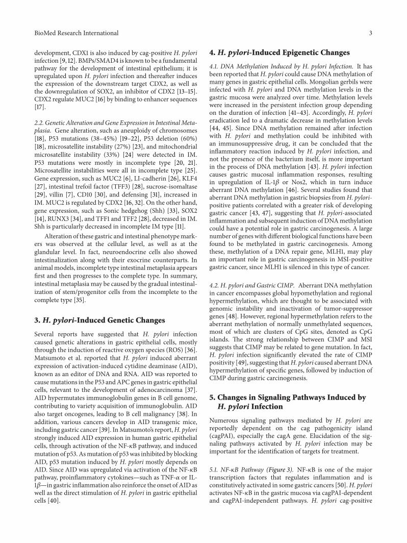
4. H. pylori-Induced Epigenetic Changes
4.1. DNA Methylation Induced by H. pylori Infection. It hasbeen reported thatH. pylori could cause DNAmethylation ofmany genes in gastric epithelial cells. Mongolian gerbils wereinfected with H. pylori and DNA methylation levels in thegastric mucosa were analyzed over time. Methylation levelswere increased in the persistent infection group dependingon the duration of infection [41–43]. Accordingly, H. pylorieradication led to a dramatic decrease in methylation levels[44, 45]. Since DNA methylation remained after infectionwith H. pylori and methylation could be inhibited withan immunosuppressive drug, it can be concluded that theinflammatory reaction induced by H. pylori infection, andnot the presence of the bacterium itself, is more importantin the process of DNA methylation [43]. H. pylori infectioncauses gastric mucosal inflammation responses, resultingin upregulation of IL-1𝛽 or Nos2, which in turn induceaberrant DNA methylation [46]. Several studies found thataberrant DNAmethylation in gastric biopsies fromH. pylori-positive patients correlated with a greater risk of developinggastric cancer [43, 47], suggesting that H. pylori-associatedinflammation and subsequent induction ofDNAmethylationcould have a potential role in gastric carcinogenesis. A largenumber of geneswith different biological functions have beenfound to be methylated in gastric carcinogenesis. Amongthese, methylation of a DNA repair gene, MLH1, may playan important role in gastric carcinogenesis in MSI-positivegastric cancer, since MLH1 is silenced in this type of cancer.
4.2. H. pylori and Gastric CIMP. Aberrant DNAmethylationin cancer encompasses global hypomethylation and regionalhypermethylation, which are thought to be associated withgenomic instability and inactivation of tumor-suppressorgenes [48]. However, regional hypermethylation refers to theaberrant methylation of normally unmethylated sequences,most of which are clusters of CpG sites, denoted as CpGislands. The strong relationship between CIMP and MSIsuggests that CIMP may be related to gene mutation. In fact,H. pylori infection significantly elevated the rate of CIMPpositivity [49], suggesting thatH. pylori caused aberrantDNAhypermethylation of specific genes, followed by induction ofCIMP during gastric carcinogenesis.
5. Changes in Signaling Pathways Induced byH. pylori Infection
Numerous signaling pathways mediated by H. pylori arereportedly dependent on the cag pathogenicity island(cagPAI), especially the cagA gene. Elucidation of the sig-naling pathways activated by H. pylori infection may beimportant for the identification of targets for treatment.
5.1. NF-𝜅B Pathway (Figure 3). NF-𝜅B is one of the majortranscription factors that regulates inflammation and isconstitutively activated in some gastric cancers [50].H. pyloriactivates NF-𝜅B in the gastric mucosa via cagPAI-dependentand cagPAI-independent pathways. H. pylori cag-positive
4 BioMed Research International
H. pylori
TRAF6 TAK1
RelA P50
P
Ubiquitination
Nod1
peptidoglycan
H. pylori
Cytoplasm TLR
MyD88
TRAF6
TAK1
RelA P50
Gene regulationNucleus RelA P50
H. pylori
Phosphorylation
H. pylori induced NF-𝜅B signaling
LPS, TNF-𝛼, IL-1𝛽
I𝜅B𝛼
IKK𝛼IKK𝛾/NEMO
IKK𝛽IKK𝛼IKK𝛾/NEMO
IKK𝛽
NF-𝜅B
NF-𝜅BInflammation cytokine IL-1𝛽, IL-6, TNF-𝛼Chemokine IL-8, MCP-1Antiapoptosis cIAPs, c-FLIP, A20, BclXLAngiogenesis VEGF, IL-8Invasion, metastasis MMP-2, MMP-9
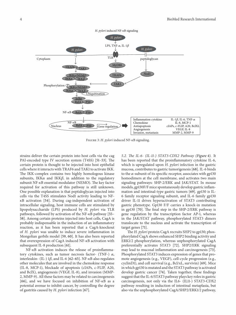
Figure 3: H. pylori induced NF-𝜅B signaling.
strains deliver the certain protein into host cells via the cagPAI-encoded type IV secretion system (T4SS) [51–53]. Thecertain protein is thought to be injected into host epithelialcells where it interacts with TRAF6 andTAK1 to activate IKK.The IKK-complex contains two highly homologous kinasesubunits, IKK𝛼 and IKK𝛽, in addition to the regulatorysubunit NF-𝜅B essential modulator (NEMO). The key factorrequired for activation of this pathway is still unknown.One possible explanation is that peptidoglycan injected intocells via the T4SS stimulates Nod1 activity leading to NF-𝜅B activation [54]. During cag-independent activation ofintracellular signaling, host immune cells are stimulated bylipopolysaccharide (LPS) produced by H. pylori via TLRpathways, followed by activation of the NF-𝜅B pathway [55–58]. Among certain proteins injected into host cells, CagA isprobably indispensable in the induction of an inflammatoryreaction, as it has been reported that a CagA-knockoutof H. pylori was unable to induce severe inflammation inMongolian gerbils model [59, 60]. It has also been reportedthat overexpression of CagA induced NF-𝜅B activation withsubsequent IL-8 production [61].
NF-𝜅B activation induces the release of proinflamma-tory cytokines, such as tumor necrosis factor- (TNF-) 𝛼,interleukin- (IL-) 1𝛽, and IL-6 [62–65]. NF-𝜅B also regulatesother molecules that are involved in the chemokine response(IL-8, MCP-1), blockade of apoptosis (cIAPs, c-FLIP, A20,and BclX), angiogenesis (VEGF, IL-8), and invasion (MMP-2, MMP-9). All these factors may be related to carcinogenesis[66], and we have focused on inhibition of NF-𝜅B as apotential avenue to inhibit cancer, by controlling the degreeof gastritis caused by H. pylori infection [67].
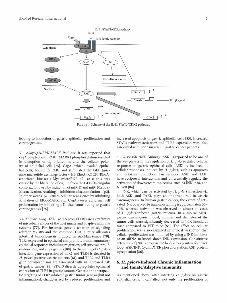
5.2. The IL-6- (IL-11-) STAT3-CDX2 Pathway (Figure 4). Ithas been reported that the proinflammatory cytokine IL-6,which is upregulated upon H. pylori infection in the gastricmucosa, contributes to gastric tumorigenesis [68]. IL-6 bindsto the 𝛼-subunit of its specific receptor, associates with gp130homodimers at the cell membrane, and activates two mainsignaling pathways: SHP-2/ERK and JAK/STAT. In mousemodels, gp130F/Fmice spontaneously develop gastric inflam-mation and intestinal-type gastric tumors [69]. gp130 is IL-6 family receptor signaling subunit, and IL-6 family gp130driver IL-11 drives hyperactivation of STAT3 contributinggastric phenotype. Gp130 F/F carries a knock-in mutationin gp130 [70]. The final step in the SHP-2/ERK pathway isgene regulation by the transcription factor AP-1, whereasin the JAK/STAT pathway, phosphorylated STAT3 dimerstranslocate to the nucleus and activate the transcription oftarget genes [71].
TheH. pylori protein CagA recruits SHP2 to gp130; phos-phorylated CagA shows enhanced SHP2 binding activity andERK1/2 phosphorylation, whereas unphosphorylated CagApreferentially activates STAT3 [72]. SHP2/ERK signalingmay lead to mucosal inflammation and carcinogenesis [73].Phosphorylated STAT3 induces expression of genes that pro-mote angiogenesis (e.g., VEGF), cell-cycle progression (e.g.,cyclinD1), and cell survival (e.g., Bcl/xL, survivin) [69]. Miceinwhich gp130 ismutated and the STAT3 pathway is activateddevelop gastric cancer [74]. Taken together, these findingssuggest that the IL-6/STAT3 pathway plays key roles in gastriccarcinogenesis, not only via the IL6- (IL11-) STAT3-CDX2pathway resulting in induction of intestinal metaplasia, butalso via the unphosphorylated CagA/SHP2/ERK1/2 pathway,
BioMed Research International 5
IL-11/STAT3/CDX2 pathway
Antiapoptotic
SHP-2Jak
Stat3
IL-6 family receptor
Stat3
PI3K
Akt
mTOR
Stat1
Smad7
Bcl2/1
Vegfa
Socs3
IL-11CagA
Cytoplasm
Nucleus
CDX2
IFN𝛾-like response
TGF𝛽 signal
Figure 4: Scheme of the IL-11/STAT3/CDX2 pathway.
leading to induction of gastric epithelial proliferation andcarcinogenesis.
5.3. c-Myc/p21/ERK-MAPK Pathway. It was reported thatcagA coupled with PAR1 (MARK) phosphorylation resultedin disruption of tight junctions and the cellular polar-ity of epithelial cells [75]. CagA, which invaded epithe-lial cells, bound to PAR1 and stimulated the GEF (gua-nine nucleotide exchange factor)–H1–RhoA–ROCK (RhoA-associated kinase)–c-Myc–microRNA–p21 axis; this wascaused by the liberation of cigulin from the GEF-H1-cingulincomplex, followed by induction of miR-17 and miR-20a by c-Myc activation, resulting in inhibition of accumulation of p21.In other words, p21 causes cellular senescence by inhibitingactivation of ERK-MAPK, and CagA causes abnormal cellproliferation by inhibiting p21, thus contributing to gastriccarcinogenesis [76].
5.4. TLR Signaling. Toll-like receptors (TLRs) are a key familyof microbial sensors of the host innate and adaptive immunesystems [77]. For instance, genetic ablation of signalingadaptor MyD88 and the common TLR in mice alleviatesintestinal tumorigenesis induced in ApcMin/+mice [78].TLRs expressed in epithelial can promote noninflammatoryepithelial responses including migration, cell survival, prolif-eration [79], and angiogenesis [80]. In the setting ofH. pyloriinfection, gene expression of TLR2 and TLR4 is elevated inH. pylori-positive gastric patients [81], and TLR2 and TLR4gene polymorphisms are associated with an increased riskof gastric cancer [82]. STAT3 directly upregulates epithelialexpression of TLR2 in gastric tumors. Genetic and therapeu-tic targeting of TLR2 inhibited gastric tumorigenesis (but notinflammation), characterized by reduced proliferation and
increased apoptosis of gastric epithelial cells [83]. IncreasedSTAT3 pathway activation and TLR2 expression were alsoassociated with poor survival in gastric cancer patients.
5.5. ROS/ASK1/JNK Pathway. ASK1 is reported to be one ofthe key players in the regulation of H. pylori-related cellularresponses in gastric epithelial cells. ASK1 is involved incellular responses induced by H. pylori, such as apoptosisand cytokine production. Furthermore, ASK1 and TAK1have reciprocal interactions and differentially regulate theactivation of downstream molecules, such as JNK, p38, andNF-𝜅B [84].
JNK, which can be activated by H. pylori infection viaboth ASK1 and TAK1, plays an important role in gastriccarcinogenesis. In human gastric cancer, the extent of acti-vated JNKobserved by immunostaining is approximately 30–40%, whereas activation was observed in almost all casesof H. pylori-infected gastric mucosa. In a mouse MNUgastric carcinogenic model, number and diameter of thetumor cells were significantly decreased in JNK knockoutmice compared to WT mice [85]. The effect on cellularproliferation was also examined in vitro; it was found thatcellular proliferation was inhibited by using a JNK inhibitoror an siRNA to knock down JNK expression. Constitutiveactivation of JNK is proposed to be due to a positive feedbackloop: ASK/JNK/CyclinD1/Rb phosphorylation/ASK proteinupregulation [86].
6. H. pylori-Induced Chronic Inflammationand Innate/Adaptive Immunity
As mentioned above, after infecting H. pylori on gastricepithelial cells, it can affect not only the proliferation of
6 BioMed Research International
gastric epithelial cells, but also the activation of intracellularsignaling, and that leads to perturbing the host’s innateand adaptive immune system [87]. Among the inflam-matory reactions induced by H. pylori infection, innateimmune system, represented by infiltration of neutrophilsand macrophages, plays key roles in production of proin-flammatory cytokines/chemokines, which promote chronicinflammation [67]. On the other hand, adaptive immunesystem plays roles not only to produce proinflammatorycytokines and cytotoxic reaction to bacterium directory, butalso in induction of anti-inflammatory cytokines, such as IL-10, to suppress the cytotoxic function of effector T cells, whichenables the bacteria to evade immune system, resulting inchronic infection [88].
7. Conclusions and Future Perspectives
Since chronic inflammation can cause epithelial cell distur-bance, the early eradication ofH. pylori could provide a basicsolution for prevention of gastric carcinogenesis caused byH. pylori. Accordingly, all patients in Japan with H. pylori-related gastritis are being recommended for eradicationmethods to decrease the risk of gastric cancer. However, as aconsiderable proportion of patients remain to be irreversible“field cancerization,” where the cancer-causing case does notcut off after H. pylori sanitization, precancerous intestinalmetaplasia even after H. pylori eradication, it is difficult toidentify those at high risk of gastric cancer. Regarding thetreatment of advanced gastric cancer, HER2 has emergedas a successful molecular target, and the treatment of otherRTK/RAS amplifications complies with the concept of onco-gene addiction, dependency of cancer on one or a few genesfor maintenance of the malignant phenotype. Elucidation ofthe mechanism of gastric carcinogenesis associated with H.pylori will aid the development of further targeted therapies,which will be accompanied by the advent of personalizedcancer medicine, a field that is developing rapidly.
Conflict of Interests
The authors declare that there is no conflict of interestsregarding the publication of this paper.
References
[1] J. Ferlay, I. Soerjomataram, R. Dikshit et al., “Cancer incidenceand mortality worldwide: sources, methods and major patternsin GLOBOCAN 2012,” International Journal of Cancer, vol. 136,no. 5, pp. E359–E386, 2015.
[2] N. Uemura, S. Okamoto, S. Yamamoto et al., “Helicobacterpylori infection and the development of gastric cancer,”TheNewEngland Journal of Medicine, vol. 345, no. 11, pp. 784–789, 2001.
[3] P. Sipponen and B. J. Marshall, “Gastritis and gastric cancer:Western countries,” Gastroenterology Clinics of North America,vol. 29, no. 3, pp. 579–592, 2000.
[4] The Cancer Genome Atlas Research Network, “Comprehensivemolecular characterization of gastric adenocarcinoma,”Nature,vol. 513, no. 7517, pp. 202–209, 2014.
[5] R. Barros, J.-N. Freund, L. David, and R. Almeida, “Gas-tric intestinal metaplasia revisited: function and regulation ofCDX2,”Trends inMolecularMedicine, vol. 18, no. 9, pp. 555–563,2012.
[6] C. A. Reis, L. David, P. Correa et al., “Intestinal metaplasia ofhuman stomach displays distinct patterns of mucin (MUC1,MUC2, MUC5AC, and MUC6) expression,” Cancer Research,vol. 59, no. 5, pp. 1003–1007, 1999.
[7] T. Niwa, Y. Ikehara, H. Nakanishi et al., “Mixed gastric-and intestinal-type metaplasia is formed by cells with dualintestinal and gastric differentiation,” Journal of Histochemistryand Cytochemistry, vol. 53, no. 1, pp. 75–85, 2005.
[8] H. Mutoh, S. Sakurai, K. Satoh et al., “Cdx1 induced intestinalmetaplasia in the transgenicmouse stomach: comparative studywith Cdx2 transgenic mice,” Gut, vol. 53, no. 10, pp. 1416–1423,2004.
[9] Y. Fujii, K. Yoshihashi, H. Suzuki et al., “CDX1 confers intestinalphenotype on gastric epithelial cells via induction of stemness-associated reprogramming factors SALL4 and KLF5,” Proceed-ings of the National Academy of Sciences of the United States ofAmerica, vol. 109, no. 50, pp. 20584–20589, 2012.
[10] H. Mutoh, H. Hayakawa, M. Sashikawa, H. Sakamoto, andK. Sugano, “Direct repression of Sonic Hedgehog expressionin the stomach by Cdx2 leads to intestinal transformation,”Biochemical Journal, vol. 427, no. 3, pp. 423–434, 2010.
[11] A. Shiotani, H. Iishi, N. Uedo et al., “Evidence that loss of sonichedgehog is an indicator ofHelicobater pylori-induced atrophicgastritis progressing to gastric cancer,” American Journal ofGastroenterology, vol. 100, no. 3, pp. 581–587, 2005.
[12] H. Lickert, C. Domon, G. Huls et al., “Wnt/𝛽-catenin signal-ing regulates the expression of the homeobox gene Cdx1 inembryonic intestine,” Development, vol. 127, no. 17, pp. 3805–3813, 2000.
[13] V. Camilo, R. Barros, S. Sousa et al., “Helicobacter pylori and theBMP pathway regulate CDX2 and SOX2 expression in gastriccells,” Carcinogenesis, vol. 33, no. 10, pp. 1985–1992, 2012.
[14] T. Tsukamoto, K. Inada, H. Tanaka et al., “Down-regulation ofa gastric transcription factor, Sox2, and ectopic expression ofintestinal homeobox genes, Cdx1 and Cdx2: inverse correlationduring progression from gastric/intestinal-mixed to completeintestinal metaplasia,” Journal of Cancer Research and ClinicalOncology, vol. 130, no. 3, pp. 135–145, 2004.
[15] S. A. Bleuming, L. L. Kodach, M. J. Garcia Leon et al., “Alteredbone morphogenetic protein signalling in the Helicobacterpylori-infected stomach,” Journal of Pathology, vol. 209, no. 2,pp. 190–197, 2006.
[16] P. Mesquita, N. Jonckheere, R. Almeida et al., “Human MUC2mucin gene is transcriptionally regulated byCdxhomeodomainproteins in gastrointestinal carcinoma cell lines,”The Journal ofBiological Chemistry, vol. 278, no. 51, pp. 51549–51556, 2003.
[17] J. K. Taylor, T. Levy, E. R. Suh, and P. G. Traber, “Activationof enhancer elements by the homeobox gene Cdx2 is cell linespecific,” Nucleic Acids Research, vol. 25, no. 12, pp. 2293–2300,1997.
[18] A. C. G. Cesar, A. A. Borim, A. Caetano, P. M. Cury, and A.E. Silva, “Aneuploidies, deletion, and overexpression of TP53gene in intestinal metaplasia of patients without gastric cancer,”Cancer Genetics and Cytogenetics, vol. 153, no. 2, pp. 127–132,2004.
[19] Y.-H. Shiao, M. Rugge, P. Correa, H. P. Lehmann, and W.D. Scheer, “p53 Alteration in gastric precancerous lesions,”American Journal of Pathology, vol. 144, no. 3, pp. 511–517, 1994.
BioMed Research International 7
[20] A. Ochiai, Y. Yamauchi, and S. Hirohashi, “p53 mutations inthe non-neoplastic mucosa of the human stomach showingintestinal metaplasia,” International Journal of Cancer, vol. 69,no. 1, pp. 28–33, 1996.
[21] Y. Gomyo, M. Osaki, N. Kaibara, and H. Ito, “Numericalaberration and point mutation of p53 gene in human gastricintestinal metaplasia and well-differentiated adenocarcinoma:analysis by fluorescence in situ hybridization (FISH) and PCR-SSCP,” International Journal of Cancer, vol. 66, no. 5, pp. 594–599, 1996.
[22] C. Morgan, G. J. S. Jenkins, T. Ashton et al., “Detection ofp53 mutations in precancerous gastric tissue,” British Journal ofCancer, vol. 89, no. 7, pp. 1314–1319, 2003.
[23] K. Kobayashi, T. Okamoto, S. Takayama, M. Akiyama, T. Ohno,and H. Yamada, “Genetic instability in intestinal metaplasia isa frequent event leading to well-differentiated early adenocarci-noma of the stomach,” European Journal of Cancer, vol. 36, no.9, pp. 1113–1119, 2000.
[24] X.-L. Ling, D.-C. Fang, R.-Q. Wang, S.-M. Yang, and L. Fang,“Mitochondrial microsatellite instability in gastric cancer andits precancerous lesions,”World Journal of Gastroenterology, vol.10, no. 6, pp. 800–803, 2004.
[25] T. Hamamoto, H. Yokozaki, S. Semba et al., “Alteredmicrosatel-lites in incomplete-type intestinal metaplasia adjacent to pri-mary gastric cancers,” Journal of Clinical Pathology, vol. 50, no.10, pp. 841–846, 1997.
[26] T. Hinoi, P. C. Lucas, R. Kuick, S. Hanash, K. R. Cho, and E.R. Fearon, “CDX2 regulates liver intestine-cadherin expressionin normal and malignant colon epithelium and intestinalmetaplasia,” Gastroenterology, vol. 123, no. 5, pp. 1565–1577,2002.
[27] B. F. Hinnebusch, A. Siddique, J. W. Henderson et al., “Ente-rocyte differentiation marker intestinal alkaline phosphataseis a target gene of the gut-enriched Kruppel-like factor,” TheAmerican Journal of Physiology—Gastrointestinal and LiverPhysiology, vol. 286, no. 1, pp. G23–G30, 2004.
[28] D. Taupin, J. Pedersen, M. Familari, G. Cook, N. Yeomans, andA. S. Giraud, “Augmented intestinal trefoil factor (TFF3) andloss of pS2 (TFF1) expression precedes metaplastic differentia-tion of gastric epithelium,” Laboratory Investigation, vol. 81, no.3, pp. 397–408, 2001.
[29] A. Zweibaum, N. Triadou, M. Kedinger et al., “Sucrase-isomaltase: a marker of foetal and malignant epithelial cells ofthe human colon,” International Journal of Cancer, vol. 32, no.4, pp. 407–412, 1983.
[30] H. Shiroshita, H. Watanabe, Y. Ajioka, G. Watanabe, K.Nishikura, and S. Kitano, “Re-evaluation of mucin phenotypesof gastric minute well-differentiated-type adenocarcinomasusing a series of HGM, MUC5AC, MUC6, M-GGMC, MUC2and CD10 stains,” Pathology International, vol. 54, no. 5, pp. 311–321, 2004.
[31] M. Tatematsu, T. Tsukamoto, and K. Inada, “Stem cells andgastric cancer: role of gastric and intestinal mixed intestinalmetaplasia,” Cancer Science, vol. 94, no. 2, pp. 135–141, 2003.
[32] H. Yamamoto, Y.-Q. Bai, and Y. Yuasa, “Homeodomain proteinCDX2 regulates goblet-specific MUC2 gene expression,” Bio-chemical and Biophysical Research Communications, vol. 300,no. 4, pp. 813–818, 2003.
[33] G. Faller and T. Kirchner, “Immunological and morphogenicbasis of gastric mucosa atrophy and metaplasia,” VirchowsArchiv, vol. 446, no. 1, pp. 1–9, 2005.
[34] Q.-L. Li, K. Ito, C. Sakakura et al., “Causal relationship betweenthe loss of RUNX3 expression and gastric cancer,” Cell, vol. 109,no. 1, pp. 113–124, 2002.
[35] T. Tsukamoto, T. Mizoshita, and M. Tatematsu, “Gastric-and-intestinal mixed-type intestinal metaplasia: aberrant expressionof transcription factors and stem cell intestinalization,” GastricCancer, vol. 9, no. 3, pp. 156–166, 2006.
[36] S. Ohnishi, N. Ma, R. Thanan et al., “DNA damage ininflammation-related carcinogenesis and cancer stem cells,”Oxidative Medicine and Cellular Longevity, vol. 2013, Article ID387014, 9 pages, 2013.
[37] Y. Matsumoto, H. Marusawa, K. Kinoshita et al., “Helicobacterpylori infection triggers aberrant expression of activation-induced cytidine deaminase in gastric epithelium,” NatureMedicine, vol. 13, no. 4, pp. 470–476, 2007.
[38] A. Yamane, W. Resch, N. Kuo et al., “Deep-sequencing identifi-cation of the genomic targets of the cytidine deaminaseAIDandits cofactor RPA in B lymphocytes,”Nature Immunology, vol. 12,no. 1, pp. 62–69, 2011.
[39] T. Honjo, K. Kinoshita, and M. Muramatsu, “Molecular mech-anism of class switch recombination: linkage with somatichypermutation,”Annual Review of Immunology, vol. 20, pp. 165–196, 2002.
[40] T. Matsumoto, H. Marusawa, Y. Endo, Y. Ueda, Y. Matsumoto,and T. Chiba, “Expression of APOBEC2 is transcriptionallyregulated by NF-𝜅B in human hepatocytes,” FEBS Letters, vol.580, no. 3, pp. 731–735, 2006.
[41] T. Maekita, K. Nakazawa, M. Mihara et al., “High levelsof aberrant DNA methylation in Helicobacter pylori-infectedgastric mucosae and its possible association with gastric cancerrisk,”Clinical Cancer Research, vol. 12, no. 3, part 1, pp. 989–995,2006.
[42] T. Nakajima, S. Yamashita, T. Maekita, T. Niwa, K. Nakazawa,and T. Ushijima, “The presence of a methylation fingerprintof Helicobacter pylori infection in human gastric mucosae,”International Journal of Cancer, vol. 124, no. 4, pp. 905–910,2009.
[43] T. Niwa, T. Tsukamoto, T. Toyoda et al., “Inflammatory pro-cesses triggered by Helicobacter pylori infection cause aberrantDNA methylation in gastric epithelial cells,” Cancer Research,vol. 70, no. 4, pp. 1430–1440, 2010.
[44] W. K. Leung, E. P. S. Man, J. Yu et al., “Effects of Helicobacterpylori eradication on methylation status of E-cadherin gene innoncancerous stomach,”Clinical Cancer Research, vol. 12, no. 10,pp. 3216–3221, 2006.
[45] F. Perri, R. Cotugno, A. Piepoli et al., “Aberrant DNA methy-lation in non-neoplastic gastric mucosa of H. pylori infectedpatients and effect of eradication,” American Journal of Gas-troenterology, vol. 102, no. 7, pp. 1361–1371, 2007.
[46] K. Hur, T. Niwa, T. Toyoda et al., “Insufficient role of cellproliferation in aberrant DNA methylation induction andinvolvement of specific types of inflammation,” Carcinogenesis,vol. 32, no. 1, pp. 35–41, 2011.
[47] T. Nakajima, S. Enomoto, S. Yamashita et al., “Persistence ofa component of DNA methylation in gastric mucosae afterHelicobacter pylori eradication,” Journal of Gastroenterology, vol.45, no. 1, pp. 37–44, 2010.
[48] S.-Y. Park, E. J. Yoo, N.-Y. Cho, N. Kim, and G. H. Kang,“Comparison of CpG island hypermethylation and repetitiveDNA hypomethylation in premalignant stages of gastric cancer,stratified forHelicobacter pylori infection,” Journal of Pathology,vol. 219, no. 4, pp. 410–416, 2009.
8 BioMed Research International
[49] L. Zong and Y. Seto, “CpG island methylator phenotype, Heli-cobacter pylori, Epstein-Barr virus, andmicrosatellite instabilityand prognosis in gastric cancer: a systematic review and meta-analysis,” PLoS ONE, vol. 9, no. 1, Article ID e86097, 2014.
[50] B. L. Lee, H. S. Lee, J. Jung et al., “Nuclear factor-kappaBactivation correlates with better prognosis and Akt activationin human gastric cancer,” Clinical Cancer Research, vol. 11, no. 7,pp. 2518–2525, 2005.
[51] J. F. Tomb, O. White, A. R. Kerlavage et al., “The completegenome sequence of the gastric pathogen Helicobacter pylori,”Nature, vol. 388, no. 6642, pp. 539–547, 1997.
[52] A. Covacci, J. L. Telford, G. Del Giudice, J. Parsonnet, and R.Rappuoli, “Helicobacter pylori virulence and genetic geography,”Science, vol. 284, no. 5418, pp. 1328–1333, 1999.
[53] S. Odenbreit, J. Puls, B. Sedlmaier, E. Gerland, W. Fischer, andR. Haas, “Translocation ofHelicobacter pylori CagA into gastricepithelial cells by type IV secretion,” Science, vol. 287, no. 5457,pp. 1497–1500, 2000.
[54] J. Viala, C. Chaput, I. G. Boneca et al., “Nod1 responds to pepti-doglycan delivered by the Helicobacter pylori cag pathogenicityisland,” Nature Immunology, vol. 5, no. 11, pp. 1166–1174, 2004.
[55] Y. Hirata, T. Ohmae, W. Shibata et al., “MyD88 and TNFreceptor-associated factor 6 are critical signal transducers inHelicobacter pylori-infected human epithelial cells,” Journal ofImmunology, vol. 176, no. 6, pp. 3796–3803, 2006.
[56] S. Maeda, H. Yoshida, K. Ogura et al., “H. pylori activates NF-𝜅B through a signaling pathway involving I𝜅B kinases, NF-𝜅B-inducing kinase, TRAF2, and TRAF6 in gastric cancer cells,”Gastroenterology, vol. 119, no. 1, pp. 97–108, 2000.
[57] A. Lamb and L.-F. Chen, “Role of the Helicobacter pylori-Induced inflammatory response in the development of gastriccancer,” Journal of Cellular Biochemistry, vol. 114, no. 3, pp. 491–497, 2013.
[58] A. Lamb and L.-F. Chen, “The many roads traveled by Heli-cobacter pylori to NF𝜅B activation,” Gut Microbes, vol. 1, no. 2,pp. 109–113, 2010.
[59] W. Shibata, Y. Hirata, S. Maeda et al., “CagA protein secretedby the intact type IV secretion system leads to gastric epithelialinflammation in theMongolian gerbilmodel,” Journal of Pathol-ogy, vol. 210, no. 3, pp. 306–314, 2006.
[60] G. Rieder, J. L. Merchant, and R. Haas, “Helicobacter pyloricag-type IV secretion system facilitates corpus colonization toinduce precancerous conditions in mongolian gerbils,” Gas-troenterology, vol. 128, no. 5, pp. 1229–1242, 2005.
[61] S. Brandt, T. Kwok, R. Hartig, W. Konig, and S. Backert, “NF-𝜅B activation and potentiation of proinflammatory responsesby the Helicobacter pylori CagA protein,” Proceedings of theNational Academy of Sciences of the United States of America,vol. 102, no. 26, pp. 9300–9305, 2005.
[62] L. A. Noach, N. B. Bosma, J. Jansen, F. J. Hoek, S. J. H. vanDeventer, and G. N. J. Tytgat, “Mucosal tumor necrosis factor-𝛼 interleukin-1𝛽, and interleukin-8 production in patientswith Helicobacter pylori infection,” Scandinavian Journal ofGastroenterology, vol. 29, no. 5, pp. 425–429, 1994.
[63] X.-G. Fan, A. Chua, X.-J. Fan, and P. W. N. Keeling, “Increasedgastric production of interleukin-8 and tumour necrosis factorin patients withHelicobacter pylori infection,” Journal of ClinicalPathology, vol. 48, no. 2, pp. 133–136, 1995.
[64] D. Basso, M. Scrigner, A. Toma et al., “Helicobacter pyloriinfection enhances mucosal interleukin-1 beta, interleukin-6,and the soluble receptor of interleukin-2,” International Journal
of Clinical and Laboratory Research, vol. 26, no. 3, pp. 207–210,1996.
[65] Y. Yamaoka, M. Kita, T. Kodama, N. Sawai, K. Kashima, and J.Imanishi, “Induction of various cytokines and development ofsevere mucosal inflammation by cagA gene positive Helicobac-ter pylori strains,” Gut, vol. 41, no. 4, pp. 442–451, 1997.
[66] S. Maeda and M. Omata, “Inflammation and cancer: role ofnuclear factor-kappaB activation,” Cancer Science, vol. 99, no.5, pp. 836–842, 2008.
[67] A. Yanai, S. Maeda, W. Shibata et al., “Activation of I𝜅B kinase 𝛽and NF-𝜅B is essential for Helicobacter pylori-induced chronicgastritis in Mongolian gerbils,” Infection and Immunity, vol. 76,no. 2, pp. 781–787, 2008.
[68] H. Kinoshita, Y. Hirata, H. Nakagawa et al., “Interleukin-6mediates epithelial-stromal interactions and promotes gastrictumorigenesis,” PLoS ONE, vol. 8, no. 4, Article ID e60914, 2013.
[69] N. C. Tebbutt, A. S. Giraud, M. Inglese et al., “Reciprocalregulation of gastrointestinal homeostasis by SHP2 and STAT-mediated trefoil gene activation in gp130 mutant mice,” NatureMedicine, vol. 8, no. 10, pp. 1089–1097, 2002.
[70] M. Howlett, T. R. Menheniott, L. M. Judd, and A. S. Giraud,“Cytokine signalling via gp130 in gastric cancer,” Biochimica etBiophysica Acta, vol. 1793, no. 11, pp. 1623–1633, 2009.
[71] N. Kanda, H. Seno, Y. Konda et al., “STAT3 is constitutivelyactivated and supports cell survival in association with survivinexpression in gastric cancer cells,” Oncogene, vol. 23, no. 28, pp.4921–4929, 2004.
[72] I. O. Lee, J. H. Kim, Y. J. Choi et al., “Helicobacter pyloriCagA phosphorylation status determines the gp130-activatedSHP2/ERK and JAK/STAT signal transduction pathways ingastric epithelial cells,”The Journal of Biological Chemistry, vol.285, no. 21, pp. 16042–16050, 2010.
[73] T. C. Wang and J. R. Goldenring, “Inflammation intersection:gp130 balances gut irritation and stomach cancer,” NatureMedicine, vol. 8, no. 10, pp. 1080–1082, 2002.
[74] M. Ernst, M. Najdovska, D. Grail et al., “STAT3 and STAT1mediate IL-11-dependent and inflammation-associated gastrictumorigenesis in gp130 receptor mutant mice,” Journal ofClinical Investigation, vol. 118, no. 5, pp. 1727–1738, 2008.
[75] I. Saadat, H. Higashi, C. Obuse et al., “Helicobacter pylori CagAtargets PAR1/MARK kinase to disrupt epithelial cell polarity,”Nature, vol. 447, no. 7142, pp. 330–333, 2007.
[76] Y. Saito, N. Murata-Kamiya, T. Hirayama, Y. Ohba, and M.Hatakeyama, “Conversion of Helicobacter pylori CagA fromsenescence inducer to oncogenic driver through polarity-dependent regulation of p21,” Journal of Experimental Medicine,vol. 207, no. 10, pp. 2157–2174, 2010.
[77] T. Kawai and S. Akira, “The role of pattern-recognition recep-tors in innate immunity: update on toll-like receptors,” NatureImmunology, vol. 11, no. 5, pp. 373–384, 2010.
[78] J. Bollrath and F. R. Greten, “IKK/NF−𝜅B and STAT3 pathways:central signalling hubs in inflammation−mediated tumourpromotion and metastasis,” The EMBO Reports, vol. 10, no. 12,pp. 1314–1319, 2009.
[79] R. Shaykhiev, J. Behr, and R. Bals, “Microbial patterns signalingvia toll-like receptors 2 and 5 contribute to epithelial repair,growth and survival,” PLoS ONE, vol. 3, no. 1, Article ID e1393,2008.
[80] X. Z. West, N. L. Malinin, A. A. Merkulova et al., “Oxidativestress induces angiogenesis by activating TLR2 with novelendogenous ligands,” Nature, vol. 467, no. 7318, pp. 972–976,2010.
BioMed Research International 9
[81] K. Uno, K. Kato, T. Atsumi et al., “Toll-like receptor (TLR) 2induced through TLR4 signaling initiated byHelicobacter pyloricooperatively amplifies iNOS induction in gastric epithelialcells,” American Journal of Physiology—Gastrointestinal andLiver Physiology, vol. 293, no. 5, pp. G1004–G1012, 2007.
[82] G. L. Hold, C. S. Rabkin, W.-H. Chow et al., “A functionalpolymorphism of toll-like receptor 4 gene increases risk ofgastric carcinoma and its precursors,”Gastroenterology, vol. 132,no. 3, pp. 905–912, 2007.
[83] H. Tye, C. L. Kennedy, M. Najdovska et al., “STAT3-drivenupregulation of TLR2 promotes gastric tumorigenesis indepen-dent of tumor inflammation,” Cancer Cell, vol. 22, no. 4, pp.466–478, 2012.
[84] Y. Hayakawa, Y. Hirata, H. Kinoshita et al., “Differential rolesof ASK1 and TAK1 in Helicobacter pylori-induced cellularresponses,” Infection and Immunity, vol. 81, no. 12, pp. 4551–4560, 2013.
[85] W. Shibata, S. Maeda, Y. Hikiba et al., “c-Jun NH2-terminalkinase 1 is a critical regulator for the development of gastriccancer in mice,” Cancer Research, vol. 68, no. 13, pp. 5031–5039,2008.
[86] Y. Hayakawa, Y. Hirata, H. Nakagawa et al., “Apoptosis signal-regulating kinase 1 and cyclin D1 compose a positive feedbackloop contributing to tumor growth in gastric cancer,” Proceed-ings of the National Academy of Sciences of the United States ofAmerica, vol. 108, no. 2, pp. 780–785, 2011.
[87] N. R. Salama, M. L. Hartung, and A. Muller, “Life in thehuman stomach: persistence strategies of the bacterial pathogenHelicobacter pylori,” Nature Reviews Microbiology, vol. 11, no. 6,pp. 385–399, 2013.
[88] H. F. Ismail, P. Fick, J. Zhang, R. G. Lynch, and D. J. Berg,“Depletion of neutrophils in IL-10-/- mice delays clearance ofgastric Helicobacter infection and decreases the Th1 immuneresponse to Helicobacter,” Journal of Immunology, vol. 170, no.7, pp. 3782–3789, 2003.

Submit your manuscripts athttp://www.hindawi.com
Stem CellsInternational
Hindawi Publishing Corporationhttp://www.hindawi.com Volume 2014
Hindawi Publishing Corporationhttp://www.hindawi.com Volume 2014
MEDIATORSINFLAMMATION
of
Hindawi Publishing Corporationhttp://www.hindawi.com Volume 2014
Behavioural Neurology
EndocrinologyInternational Journal of
Hindawi Publishing Corporationhttp://www.hindawi.com Volume 2014
Hindawi Publishing Corporationhttp://www.hindawi.com Volume 2014
Disease Markers
Hindawi Publishing Corporationhttp://www.hindawi.com Volume 2014
BioMed Research International
OncologyJournal of
Hindawi Publishing Corporationhttp://www.hindawi.com Volume 2014
Hindawi Publishing Corporationhttp://www.hindawi.com Volume 2014
Oxidative Medicine and Cellular Longevity
Hindawi Publishing Corporationhttp://www.hindawi.com Volume 2014
PPAR Research
The Scientific World JournalHindawi Publishing Corporation http://www.hindawi.com Volume 2014
Immunology ResearchHindawi Publishing Corporationhttp://www.hindawi.com Volume 2014
Journal of
ObesityJournal of
Hindawi Publishing Corporationhttp://www.hindawi.com Volume 2014
Hindawi Publishing Corporationhttp://www.hindawi.com Volume 2014
Computational and Mathematical Methods in Medicine
OphthalmologyJournal of
Hindawi Publishing Corporationhttp://www.hindawi.com Volume 2014
Diabetes ResearchJournal of
Hindawi Publishing Corporationhttp://www.hindawi.com Volume 2014
Hindawi Publishing Corporationhttp://www.hindawi.com Volume 2014
Research and TreatmentAIDS
Hindawi Publishing Corporationhttp://www.hindawi.com Volume 2014
Gastroenterology Research and Practice
Hindawi Publishing Corporationhttp://www.hindawi.com Volume 2014
Parkinson’s Disease
Evidence-Based Complementary and Alternative Medicine
Volume 2014Hindawi Publishing Corporationhttp://www.hindawi.com