Embed Size (px)
Citation preview
Respiratory
System
Ist
Practical Pulmonary
Pathology
A Diagnostc Approach 2005
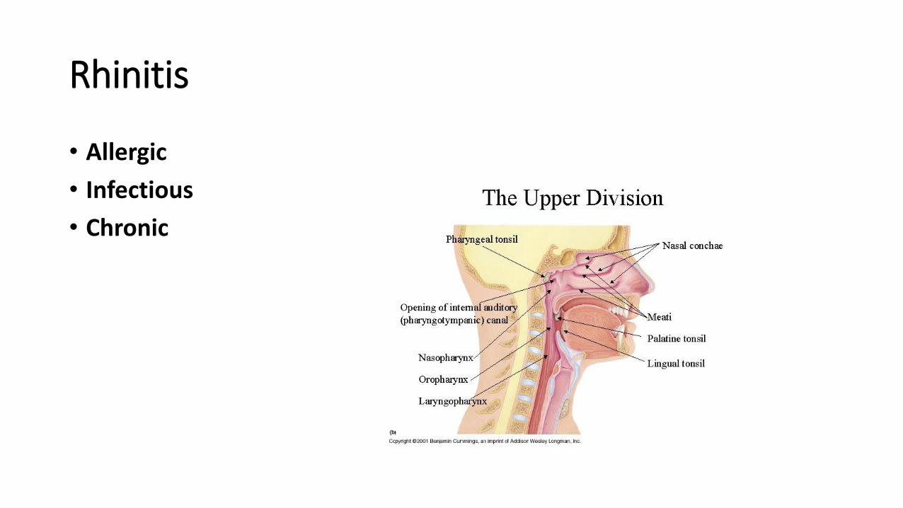
Rhinitis
• Allergic
• Infectious
• Chronic
Allergic rhinitis
• Also called hay fever
• Due to exposure to plant pollens, fungi, dust mites, animal allergens
• IgE mediated hypersensitivity reaction type I.

Infectious rhinitis
Also called “common cold”
Due to adenovirus, echovirus and rhinoviruses
Symptom: catarrhal discharge
Chronic rhinitis
Sequel to acute rhinitis with development of secondary bacterial infection
Associated with deviated septum or nasal polyps
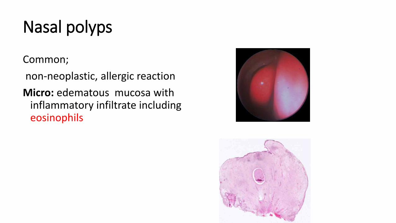
Nasal polyps
Common;
non-neoplastic, allergic reaction
Micro: edematous mucosa with inflammatory infiltrate including eosinophils
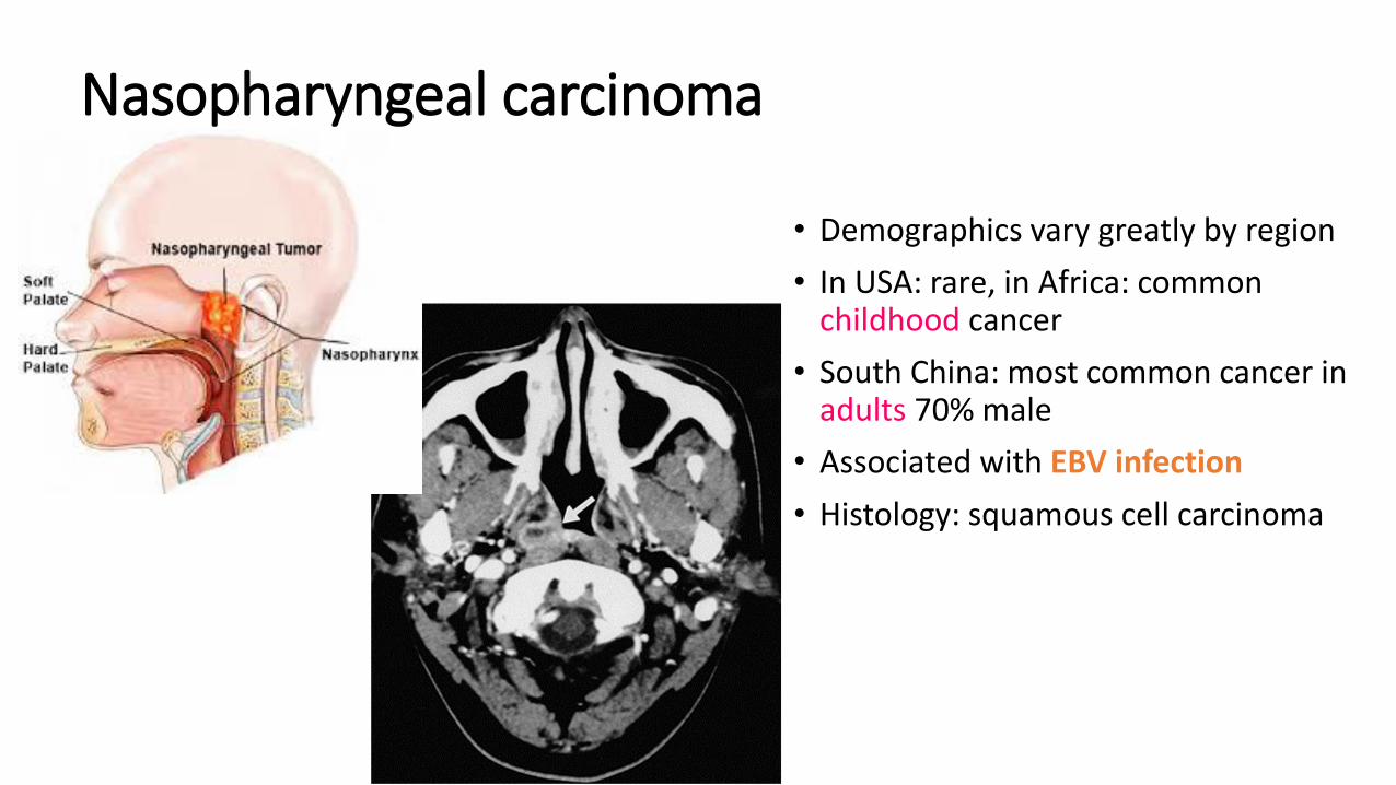
• Demographics vary greatly by region
• In USA: rare, in Africa: commonchildhood cancer
• South China: most common cancer in adults 70% male
• Associated with EBV infection
• Histology: squamous cell carcinoma
Nasopharyngeal carcinoma
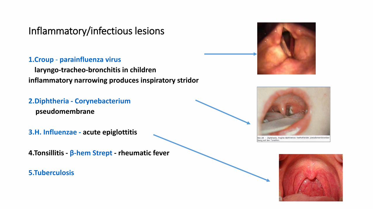
Inflammatory/infectious lesions
1.Croup - parainfluenza virus
laryngo-tracheo-bronchitis in children
inflammatory narrowing produces inspiratory stridor
2.Diphtheria - Corynebacterium
pseudomembrane
3.H. Influenzae - acute epiglottitis
4.Tonsillitis - β-hem Strept - rheumatic fever
5.Tuberculosis
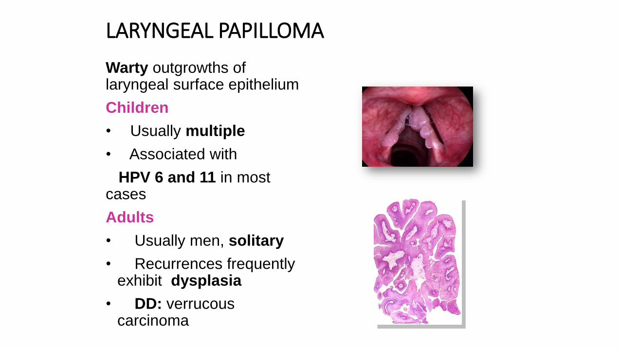
LARYNGEAL PAPILLOMA
Warty outgrowths of laryngeal surface epithelium
Children
• Usually multiple
• Associated with
HPV 6 and 11 in most cases
Adults
• Usually men, solitary
• Recurrences frequentlyexhibit dysplasia
• DD: verrucouscarcinoma
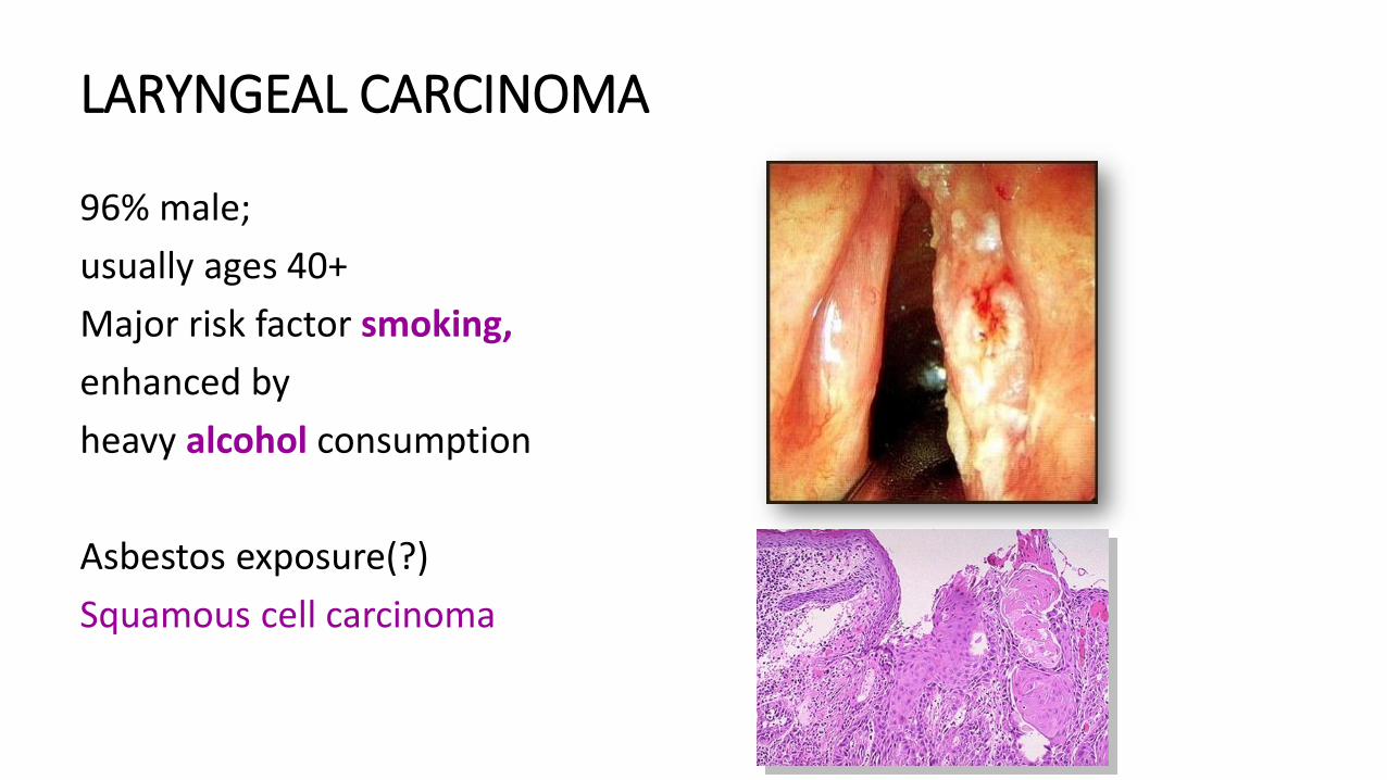
LARYNGEAL CARCINOMA
96% male;
usually ages 40+
Major risk factor smoking,
enhanced by
heavy alcohol consumption
Asbestos exposure(?)
Squamous cell carcinoma
LARYNGEAL CARCINOMA
Site influences histology and clinical behavior – either glottic, supraglottic or subglottic
• Spread is limited by tough membranes / ligaments
Metastases to regional lymph nodes and lungs; direct extension to thyroid gland and jugular vein
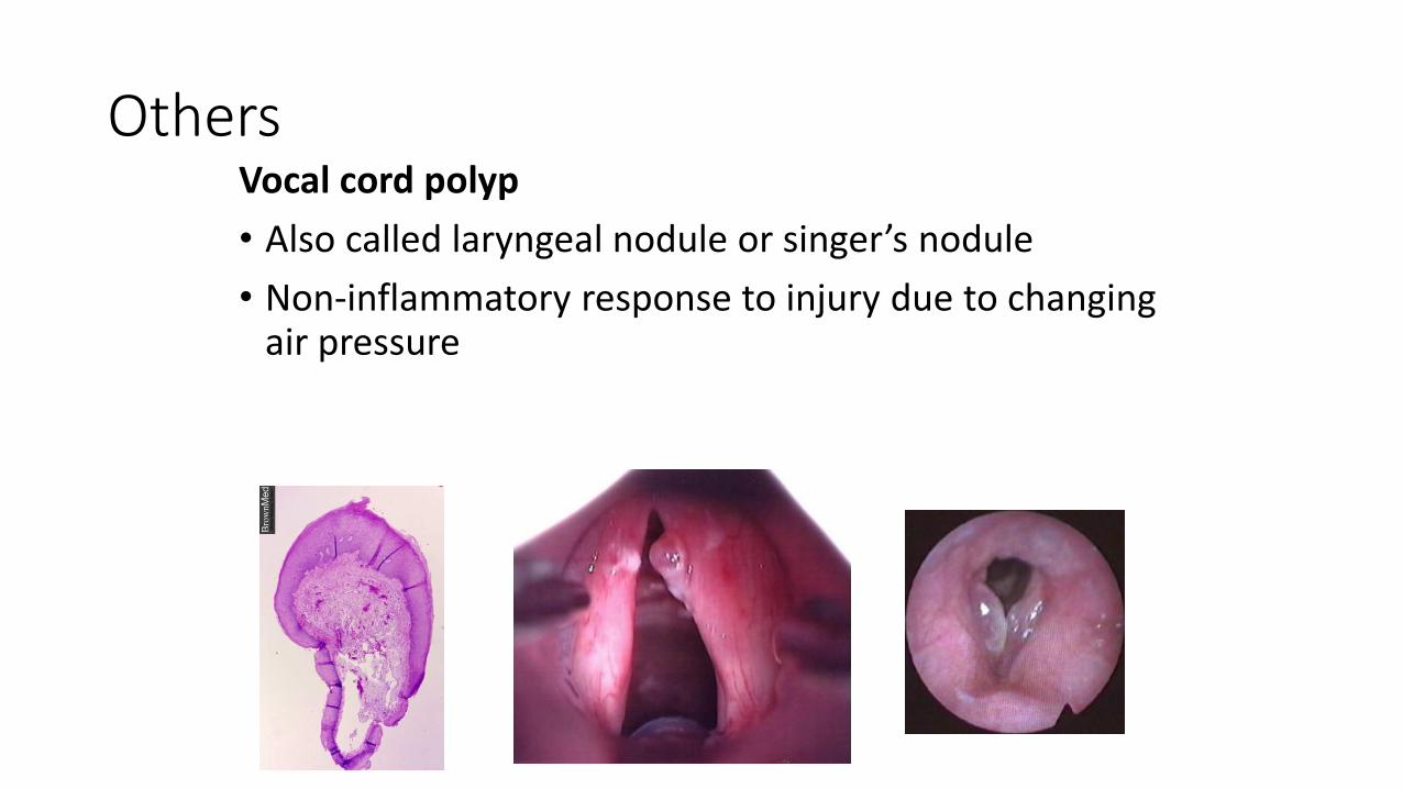
OthersVocal cord polyp
• Also called laryngeal nodule or singer’s nodule
• Non-inflammatory response to injury due to changingair pressure
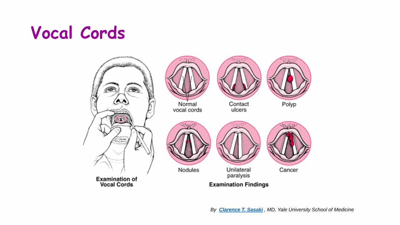
Vocal Cords
By Clarence T. Sasaki , MD, Yale University School of Medicine

TRACHEA – WINDPIPE
• Developmental
• Iflammation - tracheitis
• Decubitus
• Rare tumors
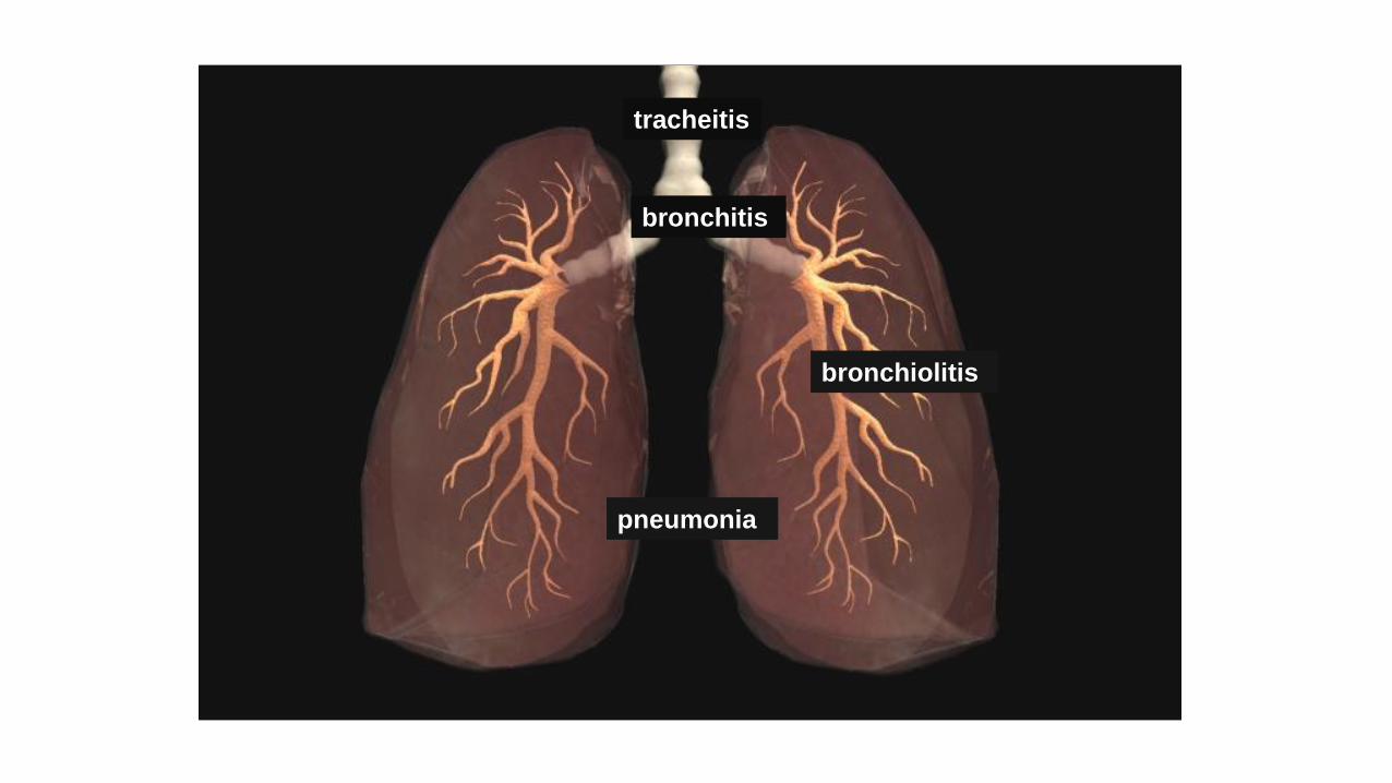
tracheitis
bronchitis
bronchiolitis
pneumonia
Lung diseases
• Acute lung injury (ARDS)
• Inflammation: pneumonia
nota bene: pneumonitis – non-organic hypersensitive reaction
• COPD (chronic obstructive lung disease)
• Restrictive lung diseases
DPLD (diffuse parenchymal lung disease)
• Neoplasma- primary & secondary
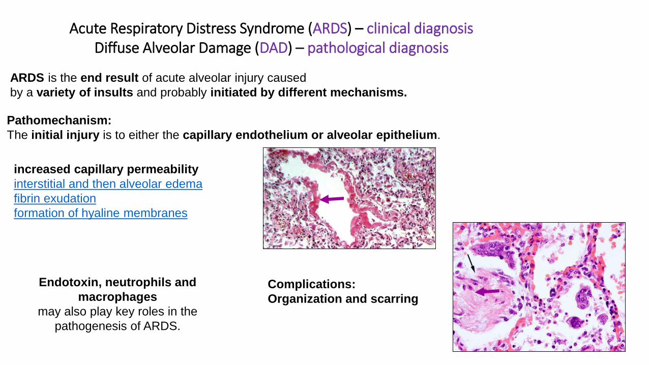
Acute Respiratory Distress Syndrome (ARDS) – clinical diagnosisDiffuse Alveolar Damage (DAD) – pathological diagnosis
ARDS is the end result of acute alveolar injury caused
by a variety of insults and probably initiated by different mechanisms.
Pathomechanism:
The initial injury is to either the capillary endothelium or alveolar epithelium.
increased capillary permeability
interstitial and then alveolar edema
fibrin exudation
formation of hyaline membranes
Complications:
Organization and scarring
Endotoxin, neutrophils and
macrophages
may also play key roles in the
pathogenesis of ARDS.
Pneumonia
• Clinical data: acute – chronic
• Pattern: broncho – lobar
• Clinical feature: atypical – hypostatic etc …..
• Type of infection: community - acquired (out of hospital) hospital–acquired nosocomial, opportunistic
• Based on agents: bacterial, viral,fungal …
• Host reaction: normal, immunocompr, illness, infants, elderly…
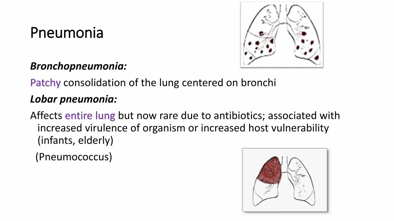
Pneumonia
Bronchopneumonia:
Patchy consolidation of the lung centered on bronchi
Lobar pneumonia:
Affects entire lung but now rare due to antibiotics; associated withincreased virulence of organism or increased host vulnerability(infants, elderly)
(Pneumococcus)
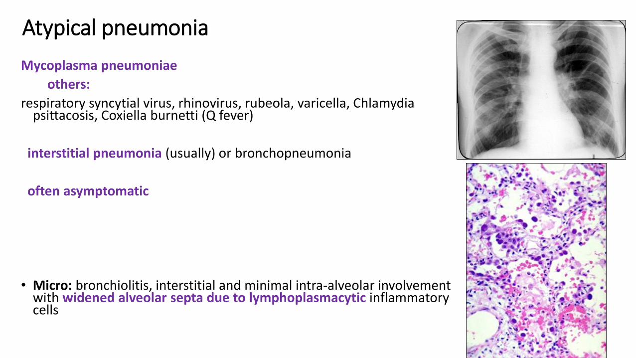
Atypical pneumonia
Mycoplasma pneumoniae
others:
respiratory syncytial virus, rhinovirus, rubeola, varicella, Chlamydia psittacosis, Coxiella burnetti (Q fever)
interstitial pneumonia (usually) or bronchopneumonia
often asymptomatic
• Micro: bronchiolitis, interstitial and minimal intra-alveolar involvementwith widened alveolar septa due to lymphoplasmacytic inflammatorycells
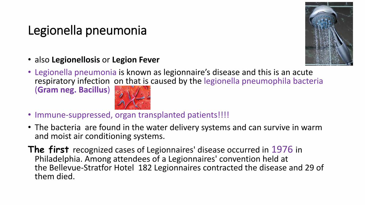
Legionella pneumonia
• also Legionellosis or Legion Fever
• Legionella pneumonia is known as legionnaire’s disease and this is an acute respiratory infection on that is caused by the legionella pneumophila bacteria(Gram neg. Bacillus)
• Immune-suppressed, organ transplanted patients!!!!
• The bacteria are found in the water delivery systems and can survive in warm and moist air conditioning systems.
The first recognized cases of Legionnaires' disease occurred in 1976 inPhiladelphia. Among attendees of a Legionnaires' convention held at the Bellevue-Stratfor Hotel 182 Legionnaires contracted the disease and 29 of them died.
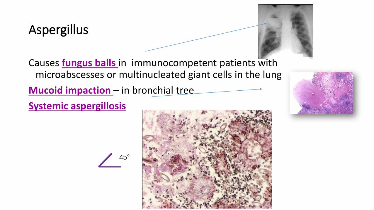
Aspergillus
Causes fungus balls in immunocompetent patients with microabscesses or multinucleated giant cells in the lung
Mucoid impaction – in bronchial tree
Systemic aspergillosis
45°
Abscess
Due to sinobronchial infections,
• dental sepsis,
• aspiration
• primary bacterial infection (Staphylococcus aureus, Klebsiella pneumonia, Streptococcus pneumonia),
• fungi,
• neoplasia induced obstruction
Aspiration induced abscesses more common on right side
Air fluid level present if there is communication with air passages
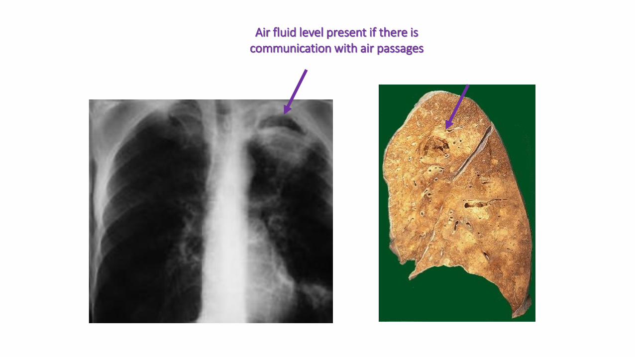
Air fluid level present if there is communication with air passages
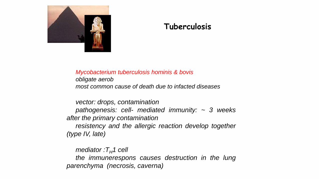
Tuberculosis
Mycobacterium tuberculosis hominis & bovis
obligate aerob
most common cause of death due to infacted diseases
vector: drops, contamination
pathogenesis: cell- mediated immunity: ~ 3 weeks
after the primary contamination
resistency and the allergic reaction develop together
(type IV, late)
mediator :TH1 cell
the immunerespons causes destruction in the lung
parenchyma (necrosis, caverna)

Type of inflammation: chronic specificgranulomatous inflammatory lesion w/wo necrosis
primary tbc – primary infectionsecunder tbc – already sensibilized patient
localizes espec in the apical parts of lung – followedby caverna
lymphatics – right heart – pulmonary arterialdissemination– miliary tbc
systemic or localized organic tbc
Tuberculosis
Tuberculosis
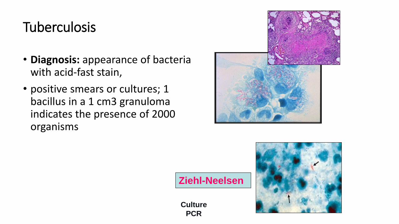
• Diagnosis: appearance of bacteria with acid-fast stain,
• positive smears or cultures; 1 bacillus in a 1 cm3 granuloma indicates the presence of 2000 organisms
Ziehl-Neelsen
Culture
PCR
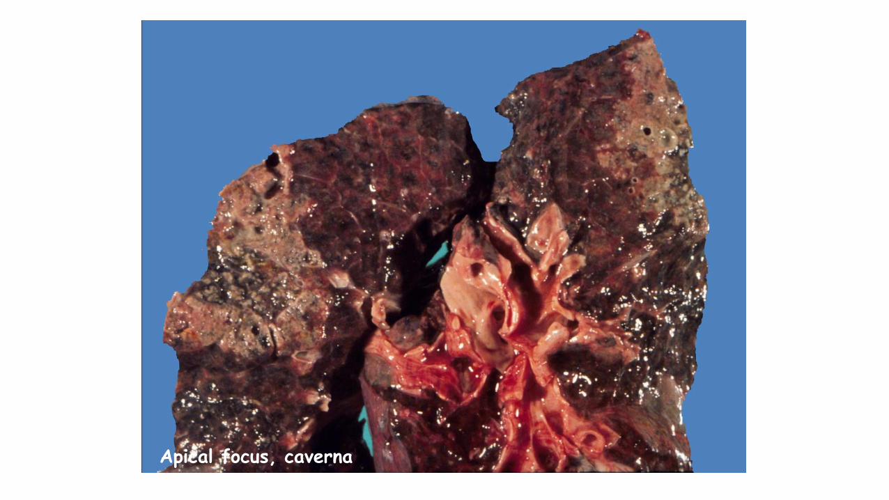
Apical focus, caverna
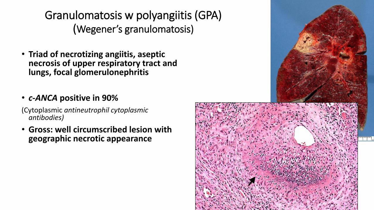
Granulomatosis w polyangiitis (GPA)(Wegener’s granulomatosis)
• Triad of necrotizing angiitis, asepticnecrosis of upper respiratory tract and lungs, focal glomerulonephritis
• c-ANCA positive in 90%
(Cytoplasmic antineutrophil cytoplasmicantibodies)
• Gross: well circumscribed lesion withgeographic necrotic appearance