Embed Size (px)
Citation preview
Histology of thetongue and teeth
Semmelweis University, Faculty of MedicineDepartment of Anatomy, Histology and Embryology
Microscopic anatomy I.
Katalin Kocsis
05/03/202006/03/2020

TONGUE
papillary surface
follicular surface
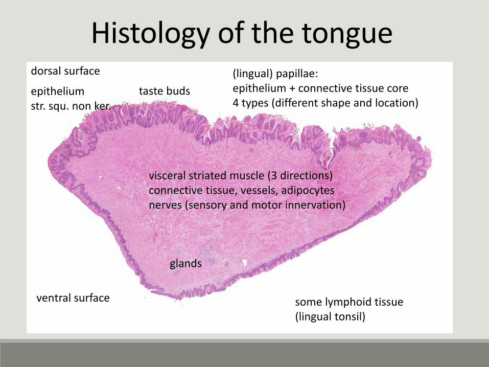
Histology of the tongue
epitheliumstr. squ. non ker.
visceral striated muscle (3 directions)connective tissue, vessels, adipocytesnerves (sensory and motor innervation)
glands
(lingual) papillae:epithelium + connective tissue core4 types (different shape and location)
dorsal surface
ventral surface some lymphoid tissue(lingual tonsil)
taste buds
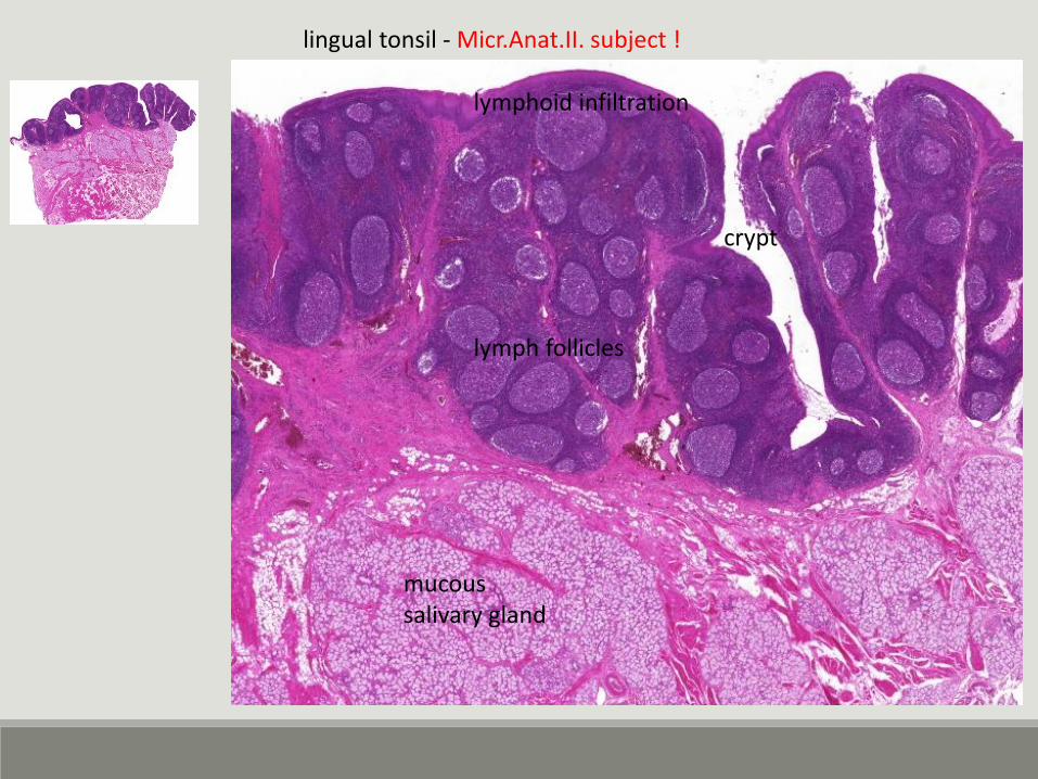
lingual tonsil - Micr.Anat.II. subject !
mucoussalivary gland
lymphoid infiltration
crypt
lymph follicles
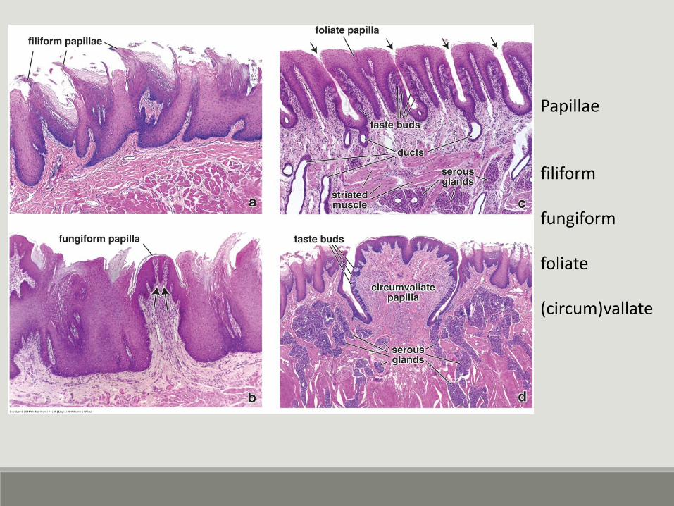
Papillae
filiform
fungiform
foliate
(circum)vallate
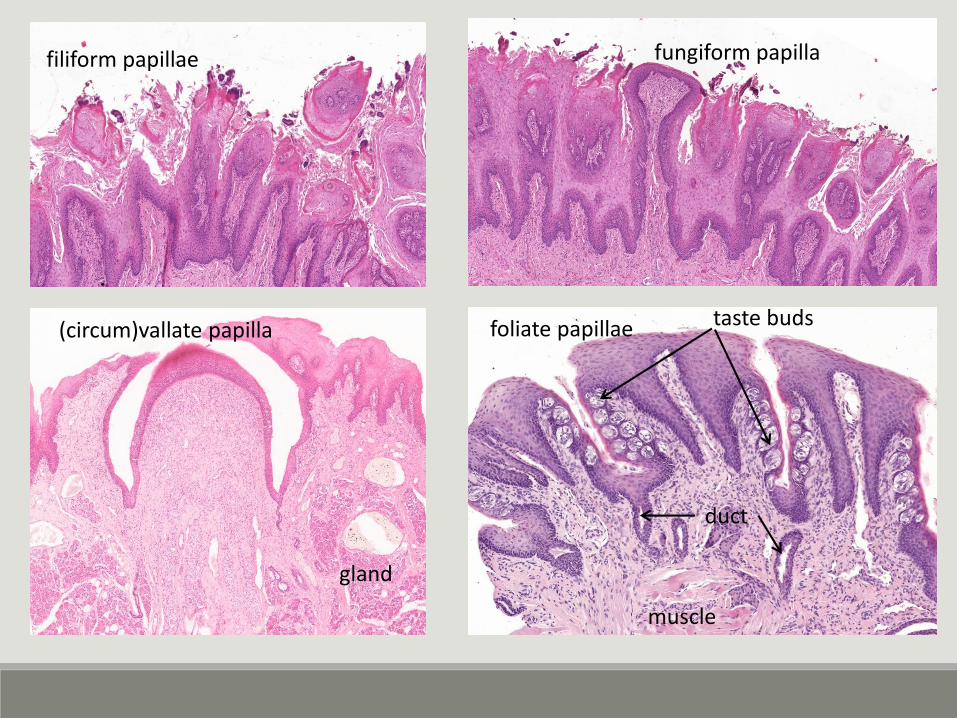
filiform papillae fungiform papilla
foliate papillae(circum)vallate papilla
gland
duct
muscle
taste buds
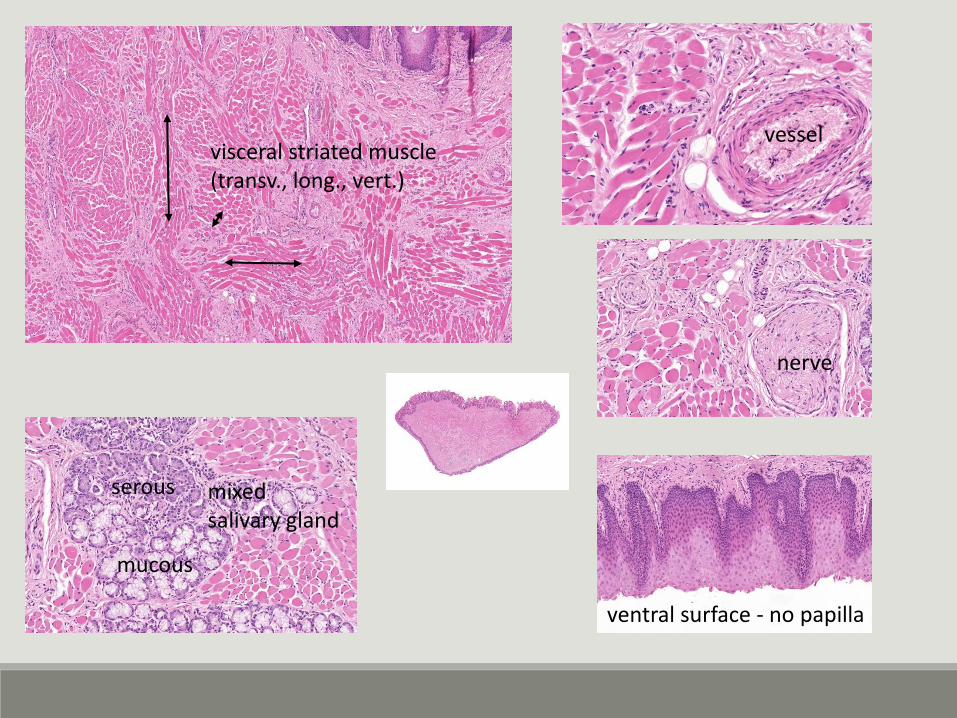
visceral striated muscle(transv., long., vert.)
mixed salivary gland
serous
mucous
nerve
vessel
ventral surface - no papilla
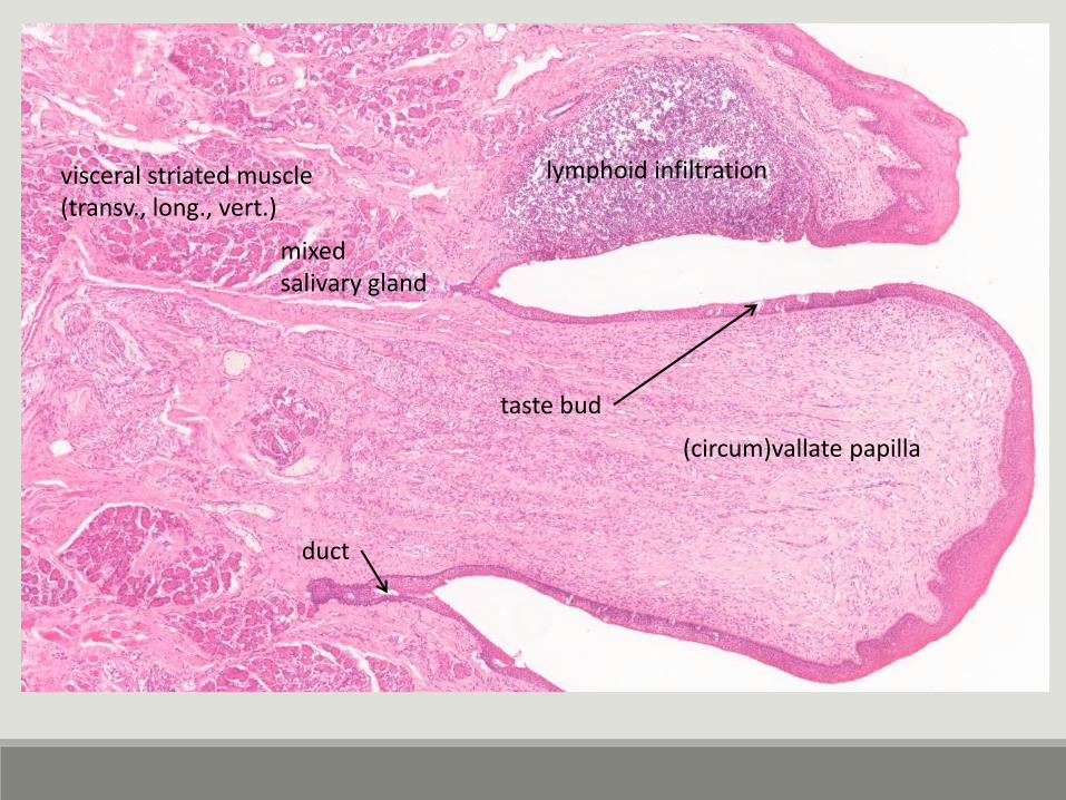
visceral striated muscle(transv., long., vert.)
mixed salivary gland
(circum)vallate papilla
taste bud
lymphoid infiltration
duct
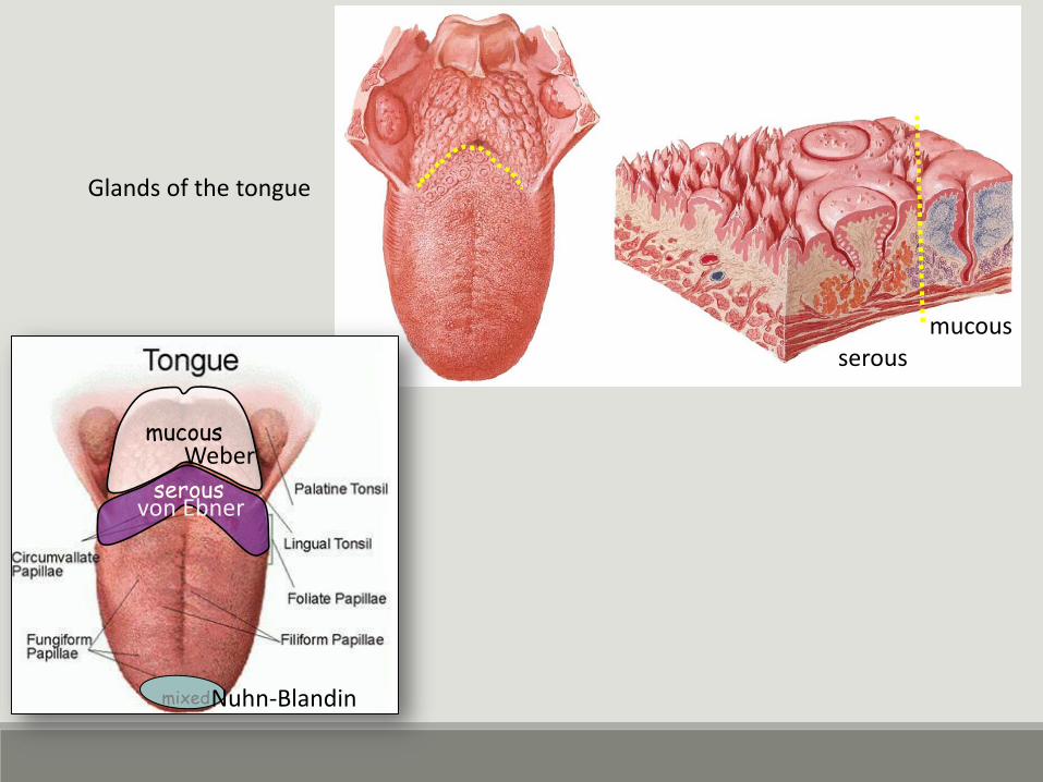
mucous
serous
mixedNuhn-Blandin
von Ebner
Weber
Glands of the tongue
serousmucous
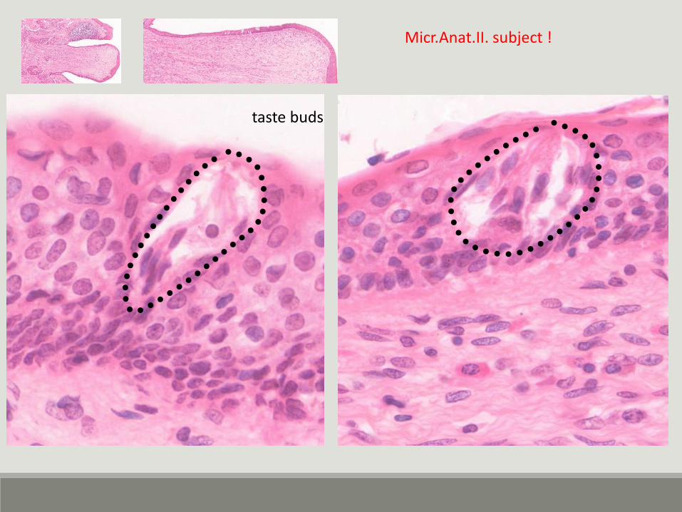
taste buds
Micr.Anat.II. subject !
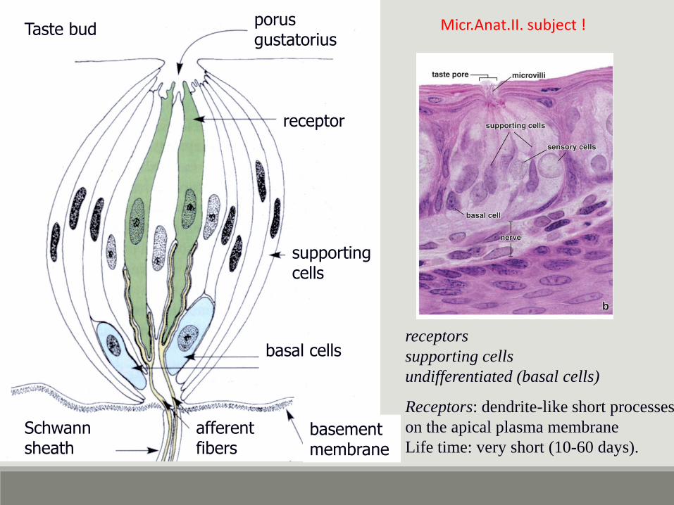
receptors
supporting cells
undifferentiated (basal cells)
Receptors: dendrite-like short processes
on the apical plasma membrane
Life time: very short (10-60 days).
porusgustatorius
receptor
supportingcells
basal cells
basementmembrane
afferentfibers
Schwannsheath
Taste bud Micr.Anat.II. subject !
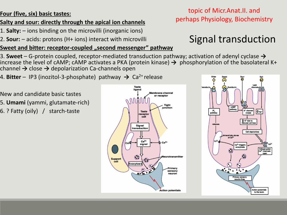
Four (five, six) basic tastes:
Salty and sour: directly through the apical ion channels
1. Salty: – ions binding on the microvilli (inorganic ions)
2. Sour: – acids: protons (H+ ions) interact with microvilli
Sweet and bitter: receptor-coupled „second messenger” pathway
3. Sweet – G-protein coupled, receptor-mediated transduction pathway; activation of adenyl cyclase →increase the level of cAMP; cAMP activates a PKA (protein kinase) → phosphorylation of the basolateral K+ channel → close → depolarization Ca-channels open
4. Bitter – IP3 (inozitol-3-phosphate) pathway → Ca2+ release
New and candidate basic tastes
5. Umami (yammi, glutamate-rich)
6. ? Fatty (oily) / starch-taste
Signal transduction
topic of Micr.Anat.II. and perhaps Physiology, Biochemistry
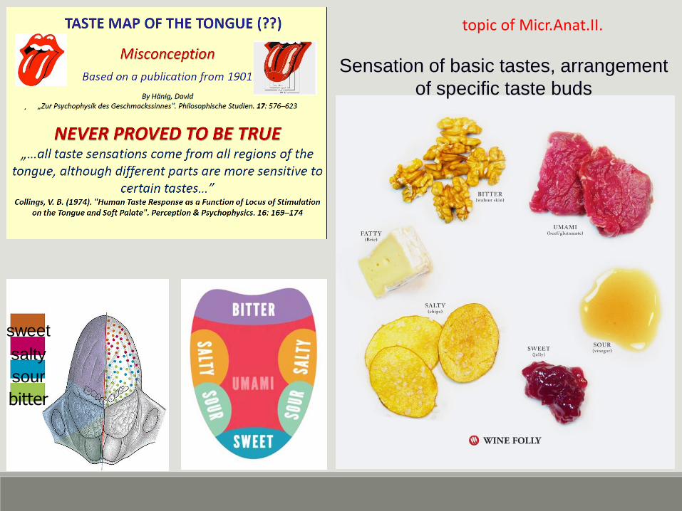
bitter
sour
salty
sweet
Sensation of basic tastes, arrangement
of specific taste buds
topic of Micr.Anat.II.
TOOTH
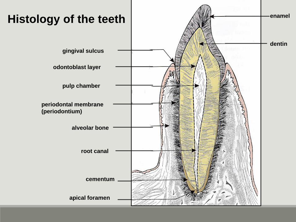
Histology of the teeth
dentin
enamel
gingival sulcus
odontoblast layer
pulp chamber
periodontal membrane
(periodontium)
alveolar bone
root canal
cementum
apical foramen
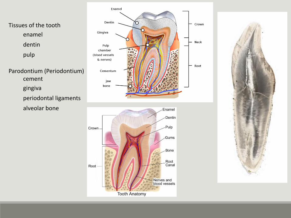
Tissues of the tooth
enamel
dentin
pulp
Parodontium (Periodontium)cement
gingiva
periodontal ligaments
alveolar bone
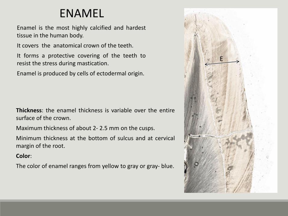
Enamel is the most highly calcified and hardesttissue in the human body.
It covers the anatomical crown of the teeth.
It forms a protective covering of the teeth toresist the stress during mastication.
Enamel is produced by cells of ectodermal origin.
E
ENAMEL
Thickness: the enamel thickness is variable over the entiresurface of the crown.
Maximum thickness of about 2- 2.5 mm on the cusps.
Minimum thickness at the bottom of sulcus and at cervicalmargin of the root.
Color:
The color of enamel ranges from yellow to gray or gray- blue.
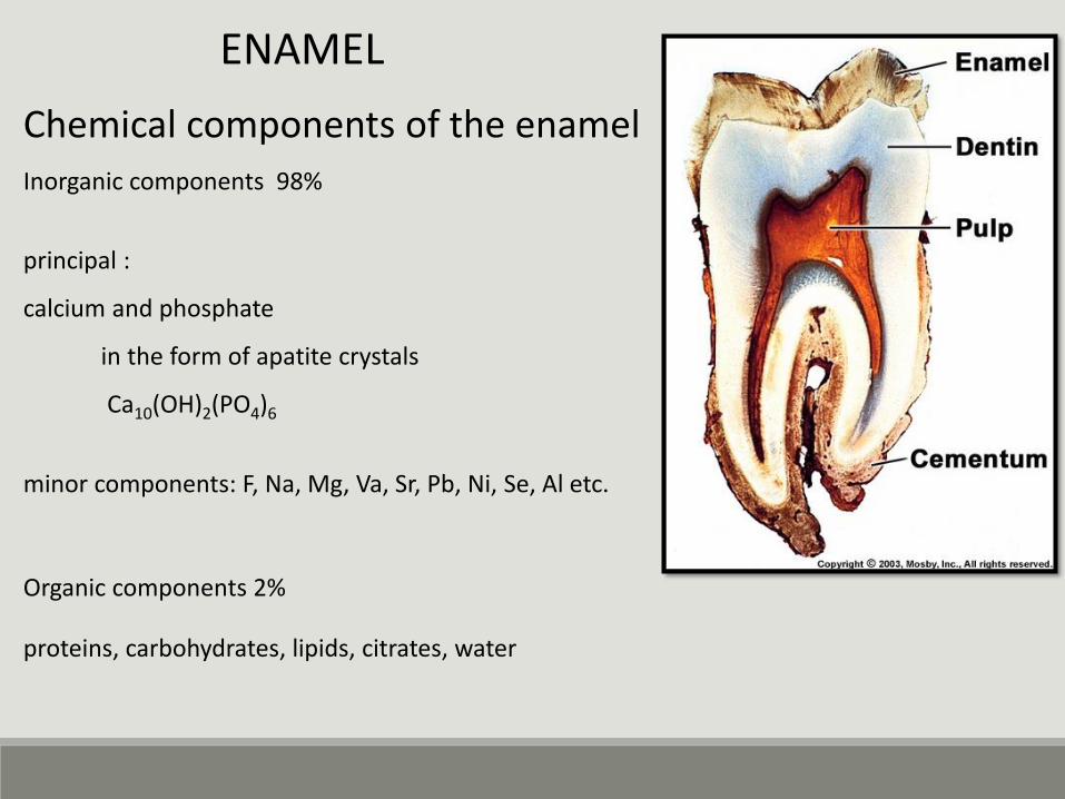
Chemical components of the enamel
Inorganic components 98%
Organic components 2%
principal :
calcium and phosphate
in the form of apatite crystals
Ca10(OH)2(PO4)6
minor components: F, Na, Mg, Va, Sr, Pb, Ni, Se, Al etc.
proteins, carbohydrates, lipids, citrates, water
ENAMEL
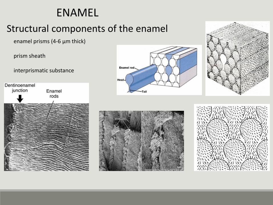
Structural components of the enamelenamel prisms (4-6 µm thick)
prism sheath
interprismatic substance
ENAMEL
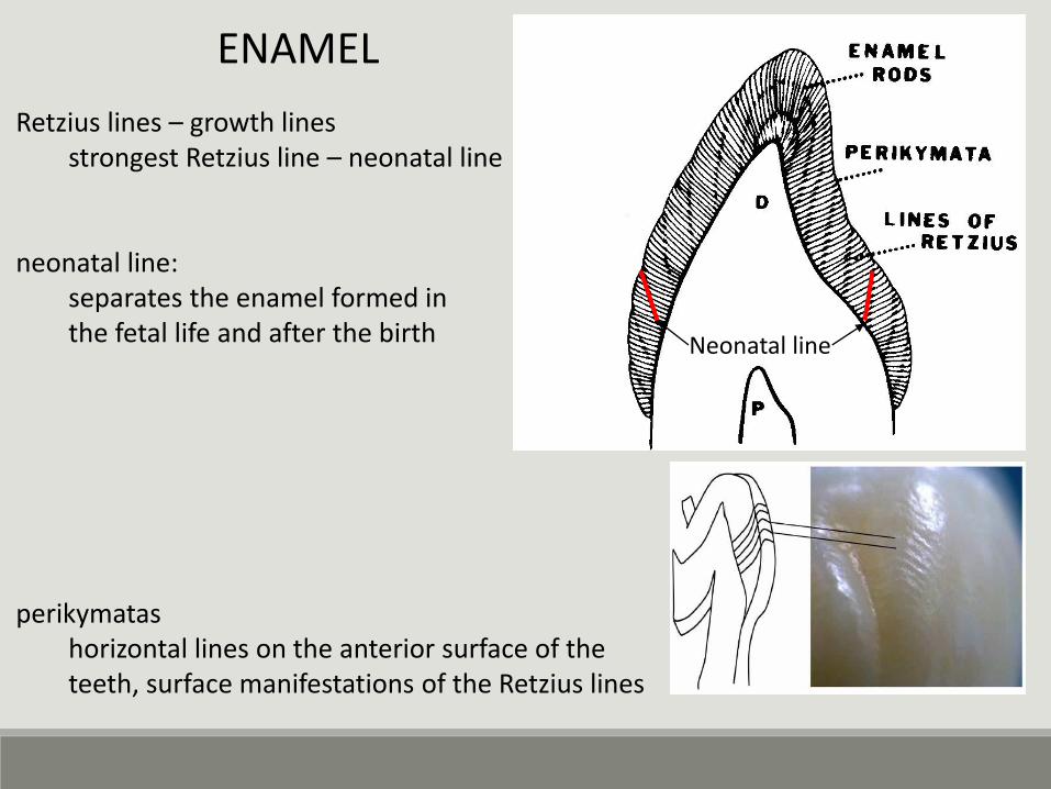
Retzius lines – growth linesstrongest Retzius line – neonatal line
neonatal line:separates the enamel formed in the fetal life and after the birth
perikymatashorizontal lines on the anterior surface of theteeth, surface manifestations of the Retzius lines
Neonatal line
ENAMEL
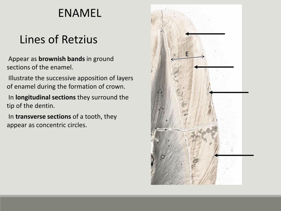
Appear as brownish bands in ground sections of the enamel.
Illustrate the successive apposition of layers of enamel during the formation of crown.
In longitudinal sections they surround the tip of the dentin.
In transverse sections of a tooth, they appear as concentric circles.
E
Lines of Retzius
ENAMEL
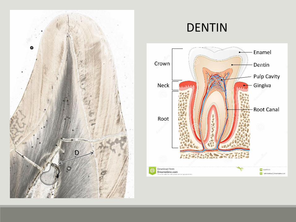
DENTIN
D
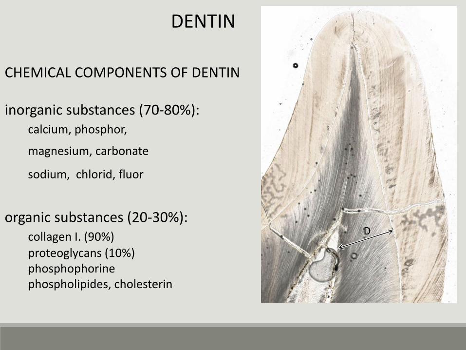
CHEMICAL COMPONENTS OF DENTIN
inorganic substances (70-80%): calcium, phosphor,
magnesium, carbonate
sodium, chlorid, fluor
organic substances (20-30%):collagen I. (90%)
proteoglycans (10%)phosphophorinephospholipides, cholesterin
D
DENTIN
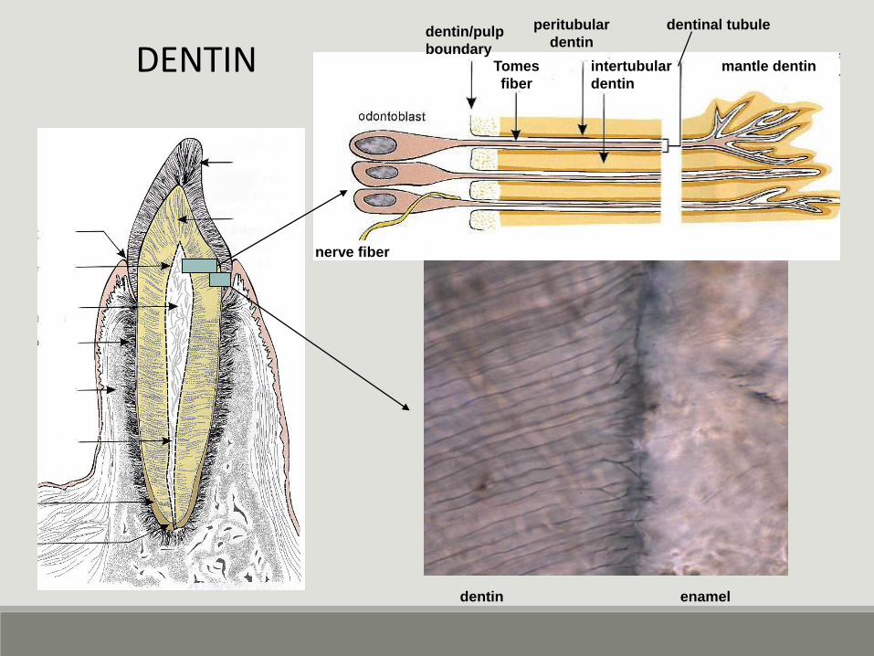
Tomes
fiber
nerve fiber
enameldentin
mantle dentin
peritubular
dentin
intertubular
dentin
dentin/pulp
boundary
dentinal tubule
DENTIN
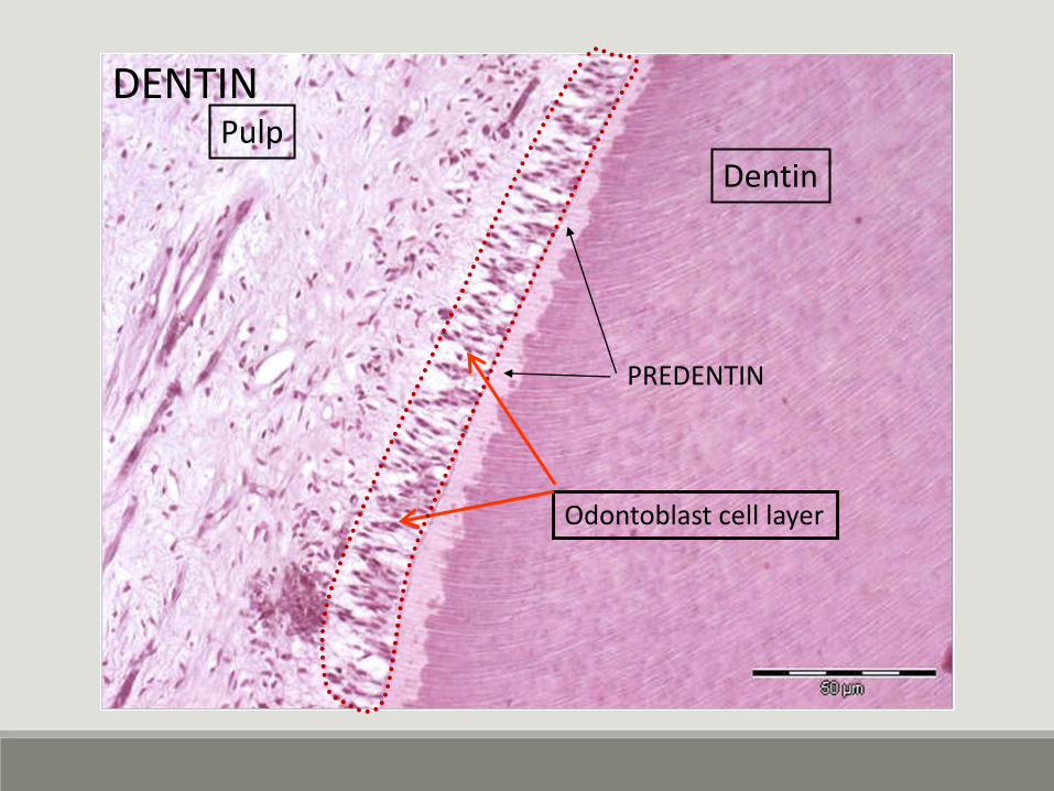
DentinPulp
Odontoblast cell layer
PREDENTIN
DENTIN
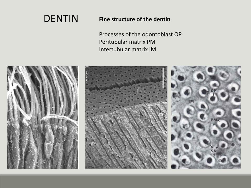
Fine structure of the dentin
Processes of the odontoblast OPPeritubular matrix PMIntertubular matrix IM
DENTIN
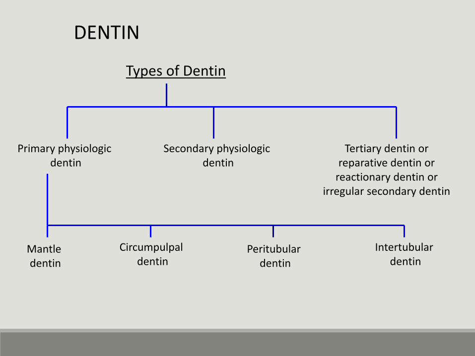
Types of Dentin
Primary physiologic dentin
Secondary physiologic dentin
Tertiary dentin orreparative dentin or
reactionary dentin orirregular secondary dentin
Mantle dentin
Circumpulpal dentin
Peritubular dentin
Intertubular dentin
DENTIN
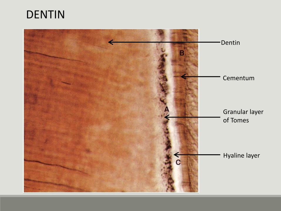
Dentin
Cementum
Granular layer of Tomes
Hyaline layer
DENTIN
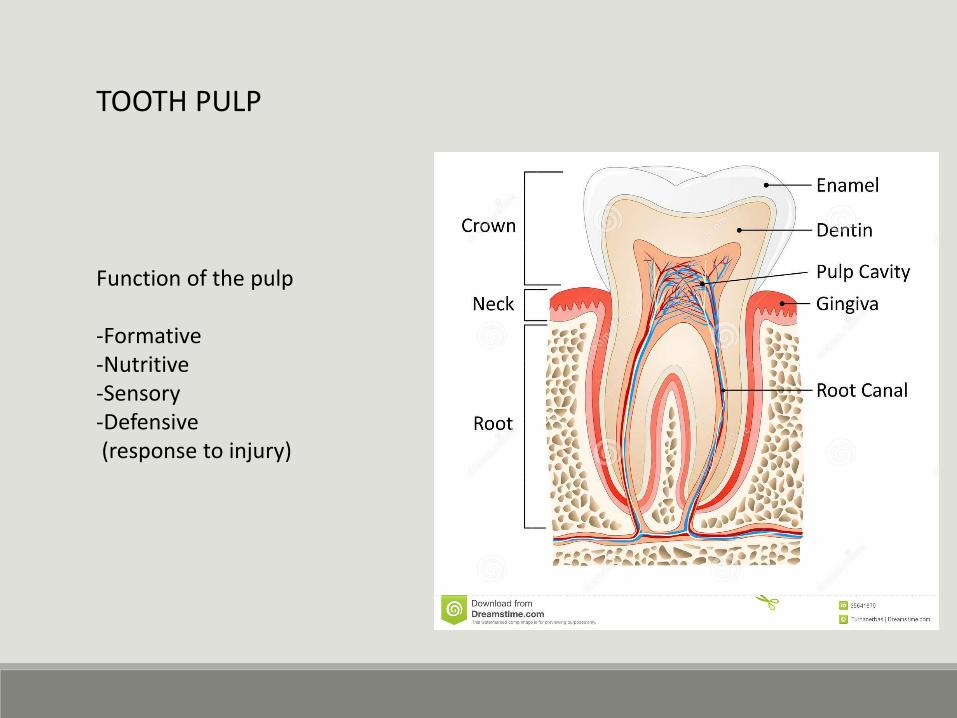
Function of the pulp
-Formative-Nutritive-Sensory-Defensive(response to injury)
TOOTH PULP
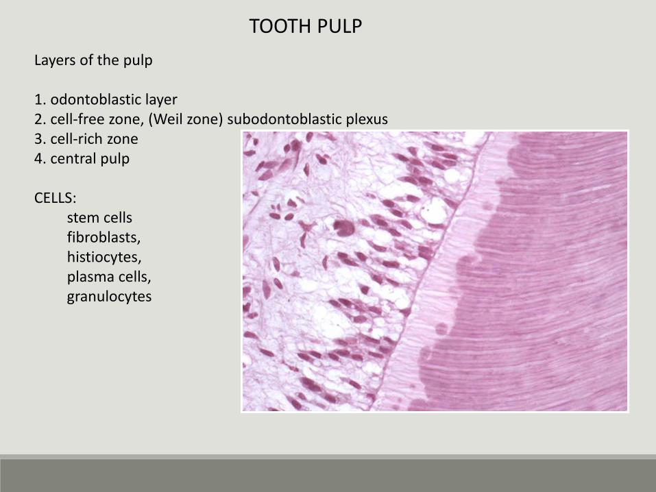
Layers of the pulp
1. odontoblastic layer2. cell-free zone, (Weil zone) subodontoblastic plexus3. cell-rich zone4. central pulp
CELLS:stem cellsfibroblasts,histiocytes,plasma cells, granulocytes
TOOTH PULP
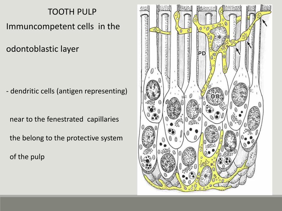
Immuncompetent cells in the
odontoblastic layer
- dendritic cells (antigen representing)
near to the fenestrated capillaries
the belong to the protective system
of the pulp
TOOTH PULP
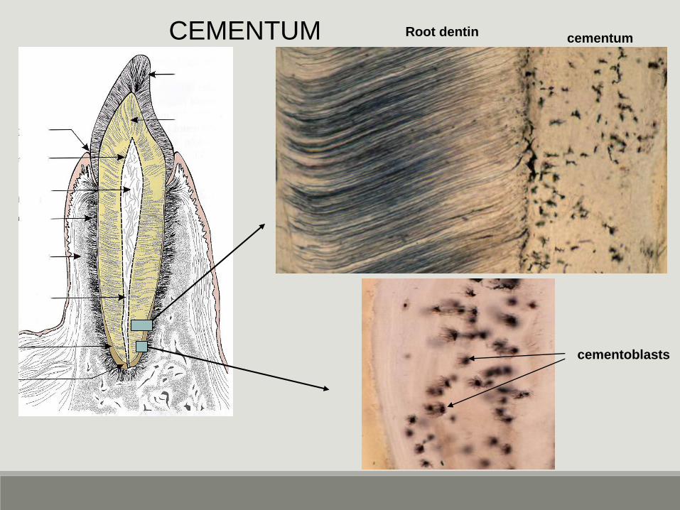
CEMENTUM Root dentincementum
cementoblasts
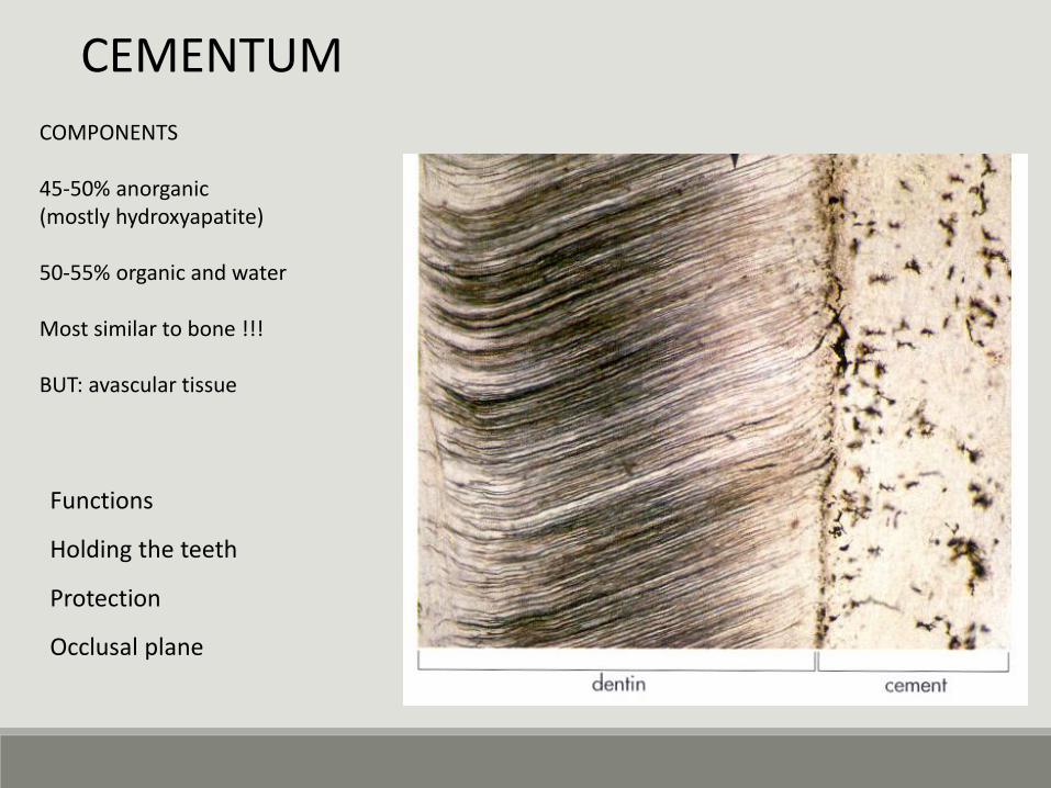
COMPONENTS
45-50% anorganic(mostly hydroxyapatite)
50-55% organic and water
Most similar to bone !!!
BUT: avascular tissue
CEMENTUM
Functions
Holding the teeth
Protection
Occlusal plane
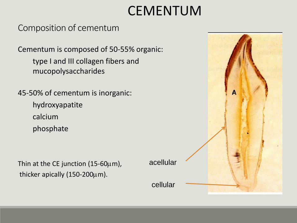
Composition of cementum
cellular
acellular
Cementum is composed of 50-55% organic:
type I and III collagen fibers and mucopolysaccharides
45-50% of cementum is inorganic:
hydroxyapatite
calcium
phosphate
Thin at the CE junction (15-60mm),
thicker apically (150-200mm).
CEMENTUM
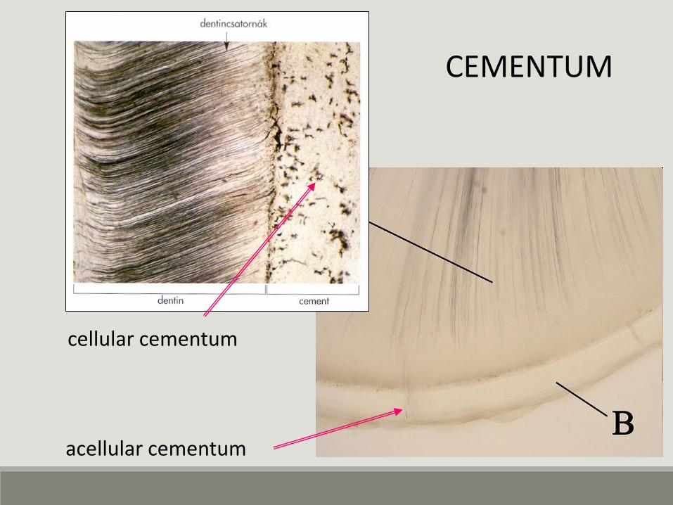
cellular cementum
acellular cementum
CEMENTUM
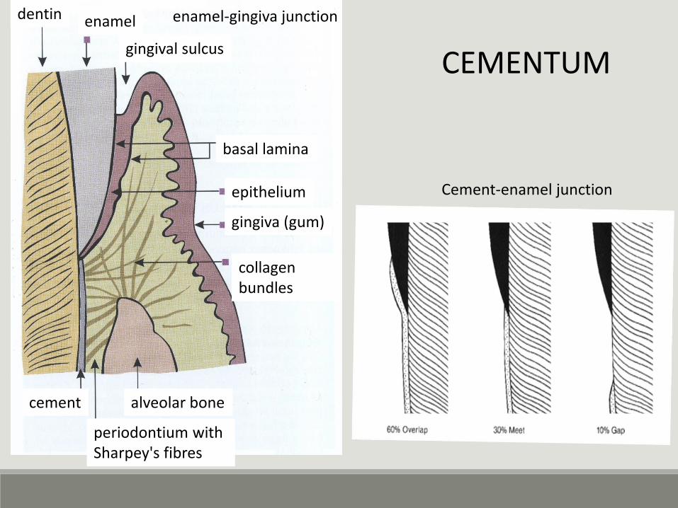
dentin enamel
gingival sulcus
basal lamina
gingiva (gum)
collagenbundles
epithelium
alveolar bonecement
periodontium withSharpey's fibres
enamel-gingiva junction
Cement-enamel junction
CEMENTUM
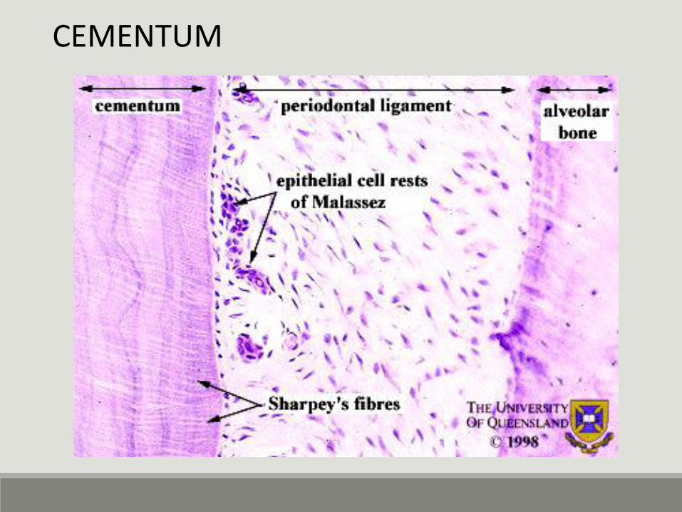
Sharpey rostok
Malassez- féle
hámszigetek
CEMENTUM
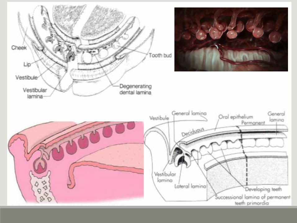
DEVELOPING TOOTHTOOTH GERM / TOOTH BUD
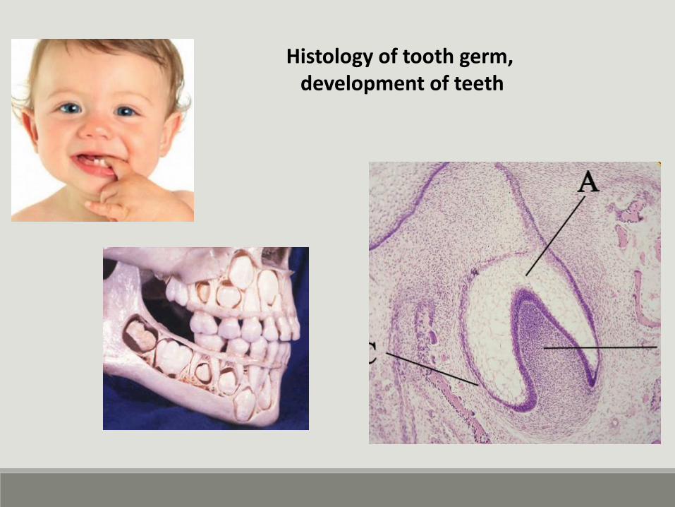
Histology of tooth germ, development of teeth
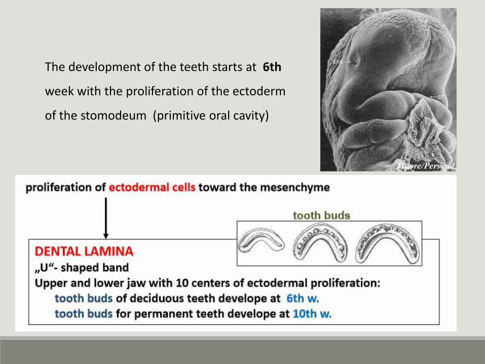
The development of the teeth starts at 6th
week with the proliferation of the ectoderm
of the stomodeum (primitive oral cavity)
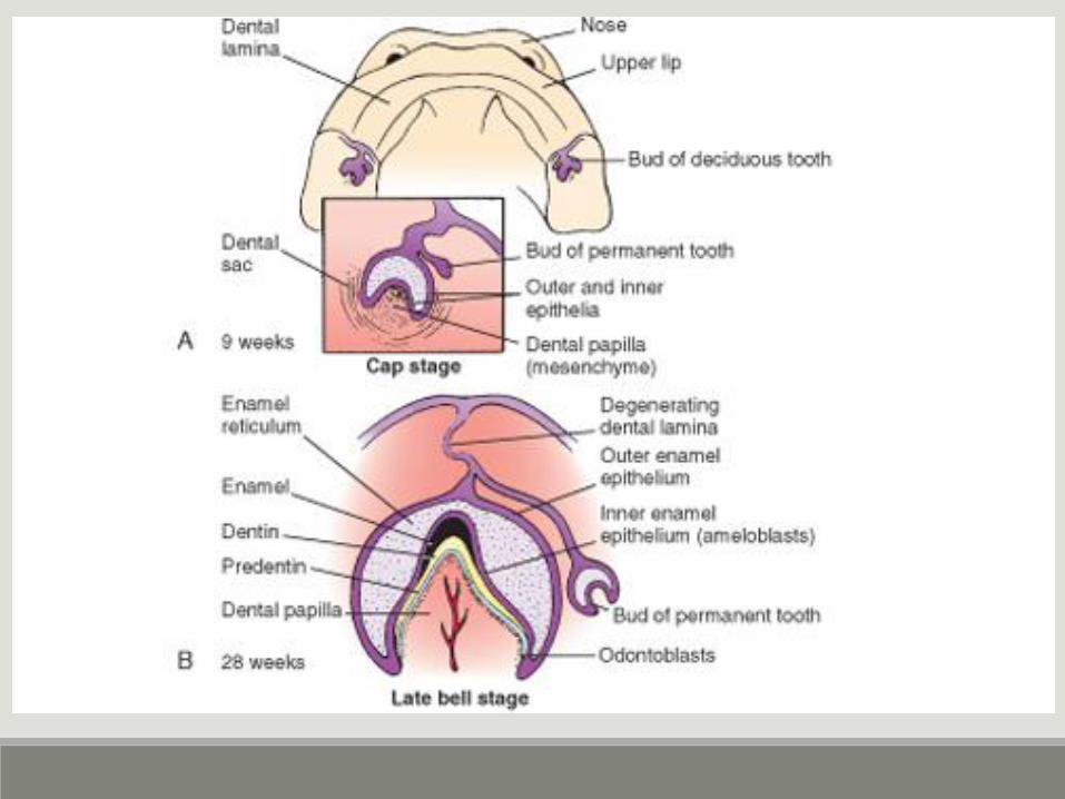
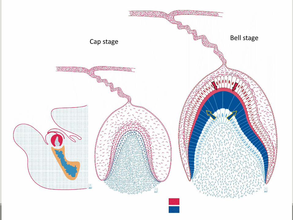
Cap stageBell stage
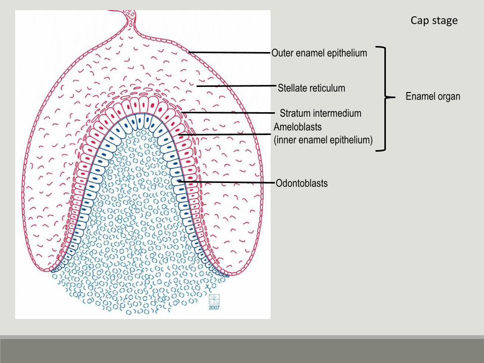
Ameloblasts
(inner enamel epithelium)
Odontoblasts
Stellate reticulum
Outer enamel epithelium
Enamel organ
Stratum intermedium
Cap stage
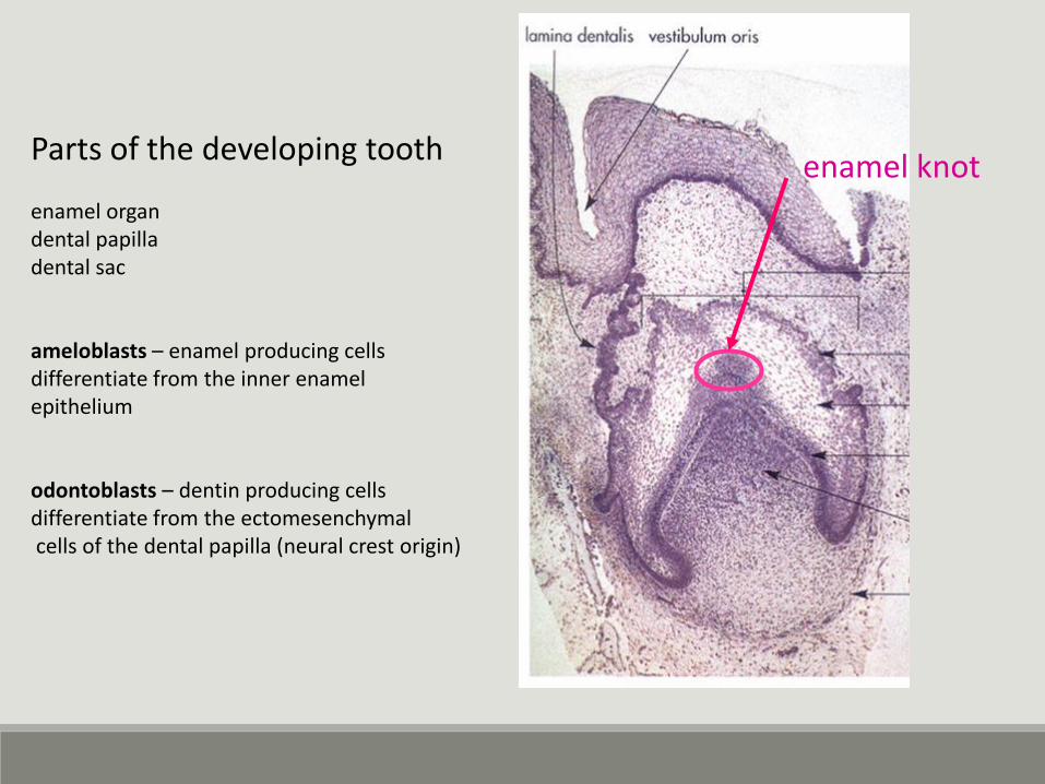
Parts of the developing tooth
enamel organdental papilladental sac
ameloblasts – enamel producing cellsdifferentiate from the inner enamelepithelium
odontoblasts – dentin producing cellsdifferentiate from the ectomesenchymalcells of the dental papilla (neural crest origin)
enamel knot
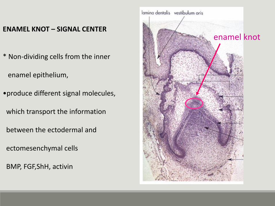
ENAMEL KNOT – SIGNAL CENTER
* Non-dividing cells from the inner
enamel epithelium,
•produce different signal molecules,
which transport the information
between the ectodermal and
ectomesenchymal cells
BMP, FGF,ShH, activin
enamel knot
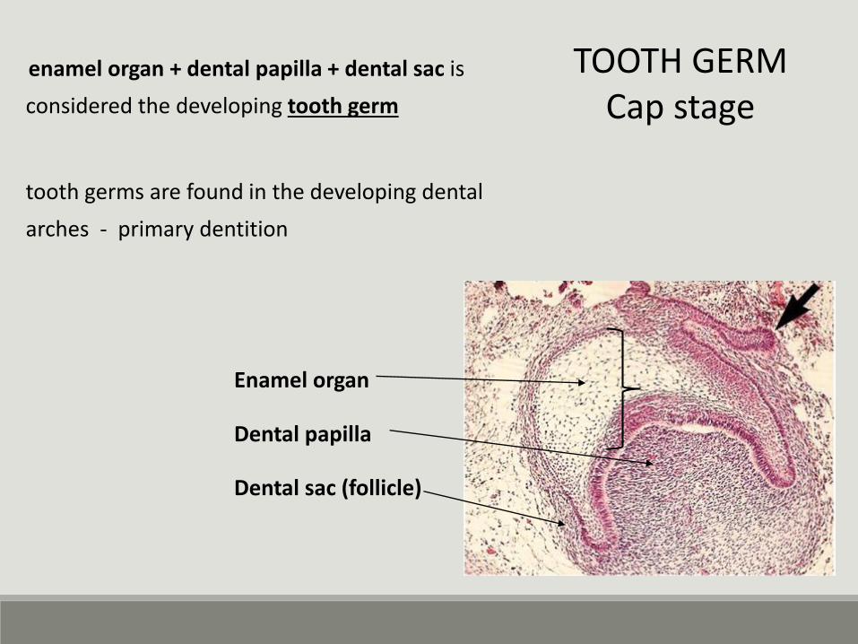
Enamel organ
Dental papilla
Dental sac (follicle)
TOOTH GERMCap stage
enamel organ + dental papilla + dental sac is
considered the developing tooth germ
tooth germs are found in the developing dental
arches - primary dentition
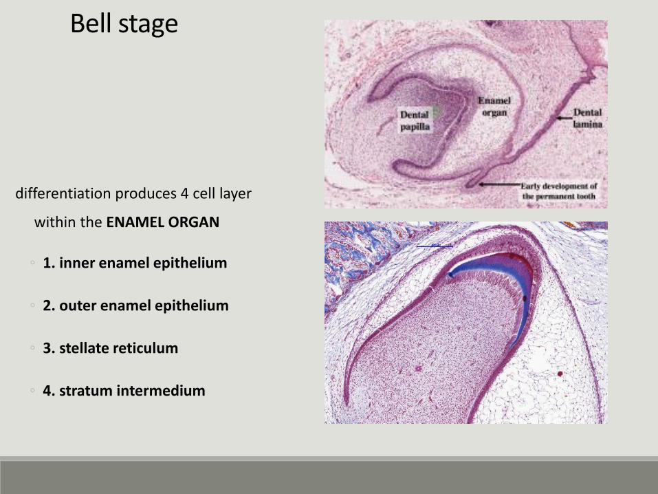
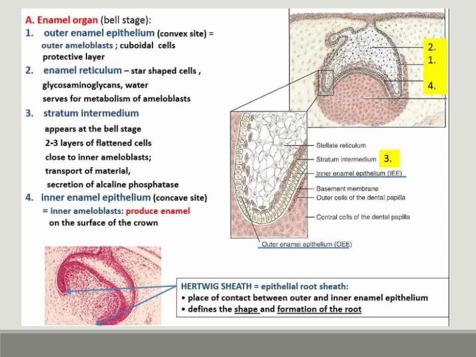
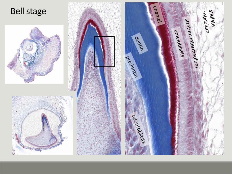
Bell stage
differentiation produces 4 cell layer
within the ENAMEL ORGAN
◦ 1. inner enamel epithelium
◦ 2. outer enamel epithelium
◦ 3. stellate reticulum
◦ 4. stratum intermedium
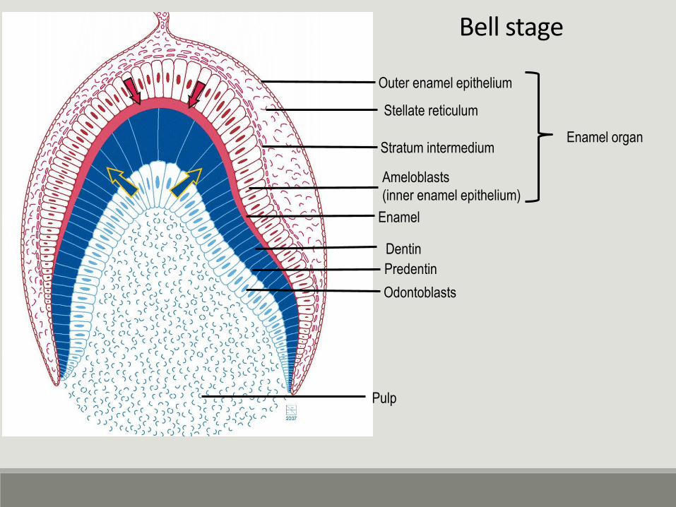
Ameloblasts
(inner enamel epithelium)
Predentin
Odontoblasts
Stratum intermedium
Stellate reticulum
Outer enamel epithelium
Enamel organ
Dentin
Enamel
Pulp
Bell stage
Bell stage
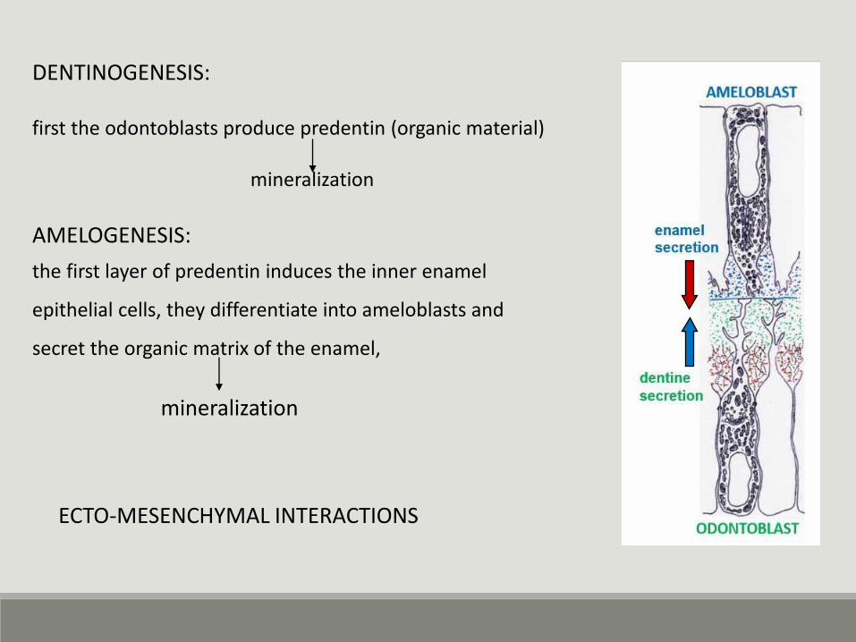
DENTINOGENESIS:
first the odontoblasts produce predentin (organic material)
mineralization
AMELOGENESIS:
the first layer of predentin induces the inner enamel
epithelial cells, they differentiate into ameloblasts and
secret the organic matrix of the enamel,
mineralization
ECTO-MESENCHYMAL INTERACTIONS
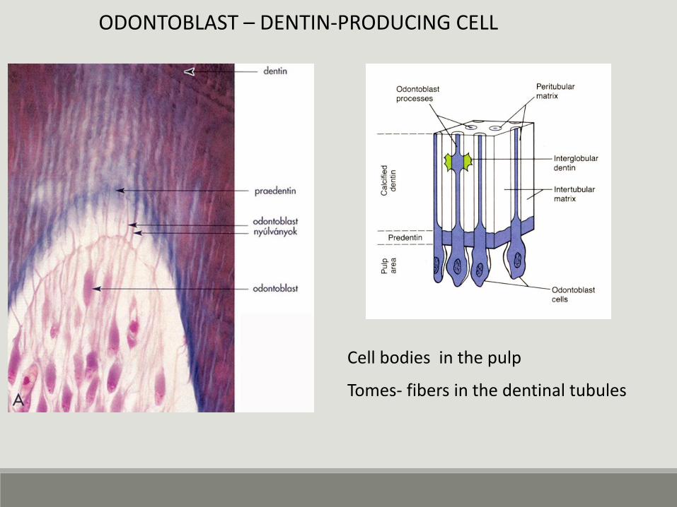
ODONTOBLAST – DENTIN-PRODUCING CELL
Cell bodies in the pulp
Tomes- fibers in the dentinal tubules
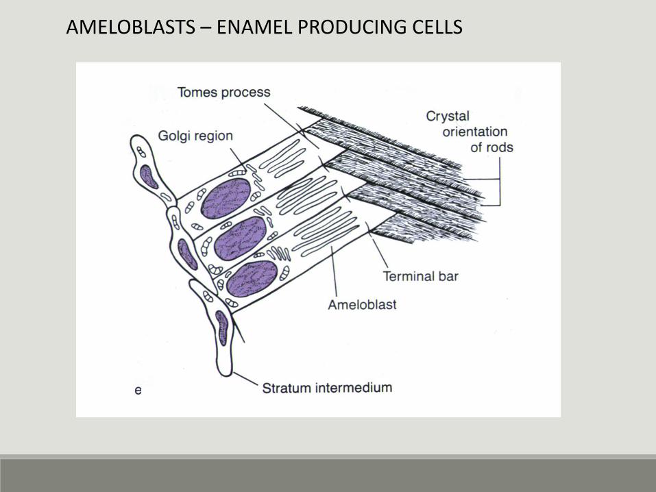
AMELOBLASTS – ENAMEL PRODUCING CELLS
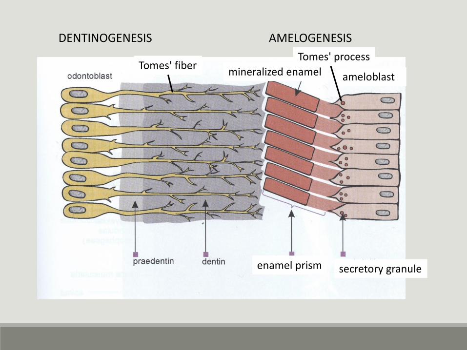
DENTINOGENESIS AMELOGENESIS
ameloblastmineralized enamel
secretory granuleenamel prism
Tomes' fiberTomes' process
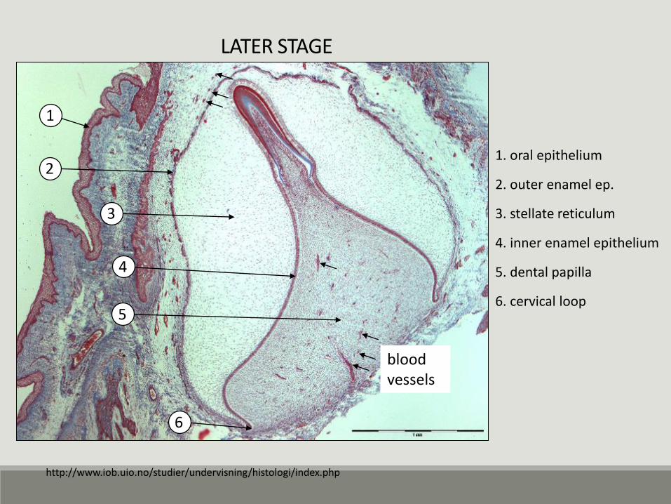
LATER STAGE
1. oral epithelium
2. outer enamel ep.
3. stellate reticulum
4. inner enamel epithelium
5. dental papilla
6. cervical loop
3
2
1
5
4
6
blood vessels
http://www.iob.uio.no/studier/undervisning/histologi/index.php
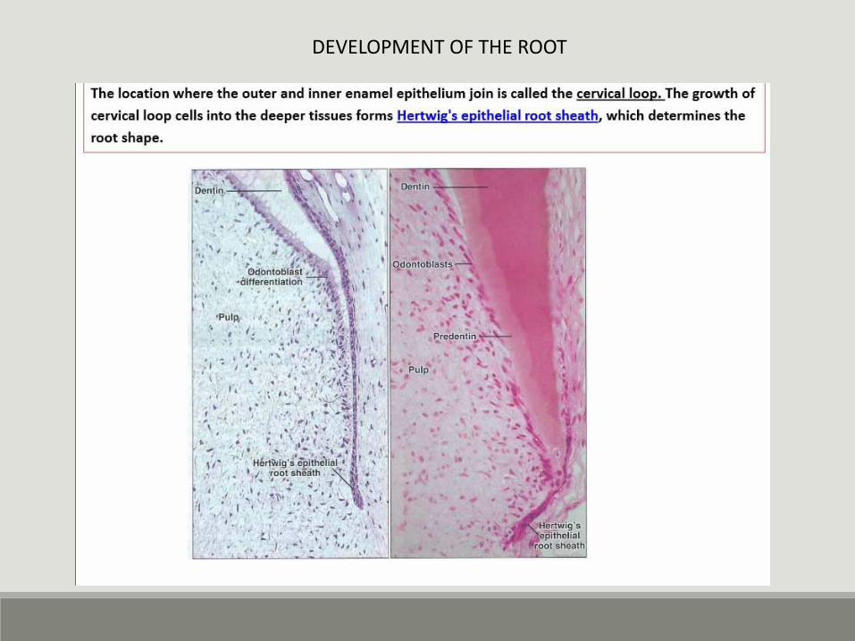
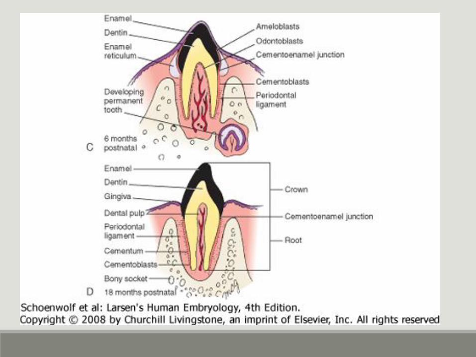
DEVELOPMENT OF THE ROOT
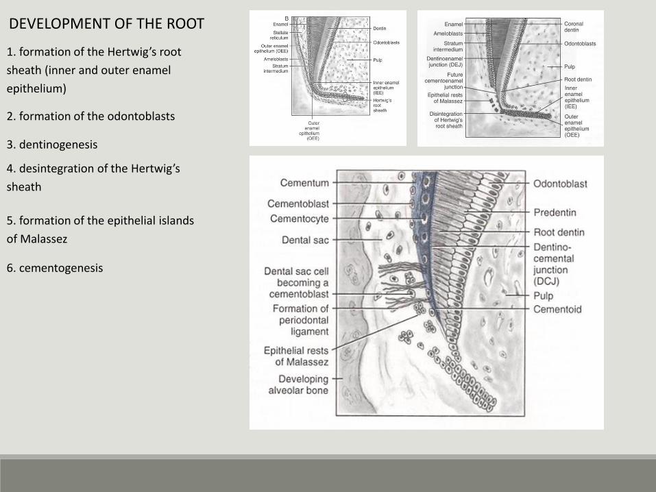
1. formation of the Hertwig’s root
sheath (inner and outer enamel
epithelium)
2. formation of the odontoblasts
DEVELOPMENT OF THE ROOT
3. dentinogenesis
4. desintegration of the Hertwig’s
sheath
5. formation of the epithelial islands
of Malassez
6. cementogenesis
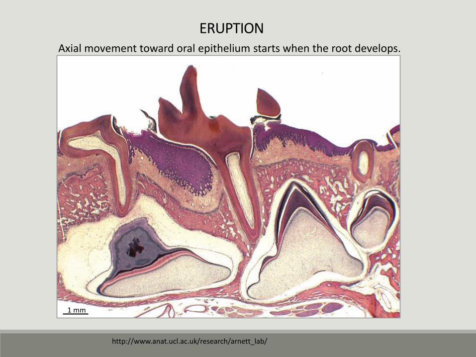
ERUPTION
http://www.anat.ucl.ac.uk/research/arnett_lab/
1 mm
Axial movement toward oral epithelium starts when the root develops.
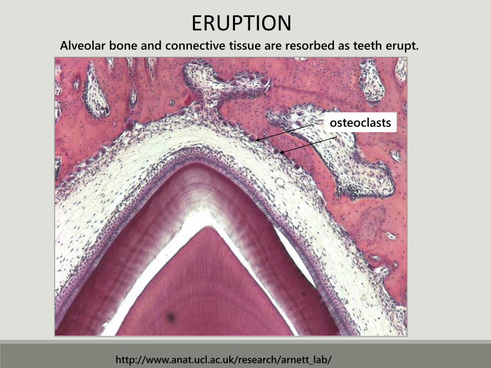
osteoclasts
Alveolar bone and connective tissue are resorbed as teeth erupt.
http://www.anat.ucl.ac.uk/research/arnett_lab/
ERUPTION