Embed Size (px)
Citation preview
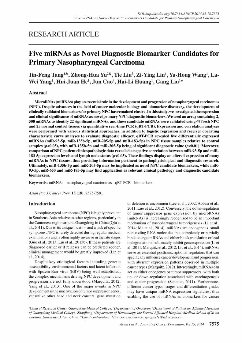
Asian Pacific Journal of Cancer Prevention, Vol 15, 2014 7575
DOI:http://dx.doi.org/10.7314/APJCP.2014.15.18.7575Five miRNAs as Novel Diagnostic Biomarkers Candidate for Primary Nasopharyngeal Carcinoma
Asian Pac J Cancer Prev, 15 (18), 7575-7581
Introduction
Nasopharyngeal carcinoma (NPC) is highly prevalent in Southeast Asia relative to other regions, particularly in the Cantonese region around Guangdong in China (Qiu et al., 2011). Due to its unique location and a lack of specific symptoms, NPC is rarely detected during regular medical examinations and is often highly invasive in the late stages (Guo et al., 2013; Liu et al., 2013b). If these patients are diagnosed earlier or if relapses can be predicted sooner, clinical management would be greatly improved (Lin et al., 2014).
Despite key etiological factors including genetic susceptibility, environmental factors and latent infection with Epstein-Barr virus (EBV) being well established, the complex mechanisms driving NPC development and progression are not fully understood (Marquitz, 2012; Yang et al., 2013). One of the major events in NPC development is the inactivation of tumor suppressor genes, yet unlike other head and neck cancers, gene mutation
1Clinical Research Center, Guangdong Medical College, 2Department of Oncology, 4Department of Pathology, Affiliated Hospital of Guangdong Medical College, Zhanjiang, 3Department of Hematology, the Second Affiliated Hospital, Medical School of Xi’an Jiaotong University, Xi’an, China &Equal contributors *For correspondence: [email protected]
Abstract
MicroRNAs (miRNAs) play an essential role in the development and progression of nasopharyngeal carcinomas (NPC). Despite advances in the field of cancer molecular biology and biomarker discovery, the development of clinically validated biomarkers for primary NPC has remained elusive. In this study, we investigated the expression and clinical significance of miRNAs as novel primary NPC diagnostic biomarkers. We used an array containing 2, 500 miRNAs to identify 22 significant miRNAs, and these candidate miRNAs were validated using 67 fresh NPC and 25 normal control tissues via quantitative real-time PCR (qRT-PCR). Expression and correlation analyses were performed with various statistical approaches, in addition to logistic regression and receiver operating characteristic curve analyses to evaluate diagnostic efficacy. qRT-PCR revealed five differentially expressed miRNAs (miR-93-5p, miR-135b-5p, miR-205-5p and miR-183-5p) in NPC tissue samples relative to control samples (p<0.05), with miR-135b-5p and miR-205-5p being of significant diagnostic value (p<0.01). Moreover, comparison of NPC patient clinicopathologic data revealed a negative correlation between miR-93-5p and miR-183-5p expression levels and lymph node status (p<0.05). These findings display an altered expression of many miRNAs in NPC tissues, thus providing information pertinent to pathophysiological and diagnostic research. Ultimately, miR-135b-5p and miR-205-5p may be implicated as novel NPC candidate biomarkers, while miR-93-5p, miR-650 and miR-183-5p may find application as relevant clinical pathology and diagnostic candidate biomarkers. Keywords: miRNAs - nasopharyngeal carcinomas - qRT-PCR - biomarkers
RESEARCH ARTICLE
Five miRNAs as Novel Diagnostic Biomarker Candidates for Primary Nasopharyngeal CarcinomaJin-Feng Tang1&, Zhong-Hua Yu2&, Tie Liu3, Zi-Ying Lin1, Ya-Hong Wang1, La-Wei Yang1, Hui-Juan He1, Jun Cao4, Hai-Li Huang1, Gang Liu1*
or deletion is uncommon (Lee et al., 2002; Abbasi et al., 2011; Luo et al., 2012). Conversely, the down-regulation of tumor suppressor gene expression by microRNAs (miRNAs) is increasingly recognized to be an important mechanism of nasopharyngeal tumorigenesis (Li et al., 2014; Ma et al., 2014). miRNAs are endogenous, small non-coding RNA molecules that completely or partially bind to target mRNAs and either block translation or lead to degradation to ultimately inhibit gene expression (Li et al., 2011; Marquitz et al., 2012; Liu et al., 2014). miRNAs serve as essential posttranscriptional regulators that can specifically influence cancer development and progression, with aberrant expression patterns observed in multiple cancer types (Marquitz, 2012). Interestingly, miRNAs can act as either oncogenes or tumor suppressors, with both up- or down-regulation associated with carcinogenesis and cancer progression (Schetter, 2011). Furthermore, different cancer types, stages and differentiation grades may have unique miRNA expression signatures, thus enabling the use of miRNAs as biomarkers for cancer
Jin-feng Tang et al
Asian Pacific Journal of Cancer Prevention, Vol 15, 20147576
detection and prognosis. In order to assess the diagnostic significance of
miRNAs in human NPC, we made miRNA profiling study about NPC tissues. We investigated the expression of miRNAs in NPC with microRNA Chip, and found that there were 22 miRNAs with significant changes. Further these miRNAs were validated by quantitative real-time PCR (qRT-PCR), and it was identified that there were five differentially expressed miRNAs in our study. Further analysis revealed that these five miRNAs might provide useful information pertaining to pathophysiological and diagnostic research.
Materials and Methods
Clinical specimensNasopharyngeal biopsy specimens were collected
in the Department of Oncology at Affiliated Hospital of Guangdong Mecical College (Zhanjiang, China) from June 2012 to October 2013. The clinicopathological characteristics of 92 patients (including 67 NPC and 25 non-cancer patients, NC) were obtained in accordance with the Institutional Ethical Review Board of the Affiliated Hospital of Guangdong Medical College in China, with consent obtained from all patients. All relevant demographic and clinical pathology details were obtained for the 92 samples of patients, with samples collected prior to any therapeutic procedures such as chemotherapy or radiotherapy, using WHO histological classification of the NPC and the TNM classification of malignant tumors (Shanmugaratnam, 1991), as shown as Table 1. Individuals with no evidence of cancer were selected as non-cancer control subjects and were matched to patients based on age and gender. Tissue samples were snap frozen in liquid nitrogen upon collection and sent directly to the laboratory for RNA extraction.
Immunohistochemical staining of tissueAll biopsies immediately were fixed in 4%
buffered paraformaldehyde, routinely processed, and embedded with paraffin. Serial 5μm histological sections were cut, mounted on glass slides coated with 3-aminopropyltriethoxysilane, and air-dried overnight at room temperature. The sections were then deparaffinised in xylene and rehydrated in ethanol. Haematoxylin and eosin (H and E) staining was performed in each specimen.
Total mRNA isolation and miRNA array analysisThree freshly frozen NPC biopsy specimens and three
normal nasopharyngeal epithelium specimens were sent to CapitalBio Corporation for noncoding RNA microarray analysis. The total RNA was extracted and purified using mirVana™ miRNA Isolation Kit (Ambion, Austin, TX) following the manufacturer’s instructions. The RNA integrity number (RIN) was assessed using an Agilent Bioanalyzer 2100 (Agilent Technologies, Santa Clara, CA), with samples with a RIN≥6.0 and 28S/18S≥0.7 used for the Agilent miRNA Chip. miRNA microarray profiles were obtained using the Agilent miRNA Microarray 19.0 containing 2047 miRNAs were comprehensive analysis of microRNA expression patterns in nasopharyngeal
carcinoma and non-tumorous (3 NPC and 3 NC). To identify differentially expresses miRNAs, the SAS system of microarray analysis was performed using a fold change value (FCV) of ≥2 or ≤0.5 and a signal value of ≥5 to determine significance. For data analysis, inter-sample variance was normalized using quantile normalization strategies. Hierarchical clustering by Euclidean distance was used to cluster samples and groups with similar miRNA profiles. Differential analysis was performed using an unpaired t-test, ANOVA, and fold-change analysis. Differentially expressed miRNAs were selected based on t-test (p≥ 0.001).
RNA isolation, reverse transcription, and quantitative real-time PCR analysis
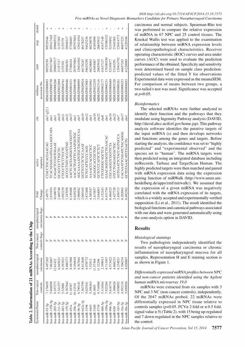
The 92 tissue samples (67 NPC and 25 non-cancer patients) were evaluated via qRT-PCR. Total RNA was extracted using TRIzol reagent (Invitrogen, Carlsbad, CA) and reverse transcribed using miRcute miRNA First-Strand cDNA Synthesis Kit (Tiangen Biotech, China). miRNA expression was quantified via qRT-PCR using designed primers by Tiangen Biotech and the information of 22 miRNA is shown in Table 2. 22 miRNAs were examed via qRT-PCR with a miRcute miRNA qRT-PCR Detection Kit (Tiangen Biotech, China) and a Rotor-Gene Q2 plex HRM (Qiagen, Germany). All samples were normalized to endogenous controls and fold changes calculated relative to control. The control miRNA U6 was used for normalization, and relative expression levels were calculated using the 2-ΔΔCTmethod (Liu et al., 2013a).
Statistical analysisThe SPSS17.0 program was used for general statistical
analysis. The Wilcoxon test was used to examine the differences of miRNA expression among nasopharyngeal
0
25.0
50.0
75.0
100.0
New
ly d
iagn
osed
with
out
trea
tmen
t
New
ly d
iagn
osed
with
tre
atm
ent
Pers
iste
nce
or r
ecur
renc
e
Rem
issi
on
Non
e
Chem
othe
rapy
Radi
othe
rapy
Conc
urre
nt c
hem
orad
iatio
n
10.3
0
12.8
30.025.0
20.310.16.3
51.7
75.051.1
30.031.354.2
46.856.3
27.625.033.130.031.3
23.738.0
31.3
Table 1. Clinical Characteristics of Nasopharyngeal Carcinoma Patients According to Training Set and Validation SetVariable Samples p-value* NPC (n=67) Control (n=25)
Sex Male 56 16 Female 11 9 Average Ages (years) 51.87±2.257 31.67±2.427 0.0001Histological types Squamous carcinoma T stages T1 12.50% T2 28.10% T3 18.8 T4 40.60% N stages N0 19.40% N1 30.60% N2 33.30% N3 16.70% TNM stages Ⅰ 3.30% Ⅱ 12.90% Ⅲ 29.00% Ⅳ 54.80% Family history yes 16.10% no 83.90% Smoking Smoking 19.40% No smoking 80.60% Region Zhanjiang 26.80% Other region 70.20%
* (NPC vs Control)
Asian Pacific Journal of Cancer Prevention, Vol 15, 2014 7577
DOI:http://dx.doi.org/10.7314/APJCP.2014.15.18.7575Five miRNAs as Novel Diagnostic Biomarkers Candidate for Primary Nasopharyngeal Carcinoma
carcinoma and normal subjects. Spearman-Rho test was performed to compare the relative expression of miRNA in 67 NPC and 25 control tissues. The Kruskal Wallis test was applied to the examination of relationship between miRNA expression levels and clinicopathological characteristics. Receiver operating characteristic (ROC) curves and area under curves (AUC) were used to evaluate the prediction performance of the obtained. Specificity and sensitivity were determined based on sample class prediction: predicted values of the fitted Y for observations Experimental data were expressed as the mean±SEM. For comparison of means between two groups, a two-tailed t-test was used. Significance was accepted at p<0.05.
BioinformaticsThe selected miRNAs were further analyzed to
identify their function and the pathways that they modulate using Ingenuity Pathway analysis (DAVID, http://david.abcc.ncifcrf.gov/home.jsp). This pathway analysis software identifies the putative targets of the input miRNA (s) and then develops networks and functions among the genes and targets. Before starting the analysis, the confidence was set to “highly predicted” and “experimental observed” and the species set to “human”. The miRNA targets were then predicted using an integrated database including miRecords, Tarbase and TargetScan Human. The highly predicted targets were then matched and paired with mRNA expression data using the expression pairing function of miRWalk (http://www.umm.uni-heidelberg.de/apps/zmf/mirwalk/). We assumed that the expression of a given miRNA was negatively correlated with the mRNA expression of its targets, which is a widely accepted and experimentally verified supposition (Li et al., 2011). The result identified the biological functions and canonical pathways associated with our data and were generated automatically using the core-analysis option in DAVID.
Results
Histological stainingsTwo pathologists independently identified the
results of nasopharyngeal carcinoma or chronic inflammation of nasopharyngeal mucosa for all samples. Representation H and E staining section is as shown in Figure 1.
Differentially expressed miRNA profiles between NPC and non-cancer patients identified using the Agilent human miRNA microarray 19.0
miRNAs were extracted from six samples with 3 NPC and 3 NC (non-cancer controls), independently. Of the 2047 miRNAs probed, 22 miRNAs were differentially expressed in NPC tissue relative to controls samples (p<0.05, FCV≥ 2 fold or ≤ 0.5 fold, signal value ≥ 5) (Table 2), with 15 being up-regulated and 7 down-regulated in the NPC samples relative to the control.
0
25.0
50.0
75.0
100.
0
Newly diagnosed without treatment
Newly diagnosed with treatment
Persistence or recurrence
Remission
None
Chemotherapy
Radiotherapy
Concurrent chemoradiation
10.3
0
12.8
30.0
25.0
20.3
10.1
6.3
51.7
75.0
51.1
30.0
31.3
54.2
46.8
56.3
27.6
25.0
33.1
30.0
31.3
23.7
38.0
31.3
0
25.0
50.0
75.0
100.
0
Newly diagnosed without treatment
Newly diagnosed with treatment
Persistence or recurrence
Remission
None
Chemotherapy
Radiotherapy
Concurrent chemoradiation
10.3
0
12.8
30.0
25.0
20.3
10.1
6.3
51.7
75.0
51.1
30.0
31.3
54.2
46.8
56.3
27.6
25.0
33.1
30.0
31.3
23.7
38.0
31.3
Tabl
e 2.
Info
rmat
ion
of 2
1 m
iRN
As A
ccor
ding
to th
e C
hip
Syst
emat
icN
ame
Non
-can
cer
naso
phar
ynge
al
Non
-can
cer
naso
phar
ynge
al
Fold
ac
tive
ch
r m
irbas
e
star
t st
op
stra
nd
cont
rols
ca
rcin
oma
cont
rols
flag
s ca
rcin
oma
flags
C
hang
e se
quen
ce
ac
cess
ion
No
hsa-
miR
-93-
5p
7.17
6659
8.
4972
65
P P
2.49
771
CTA
CC
TGC
AC
GA
AC
AG
ch
r7 q
22.1
M
IMAT
0000
093
9952
9359
99
5293
44
+hs
a-m
iR-1
35b-
5p
5.66
6698
7.
0626
67
P P
2.63
1654
TC
AC
ATA
GG
AAT
GA
AA
AG
CC
ATA
ch
r1
MIM
AT00
0075
8 20
5417
467
2054
1744
5 +
hsa-
miR
-205
-5p
9.19
7273
12
.614
22
P P
10.6
8079
C
AG
AC
TCC
GG
TGG
AAT
ch
r1
MIM
AT00
0026
6 20
9605
517
2096
0553
2 -
hsa-
miR
-211
-3p
5.34
1257
6.
6456
21
P P
2.46
9748
G
CA
CC
CC
TTTG
CTG
ch
r15
MIM
AT00
2269
4 31
3573
17
3135
7305
+
hsa-
miR
-483
-5p
5.52
888
6.59
3921
P
P 2.
0922
29
CTC
CC
TTC
TTTC
CTC
ch
r11
MIM
AT00
0476
1 21
5539
2 21
5537
9 +
hsa-
miR
-155
-5p
8.36
7844
8.
6863
73
P P
1.24
7058
A
CC
CC
TATC
AC
GAT
TAG
ch
r21
MIM
AT00
0064
6 26
9463
01
2694
6317
-
hsa-
miR
-451
a 11
.183
87
8.44
1968
P
P 0.
1494
88
AA
CTC
AG
TAAT
GG
TAA
CG
GTT
T ch
r17
MIM
AT00
0163
1 27
1884
24
2718
8403
+
hsa-
miR
-29b
-3p
10.1
2516
9.
4568
18
P P
0.62
9228
A
AC
AC
TGAT
TTC
AA
ATG
GTG
C
chr1
M
IMAT
0000
100
2079
7586
1 20
7975
841
+hs
a-m
iR-9
6-5p
6.
7901
41
8.36
7844
P
P 2.
9849
42
AG
CA
AA
AAT
GTG
CTA
GTG
CC
AA
ch
r7
MIM
AT00
0009
5 12
9414
562
1294
1454
1 +
hsa-
miR
-642
a-3p
7.
5637
99
8.61
2848
P
P 2.
0691
65
GG
TTC
CC
TCTC
CA
AAT
ch
r19
MIM
AT00
2092
4 46
1782
43
4617
8257
-
hsa-
miR
-319
8 5.
0255
41
6.97
8772
P
P 3.
8724
08
TCTC
CAT
TCC
CC
AG
G
chr1
2 M
IMAT
0015
083
5462
5250
54
6252
36
+hs
a-m
iR-4
443
5.39
6363
7.
5291
2 P
P 4.
3855
47
AA
AA
CC
CA
CG
CC
TCC
ch
r3
MIM
AT00
1896
1 48
2380
65
4823
8078
-
hsa-
miR
-510
0 10
.300
5 11
.573
04
P P
2.41
5857
A
GA
GG
CA
CC
GC
TGG
ch
r10
MIM
AT00
2225
9 43
4930
87
4349
3099
-
hsa-
miR
-573
9 9.
7169
88
11.3
2296
P
P 3.
0440
03
GC
TCC
CC
ATTC
TCTC
T ch
r22
MIM
AT00
2311
6 28
8559
18
2885
5932
-
hsa-
miR
-486
-5p
5.11
4564
2.
3990
13
P P
0.15
2243
C
TCG
GG
GC
AG
CTC
A
chr8
M
IMAT
0002
177
4163
7127
41
6371
40
-hs
a-m
iR-1
99a-
5p
7.92
5284
6.
7135
67
P P
0.43
1754
G
AA
CA
GG
TAG
TCTG
AA
CA
C
chr1
M
IMAT
0000
231
1703
8035
0 17
0380
332
+hs
a-m
iR-9
9b-5
p 5.
6187
79
4.57
3422
P
P 0.
4845
25
CG
CA
AG
GTC
GG
TTC
TA
chr1
9 M
IMAT
0000
689
5219
5877
52
1958
92
-hs
a-m
iR-6
50
3.38
8626
4.
6682
33
P P
2.42
7728
G
TCC
TGA
GA
GC
GC
TGC
ch
r22
MIM
AT00
0332
0 23
1652
91
2316
5305
-
hsa-
miR
-183
-5p
4.93
8092
6.
2348
21
P P
2.45
6713
A
GTG
AAT
TCTA
CC
AG
TGC
CA
ch
r7
MIM
AT00
0026
1 12
9414
792
1294
1477
3 +
hsa-
miR
-10a
-5p
4.59
2134
2.
7662
34
P P
0.28
2065
C
AC
AA
ATTC
GG
ATC
TAC
AG
GG
ch
r17
MIM
AT00
0025
3 46
6572
43
4665
7223
+
hsa-
miR
-30e
-3p
5.05
0956
4.
0288
07
P P
0.49
2382
G
CTG
TAA
AC
ATC
CG
AC
TG
chr1
M
IMAT
0000
693
4122
0089
41
2201
06
-
Jin-feng Tang et al
Asian Pacific Journal of Cancer Prevention, Vol 15, 20147578
Validation of differentially expressed miRNAs using qRT-PCR
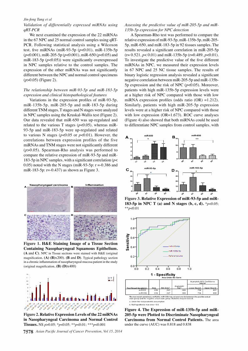
We next examined the expression of the 22 miRNAs in the 67 NPC and 25 normal control samples using qRT-PCR. Following statistical analysis using a Wilcoxon test, five miRNAs (miR-93-5p (p<0.01), miR-135b-5p (p<0.001), miR-205-5p (p<0.001), miR-650 (p<0.05) and miR-183-5p (p<0.05)) were significantly overexpressed in NPC samples relative to the control samples. The expression of the other miRNAs was not significantly different between the NPC and normal control specimens (p>0.05) (Figure 2).
The relationship between miR-93-5p and miR-183-5p expression and clinical histopathological features
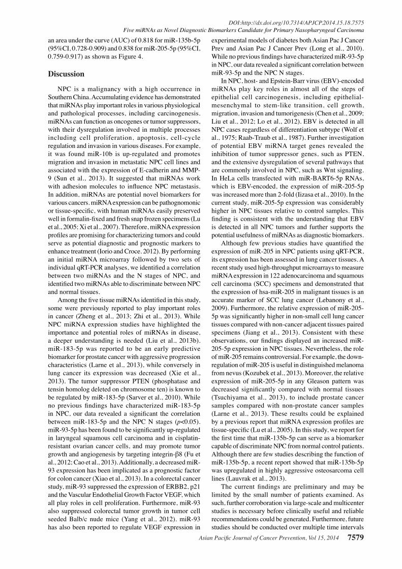
Variations in the expression profiles of miR-93-5p, miR-135b-5p, miR-205-5p and miR-183-5p during different TNM stages, T stages and N stages were analyzed in NPC samples using the Kruskal-Wallis test (Figure 2). Our data revealed that miR-650 was up-regulated and related to the various T stages (p<0.05), whereas miR-93-5p and miR-183-5p were up-regulated and related to various N stages (p<0.05 or p<0.01). However, the correlations between expression profiles of the five miRNAs and TNM stages were not significantly different (p>0.05). Spearman-Rho analysis was performed to compare the relative expression of miR-93-5p and miR-183-5p in NPC samples, with a significant correlation (p< 0.05) noted with the N stages (miR-93-5p: r =-0.386 and miR-183-5p: r=-0.437) as shown as Figure 3.
Assessing the predictive value of miR-205-5p and miR-135b-5p expression for NPC detection
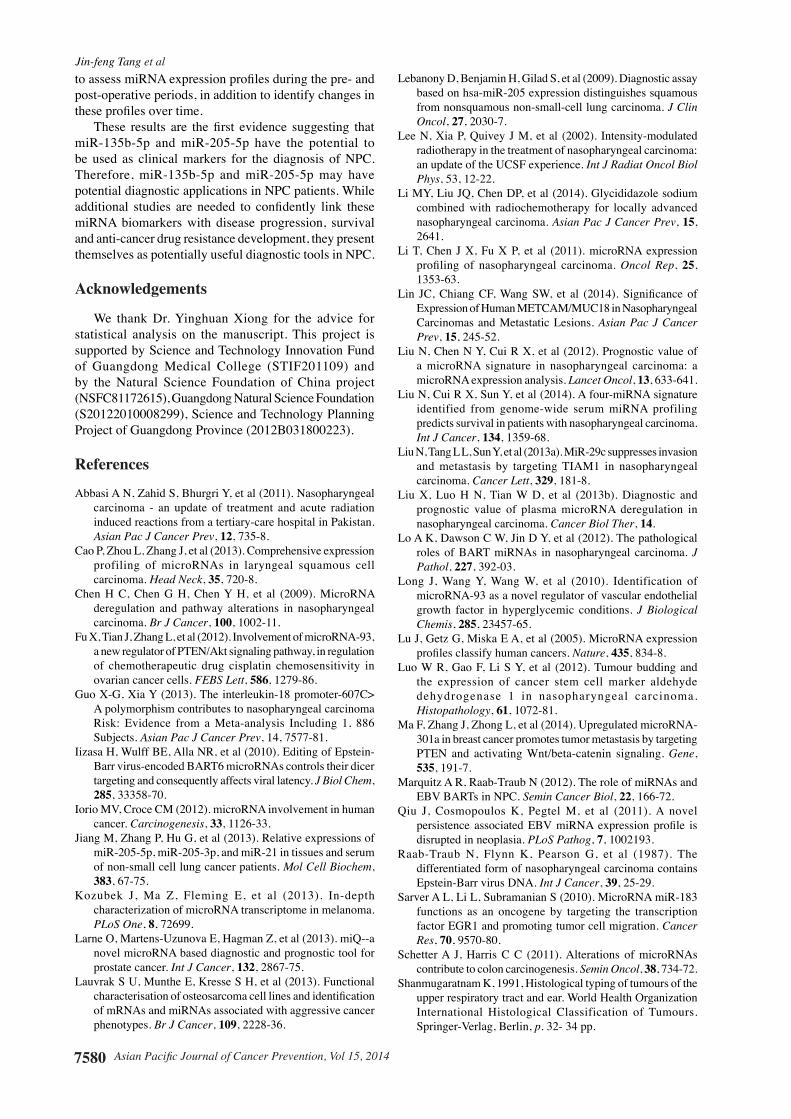
A Spearman-Rho test was performed to compare the relative expression of miR-93-5p, miR-135b-5p, miR-205-5p, miR-650, and miR-183-5p in 92 tissues samples. The results revealed a significant correlation in miR-205-5p (r= 0.521, p< 0.01) and miR-135b-5p (r=0.489, p<0.01). To investigate the predictive value of the five different miRNAs in NPC, we measured their expression levels in 67 NPC and 25 NC tissue samples. The results of binary logistic regression analysis revealed a significant negative correlation between miR-205-5p and miR-135b-5p expression and the risk of NPC (p<0.05). Moreover, patients with high miR-135b-5p expression levels were at a higher risk of NPC compared with those with low miRNA expression profiles (odds ratio (OR) =1.212). Similarly, patients with high miR-205-5p expression levels were at a higher risk of NPC compared with those with low expression (OR=1.673). ROC curve analyses (Figure 4) also showed that both miRNAs could be used to differentiate NPC samples from control samples, with
Figure 1. H&E Staining Image of a Tissue Section Containing Nasopharyngeal Squamous Epithelium. (A and C). NPC in Tissue sections were stained with H&E (original magnification, (A) (B)x200). (B and D). Typical pathology section in a chronic inflammation of nasopharyngeal mucosa patient in the study (original magnification, (B) (D)x400)
Figure 2. Relative Expression Levels of the 22 miRNAs in Nasopharyngeal Carcinoma and Normal Control Tissues. NS p>0.05; *p<0.05; **p<0.01; ***p<0.001
Figure 3. Relative Expression of miR-93-5p and miR-183-5p in NPC T (a) and N stages (b, c, d). *p<0.05; **p<0.01
Figure 4. The Expression of miR-135b-5p and miR-205-5p were Plotted to Discriminate Nasopharyngeal Carcinoma from Normal Control Patients. The area under the curve (AUC) was 0.818 and 0.838
Asian Pacific Journal of Cancer Prevention, Vol 15, 2014 7579
DOI:http://dx.doi.org/10.7314/APJCP.2014.15.18.7575Five miRNAs as Novel Diagnostic Biomarkers Candidate for Primary Nasopharyngeal Carcinoma
an area under the curve (AUC) of 0.818 for miR-135b-5p (95%CI, 0.728-0.909) and 0.838 for miR-205-5p (95%CI, 0.759-0.917) as shown as Figure 4.
Discussion
NPC is a malignancy with a high occurrence in Southern China. Accumulating evidence has demonstrated that miRNAs play important roles in various physiological and pathological processes, including carcinogenesis. miRNAs can function as oncogenes or tumor suppressors, with their dysregulation involved in multiple processes including cell proliferation, apoptosis, cell-cycle regulation and invasion in various diseases. For example, it was found miR-10b is up-regulated and promotes migration and invasion in metastatic NPC cell lines and associated with the expression of E-cadherin and MMP-9 (Sun et al., 2013). It suggested that miRNAs work with adhesion molecules to influence NPC metastasis. In addition, miRNAs are potential novel biomarkers for various cancers. miRNA expression can be pathognomonic or tissue-specific, with human miRNAs easily preserved well in formalin-fixed and fresh snap frozen specimens (Lu et al., 2005; Xi et al., 2007). Therefore, miRNA expression profiles are promising for characterizing tumors and could serve as potential diagnostic and prognostic markers to enhance treatment (Iorio and Croce. 2012). By performing an initial miRNA microarray followed by two sets of individual qRT-PCR analyses, we identified a correlation between two miRNAs and the N stages of NPC, and identified two miRNAs able to discriminate between NPC and normal tissues.
Among the five tissue miRNAs identified in this study, some were previously reported to play important roles in cancer (Zheng et al., 2013; Zhi et al., 2013). While NPC miRNA expression studies have highlighted the importance and potential roles of miRNAs in disease, a deeper understanding is needed (Liu et al., 2013b). miR-183-5p was reported to be an early predictive biomarker for prostate cancer with aggressive progression characteristics (Larne et al., 2013), while conversely in lung cancer its expression was decreased (Xie et al., 2013). The tumor suppressor PTEN (phosphatase and tensin homolog deleted on chromosome ten) is known to be regulated by miR-183-5p (Sarver et al., 2010). While no previous findings have characterized miR-183-5p in NPC, our data revealed a significant the correlation between miR-183-5p and the NPC N stages (p<0.05). miR-93-5p has been found to be significantly up-regulated in laryngeal squamous cell carcinoma and in cisplatin-resistant ovarian cancer cells, and may promote tumor growth and angiogenesis by targeting integrin-β8 (Fu et al., 2012; Cao et al., 2013). Additionally, a decreased miR-93 expression has been implicated as a prognostic factor for colon cancer (Xiao et al., 2013). In a colorectal cancer study, miR-93 suppressed the expression of ERBB2, p21 and the Vascular Endothelial Growth Factor VEGF, which all play roles in cell proliferation. Furthermore, miR-93 also suppressed colorectal tumor growth in tumor cell seeded Balb/c nude mice (Yang et al., 2012). miR-93 has also been reported to regulate VEGF expression in
experimental models of diabetes both Asian Pac J Cancer Prev and Asian Pac J Cancer Prev (Long et al., 2010). While no previous findings have characterized miR-93-5p in NPC, our data revealed a significant correlation between miR-93-5p and the NPC N stages.
In NPC, host- and Epstein-Barr virus (EBV)-encoded miRNAs play key roles in almost all of the steps of epithelial cell carcinogenesis, including epithelial-mesenchymal to stem-like transition, cell growth, migration, invasion and tumorigenesis (Chen et al., 2009; Liu et al., 2012; Lo et al., 2012). EBV is detected in all NPC cases regardless of differentiation subtype (Wolf et al., 1975; Raab-Traub et al., 1987). Further investigation of potential EBV miRNA target genes revealed the inhibition of tumor suppressor genes, such as PTEN, and the extensive dysregulation of several pathways that are commonly involved in NPC, such as Wnt signaling. In HeLa cells transfected with miR-BART6-5p RNAs, which is EBV-encoded, the expression of miR-205-5p was increased more than 2-fold (Iizasa et al., 2010). In the current study, miR-205-5p expression was considerably higher in NPC tissues relative to control samples. This finding is consistent with the understanding that EBV is detected in all NPC tumors and further supports the potential usefulness of miRNAs as diagnostic biomarkers.
Although few previous studies have quantified the expression of miR-205 in NPC patients using qRT-PCR, its expression has been assessed in lung cancer tissues. A recent study used high-throughput microarrays to measure miRNA expression in 122 adenocarcinoma and squamous cell carcinoma (SCC) specimens and demonstrated that the expression of hsa-miR-205 in malignant tissues is an accurate marker of SCC lung cancer (Lebanony et al., 2009). Furthermore, the relative expression of miR-205-5p was significantly higher in non-small cell lung cancer tissues compared with non-cancer adjacent tissues paired specimens (Jiang et al., 2013). Consistent with these observations, our findings displayed an increased miR-205-5p expression in NPC tissues. Nevertheless, the role of miR-205 remains controversial. For example, the down-regulation of miR-205 is useful in distinguished melanoma from nevus (Kozubek et al., 2013). Moreover, the relative expression of miR-205-5p in any Gleason pattern was decreased significantly compared with normal tissues (Tsuchiyama et al., 2013), to include prostate cancer samples compared with non-prostate cancer samples (Larne et al., 2013). These results could be explained by a previous report that miRNA expression profiles are tissue-specific (Lu et al., 2005). In this study, we report for the first time that miR-135b-5p can serve as a biomarker capable of discriminate NPC from normal control patients. Although there are few studies describing the function of miR-135b-5p, a recent report showed that miR-135b-5p was upregulated in highly aggressive osteosarcoma cell lines (Lauvrak et al., 2013).
The current findings are preliminary and may be limited by the small number of patients examined. As such, further corroboration via large-scale and multicenter studies is necessary before clinically useful and reliable recommendations could be generated. Furthermore, future studies should be conducted over multiple time intervals
Jin-feng Tang et al
Asian Pacific Journal of Cancer Prevention, Vol 15, 20147580
to assess miRNA expression profiles during the pre- and post-operative periods, in addition to identify changes in these profiles over time.
These results are the first evidence suggesting that miR-135b-5p and miR-205-5p have the potential to be used as clinical markers for the diagnosis of NPC. Therefore, miR-135b-5p and miR-205-5p may have potential diagnostic applications in NPC patients. While additional studies are needed to confidently link these miRNA biomarkers with disease progression, survival and anti-cancer drug resistance development, they present themselves as potentially useful diagnostic tools in NPC.
Acknowledgements
We thank Dr. Yinghuan Xiong for the advice for statistical analysis on the manuscript. This project is supported by Science and Technology Innovation Fund of Guangdong Medical College (STIF201109) and by the Natural Science Foundation of China project (NSFC81172615), Guangdong Natural Science Foundation (S20122010008299), Science and Technology Planning Project of Guangdong Province (2012B031800223).
References
Abbasi A N, Zahid S, Bhurgri Y, et al (2011). Nasopharyngeal carcinoma - an update of treatment and acute radiation induced reactions from a tertiary-care hospital in Pakistan. Asian Pac J Cancer Prev, 12, 735-8.
Cao P, Zhou L, Zhang J, et al (2013). Comprehensive expression profiling of microRNAs in laryngeal squamous cell carcinoma. Head Neck, 35, 720-8.
Chen H C, Chen G H, Chen Y H, et al (2009). MicroRNA deregulation and pathway alterations in nasopharyngeal carcinoma. Br J Cancer, 100, 1002-11.
Fu X, Tian J, Zhang L, et al (2012). Involvement of microRNA-93, a new regulator of PTEN/Akt signaling pathway, in regulation of chemotherapeutic drug cisplatin chemosensitivity in ovarian cancer cells. FEBS Lett, 586, 1279-86.
Guo X-G, Xia Y (2013). The interleukin-18 promoter-607C> A polymorphism contributes to nasopharyngeal carcinoma Risk: Evidence from a Meta-analysis Including 1, 886 Subjects. Asian Pac J Cancer Prev, 14, 7577-81.
Iizasa H, Wulff BE, Alla NR, et al (2010). Editing of Epstein-Barr virus-encoded BART6 microRNAs controls their dicer targeting and consequently affects viral latency. J Biol Chem, 285, 33358-70.
Iorio MV, Croce CM (2012). microRNA involvement in human cancer. Carcinogenesis, 33, 1126-33.
Jiang M, Zhang P, Hu G, et al (2013). Relative expressions of miR-205-5p, miR-205-3p, and miR-21 in tissues and serum of non-small cell lung cancer patients. Mol Cell Biochem, 383, 67-75.
Kozubek J, Ma Z, Fleming E, et al (2013). In-depth characterization of microRNA transcriptome in melanoma. PLoS One, 8, 72699.
Larne O, Martens-Uzunova E, Hagman Z, et al (2013). miQ--a novel microRNA based diagnostic and prognostic tool for prostate cancer. Int J Cancer, 132, 2867-75.
Lauvrak S U, Munthe E, Kresse S H, et al (2013). Functional characterisation of osteosarcoma cell lines and identification of mRNAs and miRNAs associated with aggressive cancer phenotypes. Br J Cancer, 109, 2228-36.
Lebanony D, Benjamin H, Gilad S, et al (2009). Diagnostic assay based on hsa-miR-205 expression distinguishes squamous from nonsquamous non-small-cell lung carcinoma. J Clin Oncol, 27, 2030-7.
Lee N, Xia P, Quivey J M, et al (2002). Intensity-modulated radiotherapy in the treatment of nasopharyngeal carcinoma: an update of the UCSF experience. Int J Radiat Oncol Biol Phys, 53, 12-22.
Li MY, Liu JQ, Chen DP, et al (2014). Glycididazole sodium combined with radiochemotherapy for locally advanced nasopharyngeal carcinoma. Asian Pac J Cancer Prev, 15, 2641.
Li T, Chen J X, Fu X P, et al (2011). microRNA expression profiling of nasopharyngeal carcinoma. Oncol Rep, 25, 1353-63.
Lin JC, Chiang CF, Wang SW, et al (2014). Significance of Expression of Human METCAM/MUC18 in Nasopharyngeal Carcinomas and Metastatic Lesions. Asian Pac J Cancer Prev, 15, 245-52.
Liu N, Chen N Y, Cui R X, et al (2012). Prognostic value of a microRNA signature in nasopharyngeal carcinoma: a microRNA expression analysis. Lancet Oncol, 13, 633-641.
Liu N, Cui R X, Sun Y, et al (2014). A four-miRNA signature identified from genome-wide serum miRNA profiling predicts survival in patients with nasopharyngeal carcinoma. Int J Cancer, 134, 1359-68.
Liu N, Tang L L, Sun Y, et al (2013a). MiR-29c suppresses invasion and metastasis by targeting TIAM1 in nasopharyngeal carcinoma. Cancer Lett, 329, 181-8.
Liu X, Luo H N, Tian W D, et al (2013b). Diagnostic and prognostic value of plasma microRNA deregulation in nasopharyngeal carcinoma. Cancer Biol Ther, 14.
Lo A K, Dawson C W, Jin D Y, et al (2012). The pathological roles of BART miRNAs in nasopharyngeal carcinoma. J Pathol, 227, 392-03.
Long J, Wang Y, Wang W, et al (2010). Identification of microRNA-93 as a novel regulator of vascular endothelial growth factor in hyperglycemic conditions. J Biological Chemis, 285, 23457-65.
Lu J, Getz G, Miska E A, et al (2005). MicroRNA expression profiles classify human cancers. Nature, 435, 834-8.
Luo W R, Gao F, Li S Y, et al (2012). Tumour budding and the expression of cancer stem cell marker aldehyde dehydrogenase 1 in nasopharyngeal carcinoma. Histopathology, 61, 1072-81.
Ma F, Zhang J, Zhong L, et al (2014). Upregulated microRNA-301a in breast cancer promotes tumor metastasis by targeting PTEN and activating Wnt/beta-catenin signaling. Gene, 535, 191-7.
Marquitz A R, Raab-Traub N (2012). The role of miRNAs and EBV BARTs in NPC. Semin Cancer Biol, 22, 166-72.
Qiu J, Cosmopoulos K, Pegtel M, et al (2011). A novel persistence associated EBV miRNA expression profile is disrupted in neoplasia. PLoS Pathog, 7, 1002193.
Raab-Traub N, Flynn K, Pearson G, et al (1987). The differentiated form of nasopharyngeal carcinoma contains Epstein-Barr virus DNA. Int J Cancer, 39, 25-29.
Sarver A L, Li L, Subramanian S (2010). MicroRNA miR-183 functions as an oncogene by targeting the transcription factor EGR1 and promoting tumor cell migration. Cancer Res, 70, 9570-80.
Schetter A J, Harris C C (2011). Alterations of microRNAs contribute to colon carcinogenesis. Semin Oncol, 38, 734-72.
Shanmugaratnam K, 1991, Histological typing of tumours of the upper respiratory tract and ear. World Health Organization International Histological Classification of Tumours. Springer-Verlag, Berlin, p. 32- 34 pp.
Asian Pacific Journal of Cancer Prevention, Vol 15, 2014 7581
DOI:http://dx.doi.org/10.7314/APJCP.2014.15.18.7575Five miRNAs as Novel Diagnostic Biomarkers Candidate for Primary Nasopharyngeal Carcinoma
Sun XJ, Liu H, Zhang P, et al (2013). miR-10b promotes migration and invasion in nasopharyngeal carcinoma cells. Asian Pac J Cancer Prev, 14, 5533-37.
Tsuchiyama K, Ito H, Taga M, et al (2013). Expression of microRNAs associated with Gleason grading system in prostate cancer: miR-182-5p is a useful marker for high grade prostate cancer. Prostate, 73, 827-34.
Wolf H, Zur Hausen H, Klein G, et al (1975). Attempts to detect virus-specific DNA sequences in human tumors. III. Epstein-Barr viral DNA in non-lymphoid nasopharyngeal carcinoma cells. Med Microbiol Immunol, 161, 15-21.
Xi Y, Nakajima G, Gavin E, et al (2007). Systematic analysis of microRNA expression of RNA extracted from fresh frozen and formalin-fixed paraffin-embedded samples. RNA, 13, 1668-74.
Xiao Z G, Deng Z S, Zhang Y D, et al (2013). Clinical significance of microRNA-93 downregulation in human colon cancer. Eur J Gastroenterol Hepatol, 25, 296-01.
Xie L, Yang Z, Li G, et al (2013). Genome-wide identification of bone metastasis-related microRNAs in lung adenocarcinoma by high-throughput sequencing. PLoS One, 8, 61212.
Yang H J, Huang T J, Yang C F, et al (2013). Comprehensive profiling of Epstein-Barr virus-encoded miRNA species associated with specific latency types in tumor cells. Virol J, 10, 314.
Yang I P, Tsai H L, Hou M F, et al (2012). MicroRNA-93 inhibits tumor growth and early relapse of human colorectal cancer by affecting genes involved in the cell cycle. Carcinogenesis, 33, 1522-30.
Zheng G, Wang H, Zhang X, et al (2013). Identification and validation of reference genes for qPCR detection of serum microRNAs in colorectal adenocarcinoma patients. PLoS One, 8, 83025.
Zhi F, Cao X, Xie X, et al (2013). Identification of circulating MicroRNAs as potential biomarkers for detecting acute myeloid leukemia. PloS one, 8, 56718.


![RESEARCH ARTICLE Open Access MicroRNAs in bovine ... · of adipogenic genes involved in adipocyte differentiation [12,13]. However, the regulatory mechanisms of micro-RNAs (miRNAs)](https://img.pdfslide.us/doc/110x75/5fd8e09740b3bb1519063f71/research-article-open-access-micrornas-in-bovine-of-adipogenic-genes-involved.jpg)