Embed Size (px)
Citation preview
Asian Pacific Journal of Cancer Prevention, Vol 15, 2014 9835
DOI:http://dx.doi.org/10.7314/APJCP.2014.15.22.9835Interference of Fisetin with Nuclear Factor-κB Signal Transduction Activated by EBV Latent Membrane
Asian Pac J Cancer Prev, 15 (22), 9835-9839
Introduction
The flavonoid fisetin (3, 3, 4, 7-tetrahydroxyflavone) is the main component of lacquer and is found in strawberries, apples, and grapes. The lacquer, lac tree extract, has been used as Chinese medicine in Asia for thousands of years. Several recent studies have demonstrated that the flavonoid has anticancer effects against several cancer types. Our preliminary data have indicated that fisetin induces cell cycle arrest and causes apoptosis in hepatocellular carcinoma cells (Li et al., 2010; 2011). However, the effect of fisetin on nasopharyngeal carcinoma (NPC) has not been studied extensively.
Epstein-Barr virus (EBV)-encoded latent membrane protein 1 (LMP1) has been known to have oncogenic properties during latent infection in nasopharyngeal carcinoma (NPC) . In human epithelial cells, LMP1 alters many functional properties that are involved in tumor progression and invasions (Kim et al., 2000; Adham et al., 2012; Bambang et al., 2010). Activation of different signal transduction pathways mediates various downstream pathological effects of LMP1 expression, including cell proliferation, anti-apoptosis and metastasis (Eliopoulos et al, 2001; Guo et al., 2012).
NF-κB participates in the induction of cellular genes encoding apoptosis molecules (Xu et al., 2012; Yang et al., 2012). In NPC cells, NF-κB plays a critical role in LMP1
1Department of Pathology and Pathophysiology, 2Department of Physiology, School of Basic Medicine Science, 3Laboratory of Physiological Science, Guangdong Medical College, Zhanjiang, PR China &Equal contributors *For correspondence: [email protected]
Abstract
Fisetin is an effective compound extracted from lacquer which has been used in the treatment of various diseases. Preliminary data indicate that it also exerts specific anti-cancer effects. However, the manner in which fisetin regulates cancer growth remains unknown. In this study, we elucidated interference of fisetin with targets of the nuclear factor κB signal transduction pathway activated by Epstein-Barr virus encoding latent membrane protein 1 (LMP1)in nasopharyngeal carcinoma (NPC) cells, Results showed that fisetin inhibited the survival rate of CNE-LMP1 cells and NF-κB activation caused by LMP1. Fisetin also suppressed nuclear translocation of NF-κB (p65) and IκBα phosphorylation, while inhibiting CyclinD1, all key targets of the NF-κB signal transduction pathway. It was suggested that interference effects of fisetin with signal transduction activated by LMP1 encoded by the Epstein-Barr virus may play an important role in its anticancer potential. Keywords: Fisetin - nasopharyngeal carcinoma - latent membrane protein 1 - NF-κB
RESEARCH ARTICLE
Interference of Fisetin with Targets of the Nuclear Factor-κB Signal Transduction Pathway Activated by Epstein-Barr Virus Encoded Latent Membrane Protein 1Rong Li1&, Hong-Ying Liang3&, Ming-Yong Li1, Chun-Yan Lin3, Meng-Jie Shi1, Xiu-Juan Zhang2*
mediated signal transduction. Rel A (p65) is the major component of NF-κB activated by LMP1. Phosphorylation of p65 at Ser 276, 529, 536 is important for its activation (Vermeulen et al., 2003).
Activated p65 then translocates into the nucleus and plays its transactivation activity. LMP1 phosphorylates IκBα, Activation of NF-κB via LMP1 leads to the expression of other related genes, such as cyclin D1 which take part in many biological processes and play critical roles in LMP1 mediated tumorigenesis in NPC.
Very few agents, especially naturally occurring, nontoxic dietary components, that inhibit NF-κB signaling transduction pathway activated by LMP1 have been reported. Thus, a naturally occurring agent that inhibits this signaling could be extremely useful for patients whose cancers are diagnosed at an early stage.
In this study, the differentiation type NPC cells CNE1, CNE-LMP1 and their response to fisetin treatment was examined. Results showed that fisetin inhibits growth of the CNE1, CNE-LMP1 and at the molecular level, regulates NF-κB signaling pathway by positively mediating p65.
Materials and Methods
ChemicalsRPMI-1640 and fetal bovine serum (FBS)
Rong Li et al
Asian Pacific Journal of Cancer Prevention, Vol 15, 20149836
were purchased from GIBCO-BRL. Gentamycin, penicillin, streptomycin, dimethylsulfoxide (DMSO), puromycin, MTT [3- (4, 5-dimethylthiazol-2-yl) -2, 5-diphenyltetrazoliumbromide], Fisetin were purchased from Sigma-Aldrich. NF-κB p65, IκBα, p- IκBα, cyclinD1and β-actin antibodies were purchased from Santa Cruz Biotechnology Inc, LipofectAMINE 2000Transfection Reagent were purchased from invitrogen, Dual-luciferase reporter assay system were purchased from Promega, Other reagents were of analytical grade and procured locally.
Cell culture and treatmentNPC cell lines CNE1 (well-differentiated) obtained
from Institute of Virology, Chinese Academy of Preventive Medicine, CNE1-LMP1 was derived from CNE1 cells transfected with EBV LMP-1 eukaryotic expression plasmid PAT-GFP-LMP1, which was established previously in department of pathology. Cells were cultured in media with 10% FBS, 100U/mL penicillin and 0.1mg/mL streptomycin at 37˚C with 5% CO2. CNE1-LMP1 cells were especially cultured in media contained 0.5µg/mL puromycin. Cells were grown to 80% confluence, then digested with 0.25% trypsin and transferred into a new plate with fresh media. Fisetin dissolved in dimethyl sulfoxide (final concentration 0.1% vol/vol) was used for the treatment of cells. The cells (60-70% confluent) were treated with fisetin (6.25-100µM) for 24h in complete growth medium.
CytotoxicityFor MTT, cells were plated at 8×10³ cells per well in
200µl of complete culture medium containing 6.25-100µM concentration of fisetin in 96-well microtiter plates for 24h. After incubation for 24h at 37˚C in a humidified incubator, MTT [5mg/ml in phosphate-buffered saline (PBS)] was added to each well and incubated for 4h. The supernatant was removed, the pellet was resuspended and 150µl DMSO was added to each well, followed by agitation at room temperature for 15 min to dissolve the precipitates. The absorbance (A) value was measured at 490 nm. The inhibition ratio was calculated as: the inhibition ratio of cell growth= (1-value A of each sample/value A of control)×100%.
Immunofluorescent stainingCover slips were seeded with CNE1 and CNE-LMP1
cells, and the cells were treat with fisetin (0-100µM) for 24h. The slips were washed with PBS, fixed with 1:1 acetone-methanol, cells were incubated overnight with the primary antibodies (NF-κB p65, 1:100) at 4℃, the antigenic sites were localized using FITC-conjugated rabbit anti-goat IgG (1:100). Images of the antigenic sites were captured by a laser scanning confocal microscope.
TransfectionTransfections were done using LipofectAMINE
2000Transfection Reagent. Cells seeded at 24-well culture plates were cultured in RPMI-1640 supplemented with 10% FBS at 37℃ for 24h. Briefly, 2µl of lipofectamine and 0.8µg of DNA were diluted in 500µl of RPMI-1640
followed by equilibration at room temperature for 5 min after mixing. The lipofectamine-DNA complex was added to cells and incubated for 6h. Cells were then washed with PBS and replenished with RPMI-1640 containing 20% serum. At 6h after transfection, the cells were incubated with 0, 25, 50, 100µM of fisetin for 24h.
Luciferase assaysCells were co-transfected with expression vectors NF-
κB promoter reporters, and the internal control plasmid TK-RL for 6h, cells were treated with fisetin for 24h and then harvested by lysis with 1×Passive Lysis Buffer. Lysate was added to each well of a non-transparent 96-well plate and mixed with luciferin. The luciferase activity was measured with a BioTek ELISA reader, followed by measurement of the firefly luciferase activity. Luciferase activity was then quenched with GLO Stop reagent, and new luciferin was added. The ratios of the two readings were calculated and interpreted as the calibrated reporter activity of the transcription gene. The data were presented as mean±standard deviation (SD), and were derived from at least three independent experiments.
Western blot analysisFollowing the treatment of CNE1 and CNE1-LMP1
cells with fisetin (0, 50µM, 24h), the media was aspirated, the cells were washed twice with ice-cold PBS and lysed in cold lysis buffer [50 mM Tris–HCl, pH 7.5, 150 mM sodium chloride, 0.5% a-cholate, 0.1% SDS, 2mM EDTA, 1% Triton X-100, and 10% glycerol], Lysates were incubated for 20 min on ice and centrifuged at 12000g for 15 min. The supernatant was collected. After the treatment of cells with fisetin, nuclear and cytoplasmic fractions of cells were extracted and the protein concentration was determined by BCA assay reagent. The protein was electrophoresed by sodium dodecyl sulfate polyacrylamide gel electrophoresis (SDS-PAGE) and then transferred onto polyvinylidene fluoride (PVDF) membranes. The membranes were blocked with 50g/l non-fat dried milk in TBST (TBS, 0.5ml/l Tween-20) for 1h at room temperature and incubated overnight at 4˚C with the first anti-body against human NF-κB (1:500) IκBα (1:500), cyclinD1 (1:1000) and a phosphorylated antibody IκBα (1:500), followed by incubation with an HRP-conjugated secondary antibody at room temperature for 1h. Enhanced chemiluminescence (ECL) was used to detect the results.
Results
Fisetin had a negative effect on CNE1, CNE1-LMP1 cell growth. We first investigated dose-and time-dependent effect of treatment with fisetin (6.25-100µM) on the growth of NPC (CNE1 and CNE1-LMP1) cells. We evaluated the effect of fisetin on the growth of these cells by MTT assay. We compared the antiproliferative effects of fisetin on CNE1 and CNE-LMP1 cells.Treatment with fisetin (6.25-100µM ) for 24h decreased cell viability in CNE1 (3, 7, 11, 16 and 22%) and CNE1-LMP1 (5, 11, 18, 24 and 32%) (Figure1). Fisetin treatment caused maximum decrease in cell viability in CNE1-LMP1 cells
Asian Pacific Journal of Cancer Prevention, Vol 15, 2014 9837
DOI:http://dx.doi.org/10.7314/APJCP.2014.15.22.9835Interference of Fisetin with Nuclear Factor-κB Signal Transduction Activated by EBV Latent Membrane
as compared with CNE1 cells.Fisetin inhibited NF-κB promoter activities of
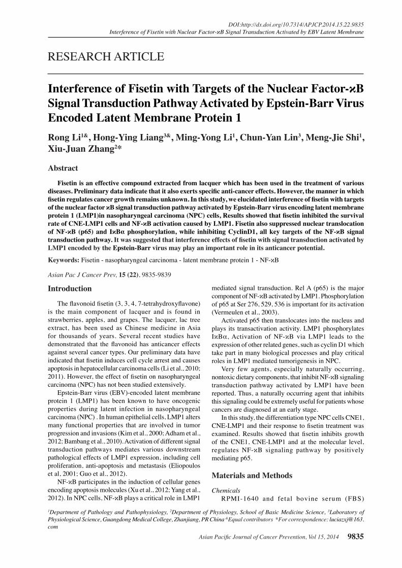
CNE1-LMP1 cells.We measured the effect of fisetin on transcriptional regulation of NF-κB using pGL6-NF-κB-luc promoter luciferase construct. The full-length NF-κB promoter coupled to a luciferase reporter gene was transfected into CNE1 and CNE1-LMP1 cells. We found that fisetin significantly reduced the activity of NF-κB promoter in a dose-dependent manner.Treatment with 25, 50, 100µM of fisetin for 24h inhibited cell NF-κB promoter activity in CNE1 (6, 18, 43%) and CNE1-LMP1 (15, 44, 60%) (Figure 2).
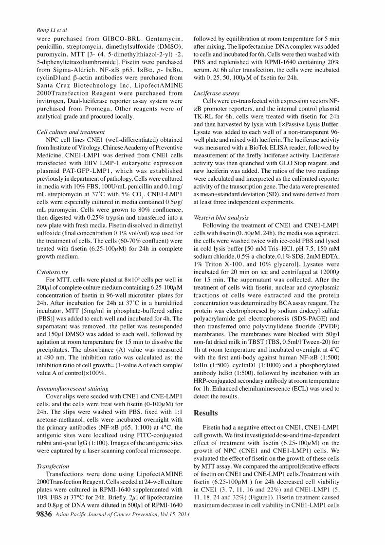
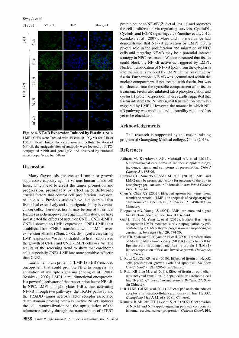
Fisetin inhibited the expression and translocation of NF-κB in CNE1-LMP1 cells. It is well known that NF- κB transcription factor is associated with cellular transformation and has been identified to be upregulated in nasopharyngeal carcinoma. In western blot analysis, we observed decreased level of p65 subunit of NF-κB in cytoplasm and concomitant decrease in p65 subunit in nuclear lysates of CNE1-LMP1 treated with fisetin, but there was no apparent change for CNE1 cells (Figure 3A, 3D). To confirm the results, we labeled fisetin-treated
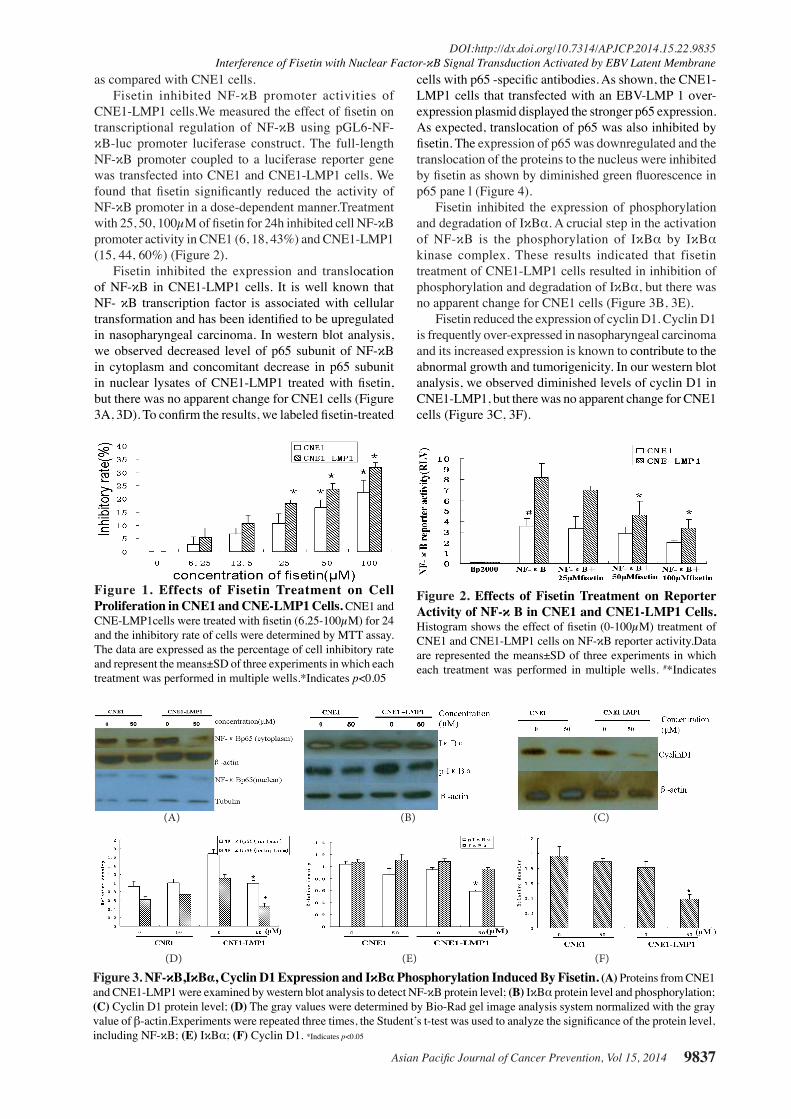
cells with p65 -specific antibodies. As shown, the CNE1-LMP1 cells that transfected with an EBV-LMP 1 over-expression plasmid displayed the stronger p65 expression. As expected, translocation of p65 was also inhibited by fisetin. The expression of p65 was downregulated and the translocation of the proteins to the nucleus were inhibited by fisetin as shown by diminished green fluorescence in p65 pane l (Figure 4).
Fisetin inhibited the expression of phosphorylation and degradation of IκBα. A crucial step in the activation of NF-κB is the phosphorylation of IκBα by IκBα kinase complex. These results indicated that fisetin treatment of CNE1-LMP1 cells resulted in inhibition of phosphorylation and degradation of IκBα, but there was no apparent change for CNE1 cells (Figure 3B, 3E).
Fisetin reduced the expression of cyclin D1. Cyclin D1 is frequently over-expressed in nasopharyngeal carcinoma and its increased expression is known to contribute to the abnormal growth and tumorigenicity. In our western blot analysis, we observed diminished levels of cyclin D1 in CNE1-LMP1, but there was no apparent change for CNE1 cells (Figure 3C, 3F).
Figure 1. Effects of Fisetin Treatment on Cell Proliferation in CNE1 and CNE-LMP1 Cells. CNE1 and CNE-LMP1cells were treated with fisetin (6.25-100µM) for 24 and the inhibitory rate of cells were determined by MTT assay. The data are expressed as the percentage of cell inhibitory rate and represent the means±SD of three experiments in which each treatment was performed in multiple wells.*Indicates p<0.05
Figure 2. Effects of Fisetin Treatment on Reporter Activity of NF-κ B in CNE1 and CNE1-LMP1 Cells. Histogram shows the effect of fisetin (0-100µM) treatment of CNE1 and CNE1-LMP1 cells on NF-κB reporter activity.Data are represented the means±SD of three experiments in which each treatment was performed in multiple wells. #*Indicates
0
25.0
50.0
75.0
100.0
New
ly d
iagn
osed
with
out
trea
tmen
t
New
ly d
iagn
osed
with
tre
atm
ent
Pers
iste
nce
or r
ecur
renc
e
Rem
issi
on
Non
e
Chem
othe
rapy
Radi
othe
rapy
Conc
urre
nt c
hem
orad
iatio
n
10.3
0
12.8
30.025.0
20.310.16.3
51.7
75.051.1
30.031.354.2
46.856.3
27.625.033.130.031.3
23.738.0
31.3
0
25.0
50.0
75.0
100.0
New
ly d
iagn
osed
with
out
trea
tmen
t
New
ly d
iagn
osed
with
tre
atm
ent
Pers
iste
nce
or r
ecur
renc
e
Rem
issi
on
Non
e
Chem
othe
rapy
Radi
othe
rapy
Conc
urre
nt c
hem
orad
iatio
n
10.3
0
12.8
30.025.0
20.310.16.3
51.7
75.051.1
30.031.354.2
46.856.3
27.625.033.130.031.3
23.738.0
31.3
Figure 3. NF-κB,IκBα, Cyclin D1 Expression and IκBα Phosphorylation Induced By Fisetin. (A) Proteins from CNE1 and CNE1-LMP1 were examined by western blot analysis to detect NF-κB protein level; (B) IκBα protein level and phosphorylation; (C) Cyclin D1 protein level; (D) The gray values were determined by Bio-Rad gel image analysis system normalized with the gray value of β-actin.Experiments were repeated three times, the Student’s t-test was used to analyze the significance of the protein level, including NF-κB; (E) IκBα; (F) Cyclin D1. *Indicates p<0.05
(A) (B) (C)
(D) (E) (F)
Rong Li et al
Asian Pacific Journal of Cancer Prevention, Vol 15, 20149838
Discussion
Many flavonoids possess anti-tumor or growth suppressive capacity against various human tumor cell lines, which lead to arrest the tumor promotion and progression, presumably by affecting or disturbing crucial factors that control cell proliferation, invasion, or apoptosis. Previous studies have demonstrated that fisetin had extensively anti-tumorigenic ability in various cancer cells. Therefore, fisetin may be one of its critical features as a chemopreventive agent. In this study, we have investigated the effects of fisetin on CNE1, CNE1-LMP1.CNE-1 showed no LMP1 expression, CNE1-LMP1 that established from CNE-1 transfected with a LMP-1 over-expression plasmid (Chen, 2002), displayed a very strong LMP1 expression. We demonstrated that fisetin suppressed the growth of CNE1 and CNE1-LMP1 cells in vitro. The results of the screening trend to show that carcinoma cells, especially CNE1-LMP1are more sensitive to fisetin than CNE1.
Latent membrane protein-1 (LMP-1) is EBV-encoded oncoprotein that could promote NPC to progress via activation of multiple signaling (Zheng et al., 2007; Yoshizaki, 2002). LMP1, a multifunctional oncoprotein, is a powerful activator of the transcription factor NF-κB. In NPC, LMP1 phosphorylates IκBα, thus activating NF-κB through two pathways: the TRAFs pathway and the TRADD (tumor necrosis factor receptor associated death domain protein) pathway. Active NF-κB induces the cell immortalization via the upregulation of the telomerase activity through the translocation of hTERT
protein bound to NF-κB (Zuo et al., 2011), and promotes the cell proliferation via regulating survivin, CyclinD1, CyclinE, and EGFR signaling, etc (Zuercher et al., 2012; Ramdass et al., 2007). More and more evidence had demonstrated that NF-κB activation by LMP1 play a pivotal role in the proliferation and migration of NPC cells and targeting NF-κB may be a potential interest strategy in NPC treatments. We demonstrated that fisetin could block the NF-κB activities triggered by LMP1. Nuclear translocation of NF-κB (p65) from the cytoplasm into the nucleus induced by LMP1 can be prevented by fisetin. Furthermore, NF- κB was accumulated within the nuclear compartment if not treated with fisetin, but was translocated into the cytosolic compartment after fisetin treatment. Fisetin also inhibited IκBα phosphorylation and cyclin D1 protein expression, These results suggested that fisetin interferes the NF-κB signal transduction pathways triggered by LMP1. However, the manner in which NF- κB pathway was modified and its stability regulated has yet to be elucidated.
Acknowledgements
This research is supported by the major training program of Guangdong Medical college, China (2013).
References
Adham M, Kurniawan AN, Muhtadi AI, et al (2012). Nasopharyngeal carcinoma in Indonesia: epidemiology, incidence, signs, and symptoms at presentation. Chin J Cancer, 31, 185-96.
Bambang H, Sunarto S, Sofia M, et al (2010). LMP1 and LMP2 may be prognostic factors for outcome of therapy in nasopharyngeal cancers in Indonesia. Asian Pac J Cancer Prev, 11, 763-6.
Chen Y, Chen XY (2002). Effect of epstein-barr virus latent membrane protein 1 (LMP1) on apoptosis of nasopharyngeal carcinoma cell line CNE1. Ai Zheng, 21, 498-503 (in Chinese).
Eliopoulos AG, Young LS (2001). LMP1 structure and signal transduction. Semin Cancer Bio, l11, 435-44.
Guo L, Tang M, Yang L, et al (2012). Epstein-Barr virus oncoprotein LMP1 mediates survivin upregulation by p53 contributing to G1/S cell cycle progression in nasopharyngeal carcinoma. Int J Mol Med, 29, 574-80.
Kim KR, Yoshizake T, Miyamori H, et al (2000). Transformation of Madin darby canine kidney (MDCK) epithelial cell by Epstein-Barr virus latent membra ne protein 1 (LMP1) induces expression of Ets1 and invasi ve growth. Oncogene, 19, 1764-77.
Li R, Li XB, Cai KR, et al (2010). Effects of fisetin on HepG2 cells proliferation, growth cycle and apoptosis. Shi Zhen Guo Yi GuoYao, 21, 3206-8 (in Chinese).
Li R, Li XB, Jing M, et al (2011). Effect of fisetin on epithelial-mesenchymal transition in hepatocellular carcinoma cell line HepG2. Chinese Pharmacologival Bulletin, 27, 91-4 (in Chinese).
Li R, Li XB, Cai KR, et al (2011). Effect of p53 on fisetin induced apoptosis in hepatocellular carcinoma cell line HepG2. Guangdong Med J, 32, 688-90 (In Chinese).
Ramdass B, Maliekal TT, Lakshmi S, et al (2007). Coexpression of Notch1 and NF-kappaB signaling pathway components in human cervical cancer progression. Gynecol Oncol, 104,
Figure 4. NF-κB Expression Induced by Fisetin. CNE1- LMP1 Cells were Treated with Fisetin (0-100µM) for 24h or DMSO alone. Image the expression and cellular location of NF-κB, the antigenic sites of antibody were located by FITC-conjugated rabbit-anti goat IgGs and observed by confocal microscope. Scale bar, 50µm
Asian Pacific Journal of Cancer Prevention, Vol 15, 2014 9839
DOI:http://dx.doi.org/10.7314/APJCP.2014.15.22.9835Interference of Fisetin with Nuclear Factor-κB Signal Transduction Activated by EBV Latent Membrane
352-61.Vermeulen L, De Wilde G, Van Damme P, et al (2003).
Transcriptional activation of the NF-κB p65 subunit by mitogen- and stress-activated protein kinase-1 (MSK1). EMBO J, 22, 1313-24.
Xu TP, Shen H, Liu LX, et al (2012). Plumbagin from plumbago zeylanica L induces apoptosis in human non-small cell lung cancer cell lines through NF-kB inactivation. Asian Pac J Cancer Prev, 14, 2325-31.
Yang XS, Liu SHA, Liu JW, et al (2012). Fucosyltransferase IV enhances expression of MMP-12 stimulated by EGF via the ERK1/2, p38 and NF-kB pathways in A431cells. Asian Pacific J Cancer Prev, 13, 1657-62.
Yoshizaki T (2002). Promotion of metastasis in nasopharyngeal carcinom a by Epstein-Barr virus latent membrane protein-1.Histol Histopathol, 17, 845-50.
Zheng H, Li LL, Hu DS, et al (2007). Role of Epstein-Barr virus encoded latent membrane protein 1 in the carcinogenesis of nasopharyngeal carcinoma. Cell Mol Immuno, l4, 185-96.
Zuercher E, Butticaz C, Wyniger J, et al (2012). Swiss HIV cohort study.genetic diversity of EBV-encoded LMP1 in the swiss HIV cohort study and implication for NF-κB activation. PLoS One, 7, 32168.
Zuo QP, Liu SK, Li ZJ, et al (2011). NF-kappaB p65 modulates the telomerase reverse transcriptase in the HepG hepatoma cell line. Eur J Pharmacol, 672, 113-20l.