Embed Size (px)
Citation preview
Int J Clin Exp Pathol 20158(12)15759-15768wwwijcepcom ISSN1936-2625IJCEP0017759
Original ArticleRole of Bcl-2 and its associated miRNAs in vasculogenic mimicry of hepatocellular carcinoma
Nan Zhao12 Bao-Cun Sun123 Xiu-Lan Zhao12 Yong Wang1 Jie Meng12 Na Che12 Xu-Yi Dong12 Qiang Gu12
1Department of Pathology Tianjin Medical University Tianjin China 2Department of Pathology General Hospital of Tianjin Medical University Tianjin China 3Department of Pathology Cancer Hospital of Tianjin Medical Univer-sity Tianjin China
Received October 13 2015 Accepted November 28 2015 Epub December 1 2015 Published December 15 2015
Abstract Objective An investigation of the role of the anti-apoptotic protein Bcl-2 and its associated miRNAs in vasculogenic mimicry (VM) in hepatocellular carcinoma Methods The Bcl-2 expression plasmid was constructed for transfection into the hepatocellular carcinoma cell line HepG2 Changes in the expression profiles of the miRNAs induced by Bcl-2 overexpression and their relationships with vasculogenic mimicry were analysed Real-time PCR was performed in frozen tissue specimens from 42 cases of hepatocellular carcinoma to analyse the relationship between Bcl-2 and miR-27a Immunohistochemical staining was performed in paraffin-embedded tissue samples from 97 cases of hepatocellular carcinoma to analyse the relationship between Bcl-2 expression and the expression of vasculogenic mimicry (VM) related molecules VEGF and HIF1A which were target genes of the Bcl-2 related miR-NAs Results Overexpression of Bcl-2 results in a significant change in the expression of a wide range of miRNAs and the target genes of these miRNAs are composed of various vasculogenic mimicry related genes Bcl-2 expres-sion was positively correlated with the expression of the miRNA target genes VEGF and HIF1A The expression of VEGF and HIF1A was significantly and positively correlated with VM and poor prognosis of patients Conclusion Bcl-2 may play a role in vasculogenic mimicry through miRNAs by targeting angiogenesis associated genes
Keywords Bcl-2 miRNA hepatocellular carcinoma vasculogenic mimicry VEGF HIF1A
Introduction
MicroRNAs (miRNAs) are members of the short non-protein coding RNA family which are widely present in eukaryotes They are single-strand-ed RNAs that consist of 19-23 nucleotides Because they can combine with mRNA 3rsquo-UTR and regulate the translation of gene expression at the post-transcriptional level with sequence specificity miRNAs play an important role in biological development fat metabolism cell differentiation proliferation and apoptosis Studies have shown that miRNAs play a key role in tumourigenesis and tumour development They not only regulate the expression of specif-ic blood vessel and tumour cell genes but also can directly act as oncogenes or tumour sup-pressors Therefore miRNAs have been identi-fied as a good target for tumour classification diagnosis prognosis and treatment [1-4]
As a crucial regulator of apoptosis Bcl-2 plays an important role in a variety of biological pro-
cesses such as embryogenesis and tissue homeostasis Studies have demonstrated that the overexpression of Bcl-2 was previously related to the malignant progression of tumours [5] Traditionally it was believed that the onco-genic properties of Bcl-2 were related to its anti-apoptotic activity The possible underlying mechanisms include that Bcl-2 can regulate the permeability of the mitochondrial membrane the release apoptotic proteins the transport of P53 across the nuclear membrane and the glu-tathione redox system [6 7] Recent studies show that Bcl-2rsquos role in promoting tumour cell survival may also be associated with its ability to induce tumour angiogenesis [8 9] In a previ-ous study by our group we found that Bcl-2 may play an important role in vasculogenic mimicry (VM) and metastasis in hepatocellular carcino-ma [10 11]
Some angiogenesis-related miRNAs have been previously reported The underlying mecha-
Bcl-2 and vasculogenic mimicry
15760 Int J Clin Exp Pathol 20158(12)15759-15768
nisms and the potential target genes of these angiogenesis-related miRNAs have been stud-
ied in depth However it is still unclear whether Bcl-2 plays a role in VM by regulating its associ-
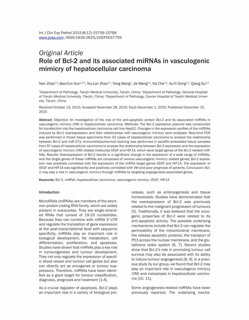
Figure 1 A A cluster analysis (part) of differentially expressed miRNAs in HepG2-Control (left S01-1-0 02 03) and HepG2-Bcl-2 (right S02-2-01 02 03) red represents upregulation green represents downregulation and black represents no significant difference B A comparison between data from the miRNA gene microarray and real-time quantitative PCR (the miRNA expression levels in the HepG2-Control was considered to be one the ratio of the level of corresponding miRNA expression in HepG2-Bcl-2 cells was considered to be its relative expression) C The rela-tionship of miR-27a and Bcl-2 in 42 cases of HCC frozen tissue specimens
Bcl-2 and vasculogenic mimicry
15761 Int J Clin Exp Pathol 20158(12)15759-15768
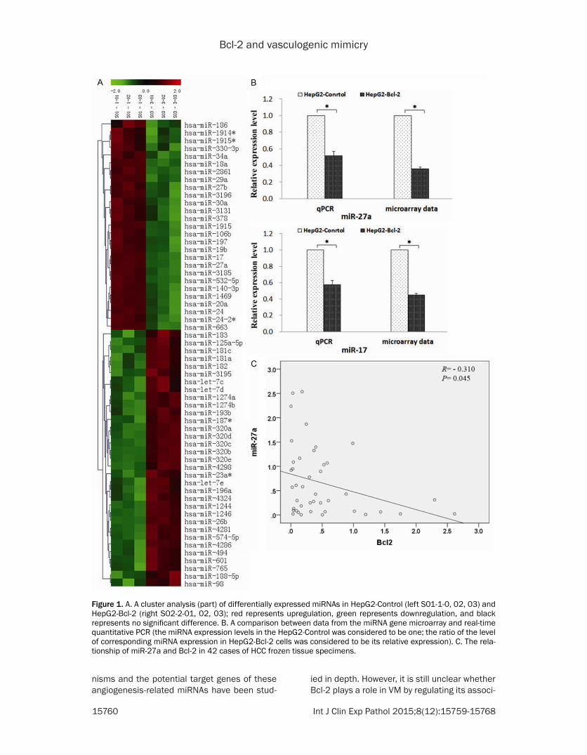
Table 1 List of miRNA identified in the HepG2-Control and HepG2-Bcl-2
No Reporter Name p-value Mean (Control)
Mean (Bcl-2)
Log2 (G2G1)
Down-regulation 256 hsa-miR-18a 887E-04 517 73 -283 300 hsa-miR-19b 170E-03 471 76 -263 507 hsa-miR-3185 618E-04 2645 458 -253 388 hsa-miR-2861 177E-04 7918 1694 -222 321 hsa-miR-20a 995E-03 5538 1584 -181 31 hsa-miR-106b 772E-04 2118 624 -176 1124 hsa-miR-663 575E-03 7189 2154 -174 197 hsa-miR-1469 780E-03 5646 1689 -174 274 hsa-miR-1915 470E-03 12722 4230 -159 232 hsa-miR-17 170E-03 4304 1544 -148 396 hsa-miR-29a 257E-03 4178 1761 -125 382 hsa-miR-27a 214E-03 9778 4384 -116 419 hsa-miR-30a 276E-03 3126 1592 -097 384 hsa-miR-27b 216E-03 4533 2591 -081 372 hsa-miR-24 850E-03 9770 6068 -069 519 hsa-miR-3196 963E-03 17007 11111 -061Up-regulation 518 hsa-miR-3195 406E-03 1342 1611 026 241 hsa-miR-182 371E-03 2591 3432 041 234 hsa-miR-181a 280E-03 609 871 052 529 hsa-miR-320a 642E-04 4806 7019 055 245 hsa-miR-183 741E-03 661 1057 068 531 hsa-miR-320c 269E-04 4045 6802 075 8 hsa-let-7d 928E-03 10839 18324 076 6 hsa-let-7c 731E-03 10469 18175 08 95 hsa-miR-125a-5p 266E-03 968 1711 082 530 hsa-miR-320b 205E-03 3329 6490 096 532 hsa-miR-320d 348E-04 2436 5016 104 533 hsa-miR-320e 254E-03 1926 4621 126 826 hsa-miR-4324 428E-03 262 693 141 286 hsa-miR-196a 411E-03 544 1523 149 380 hsa-miR-26b 140E-03 918 2873 165 10 hsa-let-7e 372E-03 3636 13029 184 778 hsa-miR-4281 439E-04 4335 17184 199 1208 hsa-miR-98 798E-03 365 2630 285 1023 hsa-miR-574-5p 986E-04 114 860 292 783 hsa-miR-4286 769E-03 94 942 333 80 hsa-miR-1246 173E-04 1160 14413 364 796 hsa-miR-4298 575E-03 49 841 41The list of miRNAs identified in the HepG2-Control and HepG2-Bcl-2 and with their mean expression values that were determined following a global normalization and a statistical analysis using studentrsquos t-test The P-values for HepG2-Control vs HepG2-Bcl-2 for each gene are lt005
ated miRNAs and the mechanism of this regu-lation is still unclear In this study we analysed
the miRNA expression profile ch- anges induced by Bcl-2 upregula-tion and validated the relationship between Bcl-2 and target genes of miRNA in human hepatocellular carcinoma (HCC)
Results
Changes of miRNA expression profiles and Bcl-2
We assessed the miRNA expres-sion profiles in hepatocellular car-cinoma HepG2 cells and HepG2 cells transfected with plasmid pcDNA-Bcl-2 The miRNA microar-ray analysis showed that thou-sands of miRNAs had been unreg-ulated or downregulated Among these 38 types of miRNA showed significant differential expression (part of the results are shown in Figure 1A and Table 1)
According to a comprehensive analysis of the target genes and related pathways two miRNAs (miR-27a and miR-17) which may be associated with tumour angio-genesis were selected as our main focus in the following valida-tion study Although there are some differences between the results of the miRNA microarray and the results of the qPCR the basic trend was consistent (Figure 1B) Therefore the authenticity of the microarray data was con-firmed The group with Bcl-2 over-expression showed that the expression of miR-27a and miR-17 was downregulated
In addition miR-27a was further verified in frozen human tissue specimens of HCC Frozen tissue specimens from 42 cases of HCC were used to extract RNA and then the RNA was reverse tran-scribed to cDNA Real-time PCR was used to detect the expression of Bcl-2 and miR-27a A Pearson correlation analysis showed that
HCC tissues with high expression of Bcl-2 were related to a decrease in the expression of miR-
Bcl-2 and vasculogenic mimicry
15762 Int J Clin Exp Pathol 20158(12)15759-15768
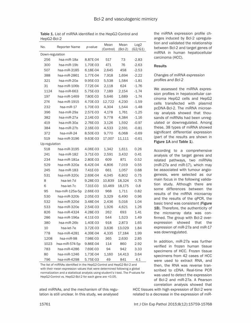
ing and currently there are no high-throughput methods for identifying miRNA targets There- fore predicting miRNA targets using theoreti-cal methods is still the ideal technique for screening and identi- fying miRNA targets TargetScan is an ideal database for predicting miRNAs using theoreti-cal methods The re- sults are as follows (1) There are 1222 types of genes that may be regulated by miR-27a Among these genes CDH5 SMAD2 TGF- BR1 VEGF are closely related to the forma-tion of VM (a portion of the result is shown in (Table 2) (2) There are 1231 types of genes that may be regu- lated by miR-17 Among these genes VEGF HIF1A and MMP2 are closely related with the formation of VM (a por-tion of the result is shown in Table 3)
Relationship between the Bcl-2 and MVD VM-PAS positive cycle
Immunohistochemistry (IHC) was performed on the paraffin-embedded tissue sections of 97 cases of HCC The posi-tive expression of Bcl-2 was shown as yellow-brown particles mainly located in the cyto-plasm of the tumour
27a and the difference was statistically signifi-cant (r=-0310 P=004) (Figure 1C)
Role of miR-27a and miR-17 in angiogenesis
Determining the target genes of miRNAs through experimentation is very time consum-
Table 2 Target genes of hsa-miR-27aGene Full nameCDH5 Vascular endothelium- cadherin (cadherin 5 CDH5)XIAP X-linked inhibitor of apoptosis proteinMAP3K14 Mitogen-activated protein kinase 14MAPK14 Mitogen-activated protein kinase 14PIK3CD Phosphoinositide-3-kinase catalytic delta polypeptideMAPKAPK3 Mitogen-activated protein kinase-activated protein kinase 3EI24 Etoposide-induced protein 24MDM4 Mouse double minute 4 homologSESN2 Sestrin-2IGF1 Insulin-like growth factor 1CDK6 Cyclin-dependent kinase 6CCNG1 Cyclin G1APAF1 Apoptotic protease activating factor 1ZMAT3 Zinc finger matrin-type 3BBC3 BCL2 binding component 3SP1 Specificity Protein 1SMAD2 Drosophila mothers against decapentaplegic protein 2VEGF Vascular endothelial growth factorTGFBR1 Transforming growth factor beta receptor 1
Table 3 Target gene of hsa-miR-17Gene Full nameMMP2 Matrix metallopeptidase 2 (gelatinase A 72 kDa type IV collagenase)VEGF-A Vascular endothelial growth factor AIRAK4 Interleukin-1 receptor-associated kinase 4MAP3K14 Mitogen-activated protein kinase 14CASP7 Caspase-7XIAP X-linked inhibitor of apoptosis protein
IL1RAP Interleukin-1 receptor accessory proteinPIK3R1 Phosphatidylinositol 3-kinase regulatory subunit polypeptide 1PLA2G6 Phospholipase A2 group VI CDK6 Cyclin-dependent kinase 6SESN2 Sestrin-2CCNG2 Cyclin G2ZMAT3 Zinc finger matrin-type 3TP73 Tumor protein 73CDKN1A Cyclin-dependent kinase inhibitor 1ARRM2 Ribonucleotide reductase M2TGFB1I1 Transforming growth factor beta-1-induced transcript 1HIF1 Hypoxia inducible factor 1 alpha subunit
cells (cytoplasm positive expression) and some positive particles could be observed around the nucleus (nucleus positive expression) (Figure 2A) The microvessel density (MVD) was determined in 97 cases of HCC and its rela-tionship with nucleus positive expression of Bcl-2 was analysed The results showed that
Bcl-2 and vasculogenic mimicry
15763 Int J Clin Exp Pathol 20158(12)15759-15768
HIF1A The difference was statistically signifi-cant (Figure 4) The analysis of the relationship of VEGF and HIF1A with VM showed that VEGF and HIF1A were significantly correlated with VM (Plt005) (Table 4) The Kaplan-Meier survival analysis was performed in 97 cases of HCC to further analyse the effect of VEGF and HIF1A on survival The results showed that the posi-tive expression of VEGF and HIF1A was nega-tively correlated with cumulative survival The median survival time of the group with VEGF positive expression was 27 months whereas that of the group with a lack of VEGF expression was 56 months the median survival time of the group with HIF1A positive expression was 29 months whereas that of the group with a lack of HIF1A expression was 42 months (Figure 5)
Discussion
According to their function miRNAs can be divided into two categories anti-angiogenic and pro-angiogenic miRNAs Anti-angiogenic
Bcl-2 expression was significantly and positive-ly correlated with MVD (Figure 2B) VM was present in 18 cases of HCC The VM-PAS posi-tive cycle was counted in the VM positive tissue sections and its relationship with nucleus posi-tive expression of Bcl-2 was analysed as well The results showed that Bcl-2 expression was significantly and positively correlated with the VM-PAS positive cycle (Figure 2C)
Relationship between expression of Bcl-2 and VEGF HIF1A
The positive expression of VEGF presented as yellow-brown particles mainly located in the cytoplasm and the positive expression of HIF1A presented as yellow-brown particles mainly located in the nucleus (Figure 3) The relationship between the nuclear expression of Bcl-2 and the expression of VEGF and HIF1A was analysed The result showed that the nuclear expression of Bcl-2 was positively relat-ed with the positive expression of VEGF and
Figure 2 A The expression of Bcl-2 in HCC tissue specimen Left panel expression of Bcl-2 in the cytoplasm Right panel expression of Bcl-2 in the nucleus mainly expressed in the perinuclear region B The relationship between Bcl-2 expression and MVD C The relationship between Bcl-2 expression and PAS
Bcl-2 and vasculogenic mimicry
15764 Int J Clin Exp Pathol 20158(12)15759-15768
miRNAs such as miR-7 inhibit angiogenesis in vitro and in vivo [12] pro-angiogenic miRNAs such as miR-145 inhibit tumour growth and angiogenesis by downregulating the expression of HIF1A and VEGF [13] Anti-angiogenic miR-NAs inhibit angiogenesis and their expression is reduced in tumour tissue Pro-angiogenic miRNAs induce angiogenesis and they are often highly expressed [14] The role of miRNAs in tumour angiogenesis is generally achieved through the regulation of cell proliferationapoptosis migration adhesion and tube forma-tion as well as other biological processes in the multi-target approach
In the present study 38 types of miRNA showed significant differential expression when Bcl-2 was overexpressed The target genes included a variety of angiogenesis related genes includ-ing CDH5 SMAD2 TGFBR1 VEGF HIF1A and MMP2 VE-cadherin (CDH5) is a superfamily of transmembrane cadherin proteins that can pro-
mote cell-cell interactions The expression of VE-cadherin is mainly located in the region of endothelial cell adherence In addition to the regulation of cell adhesion and the mainte-nance of vascular permeability VE-cadherin plays an important role in angiogenesis and the stability of the intravascular environment Hendrix et al [15] reported VE-cadherin expres-sion in highly invasive melanoma cells but its expression was barely detectable in poorly invasive melanoma cells Downregulating the expression of VE-cadherin resulted in the loss of the ability to form VM in highly invasive mela-noma cells which suggests the important role of VE-cadherin in VM formation The high expression of VEGF was closely related with microvessel density the degree of malignancy and a poor prognosis [16 17] VEGF-A belongs to the VEGF family and is the most important regulator of angiogenesis Previous studies in our laboratory also found that the capability of ovarian cancer cells to form the vessel-like
Figure 3 A The high expression of VEGF in HCC tissue specimen B The low expression of VEGF in HCC tissue specimen C The high expression of HIF1A in HCC tissue specimen D The low expression of HIF1A in HCC tissue specimen (40times)
Bcl-2 and vasculogenic mimicry
15765 Int J Clin Exp Pathol 20158(12)15759-15768
structure of VM in the three-dimensional cul-ture was enhanced after adding exogenous VEGF-A in the medium An in vitro invasion assay and wound-healing assay showed that the invasiveness and migration of ovarian can-cer cells was significantly enhanced These results confirmed that VEGF-A promoted inva-sion migration and VM formation in ovarian cancer cells The possible molecular pathway may be VEGF-ararrEphA2rarrMMPsrarrVM [18] A previous study by our group confirmed that a high expression of HIF1A was positively corre-lated with VM in melanoma and its effect may be achieved by promoting the expression of VEGF [19] Gao N also reported that HIF1A acti-vated the expression of VEGF in tumour angio-genesis and played an important role in tumour angiogenesis and tumour progression [20]
In the present study our results showed that the expression of miR-27a and miR-17 was reduced in HepG2 cells that overexpressed Bcl-2 Because miRNA negatively regulates the expression of its target genes the downregula-tion of miR-27a and miR-17 may result in the high expression of their target genes including
VE-cadherin MMP2 HIF1A and VEGF-A which can induce formation of VM The results from the human HCC specimens also confirmed that expression of Bcl-2 was positively correlated with VEGF and HIF1A which are target genes of miR-27a and miR-17 and the positive expres-sion of VEGF and HIF1A was positively related with the poor prognosis of patients
In summary the results of the present study suggest that Bcl-2 may regulate the expression of miRNA through a variety of mechanisms This regulation may reduce the expression of miR-27a and miR-17 and weaken the miRNA-induced inhibition of target genes Furthermore the expression of tumour angiogenesis related genes such as VE-cadherin MMP2 HIF1A and VEGF-A was enhanced Ultimately these changes promote the formation of VM and may affect the prognosis of patients Further study of Bcl-2 in tumour angiogenesis may provide a new target for clinical anti-tumour therapy
Materials and methods
Plasmid
The pcDNA3-Bcl-2 plasmids were obtained by cDNA-library subcloning and were verified by sequencing
Cell culture and transfection
The HepG2 cell line was cultured in Dulbeccorsquos modified Eaglersquos medium (HyClone) supple-mented with 10 foetal bovine serum (FBS HyClone) The plasmid vectors were transfect-
Figure 4 A The relationship between Bcl-2 expression and VEGF expression B The relationship between of Bcl-2 expression and HIF1A expression
Table 4 Relationship between VM and VEGF and HIF-1Aexpression
VariantTissue samples
X2 P-valueNon-VM VM
HIF-1A Negative 32 2 5564 0018Positive 47 16
VEGF Negative 37 3 5506 0019Positive 42 15
Bcl-2 and vasculogenic mimicry
15766 Int J Clin Exp Pathol 20158(12)15759-15768
ed into the cells with polyethylenimine (PEI) (PolyScience Inc Cat23966)
miRNA microarray analysis
HepG2-Control and HepG2-Bcl-2 cells were used The miRNA microarray was a double channel fluorescence chip all of the oligonucle-otide probes were labelled with Cy3 (green colour) and Cy5 (red colour) fluorescent dyes The fluorescence scanning used a double-channel laser scanner Then the figure signal was transformed to a digital signal using image analysis software (LuxScan 30 CapitalBio Beijing China)
Quantitative reverse transcription (RT)-PCR
The cDNA was synthesized from 2 μg of total RNA using Quant reverse transcriptase (Tiangen China) The RT reactions were per-formed for 1 h at 37degC Quantitative Reverse PCR was conducted in an ABI 7500 Real-Time PCR System (Applied Biosystems Foster City CA) The fluorescence intensity of the amplified products was measured at the end of each PCR cycle Two runs were performed for each data point run in triplicate The results were normal-ized to the internal control GAPDH mRNA
Western blot
The samples were transferred to polyvinylidene difluoride membranes (PVDF Millipore) The blots were blocked with 5 non-fat milk for 1 h at room temperature and then incubated with the primary antibody for 1 h at room tempera-
ture with agitation The blots were then incu-bated with horseradish peroxidase-conjugated secondary antibodies (12000 Santa Cruz) The blots were developed using an en- hanced chemiluminescence detection kit (ECL Amersham Pharmacia Biotech Piscataway NJ) We used rabbit polyclonal β-actin antibod-ies (sc1616-R 1200 Santa Cruz) for protein loading analyses The intensity of the protein bands was determined by densitometry using a Gene Genius Super System (Gene Company Limited EN)
Tissue collection
Ninety-seven HCC paraffin-embedded tissue samples surgically resected from January 2001 to December 2005 and forty-two HCC frozen tissue specimens surgically resected from January 2014 to December 2015 were obtained from the Tumor Tissue Bank of Tianjin Cancer Hospital The diagnosis of HCC was veri-fied by pathologists The use of these tissues was approved by the Institutional Research Committee
Immunohistochemical staining
Prior to immunostaining 4-μm paraffin sec-tions were deparaffinized in xylene and rehy-drated by a graded series of aqueous ethanol solutions The endogenous peroxidase activity was blocked with 3 hydrogen peroxide in 100 methanol for 15 minutes at room tem-perature The sections were washed with phos-phate-buffered saline (PBS) and then pre-treat-ed with citrate buffer (001 molL citric acid pH
Figure 5 A The relationship between VEGF expression and cumulative survival B The relationship between HIF1A expression and cumulative survival
Bcl-2 and vasculogenic mimicry
15767 Int J Clin Exp Pathol 20158(12)15759-15768
60) for 20 minutes at 95degC in a microwave oven to expose the antigens After the nonspe-cific binding sites were blocked by incubation in 10 normal goat serum in PBS for 20 minutes at 37degC the sections were incubated overnight at 4degC with the primary antibodies The next day the sections were incubated with a compatible horseradish peroxidase (HRP)-conjugated secondary antibody for 30 minutes at 37degC followed by the chromogen 33rsquo-diami-nobenzidine for 5 to 10 minutes at room tem-perature Finally the sections were lightly coun-terstained with haematoxylin for 1 minute followed by dehydration and mounting on the coverslips For the negative controls PBS was used instead of the primary antibodies The staining systems used in this study were PicTure PV6000 and Elivision Plus (Zhongshan Chemical Co Beijing China)
Immunohistochemical scoring
The evaluation of sections was performed by two independent pathologists that were blind-ed to the clinical information The expression of each marker was semi-quantitatively assessed according to both the extension of the stained cells and the intensity of the immunostaining in the individual tumour cells [21 22] More than 10 microscopic fields in each section were counted with approximately 100 tumour cells per field under light microscopy The extent of positivity (ldquoextent of distributionrdquo of positive cells) was graded on the following scale 0 for less than 10 positive cells 1 for less than 25 positive cells 2 for less than 50 positive cells and 3 for more than 50 positive cells The intensity of staining was scored on a scale of 0 to 3 as follows 0 no appreciable staining in the tumour cells 1 barely detectable stain-ing in the cytoplasm andor nucleus compared to the stromal elements 2 readily visible brown staining and 3 dark brown staining in the tumour cells obscuring the cytoplasm andor nucleus The minimum score when summed (extension + intensity) was 0 and the maxi-mum score was 6 For statistical analysis a total score of 0 to 2 were considered negativelow expression whereas scores of 3 to 6 were considered positivehigh expression
Microvessel density quantification
To identify the hotspots containing the greatest number of stained vessels a vascularity assess- ment was performed by first scanning the sec-
tion at a low power (times40) using a light micro-scope For manual counts 5 non-overlapping fields in each section were considered to acquire the MVD In each section 5 areas with the highest vascularization were selected [23]
Statistical analysis
All of the data were evaluated using SPSS 135 (SPSS Inc Chicago USA) The differences were considered significant at Plt005 The signifi-cant groups are marked with an asterisk in the figures
Acknowledgements
This study was funded by the Key Project of the National Nature Science Foundation of China (No 81230050) Project of National Nature Science Foundation of China (No 81301813 81172046 81173091) the Tianjin Natural Science Foundation (14JCYBJC27700) and the Natural Science Foundation of Tianjin Education Commission (20120103)
Disclosure of conflict of interest
None
Address correspondence to Dr Bao-Cun Sun De- partment of Pathology of Tianjin Medical Univer- sity Tianjin 300070 RP China Tel +86 136- 02111192 Fax 0086-22-83336813 E-mail bao-cunsunaliyuncom
References
[1] Cui SY Wang R Chen LB MicroRNA-145 a po-tent tumour suppressor that regulates multiple cellular pathways J Cell Mol Med 2014 18 1913-26
[2] Lu J Getz G Miska EA Alvarez-Saavedra E Lamb J Peck D Sweet-Cordero A Ebert BL Mak RH Ferrando AA Downing JR Jacks T Horvitz HR Golub TR MicroRNA expression profiles classify human cancers Nature 2005 435 834-8
[3] Zaravinos A The regulatory role of micrornas in emt and cancer J Oncol 2015 2015 865816
[4] Zhang S Lu Z Unruh AK Ivan C Baggerly KA Calin GA Li Z Bast RC Jr Le XF Clinically Rel-evant microRNAs in Ovarian Cancer Mol Can-cer Res 2015 13 393-401
[5] Correia C Lee SH Meng XW Vincelette ND Knorr KL Ding H Nowakowski GS Dai H Kaufmann SH Emerging understanding of Bcl-2 biology Implications for neoplastic progres-sion and treatment Biochim Biophys Acta 2015 1853 1658-71
Bcl-2 and vasculogenic mimicry
15768 Int J Clin Exp Pathol 20158(12)15759-15768
[6] Mora N Markovic J De la Concepcion N et al Mitochondrial and nuclear relationship in apoptosis resistance The importance of bcl-2 and redox environment FEBS J 2010 277 220-21
[7] Fulda S Targeting apoptosis for anticancer therapy Semin Cancer Biol 2015 31 84-8
[8] Iervolino A Trisciuoglio D Ribatti D Candiloro A Biroccio A Zupi G Del Bufalo D Bcl-2 over-expression in human melanoma cells increas-es angiogenesis through VEGF mRNA stabiliza-tion and HIF-1-mediated transcriptional activity FASEB J 2002 16 1453-5
[9] Gabellini C De Luca T Trisciuoglio D Desideri M Di Martile M Passeri D Candiloro A Biffoni M Rizzo MG Orlandi A Del Bufalo D BH4 do-main of bcl-2 protein is required for its proan-giogenic function under hypoxic condition Car-cinogenesis 2013 34 2558-67
[10] Sun T Sun BC Zhao XL Zhao N Dong XY Che N Yao Z Ma YM Gu Q Zong WK Liu ZY Pro-motion of tumor cell metastasis and vasculo-genic mimicry by way of transcription coactiva-tion by Bcl-2 and Twist1 a study of hepatocellular carcinoma Hepatology 2011 54 1690-706
[11] Zhao N Sun BC Zhao XL Liu ZY Sun T Qiu ZQ Gu Q Che N Dong XY Coexpression of Bcl-2 with epithelial-mesenchymal transition regula-tors is a prognostic indicator in hepatocellular carcinoma Med Oncol 2012 29 2780-92
[12] Babae N Bourajjaj M Liu Y Van Beijnum JR Cerisoli F Scaria PV Verheul M Van Berkel MP Pieters EH Van Haastert RJ Yousefi AMastrobattista E Storm G Berezikov E Cup-pen E Woodle M Schaapveld RQ Prevost GP Griffioen AW Van Noort PI Schiffelers RM Sys-temic miRNA-7 delivery inhibits tumor angio-genesis and growth in murine xenograft glio-blastoma Oncotarget 2014 5 6687-700
[13] Zou C Xu Q Mao F Li D Bian C Liu LZ Jiang Y Chen X Qi Y Zhang X Wang X Sun Q Kung HF Lin MC Dress A Wardle F Jiang BH Lai L MiR-145 inhibits tumor angiogenesis and growth by N-RAS and VEGF Cell Cycle 2012 11 2137-45
[14] Fasanaro P DrsquoAlessandra Y Di Stefano V Mel-chionna R Romani S Pompilio G Capogrossi MC Martelli F MicroRNA-210 modulates endo-thelial cell response to hypoxia and inhibits the receptor tyrosine kinase ligand Ephrin-A3 J Biol Chem 2008 283 15878-83
[15] Maniotis AJ Folberg R Hess A Seftor EA Gard-ner LM Persquoer J Trent JM Meltzer PS Hendrix MJ Vascular channel formation by human melanoma cells in vivo and in vitro vasculo-genic mimicry Am J Pathol 1999 155 739-52
[16] Scartozzi M Faloppi L Svegliati Baroni G Lore-telli C Piscaglia F Iavarone M Toniutto P Fava G De Minicis S Mandolesi A Bianconi M Gi-ampieri R Granito A Facchetti F Bitetto D Marinelli S Venerandi L Vavassori S Gemini S DrsquoErrico A Colombo M Bolondi L Bearzi I Benedetti A Cascinu S VEGF and VEGFR ge-notyping in the prediction of clinical outcome for HCC patients receiving sorafenib the AL-ICE-1 study Int J Cancer 2014 135 1247-56
[17] Anannamcharoen S and Nimmanon T Study of the vascular endothelial growth factor (VEGF) expression and microvascular density (MVD) in primary colorectal cancer specimens J Med Assoc Thai 2012 95 1041-7
[18] Wang JY Sun T Zhao XL Zhang SW Zhang DF Gu Q Wang XH Zhao N Qie S Sun BC Func-tional significance of VEGF-a in human ovarian carcinoma role in vasculogenic mimicry Can-cer Biol Ther 2008 7 758-66
[19] Sun B Zhang D Zhang S Zhang W Guo H Zhao X Hypoxia influences vasculogenic mim-icry channel formation and tumor invasion-re-lated protein expression in melanoma Cancer Lett 2007 249 188-97
[20] Gao N Ding M Zheng JZ Zhang Z Leonard SS Liu KJ Shi X Jiang BH Vanadate-induced ex-pression of hypoxia-inducible factor 1 alpha and vascular endothelial growth factor through phosphatidylinositol 3-kinaseAkt pathway and reactive oxygen species J Biol Chem 2002 277 31963-71
[21] Rahman MA Dhar DK Yamaguchi E Maruyama S Sato T Hayashi H Ono T Yaman-oi A Kohno H Nagasue N Coexpression of in-ducible nitric oxide synthase and COX-2 in he-patocellular carcinoma and surrounding liver possible involvement of COX-2 in the angiogen-esis of hepatitis C virus-positive cases Clin Cancer Res 2001 7 1325-32
[22] Cheng AL Huang WG Chen ZC Peng F Zhang PF Li MY Li F Li JL Li C Yi H Yi B Xiao ZQ Identification of novel nasopharyngeal carci-noma biomarkers by laser capture microdis-section and proteomic analysis Clin Cancer Res 2008 14 435-45
[23] Foote RL Weidner N Harris J Hammond E Lewis JE Vuong T Ang KK Fu KK Evaluation of tumor angiogenesis measured with microves-sel density (MVD) as a prognostic indicator in nasopharyngeal carcinoma results of RTOG 9505 Int J Radiat Oncol Biol Phys 2005 61 745-53
Bcl-2 and vasculogenic mimicry
15760 Int J Clin Exp Pathol 20158(12)15759-15768
nisms and the potential target genes of these angiogenesis-related miRNAs have been stud-
ied in depth However it is still unclear whether Bcl-2 plays a role in VM by regulating its associ-
Figure 1 A A cluster analysis (part) of differentially expressed miRNAs in HepG2-Control (left S01-1-0 02 03) and HepG2-Bcl-2 (right S02-2-01 02 03) red represents upregulation green represents downregulation and black represents no significant difference B A comparison between data from the miRNA gene microarray and real-time quantitative PCR (the miRNA expression levels in the HepG2-Control was considered to be one the ratio of the level of corresponding miRNA expression in HepG2-Bcl-2 cells was considered to be its relative expression) C The rela-tionship of miR-27a and Bcl-2 in 42 cases of HCC frozen tissue specimens
Bcl-2 and vasculogenic mimicry
15761 Int J Clin Exp Pathol 20158(12)15759-15768
Table 1 List of miRNA identified in the HepG2-Control and HepG2-Bcl-2
No Reporter Name p-value Mean (Control)
Mean (Bcl-2)
Log2 (G2G1)
Down-regulation 256 hsa-miR-18a 887E-04 517 73 -283 300 hsa-miR-19b 170E-03 471 76 -263 507 hsa-miR-3185 618E-04 2645 458 -253 388 hsa-miR-2861 177E-04 7918 1694 -222 321 hsa-miR-20a 995E-03 5538 1584 -181 31 hsa-miR-106b 772E-04 2118 624 -176 1124 hsa-miR-663 575E-03 7189 2154 -174 197 hsa-miR-1469 780E-03 5646 1689 -174 274 hsa-miR-1915 470E-03 12722 4230 -159 232 hsa-miR-17 170E-03 4304 1544 -148 396 hsa-miR-29a 257E-03 4178 1761 -125 382 hsa-miR-27a 214E-03 9778 4384 -116 419 hsa-miR-30a 276E-03 3126 1592 -097 384 hsa-miR-27b 216E-03 4533 2591 -081 372 hsa-miR-24 850E-03 9770 6068 -069 519 hsa-miR-3196 963E-03 17007 11111 -061Up-regulation 518 hsa-miR-3195 406E-03 1342 1611 026 241 hsa-miR-182 371E-03 2591 3432 041 234 hsa-miR-181a 280E-03 609 871 052 529 hsa-miR-320a 642E-04 4806 7019 055 245 hsa-miR-183 741E-03 661 1057 068 531 hsa-miR-320c 269E-04 4045 6802 075 8 hsa-let-7d 928E-03 10839 18324 076 6 hsa-let-7c 731E-03 10469 18175 08 95 hsa-miR-125a-5p 266E-03 968 1711 082 530 hsa-miR-320b 205E-03 3329 6490 096 532 hsa-miR-320d 348E-04 2436 5016 104 533 hsa-miR-320e 254E-03 1926 4621 126 826 hsa-miR-4324 428E-03 262 693 141 286 hsa-miR-196a 411E-03 544 1523 149 380 hsa-miR-26b 140E-03 918 2873 165 10 hsa-let-7e 372E-03 3636 13029 184 778 hsa-miR-4281 439E-04 4335 17184 199 1208 hsa-miR-98 798E-03 365 2630 285 1023 hsa-miR-574-5p 986E-04 114 860 292 783 hsa-miR-4286 769E-03 94 942 333 80 hsa-miR-1246 173E-04 1160 14413 364 796 hsa-miR-4298 575E-03 49 841 41The list of miRNAs identified in the HepG2-Control and HepG2-Bcl-2 and with their mean expression values that were determined following a global normalization and a statistical analysis using studentrsquos t-test The P-values for HepG2-Control vs HepG2-Bcl-2 for each gene are lt005
ated miRNAs and the mechanism of this regu-lation is still unclear In this study we analysed
the miRNA expression profile ch- anges induced by Bcl-2 upregula-tion and validated the relationship between Bcl-2 and target genes of miRNA in human hepatocellular carcinoma (HCC)
Results
Changes of miRNA expression profiles and Bcl-2
We assessed the miRNA expres-sion profiles in hepatocellular car-cinoma HepG2 cells and HepG2 cells transfected with plasmid pcDNA-Bcl-2 The miRNA microar-ray analysis showed that thou-sands of miRNAs had been unreg-ulated or downregulated Among these 38 types of miRNA showed significant differential expression (part of the results are shown in Figure 1A and Table 1)
According to a comprehensive analysis of the target genes and related pathways two miRNAs (miR-27a and miR-17) which may be associated with tumour angio-genesis were selected as our main focus in the following valida-tion study Although there are some differences between the results of the miRNA microarray and the results of the qPCR the basic trend was consistent (Figure 1B) Therefore the authenticity of the microarray data was con-firmed The group with Bcl-2 over-expression showed that the expression of miR-27a and miR-17 was downregulated
In addition miR-27a was further verified in frozen human tissue specimens of HCC Frozen tissue specimens from 42 cases of HCC were used to extract RNA and then the RNA was reverse tran-scribed to cDNA Real-time PCR was used to detect the expression of Bcl-2 and miR-27a A Pearson correlation analysis showed that
HCC tissues with high expression of Bcl-2 were related to a decrease in the expression of miR-
Bcl-2 and vasculogenic mimicry
15762 Int J Clin Exp Pathol 20158(12)15759-15768
ing and currently there are no high-throughput methods for identifying miRNA targets There- fore predicting miRNA targets using theoreti-cal methods is still the ideal technique for screening and identi- fying miRNA targets TargetScan is an ideal database for predicting miRNAs using theoreti-cal methods The re- sults are as follows (1) There are 1222 types of genes that may be regulated by miR-27a Among these genes CDH5 SMAD2 TGF- BR1 VEGF are closely related to the forma-tion of VM (a portion of the result is shown in (Table 2) (2) There are 1231 types of genes that may be regu- lated by miR-17 Among these genes VEGF HIF1A and MMP2 are closely related with the formation of VM (a por-tion of the result is shown in Table 3)
Relationship between the Bcl-2 and MVD VM-PAS positive cycle
Immunohistochemistry (IHC) was performed on the paraffin-embedded tissue sections of 97 cases of HCC The posi-tive expression of Bcl-2 was shown as yellow-brown particles mainly located in the cyto-plasm of the tumour
27a and the difference was statistically signifi-cant (r=-0310 P=004) (Figure 1C)
Role of miR-27a and miR-17 in angiogenesis
Determining the target genes of miRNAs through experimentation is very time consum-
Table 2 Target genes of hsa-miR-27aGene Full nameCDH5 Vascular endothelium- cadherin (cadherin 5 CDH5)XIAP X-linked inhibitor of apoptosis proteinMAP3K14 Mitogen-activated protein kinase 14MAPK14 Mitogen-activated protein kinase 14PIK3CD Phosphoinositide-3-kinase catalytic delta polypeptideMAPKAPK3 Mitogen-activated protein kinase-activated protein kinase 3EI24 Etoposide-induced protein 24MDM4 Mouse double minute 4 homologSESN2 Sestrin-2IGF1 Insulin-like growth factor 1CDK6 Cyclin-dependent kinase 6CCNG1 Cyclin G1APAF1 Apoptotic protease activating factor 1ZMAT3 Zinc finger matrin-type 3BBC3 BCL2 binding component 3SP1 Specificity Protein 1SMAD2 Drosophila mothers against decapentaplegic protein 2VEGF Vascular endothelial growth factorTGFBR1 Transforming growth factor beta receptor 1
Table 3 Target gene of hsa-miR-17Gene Full nameMMP2 Matrix metallopeptidase 2 (gelatinase A 72 kDa type IV collagenase)VEGF-A Vascular endothelial growth factor AIRAK4 Interleukin-1 receptor-associated kinase 4MAP3K14 Mitogen-activated protein kinase 14CASP7 Caspase-7XIAP X-linked inhibitor of apoptosis protein
IL1RAP Interleukin-1 receptor accessory proteinPIK3R1 Phosphatidylinositol 3-kinase regulatory subunit polypeptide 1PLA2G6 Phospholipase A2 group VI CDK6 Cyclin-dependent kinase 6SESN2 Sestrin-2CCNG2 Cyclin G2ZMAT3 Zinc finger matrin-type 3TP73 Tumor protein 73CDKN1A Cyclin-dependent kinase inhibitor 1ARRM2 Ribonucleotide reductase M2TGFB1I1 Transforming growth factor beta-1-induced transcript 1HIF1 Hypoxia inducible factor 1 alpha subunit
cells (cytoplasm positive expression) and some positive particles could be observed around the nucleus (nucleus positive expression) (Figure 2A) The microvessel density (MVD) was determined in 97 cases of HCC and its rela-tionship with nucleus positive expression of Bcl-2 was analysed The results showed that
Bcl-2 and vasculogenic mimicry
15763 Int J Clin Exp Pathol 20158(12)15759-15768
HIF1A The difference was statistically signifi-cant (Figure 4) The analysis of the relationship of VEGF and HIF1A with VM showed that VEGF and HIF1A were significantly correlated with VM (Plt005) (Table 4) The Kaplan-Meier survival analysis was performed in 97 cases of HCC to further analyse the effect of VEGF and HIF1A on survival The results showed that the posi-tive expression of VEGF and HIF1A was nega-tively correlated with cumulative survival The median survival time of the group with VEGF positive expression was 27 months whereas that of the group with a lack of VEGF expression was 56 months the median survival time of the group with HIF1A positive expression was 29 months whereas that of the group with a lack of HIF1A expression was 42 months (Figure 5)
Discussion
According to their function miRNAs can be divided into two categories anti-angiogenic and pro-angiogenic miRNAs Anti-angiogenic
Bcl-2 expression was significantly and positive-ly correlated with MVD (Figure 2B) VM was present in 18 cases of HCC The VM-PAS posi-tive cycle was counted in the VM positive tissue sections and its relationship with nucleus posi-tive expression of Bcl-2 was analysed as well The results showed that Bcl-2 expression was significantly and positively correlated with the VM-PAS positive cycle (Figure 2C)
Relationship between expression of Bcl-2 and VEGF HIF1A
The positive expression of VEGF presented as yellow-brown particles mainly located in the cytoplasm and the positive expression of HIF1A presented as yellow-brown particles mainly located in the nucleus (Figure 3) The relationship between the nuclear expression of Bcl-2 and the expression of VEGF and HIF1A was analysed The result showed that the nuclear expression of Bcl-2 was positively relat-ed with the positive expression of VEGF and
Figure 2 A The expression of Bcl-2 in HCC tissue specimen Left panel expression of Bcl-2 in the cytoplasm Right panel expression of Bcl-2 in the nucleus mainly expressed in the perinuclear region B The relationship between Bcl-2 expression and MVD C The relationship between Bcl-2 expression and PAS
Bcl-2 and vasculogenic mimicry
15764 Int J Clin Exp Pathol 20158(12)15759-15768
miRNAs such as miR-7 inhibit angiogenesis in vitro and in vivo [12] pro-angiogenic miRNAs such as miR-145 inhibit tumour growth and angiogenesis by downregulating the expression of HIF1A and VEGF [13] Anti-angiogenic miR-NAs inhibit angiogenesis and their expression is reduced in tumour tissue Pro-angiogenic miRNAs induce angiogenesis and they are often highly expressed [14] The role of miRNAs in tumour angiogenesis is generally achieved through the regulation of cell proliferationapoptosis migration adhesion and tube forma-tion as well as other biological processes in the multi-target approach
In the present study 38 types of miRNA showed significant differential expression when Bcl-2 was overexpressed The target genes included a variety of angiogenesis related genes includ-ing CDH5 SMAD2 TGFBR1 VEGF HIF1A and MMP2 VE-cadherin (CDH5) is a superfamily of transmembrane cadherin proteins that can pro-
mote cell-cell interactions The expression of VE-cadherin is mainly located in the region of endothelial cell adherence In addition to the regulation of cell adhesion and the mainte-nance of vascular permeability VE-cadherin plays an important role in angiogenesis and the stability of the intravascular environment Hendrix et al [15] reported VE-cadherin expres-sion in highly invasive melanoma cells but its expression was barely detectable in poorly invasive melanoma cells Downregulating the expression of VE-cadherin resulted in the loss of the ability to form VM in highly invasive mela-noma cells which suggests the important role of VE-cadherin in VM formation The high expression of VEGF was closely related with microvessel density the degree of malignancy and a poor prognosis [16 17] VEGF-A belongs to the VEGF family and is the most important regulator of angiogenesis Previous studies in our laboratory also found that the capability of ovarian cancer cells to form the vessel-like
Figure 3 A The high expression of VEGF in HCC tissue specimen B The low expression of VEGF in HCC tissue specimen C The high expression of HIF1A in HCC tissue specimen D The low expression of HIF1A in HCC tissue specimen (40times)
Bcl-2 and vasculogenic mimicry
15765 Int J Clin Exp Pathol 20158(12)15759-15768
structure of VM in the three-dimensional cul-ture was enhanced after adding exogenous VEGF-A in the medium An in vitro invasion assay and wound-healing assay showed that the invasiveness and migration of ovarian can-cer cells was significantly enhanced These results confirmed that VEGF-A promoted inva-sion migration and VM formation in ovarian cancer cells The possible molecular pathway may be VEGF-ararrEphA2rarrMMPsrarrVM [18] A previous study by our group confirmed that a high expression of HIF1A was positively corre-lated with VM in melanoma and its effect may be achieved by promoting the expression of VEGF [19] Gao N also reported that HIF1A acti-vated the expression of VEGF in tumour angio-genesis and played an important role in tumour angiogenesis and tumour progression [20]
In the present study our results showed that the expression of miR-27a and miR-17 was reduced in HepG2 cells that overexpressed Bcl-2 Because miRNA negatively regulates the expression of its target genes the downregula-tion of miR-27a and miR-17 may result in the high expression of their target genes including
VE-cadherin MMP2 HIF1A and VEGF-A which can induce formation of VM The results from the human HCC specimens also confirmed that expression of Bcl-2 was positively correlated with VEGF and HIF1A which are target genes of miR-27a and miR-17 and the positive expres-sion of VEGF and HIF1A was positively related with the poor prognosis of patients
In summary the results of the present study suggest that Bcl-2 may regulate the expression of miRNA through a variety of mechanisms This regulation may reduce the expression of miR-27a and miR-17 and weaken the miRNA-induced inhibition of target genes Furthermore the expression of tumour angiogenesis related genes such as VE-cadherin MMP2 HIF1A and VEGF-A was enhanced Ultimately these changes promote the formation of VM and may affect the prognosis of patients Further study of Bcl-2 in tumour angiogenesis may provide a new target for clinical anti-tumour therapy
Materials and methods
Plasmid
The pcDNA3-Bcl-2 plasmids were obtained by cDNA-library subcloning and were verified by sequencing
Cell culture and transfection
The HepG2 cell line was cultured in Dulbeccorsquos modified Eaglersquos medium (HyClone) supple-mented with 10 foetal bovine serum (FBS HyClone) The plasmid vectors were transfect-
Figure 4 A The relationship between Bcl-2 expression and VEGF expression B The relationship between of Bcl-2 expression and HIF1A expression
Table 4 Relationship between VM and VEGF and HIF-1Aexpression
VariantTissue samples
X2 P-valueNon-VM VM
HIF-1A Negative 32 2 5564 0018Positive 47 16
VEGF Negative 37 3 5506 0019Positive 42 15
Bcl-2 and vasculogenic mimicry
15766 Int J Clin Exp Pathol 20158(12)15759-15768
ed into the cells with polyethylenimine (PEI) (PolyScience Inc Cat23966)
miRNA microarray analysis
HepG2-Control and HepG2-Bcl-2 cells were used The miRNA microarray was a double channel fluorescence chip all of the oligonucle-otide probes were labelled with Cy3 (green colour) and Cy5 (red colour) fluorescent dyes The fluorescence scanning used a double-channel laser scanner Then the figure signal was transformed to a digital signal using image analysis software (LuxScan 30 CapitalBio Beijing China)
Quantitative reverse transcription (RT)-PCR
The cDNA was synthesized from 2 μg of total RNA using Quant reverse transcriptase (Tiangen China) The RT reactions were per-formed for 1 h at 37degC Quantitative Reverse PCR was conducted in an ABI 7500 Real-Time PCR System (Applied Biosystems Foster City CA) The fluorescence intensity of the amplified products was measured at the end of each PCR cycle Two runs were performed for each data point run in triplicate The results were normal-ized to the internal control GAPDH mRNA
Western blot
The samples were transferred to polyvinylidene difluoride membranes (PVDF Millipore) The blots were blocked with 5 non-fat milk for 1 h at room temperature and then incubated with the primary antibody for 1 h at room tempera-
ture with agitation The blots were then incu-bated with horseradish peroxidase-conjugated secondary antibodies (12000 Santa Cruz) The blots were developed using an en- hanced chemiluminescence detection kit (ECL Amersham Pharmacia Biotech Piscataway NJ) We used rabbit polyclonal β-actin antibod-ies (sc1616-R 1200 Santa Cruz) for protein loading analyses The intensity of the protein bands was determined by densitometry using a Gene Genius Super System (Gene Company Limited EN)
Tissue collection
Ninety-seven HCC paraffin-embedded tissue samples surgically resected from January 2001 to December 2005 and forty-two HCC frozen tissue specimens surgically resected from January 2014 to December 2015 were obtained from the Tumor Tissue Bank of Tianjin Cancer Hospital The diagnosis of HCC was veri-fied by pathologists The use of these tissues was approved by the Institutional Research Committee
Immunohistochemical staining
Prior to immunostaining 4-μm paraffin sec-tions were deparaffinized in xylene and rehy-drated by a graded series of aqueous ethanol solutions The endogenous peroxidase activity was blocked with 3 hydrogen peroxide in 100 methanol for 15 minutes at room tem-perature The sections were washed with phos-phate-buffered saline (PBS) and then pre-treat-ed with citrate buffer (001 molL citric acid pH
Figure 5 A The relationship between VEGF expression and cumulative survival B The relationship between HIF1A expression and cumulative survival
Bcl-2 and vasculogenic mimicry
15767 Int J Clin Exp Pathol 20158(12)15759-15768
60) for 20 minutes at 95degC in a microwave oven to expose the antigens After the nonspe-cific binding sites were blocked by incubation in 10 normal goat serum in PBS for 20 minutes at 37degC the sections were incubated overnight at 4degC with the primary antibodies The next day the sections were incubated with a compatible horseradish peroxidase (HRP)-conjugated secondary antibody for 30 minutes at 37degC followed by the chromogen 33rsquo-diami-nobenzidine for 5 to 10 minutes at room tem-perature Finally the sections were lightly coun-terstained with haematoxylin for 1 minute followed by dehydration and mounting on the coverslips For the negative controls PBS was used instead of the primary antibodies The staining systems used in this study were PicTure PV6000 and Elivision Plus (Zhongshan Chemical Co Beijing China)
Immunohistochemical scoring
The evaluation of sections was performed by two independent pathologists that were blind-ed to the clinical information The expression of each marker was semi-quantitatively assessed according to both the extension of the stained cells and the intensity of the immunostaining in the individual tumour cells [21 22] More than 10 microscopic fields in each section were counted with approximately 100 tumour cells per field under light microscopy The extent of positivity (ldquoextent of distributionrdquo of positive cells) was graded on the following scale 0 for less than 10 positive cells 1 for less than 25 positive cells 2 for less than 50 positive cells and 3 for more than 50 positive cells The intensity of staining was scored on a scale of 0 to 3 as follows 0 no appreciable staining in the tumour cells 1 barely detectable stain-ing in the cytoplasm andor nucleus compared to the stromal elements 2 readily visible brown staining and 3 dark brown staining in the tumour cells obscuring the cytoplasm andor nucleus The minimum score when summed (extension + intensity) was 0 and the maxi-mum score was 6 For statistical analysis a total score of 0 to 2 were considered negativelow expression whereas scores of 3 to 6 were considered positivehigh expression
Microvessel density quantification
To identify the hotspots containing the greatest number of stained vessels a vascularity assess- ment was performed by first scanning the sec-
tion at a low power (times40) using a light micro-scope For manual counts 5 non-overlapping fields in each section were considered to acquire the MVD In each section 5 areas with the highest vascularization were selected [23]
Statistical analysis
All of the data were evaluated using SPSS 135 (SPSS Inc Chicago USA) The differences were considered significant at Plt005 The signifi-cant groups are marked with an asterisk in the figures
Acknowledgements
This study was funded by the Key Project of the National Nature Science Foundation of China (No 81230050) Project of National Nature Science Foundation of China (No 81301813 81172046 81173091) the Tianjin Natural Science Foundation (14JCYBJC27700) and the Natural Science Foundation of Tianjin Education Commission (20120103)
Disclosure of conflict of interest
None
Address correspondence to Dr Bao-Cun Sun De- partment of Pathology of Tianjin Medical Univer- sity Tianjin 300070 RP China Tel +86 136- 02111192 Fax 0086-22-83336813 E-mail bao-cunsunaliyuncom
References
[1] Cui SY Wang R Chen LB MicroRNA-145 a po-tent tumour suppressor that regulates multiple cellular pathways J Cell Mol Med 2014 18 1913-26
[2] Lu J Getz G Miska EA Alvarez-Saavedra E Lamb J Peck D Sweet-Cordero A Ebert BL Mak RH Ferrando AA Downing JR Jacks T Horvitz HR Golub TR MicroRNA expression profiles classify human cancers Nature 2005 435 834-8
[3] Zaravinos A The regulatory role of micrornas in emt and cancer J Oncol 2015 2015 865816
[4] Zhang S Lu Z Unruh AK Ivan C Baggerly KA Calin GA Li Z Bast RC Jr Le XF Clinically Rel-evant microRNAs in Ovarian Cancer Mol Can-cer Res 2015 13 393-401
[5] Correia C Lee SH Meng XW Vincelette ND Knorr KL Ding H Nowakowski GS Dai H Kaufmann SH Emerging understanding of Bcl-2 biology Implications for neoplastic progres-sion and treatment Biochim Biophys Acta 2015 1853 1658-71
Bcl-2 and vasculogenic mimicry
15768 Int J Clin Exp Pathol 20158(12)15759-15768
[6] Mora N Markovic J De la Concepcion N et al Mitochondrial and nuclear relationship in apoptosis resistance The importance of bcl-2 and redox environment FEBS J 2010 277 220-21
[7] Fulda S Targeting apoptosis for anticancer therapy Semin Cancer Biol 2015 31 84-8
[8] Iervolino A Trisciuoglio D Ribatti D Candiloro A Biroccio A Zupi G Del Bufalo D Bcl-2 over-expression in human melanoma cells increas-es angiogenesis through VEGF mRNA stabiliza-tion and HIF-1-mediated transcriptional activity FASEB J 2002 16 1453-5
[9] Gabellini C De Luca T Trisciuoglio D Desideri M Di Martile M Passeri D Candiloro A Biffoni M Rizzo MG Orlandi A Del Bufalo D BH4 do-main of bcl-2 protein is required for its proan-giogenic function under hypoxic condition Car-cinogenesis 2013 34 2558-67
[10] Sun T Sun BC Zhao XL Zhao N Dong XY Che N Yao Z Ma YM Gu Q Zong WK Liu ZY Pro-motion of tumor cell metastasis and vasculo-genic mimicry by way of transcription coactiva-tion by Bcl-2 and Twist1 a study of hepatocellular carcinoma Hepatology 2011 54 1690-706
[11] Zhao N Sun BC Zhao XL Liu ZY Sun T Qiu ZQ Gu Q Che N Dong XY Coexpression of Bcl-2 with epithelial-mesenchymal transition regula-tors is a prognostic indicator in hepatocellular carcinoma Med Oncol 2012 29 2780-92
[12] Babae N Bourajjaj M Liu Y Van Beijnum JR Cerisoli F Scaria PV Verheul M Van Berkel MP Pieters EH Van Haastert RJ Yousefi AMastrobattista E Storm G Berezikov E Cup-pen E Woodle M Schaapveld RQ Prevost GP Griffioen AW Van Noort PI Schiffelers RM Sys-temic miRNA-7 delivery inhibits tumor angio-genesis and growth in murine xenograft glio-blastoma Oncotarget 2014 5 6687-700
[13] Zou C Xu Q Mao F Li D Bian C Liu LZ Jiang Y Chen X Qi Y Zhang X Wang X Sun Q Kung HF Lin MC Dress A Wardle F Jiang BH Lai L MiR-145 inhibits tumor angiogenesis and growth by N-RAS and VEGF Cell Cycle 2012 11 2137-45
[14] Fasanaro P DrsquoAlessandra Y Di Stefano V Mel-chionna R Romani S Pompilio G Capogrossi MC Martelli F MicroRNA-210 modulates endo-thelial cell response to hypoxia and inhibits the receptor tyrosine kinase ligand Ephrin-A3 J Biol Chem 2008 283 15878-83
[15] Maniotis AJ Folberg R Hess A Seftor EA Gard-ner LM Persquoer J Trent JM Meltzer PS Hendrix MJ Vascular channel formation by human melanoma cells in vivo and in vitro vasculo-genic mimicry Am J Pathol 1999 155 739-52
[16] Scartozzi M Faloppi L Svegliati Baroni G Lore-telli C Piscaglia F Iavarone M Toniutto P Fava G De Minicis S Mandolesi A Bianconi M Gi-ampieri R Granito A Facchetti F Bitetto D Marinelli S Venerandi L Vavassori S Gemini S DrsquoErrico A Colombo M Bolondi L Bearzi I Benedetti A Cascinu S VEGF and VEGFR ge-notyping in the prediction of clinical outcome for HCC patients receiving sorafenib the AL-ICE-1 study Int J Cancer 2014 135 1247-56
[17] Anannamcharoen S and Nimmanon T Study of the vascular endothelial growth factor (VEGF) expression and microvascular density (MVD) in primary colorectal cancer specimens J Med Assoc Thai 2012 95 1041-7
[18] Wang JY Sun T Zhao XL Zhang SW Zhang DF Gu Q Wang XH Zhao N Qie S Sun BC Func-tional significance of VEGF-a in human ovarian carcinoma role in vasculogenic mimicry Can-cer Biol Ther 2008 7 758-66
[19] Sun B Zhang D Zhang S Zhang W Guo H Zhao X Hypoxia influences vasculogenic mim-icry channel formation and tumor invasion-re-lated protein expression in melanoma Cancer Lett 2007 249 188-97
[20] Gao N Ding M Zheng JZ Zhang Z Leonard SS Liu KJ Shi X Jiang BH Vanadate-induced ex-pression of hypoxia-inducible factor 1 alpha and vascular endothelial growth factor through phosphatidylinositol 3-kinaseAkt pathway and reactive oxygen species J Biol Chem 2002 277 31963-71
[21] Rahman MA Dhar DK Yamaguchi E Maruyama S Sato T Hayashi H Ono T Yaman-oi A Kohno H Nagasue N Coexpression of in-ducible nitric oxide synthase and COX-2 in he-patocellular carcinoma and surrounding liver possible involvement of COX-2 in the angiogen-esis of hepatitis C virus-positive cases Clin Cancer Res 2001 7 1325-32
[22] Cheng AL Huang WG Chen ZC Peng F Zhang PF Li MY Li F Li JL Li C Yi H Yi B Xiao ZQ Identification of novel nasopharyngeal carci-noma biomarkers by laser capture microdis-section and proteomic analysis Clin Cancer Res 2008 14 435-45
[23] Foote RL Weidner N Harris J Hammond E Lewis JE Vuong T Ang KK Fu KK Evaluation of tumor angiogenesis measured with microves-sel density (MVD) as a prognostic indicator in nasopharyngeal carcinoma results of RTOG 9505 Int J Radiat Oncol Biol Phys 2005 61 745-53
Bcl-2 and vasculogenic mimicry
15762 Int J Clin Exp Pathol 20158(12)15759-15768
ing and currently there are no high-throughput methods for identifying miRNA targets There- fore predicting miRNA targets using theoreti-cal methods is still the ideal technique for screening and identi- fying miRNA targets TargetScan is an ideal database for predicting miRNAs using theoreti-cal methods The re- sults are as follows (1) There are 1222 types of genes that may be regulated by miR-27a Among these genes CDH5 SMAD2 TGF- BR1 VEGF are closely related to the forma-tion of VM (a portion of the result is shown in (Table 2) (2) There are 1231 types of genes that may be regu- lated by miR-17 Among these genes VEGF HIF1A and MMP2 are closely related with the formation of VM (a por-tion of the result is shown in Table 3)
Relationship between the Bcl-2 and MVD VM-PAS positive cycle
Immunohistochemistry (IHC) was performed on the paraffin-embedded tissue sections of 97 cases of HCC The posi-tive expression of Bcl-2 was shown as yellow-brown particles mainly located in the cyto-plasm of the tumour
27a and the difference was statistically signifi-cant (r=-0310 P=004) (Figure 1C)
Role of miR-27a and miR-17 in angiogenesis
Determining the target genes of miRNAs through experimentation is very time consum-
Table 2 Target genes of hsa-miR-27aGene Full nameCDH5 Vascular endothelium- cadherin (cadherin 5 CDH5)XIAP X-linked inhibitor of apoptosis proteinMAP3K14 Mitogen-activated protein kinase 14MAPK14 Mitogen-activated protein kinase 14PIK3CD Phosphoinositide-3-kinase catalytic delta polypeptideMAPKAPK3 Mitogen-activated protein kinase-activated protein kinase 3EI24 Etoposide-induced protein 24MDM4 Mouse double minute 4 homologSESN2 Sestrin-2IGF1 Insulin-like growth factor 1CDK6 Cyclin-dependent kinase 6CCNG1 Cyclin G1APAF1 Apoptotic protease activating factor 1ZMAT3 Zinc finger matrin-type 3BBC3 BCL2 binding component 3SP1 Specificity Protein 1SMAD2 Drosophila mothers against decapentaplegic protein 2VEGF Vascular endothelial growth factorTGFBR1 Transforming growth factor beta receptor 1
Table 3 Target gene of hsa-miR-17Gene Full nameMMP2 Matrix metallopeptidase 2 (gelatinase A 72 kDa type IV collagenase)VEGF-A Vascular endothelial growth factor AIRAK4 Interleukin-1 receptor-associated kinase 4MAP3K14 Mitogen-activated protein kinase 14CASP7 Caspase-7XIAP X-linked inhibitor of apoptosis protein
IL1RAP Interleukin-1 receptor accessory proteinPIK3R1 Phosphatidylinositol 3-kinase regulatory subunit polypeptide 1PLA2G6 Phospholipase A2 group VI CDK6 Cyclin-dependent kinase 6SESN2 Sestrin-2CCNG2 Cyclin G2ZMAT3 Zinc finger matrin-type 3TP73 Tumor protein 73CDKN1A Cyclin-dependent kinase inhibitor 1ARRM2 Ribonucleotide reductase M2TGFB1I1 Transforming growth factor beta-1-induced transcript 1HIF1 Hypoxia inducible factor 1 alpha subunit
cells (cytoplasm positive expression) and some positive particles could be observed around the nucleus (nucleus positive expression) (Figure 2A) The microvessel density (MVD) was determined in 97 cases of HCC and its rela-tionship with nucleus positive expression of Bcl-2 was analysed The results showed that
Bcl-2 and vasculogenic mimicry
15763 Int J Clin Exp Pathol 20158(12)15759-15768
HIF1A The difference was statistically signifi-cant (Figure 4) The analysis of the relationship of VEGF and HIF1A with VM showed that VEGF and HIF1A were significantly correlated with VM (Plt005) (Table 4) The Kaplan-Meier survival analysis was performed in 97 cases of HCC to further analyse the effect of VEGF and HIF1A on survival The results showed that the posi-tive expression of VEGF and HIF1A was nega-tively correlated with cumulative survival The median survival time of the group with VEGF positive expression was 27 months whereas that of the group with a lack of VEGF expression was 56 months the median survival time of the group with HIF1A positive expression was 29 months whereas that of the group with a lack of HIF1A expression was 42 months (Figure 5)
Discussion
According to their function miRNAs can be divided into two categories anti-angiogenic and pro-angiogenic miRNAs Anti-angiogenic
Bcl-2 expression was significantly and positive-ly correlated with MVD (Figure 2B) VM was present in 18 cases of HCC The VM-PAS posi-tive cycle was counted in the VM positive tissue sections and its relationship with nucleus posi-tive expression of Bcl-2 was analysed as well The results showed that Bcl-2 expression was significantly and positively correlated with the VM-PAS positive cycle (Figure 2C)
Relationship between expression of Bcl-2 and VEGF HIF1A
The positive expression of VEGF presented as yellow-brown particles mainly located in the cytoplasm and the positive expression of HIF1A presented as yellow-brown particles mainly located in the nucleus (Figure 3) The relationship between the nuclear expression of Bcl-2 and the expression of VEGF and HIF1A was analysed The result showed that the nuclear expression of Bcl-2 was positively relat-ed with the positive expression of VEGF and
Figure 2 A The expression of Bcl-2 in HCC tissue specimen Left panel expression of Bcl-2 in the cytoplasm Right panel expression of Bcl-2 in the nucleus mainly expressed in the perinuclear region B The relationship between Bcl-2 expression and MVD C The relationship between Bcl-2 expression and PAS
Bcl-2 and vasculogenic mimicry
15764 Int J Clin Exp Pathol 20158(12)15759-15768
miRNAs such as miR-7 inhibit angiogenesis in vitro and in vivo [12] pro-angiogenic miRNAs such as miR-145 inhibit tumour growth and angiogenesis by downregulating the expression of HIF1A and VEGF [13] Anti-angiogenic miR-NAs inhibit angiogenesis and their expression is reduced in tumour tissue Pro-angiogenic miRNAs induce angiogenesis and they are often highly expressed [14] The role of miRNAs in tumour angiogenesis is generally achieved through the regulation of cell proliferationapoptosis migration adhesion and tube forma-tion as well as other biological processes in the multi-target approach
In the present study 38 types of miRNA showed significant differential expression when Bcl-2 was overexpressed The target genes included a variety of angiogenesis related genes includ-ing CDH5 SMAD2 TGFBR1 VEGF HIF1A and MMP2 VE-cadherin (CDH5) is a superfamily of transmembrane cadherin proteins that can pro-
mote cell-cell interactions The expression of VE-cadherin is mainly located in the region of endothelial cell adherence In addition to the regulation of cell adhesion and the mainte-nance of vascular permeability VE-cadherin plays an important role in angiogenesis and the stability of the intravascular environment Hendrix et al [15] reported VE-cadherin expres-sion in highly invasive melanoma cells but its expression was barely detectable in poorly invasive melanoma cells Downregulating the expression of VE-cadherin resulted in the loss of the ability to form VM in highly invasive mela-noma cells which suggests the important role of VE-cadherin in VM formation The high expression of VEGF was closely related with microvessel density the degree of malignancy and a poor prognosis [16 17] VEGF-A belongs to the VEGF family and is the most important regulator of angiogenesis Previous studies in our laboratory also found that the capability of ovarian cancer cells to form the vessel-like
Figure 3 A The high expression of VEGF in HCC tissue specimen B The low expression of VEGF in HCC tissue specimen C The high expression of HIF1A in HCC tissue specimen D The low expression of HIF1A in HCC tissue specimen (40times)
Bcl-2 and vasculogenic mimicry
15765 Int J Clin Exp Pathol 20158(12)15759-15768
structure of VM in the three-dimensional cul-ture was enhanced after adding exogenous VEGF-A in the medium An in vitro invasion assay and wound-healing assay showed that the invasiveness and migration of ovarian can-cer cells was significantly enhanced These results confirmed that VEGF-A promoted inva-sion migration and VM formation in ovarian cancer cells The possible molecular pathway may be VEGF-ararrEphA2rarrMMPsrarrVM [18] A previous study by our group confirmed that a high expression of HIF1A was positively corre-lated with VM in melanoma and its effect may be achieved by promoting the expression of VEGF [19] Gao N also reported that HIF1A acti-vated the expression of VEGF in tumour angio-genesis and played an important role in tumour angiogenesis and tumour progression [20]
In the present study our results showed that the expression of miR-27a and miR-17 was reduced in HepG2 cells that overexpressed Bcl-2 Because miRNA negatively regulates the expression of its target genes the downregula-tion of miR-27a and miR-17 may result in the high expression of their target genes including
VE-cadherin MMP2 HIF1A and VEGF-A which can induce formation of VM The results from the human HCC specimens also confirmed that expression of Bcl-2 was positively correlated with VEGF and HIF1A which are target genes of miR-27a and miR-17 and the positive expres-sion of VEGF and HIF1A was positively related with the poor prognosis of patients
In summary the results of the present study suggest that Bcl-2 may regulate the expression of miRNA through a variety of mechanisms This regulation may reduce the expression of miR-27a and miR-17 and weaken the miRNA-induced inhibition of target genes Furthermore the expression of tumour angiogenesis related genes such as VE-cadherin MMP2 HIF1A and VEGF-A was enhanced Ultimately these changes promote the formation of VM and may affect the prognosis of patients Further study of Bcl-2 in tumour angiogenesis may provide a new target for clinical anti-tumour therapy
Materials and methods
Plasmid
The pcDNA3-Bcl-2 plasmids were obtained by cDNA-library subcloning and were verified by sequencing
Cell culture and transfection
The HepG2 cell line was cultured in Dulbeccorsquos modified Eaglersquos medium (HyClone) supple-mented with 10 foetal bovine serum (FBS HyClone) The plasmid vectors were transfect-
Figure 4 A The relationship between Bcl-2 expression and VEGF expression B The relationship between of Bcl-2 expression and HIF1A expression
Table 4 Relationship between VM and VEGF and HIF-1Aexpression
VariantTissue samples
X2 P-valueNon-VM VM
HIF-1A Negative 32 2 5564 0018Positive 47 16
VEGF Negative 37 3 5506 0019Positive 42 15
Bcl-2 and vasculogenic mimicry
15766 Int J Clin Exp Pathol 20158(12)15759-15768
ed into the cells with polyethylenimine (PEI) (PolyScience Inc Cat23966)
miRNA microarray analysis
HepG2-Control and HepG2-Bcl-2 cells were used The miRNA microarray was a double channel fluorescence chip all of the oligonucle-otide probes were labelled with Cy3 (green colour) and Cy5 (red colour) fluorescent dyes The fluorescence scanning used a double-channel laser scanner Then the figure signal was transformed to a digital signal using image analysis software (LuxScan 30 CapitalBio Beijing China)
Quantitative reverse transcription (RT)-PCR
The cDNA was synthesized from 2 μg of total RNA using Quant reverse transcriptase (Tiangen China) The RT reactions were per-formed for 1 h at 37degC Quantitative Reverse PCR was conducted in an ABI 7500 Real-Time PCR System (Applied Biosystems Foster City CA) The fluorescence intensity of the amplified products was measured at the end of each PCR cycle Two runs were performed for each data point run in triplicate The results were normal-ized to the internal control GAPDH mRNA
Western blot
The samples were transferred to polyvinylidene difluoride membranes (PVDF Millipore) The blots were blocked with 5 non-fat milk for 1 h at room temperature and then incubated with the primary antibody for 1 h at room tempera-
ture with agitation The blots were then incu-bated with horseradish peroxidase-conjugated secondary antibodies (12000 Santa Cruz) The blots were developed using an en- hanced chemiluminescence detection kit (ECL Amersham Pharmacia Biotech Piscataway NJ) We used rabbit polyclonal β-actin antibod-ies (sc1616-R 1200 Santa Cruz) for protein loading analyses The intensity of the protein bands was determined by densitometry using a Gene Genius Super System (Gene Company Limited EN)
Tissue collection
Ninety-seven HCC paraffin-embedded tissue samples surgically resected from January 2001 to December 2005 and forty-two HCC frozen tissue specimens surgically resected from January 2014 to December 2015 were obtained from the Tumor Tissue Bank of Tianjin Cancer Hospital The diagnosis of HCC was veri-fied by pathologists The use of these tissues was approved by the Institutional Research Committee
Immunohistochemical staining
Prior to immunostaining 4-μm paraffin sec-tions were deparaffinized in xylene and rehy-drated by a graded series of aqueous ethanol solutions The endogenous peroxidase activity was blocked with 3 hydrogen peroxide in 100 methanol for 15 minutes at room tem-perature The sections were washed with phos-phate-buffered saline (PBS) and then pre-treat-ed with citrate buffer (001 molL citric acid pH
Figure 5 A The relationship between VEGF expression and cumulative survival B The relationship between HIF1A expression and cumulative survival
Bcl-2 and vasculogenic mimicry
15767 Int J Clin Exp Pathol 20158(12)15759-15768
60) for 20 minutes at 95degC in a microwave oven to expose the antigens After the nonspe-cific binding sites were blocked by incubation in 10 normal goat serum in PBS for 20 minutes at 37degC the sections were incubated overnight at 4degC with the primary antibodies The next day the sections were incubated with a compatible horseradish peroxidase (HRP)-conjugated secondary antibody for 30 minutes at 37degC followed by the chromogen 33rsquo-diami-nobenzidine for 5 to 10 minutes at room tem-perature Finally the sections were lightly coun-terstained with haematoxylin for 1 minute followed by dehydration and mounting on the coverslips For the negative controls PBS was used instead of the primary antibodies The staining systems used in this study were PicTure PV6000 and Elivision Plus (Zhongshan Chemical Co Beijing China)
Immunohistochemical scoring
The evaluation of sections was performed by two independent pathologists that were blind-ed to the clinical information The expression of each marker was semi-quantitatively assessed according to both the extension of the stained cells and the intensity of the immunostaining in the individual tumour cells [21 22] More than 10 microscopic fields in each section were counted with approximately 100 tumour cells per field under light microscopy The extent of positivity (ldquoextent of distributionrdquo of positive cells) was graded on the following scale 0 for less than 10 positive cells 1 for less than 25 positive cells 2 for less than 50 positive cells and 3 for more than 50 positive cells The intensity of staining was scored on a scale of 0 to 3 as follows 0 no appreciable staining in the tumour cells 1 barely detectable stain-ing in the cytoplasm andor nucleus compared to the stromal elements 2 readily visible brown staining and 3 dark brown staining in the tumour cells obscuring the cytoplasm andor nucleus The minimum score when summed (extension + intensity) was 0 and the maxi-mum score was 6 For statistical analysis a total score of 0 to 2 were considered negativelow expression whereas scores of 3 to 6 were considered positivehigh expression
Microvessel density quantification
To identify the hotspots containing the greatest number of stained vessels a vascularity assess- ment was performed by first scanning the sec-
tion at a low power (times40) using a light micro-scope For manual counts 5 non-overlapping fields in each section were considered to acquire the MVD In each section 5 areas with the highest vascularization were selected [23]
Statistical analysis
All of the data were evaluated using SPSS 135 (SPSS Inc Chicago USA) The differences were considered significant at Plt005 The signifi-cant groups are marked with an asterisk in the figures
Acknowledgements
This study was funded by the Key Project of the National Nature Science Foundation of China (No 81230050) Project of National Nature Science Foundation of China (No 81301813 81172046 81173091) the Tianjin Natural Science Foundation (14JCYBJC27700) and the Natural Science Foundation of Tianjin Education Commission (20120103)
Disclosure of conflict of interest
None
Address correspondence to Dr Bao-Cun Sun De- partment of Pathology of Tianjin Medical Univer- sity Tianjin 300070 RP China Tel +86 136- 02111192 Fax 0086-22-83336813 E-mail bao-cunsunaliyuncom
References
[1] Cui SY Wang R Chen LB MicroRNA-145 a po-tent tumour suppressor that regulates multiple cellular pathways J Cell Mol Med 2014 18 1913-26
[2] Lu J Getz G Miska EA Alvarez-Saavedra E Lamb J Peck D Sweet-Cordero A Ebert BL Mak RH Ferrando AA Downing JR Jacks T Horvitz HR Golub TR MicroRNA expression profiles classify human cancers Nature 2005 435 834-8
[3] Zaravinos A The regulatory role of micrornas in emt and cancer J Oncol 2015 2015 865816
[4] Zhang S Lu Z Unruh AK Ivan C Baggerly KA Calin GA Li Z Bast RC Jr Le XF Clinically Rel-evant microRNAs in Ovarian Cancer Mol Can-cer Res 2015 13 393-401
[5] Correia C Lee SH Meng XW Vincelette ND Knorr KL Ding H Nowakowski GS Dai H Kaufmann SH Emerging understanding of Bcl-2 biology Implications for neoplastic progres-sion and treatment Biochim Biophys Acta 2015 1853 1658-71
Bcl-2 and vasculogenic mimicry
15768 Int J Clin Exp Pathol 20158(12)15759-15768
[6] Mora N Markovic J De la Concepcion N et al Mitochondrial and nuclear relationship in apoptosis resistance The importance of bcl-2 and redox environment FEBS J 2010 277 220-21
[7] Fulda S Targeting apoptosis for anticancer therapy Semin Cancer Biol 2015 31 84-8
[8] Iervolino A Trisciuoglio D Ribatti D Candiloro A Biroccio A Zupi G Del Bufalo D Bcl-2 over-expression in human melanoma cells increas-es angiogenesis through VEGF mRNA stabiliza-tion and HIF-1-mediated transcriptional activity FASEB J 2002 16 1453-5
[9] Gabellini C De Luca T Trisciuoglio D Desideri M Di Martile M Passeri D Candiloro A Biffoni M Rizzo MG Orlandi A Del Bufalo D BH4 do-main of bcl-2 protein is required for its proan-giogenic function under hypoxic condition Car-cinogenesis 2013 34 2558-67
[10] Sun T Sun BC Zhao XL Zhao N Dong XY Che N Yao Z Ma YM Gu Q Zong WK Liu ZY Pro-motion of tumor cell metastasis and vasculo-genic mimicry by way of transcription coactiva-tion by Bcl-2 and Twist1 a study of hepatocellular carcinoma Hepatology 2011 54 1690-706
[11] Zhao N Sun BC Zhao XL Liu ZY Sun T Qiu ZQ Gu Q Che N Dong XY Coexpression of Bcl-2 with epithelial-mesenchymal transition regula-tors is a prognostic indicator in hepatocellular carcinoma Med Oncol 2012 29 2780-92
[12] Babae N Bourajjaj M Liu Y Van Beijnum JR Cerisoli F Scaria PV Verheul M Van Berkel MP Pieters EH Van Haastert RJ Yousefi AMastrobattista E Storm G Berezikov E Cup-pen E Woodle M Schaapveld RQ Prevost GP Griffioen AW Van Noort PI Schiffelers RM Sys-temic miRNA-7 delivery inhibits tumor angio-genesis and growth in murine xenograft glio-blastoma Oncotarget 2014 5 6687-700
[13] Zou C Xu Q Mao F Li D Bian C Liu LZ Jiang Y Chen X Qi Y Zhang X Wang X Sun Q Kung HF Lin MC Dress A Wardle F Jiang BH Lai L MiR-145 inhibits tumor angiogenesis and growth by N-RAS and VEGF Cell Cycle 2012 11 2137-45
[14] Fasanaro P DrsquoAlessandra Y Di Stefano V Mel-chionna R Romani S Pompilio G Capogrossi MC Martelli F MicroRNA-210 modulates endo-thelial cell response to hypoxia and inhibits the receptor tyrosine kinase ligand Ephrin-A3 J Biol Chem 2008 283 15878-83
[15] Maniotis AJ Folberg R Hess A Seftor EA Gard-ner LM Persquoer J Trent JM Meltzer PS Hendrix MJ Vascular channel formation by human melanoma cells in vivo and in vitro vasculo-genic mimicry Am J Pathol 1999 155 739-52
[16] Scartozzi M Faloppi L Svegliati Baroni G Lore-telli C Piscaglia F Iavarone M Toniutto P Fava G De Minicis S Mandolesi A Bianconi M Gi-ampieri R Granito A Facchetti F Bitetto D Marinelli S Venerandi L Vavassori S Gemini S DrsquoErrico A Colombo M Bolondi L Bearzi I Benedetti A Cascinu S VEGF and VEGFR ge-notyping in the prediction of clinical outcome for HCC patients receiving sorafenib the AL-ICE-1 study Int J Cancer 2014 135 1247-56
[17] Anannamcharoen S and Nimmanon T Study of the vascular endothelial growth factor (VEGF) expression and microvascular density (MVD) in primary colorectal cancer specimens J Med Assoc Thai 2012 95 1041-7
[18] Wang JY Sun T Zhao XL Zhang SW Zhang DF Gu Q Wang XH Zhao N Qie S Sun BC Func-tional significance of VEGF-a in human ovarian carcinoma role in vasculogenic mimicry Can-cer Biol Ther 2008 7 758-66
[19] Sun B Zhang D Zhang S Zhang W Guo H Zhao X Hypoxia influences vasculogenic mim-icry channel formation and tumor invasion-re-lated protein expression in melanoma Cancer Lett 2007 249 188-97
[20] Gao N Ding M Zheng JZ Zhang Z Leonard SS Liu KJ Shi X Jiang BH Vanadate-induced ex-pression of hypoxia-inducible factor 1 alpha and vascular endothelial growth factor through phosphatidylinositol 3-kinaseAkt pathway and reactive oxygen species J Biol Chem 2002 277 31963-71
[21] Rahman MA Dhar DK Yamaguchi E Maruyama S Sato T Hayashi H Ono T Yaman-oi A Kohno H Nagasue N Coexpression of in-ducible nitric oxide synthase and COX-2 in he-patocellular carcinoma and surrounding liver possible involvement of COX-2 in the angiogen-esis of hepatitis C virus-positive cases Clin Cancer Res 2001 7 1325-32
[22] Cheng AL Huang WG Chen ZC Peng F Zhang PF Li MY Li F Li JL Li C Yi H Yi B Xiao ZQ Identification of novel nasopharyngeal carci-noma biomarkers by laser capture microdis-section and proteomic analysis Clin Cancer Res 2008 14 435-45
[23] Foote RL Weidner N Harris J Hammond E Lewis JE Vuong T Ang KK Fu KK Evaluation of tumor angiogenesis measured with microves-sel density (MVD) as a prognostic indicator in nasopharyngeal carcinoma results of RTOG 9505 Int J Radiat Oncol Biol Phys 2005 61 745-53
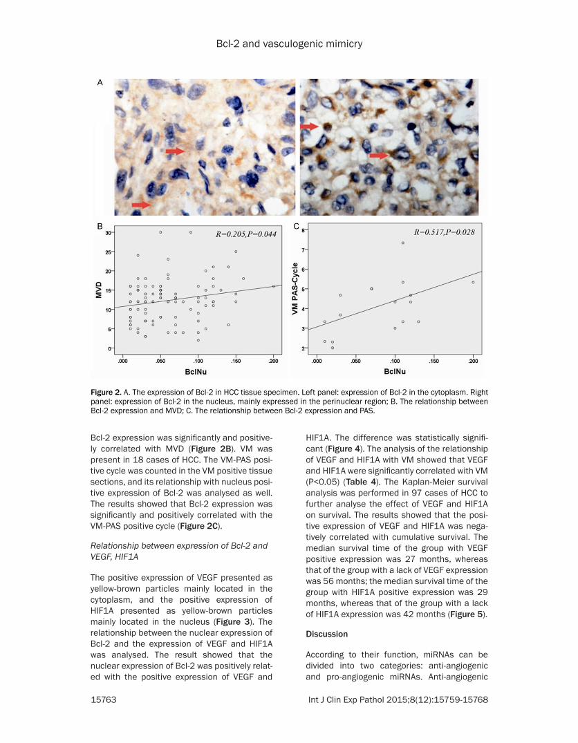
Bcl-2 and vasculogenic mimicry
15763 Int J Clin Exp Pathol 20158(12)15759-15768
HIF1A The difference was statistically signifi-cant (Figure 4) The analysis of the relationship of VEGF and HIF1A with VM showed that VEGF and HIF1A were significantly correlated with VM (Plt005) (Table 4) The Kaplan-Meier survival analysis was performed in 97 cases of HCC to further analyse the effect of VEGF and HIF1A on survival The results showed that the posi-tive expression of VEGF and HIF1A was nega-tively correlated with cumulative survival The median survival time of the group with VEGF positive expression was 27 months whereas that of the group with a lack of VEGF expression was 56 months the median survival time of the group with HIF1A positive expression was 29 months whereas that of the group with a lack of HIF1A expression was 42 months (Figure 5)
Discussion
According to their function miRNAs can be divided into two categories anti-angiogenic and pro-angiogenic miRNAs Anti-angiogenic
Bcl-2 expression was significantly and positive-ly correlated with MVD (Figure 2B) VM was present in 18 cases of HCC The VM-PAS posi-tive cycle was counted in the VM positive tissue sections and its relationship with nucleus posi-tive expression of Bcl-2 was analysed as well The results showed that Bcl-2 expression was significantly and positively correlated with the VM-PAS positive cycle (Figure 2C)
Relationship between expression of Bcl-2 and VEGF HIF1A
The positive expression of VEGF presented as yellow-brown particles mainly located in the cytoplasm and the positive expression of HIF1A presented as yellow-brown particles mainly located in the nucleus (Figure 3) The relationship between the nuclear expression of Bcl-2 and the expression of VEGF and HIF1A was analysed The result showed that the nuclear expression of Bcl-2 was positively relat-ed with the positive expression of VEGF and
Figure 2 A The expression of Bcl-2 in HCC tissue specimen Left panel expression of Bcl-2 in the cytoplasm Right panel expression of Bcl-2 in the nucleus mainly expressed in the perinuclear region B The relationship between Bcl-2 expression and MVD C The relationship between Bcl-2 expression and PAS
Bcl-2 and vasculogenic mimicry
15764 Int J Clin Exp Pathol 20158(12)15759-15768
miRNAs such as miR-7 inhibit angiogenesis in vitro and in vivo [12] pro-angiogenic miRNAs such as miR-145 inhibit tumour growth and angiogenesis by downregulating the expression of HIF1A and VEGF [13] Anti-angiogenic miR-NAs inhibit angiogenesis and their expression is reduced in tumour tissue Pro-angiogenic miRNAs induce angiogenesis and they are often highly expressed [14] The role of miRNAs in tumour angiogenesis is generally achieved through the regulation of cell proliferationapoptosis migration adhesion and tube forma-tion as well as other biological processes in the multi-target approach
In the present study 38 types of miRNA showed significant differential expression when Bcl-2 was overexpressed The target genes included a variety of angiogenesis related genes includ-ing CDH5 SMAD2 TGFBR1 VEGF HIF1A and MMP2 VE-cadherin (CDH5) is a superfamily of transmembrane cadherin proteins that can pro-
mote cell-cell interactions The expression of VE-cadherin is mainly located in the region of endothelial cell adherence In addition to the regulation of cell adhesion and the mainte-nance of vascular permeability VE-cadherin plays an important role in angiogenesis and the stability of the intravascular environment Hendrix et al [15] reported VE-cadherin expres-sion in highly invasive melanoma cells but its expression was barely detectable in poorly invasive melanoma cells Downregulating the expression of VE-cadherin resulted in the loss of the ability to form VM in highly invasive mela-noma cells which suggests the important role of VE-cadherin in VM formation The high expression of VEGF was closely related with microvessel density the degree of malignancy and a poor prognosis [16 17] VEGF-A belongs to the VEGF family and is the most important regulator of angiogenesis Previous studies in our laboratory also found that the capability of ovarian cancer cells to form the vessel-like
Figure 3 A The high expression of VEGF in HCC tissue specimen B The low expression of VEGF in HCC tissue specimen C The high expression of HIF1A in HCC tissue specimen D The low expression of HIF1A in HCC tissue specimen (40times)
Bcl-2 and vasculogenic mimicry
15765 Int J Clin Exp Pathol 20158(12)15759-15768
structure of VM in the three-dimensional cul-ture was enhanced after adding exogenous VEGF-A in the medium An in vitro invasion assay and wound-healing assay showed that the invasiveness and migration of ovarian can-cer cells was significantly enhanced These results confirmed that VEGF-A promoted inva-sion migration and VM formation in ovarian cancer cells The possible molecular pathway may be VEGF-ararrEphA2rarrMMPsrarrVM [18] A previous study by our group confirmed that a high expression of HIF1A was positively corre-lated with VM in melanoma and its effect may be achieved by promoting the expression of VEGF [19] Gao N also reported that HIF1A acti-vated the expression of VEGF in tumour angio-genesis and played an important role in tumour angiogenesis and tumour progression [20]
In the present study our results showed that the expression of miR-27a and miR-17 was reduced in HepG2 cells that overexpressed Bcl-2 Because miRNA negatively regulates the expression of its target genes the downregula-tion of miR-27a and miR-17 may result in the high expression of their target genes including
VE-cadherin MMP2 HIF1A and VEGF-A which can induce formation of VM The results from the human HCC specimens also confirmed that expression of Bcl-2 was positively correlated with VEGF and HIF1A which are target genes of miR-27a and miR-17 and the positive expres-sion of VEGF and HIF1A was positively related with the poor prognosis of patients
In summary the results of the present study suggest that Bcl-2 may regulate the expression of miRNA through a variety of mechanisms This regulation may reduce the expression of miR-27a and miR-17 and weaken the miRNA-induced inhibition of target genes Furthermore the expression of tumour angiogenesis related genes such as VE-cadherin MMP2 HIF1A and VEGF-A was enhanced Ultimately these changes promote the formation of VM and may affect the prognosis of patients Further study of Bcl-2 in tumour angiogenesis may provide a new target for clinical anti-tumour therapy
Materials and methods
Plasmid
The pcDNA3-Bcl-2 plasmids were obtained by cDNA-library subcloning and were verified by sequencing
Cell culture and transfection
The HepG2 cell line was cultured in Dulbeccorsquos modified Eaglersquos medium (HyClone) supple-mented with 10 foetal bovine serum (FBS HyClone) The plasmid vectors were transfect-
Figure 4 A The relationship between Bcl-2 expression and VEGF expression B The relationship between of Bcl-2 expression and HIF1A expression
Table 4 Relationship between VM and VEGF and HIF-1Aexpression
VariantTissue samples
X2 P-valueNon-VM VM
HIF-1A Negative 32 2 5564 0018Positive 47 16
VEGF Negative 37 3 5506 0019Positive 42 15
Bcl-2 and vasculogenic mimicry
15766 Int J Clin Exp Pathol 20158(12)15759-15768
ed into the cells with polyethylenimine (PEI) (PolyScience Inc Cat23966)
miRNA microarray analysis
HepG2-Control and HepG2-Bcl-2 cells were used The miRNA microarray was a double channel fluorescence chip all of the oligonucle-otide probes were labelled with Cy3 (green colour) and Cy5 (red colour) fluorescent dyes The fluorescence scanning used a double-channel laser scanner Then the figure signal was transformed to a digital signal using image analysis software (LuxScan 30 CapitalBio Beijing China)
Quantitative reverse transcription (RT)-PCR
The cDNA was synthesized from 2 μg of total RNA using Quant reverse transcriptase (Tiangen China) The RT reactions were per-formed for 1 h at 37degC Quantitative Reverse PCR was conducted in an ABI 7500 Real-Time PCR System (Applied Biosystems Foster City CA) The fluorescence intensity of the amplified products was measured at the end of each PCR cycle Two runs were performed for each data point run in triplicate The results were normal-ized to the internal control GAPDH mRNA
Western blot
The samples were transferred to polyvinylidene difluoride membranes (PVDF Millipore) The blots were blocked with 5 non-fat milk for 1 h at room temperature and then incubated with the primary antibody for 1 h at room tempera-
ture with agitation The blots were then incu-bated with horseradish peroxidase-conjugated secondary antibodies (12000 Santa Cruz) The blots were developed using an en- hanced chemiluminescence detection kit (ECL Amersham Pharmacia Biotech Piscataway NJ) We used rabbit polyclonal β-actin antibod-ies (sc1616-R 1200 Santa Cruz) for protein loading analyses The intensity of the protein bands was determined by densitometry using a Gene Genius Super System (Gene Company Limited EN)
Tissue collection
Ninety-seven HCC paraffin-embedded tissue samples surgically resected from January 2001 to December 2005 and forty-two HCC frozen tissue specimens surgically resected from January 2014 to December 2015 were obtained from the Tumor Tissue Bank of Tianjin Cancer Hospital The diagnosis of HCC was veri-fied by pathologists The use of these tissues was approved by the Institutional Research Committee
Immunohistochemical staining
Prior to immunostaining 4-μm paraffin sec-tions were deparaffinized in xylene and rehy-drated by a graded series of aqueous ethanol solutions The endogenous peroxidase activity was blocked with 3 hydrogen peroxide in 100 methanol for 15 minutes at room tem-perature The sections were washed with phos-phate-buffered saline (PBS) and then pre-treat-ed with citrate buffer (001 molL citric acid pH
Figure 5 A The relationship between VEGF expression and cumulative survival B The relationship between HIF1A expression and cumulative survival
Bcl-2 and vasculogenic mimicry
15767 Int J Clin Exp Pathol 20158(12)15759-15768
60) for 20 minutes at 95degC in a microwave oven to expose the antigens After the nonspe-cific binding sites were blocked by incubation in 10 normal goat serum in PBS for 20 minutes at 37degC the sections were incubated overnight at 4degC with the primary antibodies The next day the sections were incubated with a compatible horseradish peroxidase (HRP)-conjugated secondary antibody for 30 minutes at 37degC followed by the chromogen 33rsquo-diami-nobenzidine for 5 to 10 minutes at room tem-perature Finally the sections were lightly coun-terstained with haematoxylin for 1 minute followed by dehydration and mounting on the coverslips For the negative controls PBS was used instead of the primary antibodies The staining systems used in this study were PicTure PV6000 and Elivision Plus (Zhongshan Chemical Co Beijing China)
Immunohistochemical scoring
The evaluation of sections was performed by two independent pathologists that were blind-ed to the clinical information The expression of each marker was semi-quantitatively assessed according to both the extension of the stained cells and the intensity of the immunostaining in the individual tumour cells [21 22] More than 10 microscopic fields in each section were counted with approximately 100 tumour cells per field under light microscopy The extent of positivity (ldquoextent of distributionrdquo of positive cells) was graded on the following scale 0 for less than 10 positive cells 1 for less than 25 positive cells 2 for less than 50 positive cells and 3 for more than 50 positive cells The intensity of staining was scored on a scale of 0 to 3 as follows 0 no appreciable staining in the tumour cells 1 barely detectable stain-ing in the cytoplasm andor nucleus compared to the stromal elements 2 readily visible brown staining and 3 dark brown staining in the tumour cells obscuring the cytoplasm andor nucleus The minimum score when summed (extension + intensity) was 0 and the maxi-mum score was 6 For statistical analysis a total score of 0 to 2 were considered negativelow expression whereas scores of 3 to 6 were considered positivehigh expression
Microvessel density quantification
To identify the hotspots containing the greatest number of stained vessels a vascularity assess- ment was performed by first scanning the sec-
tion at a low power (times40) using a light micro-scope For manual counts 5 non-overlapping fields in each section were considered to acquire the MVD In each section 5 areas with the highest vascularization were selected [23]
Statistical analysis
All of the data were evaluated using SPSS 135 (SPSS Inc Chicago USA) The differences were considered significant at Plt005 The signifi-cant groups are marked with an asterisk in the figures
Acknowledgements
This study was funded by the Key Project of the National Nature Science Foundation of China (No 81230050) Project of National Nature Science Foundation of China (No 81301813 81172046 81173091) the Tianjin Natural Science Foundation (14JCYBJC27700) and the Natural Science Foundation of Tianjin Education Commission (20120103)
Disclosure of conflict of interest
None
Address correspondence to Dr Bao-Cun Sun De- partment of Pathology of Tianjin Medical Univer- sity Tianjin 300070 RP China Tel +86 136- 02111192 Fax 0086-22-83336813 E-mail bao-cunsunaliyuncom
References
[1] Cui SY Wang R Chen LB MicroRNA-145 a po-tent tumour suppressor that regulates multiple cellular pathways J Cell Mol Med 2014 18 1913-26
[2] Lu J Getz G Miska EA Alvarez-Saavedra E Lamb J Peck D Sweet-Cordero A Ebert BL Mak RH Ferrando AA Downing JR Jacks T Horvitz HR Golub TR MicroRNA expression profiles classify human cancers Nature 2005 435 834-8
[3] Zaravinos A The regulatory role of micrornas in emt and cancer J Oncol 2015 2015 865816
[4] Zhang S Lu Z Unruh AK Ivan C Baggerly KA Calin GA Li Z Bast RC Jr Le XF Clinically Rel-evant microRNAs in Ovarian Cancer Mol Can-cer Res 2015 13 393-401
[5] Correia C Lee SH Meng XW Vincelette ND Knorr KL Ding H Nowakowski GS Dai H Kaufmann SH Emerging understanding of Bcl-2 biology Implications for neoplastic progres-sion and treatment Biochim Biophys Acta 2015 1853 1658-71
Bcl-2 and vasculogenic mimicry
15768 Int J Clin Exp Pathol 20158(12)15759-15768
[6] Mora N Markovic J De la Concepcion N et al Mitochondrial and nuclear relationship in apoptosis resistance The importance of bcl-2 and redox environment FEBS J 2010 277 220-21
[7] Fulda S Targeting apoptosis for anticancer therapy Semin Cancer Biol 2015 31 84-8
[8] Iervolino A Trisciuoglio D Ribatti D Candiloro A Biroccio A Zupi G Del Bufalo D Bcl-2 over-expression in human melanoma cells increas-es angiogenesis through VEGF mRNA stabiliza-tion and HIF-1-mediated transcriptional activity FASEB J 2002 16 1453-5
[9] Gabellini C De Luca T Trisciuoglio D Desideri M Di Martile M Passeri D Candiloro A Biffoni M Rizzo MG Orlandi A Del Bufalo D BH4 do-main of bcl-2 protein is required for its proan-giogenic function under hypoxic condition Car-cinogenesis 2013 34 2558-67
[10] Sun T Sun BC Zhao XL Zhao N Dong XY Che N Yao Z Ma YM Gu Q Zong WK Liu ZY Pro-motion of tumor cell metastasis and vasculo-genic mimicry by way of transcription coactiva-tion by Bcl-2 and Twist1 a study of hepatocellular carcinoma Hepatology 2011 54 1690-706
[11] Zhao N Sun BC Zhao XL Liu ZY Sun T Qiu ZQ Gu Q Che N Dong XY Coexpression of Bcl-2 with epithelial-mesenchymal transition regula-tors is a prognostic indicator in hepatocellular carcinoma Med Oncol 2012 29 2780-92
[12] Babae N Bourajjaj M Liu Y Van Beijnum JR Cerisoli F Scaria PV Verheul M Van Berkel MP Pieters EH Van Haastert RJ Yousefi AMastrobattista E Storm G Berezikov E Cup-pen E Woodle M Schaapveld RQ Prevost GP Griffioen AW Van Noort PI Schiffelers RM Sys-temic miRNA-7 delivery inhibits tumor angio-genesis and growth in murine xenograft glio-blastoma Oncotarget 2014 5 6687-700
[13] Zou C Xu Q Mao F Li D Bian C Liu LZ Jiang Y Chen X Qi Y Zhang X Wang X Sun Q Kung HF Lin MC Dress A Wardle F Jiang BH Lai L MiR-145 inhibits tumor angiogenesis and growth by N-RAS and VEGF Cell Cycle 2012 11 2137-45
[14] Fasanaro P DrsquoAlessandra Y Di Stefano V Mel-chionna R Romani S Pompilio G Capogrossi MC Martelli F MicroRNA-210 modulates endo-thelial cell response to hypoxia and inhibits the receptor tyrosine kinase ligand Ephrin-A3 J Biol Chem 2008 283 15878-83
[15] Maniotis AJ Folberg R Hess A Seftor EA Gard-ner LM Persquoer J Trent JM Meltzer PS Hendrix MJ Vascular channel formation by human melanoma cells in vivo and in vitro vasculo-genic mimicry Am J Pathol 1999 155 739-52
[16] Scartozzi M Faloppi L Svegliati Baroni G Lore-telli C Piscaglia F Iavarone M Toniutto P Fava G De Minicis S Mandolesi A Bianconi M Gi-ampieri R Granito A Facchetti F Bitetto D Marinelli S Venerandi L Vavassori S Gemini S DrsquoErrico A Colombo M Bolondi L Bearzi I Benedetti A Cascinu S VEGF and VEGFR ge-notyping in the prediction of clinical outcome for HCC patients receiving sorafenib the AL-ICE-1 study Int J Cancer 2014 135 1247-56
[17] Anannamcharoen S and Nimmanon T Study of the vascular endothelial growth factor (VEGF) expression and microvascular density (MVD) in primary colorectal cancer specimens J Med Assoc Thai 2012 95 1041-7
[18] Wang JY Sun T Zhao XL Zhang SW Zhang DF Gu Q Wang XH Zhao N Qie S Sun BC Func-tional significance of VEGF-a in human ovarian carcinoma role in vasculogenic mimicry Can-cer Biol Ther 2008 7 758-66
[19] Sun B Zhang D Zhang S Zhang W Guo H Zhao X Hypoxia influences vasculogenic mim-icry channel formation and tumor invasion-re-lated protein expression in melanoma Cancer Lett 2007 249 188-97
[20] Gao N Ding M Zheng JZ Zhang Z Leonard SS Liu KJ Shi X Jiang BH Vanadate-induced ex-pression of hypoxia-inducible factor 1 alpha and vascular endothelial growth factor through phosphatidylinositol 3-kinaseAkt pathway and reactive oxygen species J Biol Chem 2002 277 31963-71
[21] Rahman MA Dhar DK Yamaguchi E Maruyama S Sato T Hayashi H Ono T Yaman-oi A Kohno H Nagasue N Coexpression of in-ducible nitric oxide synthase and COX-2 in he-patocellular carcinoma and surrounding liver possible involvement of COX-2 in the angiogen-esis of hepatitis C virus-positive cases Clin Cancer Res 2001 7 1325-32
[22] Cheng AL Huang WG Chen ZC Peng F Zhang PF Li MY Li F Li JL Li C Yi H Yi B Xiao ZQ Identification of novel nasopharyngeal carci-noma biomarkers by laser capture microdis-section and proteomic analysis Clin Cancer Res 2008 14 435-45
[23] Foote RL Weidner N Harris J Hammond E Lewis JE Vuong T Ang KK Fu KK Evaluation of tumor angiogenesis measured with microves-sel density (MVD) as a prognostic indicator in nasopharyngeal carcinoma results of RTOG 9505 Int J Radiat Oncol Biol Phys 2005 61 745-53
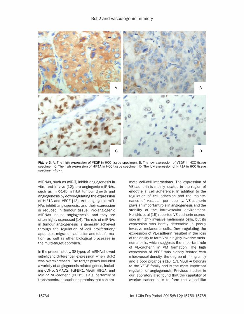
Bcl-2 and vasculogenic mimicry
15764 Int J Clin Exp Pathol 20158(12)15759-15768
miRNAs such as miR-7 inhibit angiogenesis in vitro and in vivo [12] pro-angiogenic miRNAs such as miR-145 inhibit tumour growth and angiogenesis by downregulating the expression of HIF1A and VEGF [13] Anti-angiogenic miR-NAs inhibit angiogenesis and their expression is reduced in tumour tissue Pro-angiogenic miRNAs induce angiogenesis and they are often highly expressed [14] The role of miRNAs in tumour angiogenesis is generally achieved through the regulation of cell proliferationapoptosis migration adhesion and tube forma-tion as well as other biological processes in the multi-target approach
In the present study 38 types of miRNA showed significant differential expression when Bcl-2 was overexpressed The target genes included a variety of angiogenesis related genes includ-ing CDH5 SMAD2 TGFBR1 VEGF HIF1A and MMP2 VE-cadherin (CDH5) is a superfamily of transmembrane cadherin proteins that can pro-
mote cell-cell interactions The expression of VE-cadherin is mainly located in the region of endothelial cell adherence In addition to the regulation of cell adhesion and the mainte-nance of vascular permeability VE-cadherin plays an important role in angiogenesis and the stability of the intravascular environment Hendrix et al [15] reported VE-cadherin expres-sion in highly invasive melanoma cells but its expression was barely detectable in poorly invasive melanoma cells Downregulating the expression of VE-cadherin resulted in the loss of the ability to form VM in highly invasive mela-noma cells which suggests the important role of VE-cadherin in VM formation The high expression of VEGF was closely related with microvessel density the degree of malignancy and a poor prognosis [16 17] VEGF-A belongs to the VEGF family and is the most important regulator of angiogenesis Previous studies in our laboratory also found that the capability of ovarian cancer cells to form the vessel-like
Figure 3 A The high expression of VEGF in HCC tissue specimen B The low expression of VEGF in HCC tissue specimen C The high expression of HIF1A in HCC tissue specimen D The low expression of HIF1A in HCC tissue specimen (40times)
Bcl-2 and vasculogenic mimicry
15765 Int J Clin Exp Pathol 20158(12)15759-15768
structure of VM in the three-dimensional cul-ture was enhanced after adding exogenous VEGF-A in the medium An in vitro invasion assay and wound-healing assay showed that the invasiveness and migration of ovarian can-cer cells was significantly enhanced These results confirmed that VEGF-A promoted inva-sion migration and VM formation in ovarian cancer cells The possible molecular pathway may be VEGF-ararrEphA2rarrMMPsrarrVM [18] A previous study by our group confirmed that a high expression of HIF1A was positively corre-lated with VM in melanoma and its effect may be achieved by promoting the expression of VEGF [19] Gao N also reported that HIF1A acti-vated the expression of VEGF in tumour angio-genesis and played an important role in tumour angiogenesis and tumour progression [20]
In the present study our results showed that the expression of miR-27a and miR-17 was reduced in HepG2 cells that overexpressed Bcl-2 Because miRNA negatively regulates the expression of its target genes the downregula-tion of miR-27a and miR-17 may result in the high expression of their target genes including
VE-cadherin MMP2 HIF1A and VEGF-A which can induce formation of VM The results from the human HCC specimens also confirmed that expression of Bcl-2 was positively correlated with VEGF and HIF1A which are target genes of miR-27a and miR-17 and the positive expres-sion of VEGF and HIF1A was positively related with the poor prognosis of patients
In summary the results of the present study suggest that Bcl-2 may regulate the expression of miRNA through a variety of mechanisms This regulation may reduce the expression of miR-27a and miR-17 and weaken the miRNA-induced inhibition of target genes Furthermore the expression of tumour angiogenesis related genes such as VE-cadherin MMP2 HIF1A and VEGF-A was enhanced Ultimately these changes promote the formation of VM and may affect the prognosis of patients Further study of Bcl-2 in tumour angiogenesis may provide a new target for clinical anti-tumour therapy
Materials and methods
Plasmid
The pcDNA3-Bcl-2 plasmids were obtained by cDNA-library subcloning and were verified by sequencing
Cell culture and transfection
The HepG2 cell line was cultured in Dulbeccorsquos modified Eaglersquos medium (HyClone) supple-mented with 10 foetal bovine serum (FBS HyClone) The plasmid vectors were transfect-
Figure 4 A The relationship between Bcl-2 expression and VEGF expression B The relationship between of Bcl-2 expression and HIF1A expression
Table 4 Relationship between VM and VEGF and HIF-1Aexpression
VariantTissue samples
X2 P-valueNon-VM VM
HIF-1A Negative 32 2 5564 0018Positive 47 16
VEGF Negative 37 3 5506 0019Positive 42 15
Bcl-2 and vasculogenic mimicry
15766 Int J Clin Exp Pathol 20158(12)15759-15768
ed into the cells with polyethylenimine (PEI) (PolyScience Inc Cat23966)
miRNA microarray analysis
HepG2-Control and HepG2-Bcl-2 cells were used The miRNA microarray was a double channel fluorescence chip all of the oligonucle-otide probes were labelled with Cy3 (green colour) and Cy5 (red colour) fluorescent dyes The fluorescence scanning used a double-channel laser scanner Then the figure signal was transformed to a digital signal using image analysis software (LuxScan 30 CapitalBio Beijing China)
Quantitative reverse transcription (RT)-PCR
The cDNA was synthesized from 2 μg of total RNA using Quant reverse transcriptase (Tiangen China) The RT reactions were per-formed for 1 h at 37degC Quantitative Reverse PCR was conducted in an ABI 7500 Real-Time PCR System (Applied Biosystems Foster City CA) The fluorescence intensity of the amplified products was measured at the end of each PCR cycle Two runs were performed for each data point run in triplicate The results were normal-ized to the internal control GAPDH mRNA
Western blot
The samples were transferred to polyvinylidene difluoride membranes (PVDF Millipore) The blots were blocked with 5 non-fat milk for 1 h at room temperature and then incubated with the primary antibody for 1 h at room tempera-
ture with agitation The blots were then incu-bated with horseradish peroxidase-conjugated secondary antibodies (12000 Santa Cruz) The blots were developed using an en- hanced chemiluminescence detection kit (ECL Amersham Pharmacia Biotech Piscataway NJ) We used rabbit polyclonal β-actin antibod-ies (sc1616-R 1200 Santa Cruz) for protein loading analyses The intensity of the protein bands was determined by densitometry using a Gene Genius Super System (Gene Company Limited EN)
Tissue collection
Ninety-seven HCC paraffin-embedded tissue samples surgically resected from January 2001 to December 2005 and forty-two HCC frozen tissue specimens surgically resected from January 2014 to December 2015 were obtained from the Tumor Tissue Bank of Tianjin Cancer Hospital The diagnosis of HCC was veri-fied by pathologists The use of these tissues was approved by the Institutional Research Committee
Immunohistochemical staining
Prior to immunostaining 4-μm paraffin sec-tions were deparaffinized in xylene and rehy-drated by a graded series of aqueous ethanol solutions The endogenous peroxidase activity was blocked with 3 hydrogen peroxide in 100 methanol for 15 minutes at room tem-perature The sections were washed with phos-phate-buffered saline (PBS) and then pre-treat-ed with citrate buffer (001 molL citric acid pH
Figure 5 A The relationship between VEGF expression and cumulative survival B The relationship between HIF1A expression and cumulative survival
Bcl-2 and vasculogenic mimicry
15767 Int J Clin Exp Pathol 20158(12)15759-15768
60) for 20 minutes at 95degC in a microwave oven to expose the antigens After the nonspe-cific binding sites were blocked by incubation in 10 normal goat serum in PBS for 20 minutes at 37degC the sections were incubated overnight at 4degC with the primary antibodies The next day the sections were incubated with a compatible horseradish peroxidase (HRP)-conjugated secondary antibody for 30 minutes at 37degC followed by the chromogen 33rsquo-diami-nobenzidine for 5 to 10 minutes at room tem-perature Finally the sections were lightly coun-terstained with haematoxylin for 1 minute followed by dehydration and mounting on the coverslips For the negative controls PBS was used instead of the primary antibodies The staining systems used in this study were PicTure PV6000 and Elivision Plus (Zhongshan Chemical Co Beijing China)
Immunohistochemical scoring
The evaluation of sections was performed by two independent pathologists that were blind-ed to the clinical information The expression of each marker was semi-quantitatively assessed according to both the extension of the stained cells and the intensity of the immunostaining in the individual tumour cells [21 22] More than 10 microscopic fields in each section were counted with approximately 100 tumour cells per field under light microscopy The extent of positivity (ldquoextent of distributionrdquo of positive cells) was graded on the following scale 0 for less than 10 positive cells 1 for less than 25 positive cells 2 for less than 50 positive cells and 3 for more than 50 positive cells The intensity of staining was scored on a scale of 0 to 3 as follows 0 no appreciable staining in the tumour cells 1 barely detectable stain-ing in the cytoplasm andor nucleus compared to the stromal elements 2 readily visible brown staining and 3 dark brown staining in the tumour cells obscuring the cytoplasm andor nucleus The minimum score when summed (extension + intensity) was 0 and the maxi-mum score was 6 For statistical analysis a total score of 0 to 2 were considered negativelow expression whereas scores of 3 to 6 were considered positivehigh expression
Microvessel density quantification
To identify the hotspots containing the greatest number of stained vessels a vascularity assess- ment was performed by first scanning the sec-
tion at a low power (times40) using a light micro-scope For manual counts 5 non-overlapping fields in each section were considered to acquire the MVD In each section 5 areas with the highest vascularization were selected [23]
Statistical analysis
All of the data were evaluated using SPSS 135 (SPSS Inc Chicago USA) The differences were considered significant at Plt005 The signifi-cant groups are marked with an asterisk in the figures
Acknowledgements
This study was funded by the Key Project of the National Nature Science Foundation of China (No 81230050) Project of National Nature Science Foundation of China (No 81301813 81172046 81173091) the Tianjin Natural Science Foundation (14JCYBJC27700) and the Natural Science Foundation of Tianjin Education Commission (20120103)
Disclosure of conflict of interest
None
Address correspondence to Dr Bao-Cun Sun De- partment of Pathology of Tianjin Medical Univer- sity Tianjin 300070 RP China Tel +86 136- 02111192 Fax 0086-22-83336813 E-mail bao-cunsunaliyuncom
References
[1] Cui SY Wang R Chen LB MicroRNA-145 a po-tent tumour suppressor that regulates multiple cellular pathways J Cell Mol Med 2014 18 1913-26
[2] Lu J Getz G Miska EA Alvarez-Saavedra E Lamb J Peck D Sweet-Cordero A Ebert BL Mak RH Ferrando AA Downing JR Jacks T Horvitz HR Golub TR MicroRNA expression profiles classify human cancers Nature 2005 435 834-8
[3] Zaravinos A The regulatory role of micrornas in emt and cancer J Oncol 2015 2015 865816
[4] Zhang S Lu Z Unruh AK Ivan C Baggerly KA Calin GA Li Z Bast RC Jr Le XF Clinically Rel-evant microRNAs in Ovarian Cancer Mol Can-cer Res 2015 13 393-401
[5] Correia C Lee SH Meng XW Vincelette ND Knorr KL Ding H Nowakowski GS Dai H Kaufmann SH Emerging understanding of Bcl-2 biology Implications for neoplastic progres-sion and treatment Biochim Biophys Acta 2015 1853 1658-71
Bcl-2 and vasculogenic mimicry
15768 Int J Clin Exp Pathol 20158(12)15759-15768
[6] Mora N Markovic J De la Concepcion N et al Mitochondrial and nuclear relationship in apoptosis resistance The importance of bcl-2 and redox environment FEBS J 2010 277 220-21
[7] Fulda S Targeting apoptosis for anticancer therapy Semin Cancer Biol 2015 31 84-8
[8] Iervolino A Trisciuoglio D Ribatti D Candiloro A Biroccio A Zupi G Del Bufalo D Bcl-2 over-expression in human melanoma cells increas-es angiogenesis through VEGF mRNA stabiliza-tion and HIF-1-mediated transcriptional activity FASEB J 2002 16 1453-5
[9] Gabellini C De Luca T Trisciuoglio D Desideri M Di Martile M Passeri D Candiloro A Biffoni M Rizzo MG Orlandi A Del Bufalo D BH4 do-main of bcl-2 protein is required for its proan-giogenic function under hypoxic condition Car-cinogenesis 2013 34 2558-67
[10] Sun T Sun BC Zhao XL Zhao N Dong XY Che N Yao Z Ma YM Gu Q Zong WK Liu ZY Pro-motion of tumor cell metastasis and vasculo-genic mimicry by way of transcription coactiva-tion by Bcl-2 and Twist1 a study of hepatocellular carcinoma Hepatology 2011 54 1690-706
[11] Zhao N Sun BC Zhao XL Liu ZY Sun T Qiu ZQ Gu Q Che N Dong XY Coexpression of Bcl-2 with epithelial-mesenchymal transition regula-tors is a prognostic indicator in hepatocellular carcinoma Med Oncol 2012 29 2780-92
[12] Babae N Bourajjaj M Liu Y Van Beijnum JR Cerisoli F Scaria PV Verheul M Van Berkel MP Pieters EH Van Haastert RJ Yousefi AMastrobattista E Storm G Berezikov E Cup-pen E Woodle M Schaapveld RQ Prevost GP Griffioen AW Van Noort PI Schiffelers RM Sys-temic miRNA-7 delivery inhibits tumor angio-genesis and growth in murine xenograft glio-blastoma Oncotarget 2014 5 6687-700
[13] Zou C Xu Q Mao F Li D Bian C Liu LZ Jiang Y Chen X Qi Y Zhang X Wang X Sun Q Kung HF Lin MC Dress A Wardle F Jiang BH Lai L MiR-145 inhibits tumor angiogenesis and growth by N-RAS and VEGF Cell Cycle 2012 11 2137-45
[14] Fasanaro P DrsquoAlessandra Y Di Stefano V Mel-chionna R Romani S Pompilio G Capogrossi MC Martelli F MicroRNA-210 modulates endo-thelial cell response to hypoxia and inhibits the receptor tyrosine kinase ligand Ephrin-A3 J Biol Chem 2008 283 15878-83
[15] Maniotis AJ Folberg R Hess A Seftor EA Gard-ner LM Persquoer J Trent JM Meltzer PS Hendrix MJ Vascular channel formation by human melanoma cells in vivo and in vitro vasculo-genic mimicry Am J Pathol 1999 155 739-52
[16] Scartozzi M Faloppi L Svegliati Baroni G Lore-telli C Piscaglia F Iavarone M Toniutto P Fava G De Minicis S Mandolesi A Bianconi M Gi-ampieri R Granito A Facchetti F Bitetto D Marinelli S Venerandi L Vavassori S Gemini S DrsquoErrico A Colombo M Bolondi L Bearzi I Benedetti A Cascinu S VEGF and VEGFR ge-notyping in the prediction of clinical outcome for HCC patients receiving sorafenib the AL-ICE-1 study Int J Cancer 2014 135 1247-56
[17] Anannamcharoen S and Nimmanon T Study of the vascular endothelial growth factor (VEGF) expression and microvascular density (MVD) in primary colorectal cancer specimens J Med Assoc Thai 2012 95 1041-7
[18] Wang JY Sun T Zhao XL Zhang SW Zhang DF Gu Q Wang XH Zhao N Qie S Sun BC Func-tional significance of VEGF-a in human ovarian carcinoma role in vasculogenic mimicry Can-cer Biol Ther 2008 7 758-66
[19] Sun B Zhang D Zhang S Zhang W Guo H Zhao X Hypoxia influences vasculogenic mim-icry channel formation and tumor invasion-re-lated protein expression in melanoma Cancer Lett 2007 249 188-97
[20] Gao N Ding M Zheng JZ Zhang Z Leonard SS Liu KJ Shi X Jiang BH Vanadate-induced ex-pression of hypoxia-inducible factor 1 alpha and vascular endothelial growth factor through phosphatidylinositol 3-kinaseAkt pathway and reactive oxygen species J Biol Chem 2002 277 31963-71
[21] Rahman MA Dhar DK Yamaguchi E Maruyama S Sato T Hayashi H Ono T Yaman-oi A Kohno H Nagasue N Coexpression of in-ducible nitric oxide synthase and COX-2 in he-patocellular carcinoma and surrounding liver possible involvement of COX-2 in the angiogen-esis of hepatitis C virus-positive cases Clin Cancer Res 2001 7 1325-32
[22] Cheng AL Huang WG Chen ZC Peng F Zhang PF Li MY Li F Li JL Li C Yi H Yi B Xiao ZQ Identification of novel nasopharyngeal carci-noma biomarkers by laser capture microdis-section and proteomic analysis Clin Cancer Res 2008 14 435-45
[23] Foote RL Weidner N Harris J Hammond E Lewis JE Vuong T Ang KK Fu KK Evaluation of tumor angiogenesis measured with microves-sel density (MVD) as a prognostic indicator in nasopharyngeal carcinoma results of RTOG 9505 Int J Radiat Oncol Biol Phys 2005 61 745-53
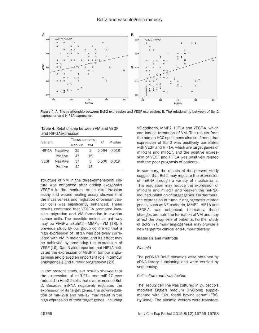
Bcl-2 and vasculogenic mimicry
15765 Int J Clin Exp Pathol 20158(12)15759-15768
structure of VM in the three-dimensional cul-ture was enhanced after adding exogenous VEGF-A in the medium An in vitro invasion assay and wound-healing assay showed that the invasiveness and migration of ovarian can-cer cells was significantly enhanced These results confirmed that VEGF-A promoted inva-sion migration and VM formation in ovarian cancer cells The possible molecular pathway may be VEGF-ararrEphA2rarrMMPsrarrVM [18] A previous study by our group confirmed that a high expression of HIF1A was positively corre-lated with VM in melanoma and its effect may be achieved by promoting the expression of VEGF [19] Gao N also reported that HIF1A acti-vated the expression of VEGF in tumour angio-genesis and played an important role in tumour angiogenesis and tumour progression [20]
In the present study our results showed that the expression of miR-27a and miR-17 was reduced in HepG2 cells that overexpressed Bcl-2 Because miRNA negatively regulates the expression of its target genes the downregula-tion of miR-27a and miR-17 may result in the high expression of their target genes including
VE-cadherin MMP2 HIF1A and VEGF-A which can induce formation of VM The results from the human HCC specimens also confirmed that expression of Bcl-2 was positively correlated with VEGF and HIF1A which are target genes of miR-27a and miR-17 and the positive expres-sion of VEGF and HIF1A was positively related with the poor prognosis of patients
In summary the results of the present study suggest that Bcl-2 may regulate the expression of miRNA through a variety of mechanisms This regulation may reduce the expression of miR-27a and miR-17 and weaken the miRNA-induced inhibition of target genes Furthermore the expression of tumour angiogenesis related genes such as VE-cadherin MMP2 HIF1A and VEGF-A was enhanced Ultimately these changes promote the formation of VM and may affect the prognosis of patients Further study of Bcl-2 in tumour angiogenesis may provide a new target for clinical anti-tumour therapy
Materials and methods
Plasmid
The pcDNA3-Bcl-2 plasmids were obtained by cDNA-library subcloning and were verified by sequencing
Cell culture and transfection
The HepG2 cell line was cultured in Dulbeccorsquos modified Eaglersquos medium (HyClone) supple-mented with 10 foetal bovine serum (FBS HyClone) The plasmid vectors were transfect-
Figure 4 A The relationship between Bcl-2 expression and VEGF expression B The relationship between of Bcl-2 expression and HIF1A expression
Table 4 Relationship between VM and VEGF and HIF-1Aexpression
VariantTissue samples
X2 P-valueNon-VM VM
HIF-1A Negative 32 2 5564 0018Positive 47 16
VEGF Negative 37 3 5506 0019Positive 42 15
Bcl-2 and vasculogenic mimicry
15766 Int J Clin Exp Pathol 20158(12)15759-15768
ed into the cells with polyethylenimine (PEI) (PolyScience Inc Cat23966)
miRNA microarray analysis
HepG2-Control and HepG2-Bcl-2 cells were used The miRNA microarray was a double channel fluorescence chip all of the oligonucle-otide probes were labelled with Cy3 (green colour) and Cy5 (red colour) fluorescent dyes The fluorescence scanning used a double-channel laser scanner Then the figure signal was transformed to a digital signal using image analysis software (LuxScan 30 CapitalBio Beijing China)
Quantitative reverse transcription (RT)-PCR
The cDNA was synthesized from 2 μg of total RNA using Quant reverse transcriptase (Tiangen China) The RT reactions were per-formed for 1 h at 37degC Quantitative Reverse PCR was conducted in an ABI 7500 Real-Time PCR System (Applied Biosystems Foster City CA) The fluorescence intensity of the amplified products was measured at the end of each PCR cycle Two runs were performed for each data point run in triplicate The results were normal-ized to the internal control GAPDH mRNA
Western blot
The samples were transferred to polyvinylidene difluoride membranes (PVDF Millipore) The blots were blocked with 5 non-fat milk for 1 h at room temperature and then incubated with the primary antibody for 1 h at room tempera-
ture with agitation The blots were then incu-bated with horseradish peroxidase-conjugated secondary antibodies (12000 Santa Cruz) The blots were developed using an en- hanced chemiluminescence detection kit (ECL Amersham Pharmacia Biotech Piscataway NJ) We used rabbit polyclonal β-actin antibod-ies (sc1616-R 1200 Santa Cruz) for protein loading analyses The intensity of the protein bands was determined by densitometry using a Gene Genius Super System (Gene Company Limited EN)
Tissue collection
Ninety-seven HCC paraffin-embedded tissue samples surgically resected from January 2001 to December 2005 and forty-two HCC frozen tissue specimens surgically resected from January 2014 to December 2015 were obtained from the Tumor Tissue Bank of Tianjin Cancer Hospital The diagnosis of HCC was veri-fied by pathologists The use of these tissues was approved by the Institutional Research Committee
Immunohistochemical staining
Prior to immunostaining 4-μm paraffin sec-tions were deparaffinized in xylene and rehy-drated by a graded series of aqueous ethanol solutions The endogenous peroxidase activity was blocked with 3 hydrogen peroxide in 100 methanol for 15 minutes at room tem-perature The sections were washed with phos-phate-buffered saline (PBS) and then pre-treat-ed with citrate buffer (001 molL citric acid pH
Figure 5 A The relationship between VEGF expression and cumulative survival B The relationship between HIF1A expression and cumulative survival
Bcl-2 and vasculogenic mimicry
15767 Int J Clin Exp Pathol 20158(12)15759-15768
60) for 20 minutes at 95degC in a microwave oven to expose the antigens After the nonspe-cific binding sites were blocked by incubation in 10 normal goat serum in PBS for 20 minutes at 37degC the sections were incubated overnight at 4degC with the primary antibodies The next day the sections were incubated with a compatible horseradish peroxidase (HRP)-conjugated secondary antibody for 30 minutes at 37degC followed by the chromogen 33rsquo-diami-nobenzidine for 5 to 10 minutes at room tem-perature Finally the sections were lightly coun-terstained with haematoxylin for 1 minute followed by dehydration and mounting on the coverslips For the negative controls PBS was used instead of the primary antibodies The staining systems used in this study were PicTure PV6000 and Elivision Plus (Zhongshan Chemical Co Beijing China)
Immunohistochemical scoring
The evaluation of sections was performed by two independent pathologists that were blind-ed to the clinical information The expression of each marker was semi-quantitatively assessed according to both the extension of the stained cells and the intensity of the immunostaining in the individual tumour cells [21 22] More than 10 microscopic fields in each section were counted with approximately 100 tumour cells per field under light microscopy The extent of positivity (ldquoextent of distributionrdquo of positive cells) was graded on the following scale 0 for less than 10 positive cells 1 for less than 25 positive cells 2 for less than 50 positive cells and 3 for more than 50 positive cells The intensity of staining was scored on a scale of 0 to 3 as follows 0 no appreciable staining in the tumour cells 1 barely detectable stain-ing in the cytoplasm andor nucleus compared to the stromal elements 2 readily visible brown staining and 3 dark brown staining in the tumour cells obscuring the cytoplasm andor nucleus The minimum score when summed (extension + intensity) was 0 and the maxi-mum score was 6 For statistical analysis a total score of 0 to 2 were considered negativelow expression whereas scores of 3 to 6 were considered positivehigh expression
Microvessel density quantification
To identify the hotspots containing the greatest number of stained vessels a vascularity assess- ment was performed by first scanning the sec-
tion at a low power (times40) using a light micro-scope For manual counts 5 non-overlapping fields in each section were considered to acquire the MVD In each section 5 areas with the highest vascularization were selected [23]
Statistical analysis
All of the data were evaluated using SPSS 135 (SPSS Inc Chicago USA) The differences were considered significant at Plt005 The signifi-cant groups are marked with an asterisk in the figures
Acknowledgements
This study was funded by the Key Project of the National Nature Science Foundation of China (No 81230050) Project of National Nature Science Foundation of China (No 81301813 81172046 81173091) the Tianjin Natural Science Foundation (14JCYBJC27700) and the Natural Science Foundation of Tianjin Education Commission (20120103)
Disclosure of conflict of interest
None
Address correspondence to Dr Bao-Cun Sun De- partment of Pathology of Tianjin Medical Univer- sity Tianjin 300070 RP China Tel +86 136- 02111192 Fax 0086-22-83336813 E-mail bao-cunsunaliyuncom
References
[1] Cui SY Wang R Chen LB MicroRNA-145 a po-tent tumour suppressor that regulates multiple cellular pathways J Cell Mol Med 2014 18 1913-26
[2] Lu J Getz G Miska EA Alvarez-Saavedra E Lamb J Peck D Sweet-Cordero A Ebert BL Mak RH Ferrando AA Downing JR Jacks T Horvitz HR Golub TR MicroRNA expression profiles classify human cancers Nature 2005 435 834-8
[3] Zaravinos A The regulatory role of micrornas in emt and cancer J Oncol 2015 2015 865816
[4] Zhang S Lu Z Unruh AK Ivan C Baggerly KA Calin GA Li Z Bast RC Jr Le XF Clinically Rel-evant microRNAs in Ovarian Cancer Mol Can-cer Res 2015 13 393-401
[5] Correia C Lee SH Meng XW Vincelette ND Knorr KL Ding H Nowakowski GS Dai H Kaufmann SH Emerging understanding of Bcl-2 biology Implications for neoplastic progres-sion and treatment Biochim Biophys Acta 2015 1853 1658-71
Bcl-2 and vasculogenic mimicry
15768 Int J Clin Exp Pathol 20158(12)15759-15768
[6] Mora N Markovic J De la Concepcion N et al Mitochondrial and nuclear relationship in apoptosis resistance The importance of bcl-2 and redox environment FEBS J 2010 277 220-21
[7] Fulda S Targeting apoptosis for anticancer therapy Semin Cancer Biol 2015 31 84-8
[8] Iervolino A Trisciuoglio D Ribatti D Candiloro A Biroccio A Zupi G Del Bufalo D Bcl-2 over-expression in human melanoma cells increas-es angiogenesis through VEGF mRNA stabiliza-tion and HIF-1-mediated transcriptional activity FASEB J 2002 16 1453-5
[9] Gabellini C De Luca T Trisciuoglio D Desideri M Di Martile M Passeri D Candiloro A Biffoni M Rizzo MG Orlandi A Del Bufalo D BH4 do-main of bcl-2 protein is required for its proan-giogenic function under hypoxic condition Car-cinogenesis 2013 34 2558-67
[10] Sun T Sun BC Zhao XL Zhao N Dong XY Che N Yao Z Ma YM Gu Q Zong WK Liu ZY Pro-motion of tumor cell metastasis and vasculo-genic mimicry by way of transcription coactiva-tion by Bcl-2 and Twist1 a study of hepatocellular carcinoma Hepatology 2011 54 1690-706
[11] Zhao N Sun BC Zhao XL Liu ZY Sun T Qiu ZQ Gu Q Che N Dong XY Coexpression of Bcl-2 with epithelial-mesenchymal transition regula-tors is a prognostic indicator in hepatocellular carcinoma Med Oncol 2012 29 2780-92
[12] Babae N Bourajjaj M Liu Y Van Beijnum JR Cerisoli F Scaria PV Verheul M Van Berkel MP Pieters EH Van Haastert RJ Yousefi AMastrobattista E Storm G Berezikov E Cup-pen E Woodle M Schaapveld RQ Prevost GP Griffioen AW Van Noort PI Schiffelers RM Sys-temic miRNA-7 delivery inhibits tumor angio-genesis and growth in murine xenograft glio-blastoma Oncotarget 2014 5 6687-700
[13] Zou C Xu Q Mao F Li D Bian C Liu LZ Jiang Y Chen X Qi Y Zhang X Wang X Sun Q Kung HF Lin MC Dress A Wardle F Jiang BH Lai L MiR-145 inhibits tumor angiogenesis and growth by N-RAS and VEGF Cell Cycle 2012 11 2137-45
[14] Fasanaro P DrsquoAlessandra Y Di Stefano V Mel-chionna R Romani S Pompilio G Capogrossi MC Martelli F MicroRNA-210 modulates endo-thelial cell response to hypoxia and inhibits the receptor tyrosine kinase ligand Ephrin-A3 J Biol Chem 2008 283 15878-83
[15] Maniotis AJ Folberg R Hess A Seftor EA Gard-ner LM Persquoer J Trent JM Meltzer PS Hendrix MJ Vascular channel formation by human melanoma cells in vivo and in vitro vasculo-genic mimicry Am J Pathol 1999 155 739-52
[16] Scartozzi M Faloppi L Svegliati Baroni G Lore-telli C Piscaglia F Iavarone M Toniutto P Fava G De Minicis S Mandolesi A Bianconi M Gi-ampieri R Granito A Facchetti F Bitetto D Marinelli S Venerandi L Vavassori S Gemini S DrsquoErrico A Colombo M Bolondi L Bearzi I Benedetti A Cascinu S VEGF and VEGFR ge-notyping in the prediction of clinical outcome for HCC patients receiving sorafenib the AL-ICE-1 study Int J Cancer 2014 135 1247-56
[17] Anannamcharoen S and Nimmanon T Study of the vascular endothelial growth factor (VEGF) expression and microvascular density (MVD) in primary colorectal cancer specimens J Med Assoc Thai 2012 95 1041-7
[18] Wang JY Sun T Zhao XL Zhang SW Zhang DF Gu Q Wang XH Zhao N Qie S Sun BC Func-tional significance of VEGF-a in human ovarian carcinoma role in vasculogenic mimicry Can-cer Biol Ther 2008 7 758-66
[19] Sun B Zhang D Zhang S Zhang W Guo H Zhao X Hypoxia influences vasculogenic mim-icry channel formation and tumor invasion-re-lated protein expression in melanoma Cancer Lett 2007 249 188-97
[20] Gao N Ding M Zheng JZ Zhang Z Leonard SS Liu KJ Shi X Jiang BH Vanadate-induced ex-pression of hypoxia-inducible factor 1 alpha and vascular endothelial growth factor through phosphatidylinositol 3-kinaseAkt pathway and reactive oxygen species J Biol Chem 2002 277 31963-71
[21] Rahman MA Dhar DK Yamaguchi E Maruyama S Sato T Hayashi H Ono T Yaman-oi A Kohno H Nagasue N Coexpression of in-ducible nitric oxide synthase and COX-2 in he-patocellular carcinoma and surrounding liver possible involvement of COX-2 in the angiogen-esis of hepatitis C virus-positive cases Clin Cancer Res 2001 7 1325-32
[22] Cheng AL Huang WG Chen ZC Peng F Zhang PF Li MY Li F Li JL Li C Yi H Yi B Xiao ZQ Identification of novel nasopharyngeal carci-noma biomarkers by laser capture microdis-section and proteomic analysis Clin Cancer Res 2008 14 435-45
[23] Foote RL Weidner N Harris J Hammond E Lewis JE Vuong T Ang KK Fu KK Evaluation of tumor angiogenesis measured with microves-sel density (MVD) as a prognostic indicator in nasopharyngeal carcinoma results of RTOG 9505 Int J Radiat Oncol Biol Phys 2005 61 745-53
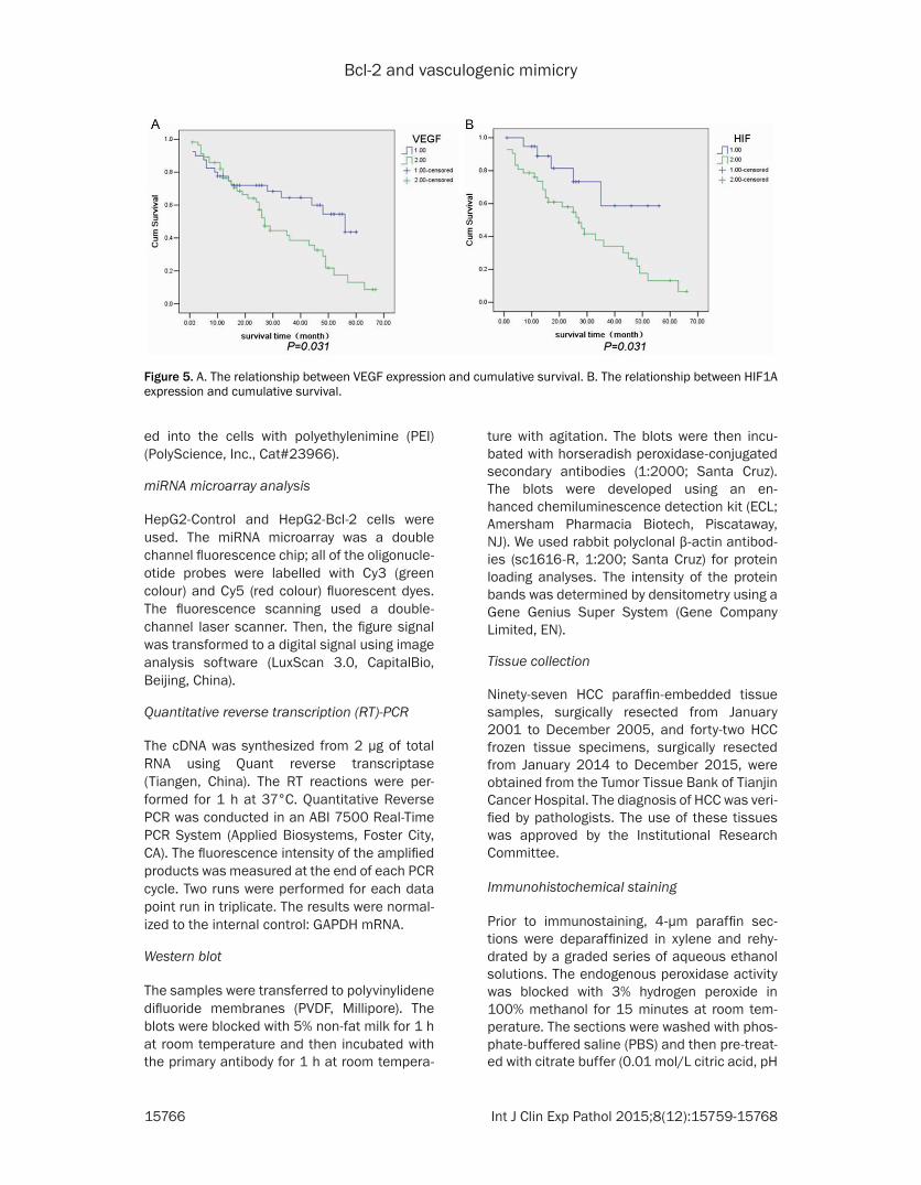
Bcl-2 and vasculogenic mimicry
15767 Int J Clin Exp Pathol 20158(12)15759-15768
60) for 20 minutes at 95degC in a microwave oven to expose the antigens After the nonspe-cific binding sites were blocked by incubation in 10 normal goat serum in PBS for 20 minutes at 37degC the sections were incubated overnight at 4degC with the primary antibodies The next day the sections were incubated with a compatible horseradish peroxidase (HRP)-conjugated secondary antibody for 30 minutes at 37degC followed by the chromogen 33rsquo-diami-nobenzidine for 5 to 10 minutes at room tem-perature Finally the sections were lightly coun-terstained with haematoxylin for 1 minute followed by dehydration and mounting on the coverslips For the negative controls PBS was used instead of the primary antibodies The staining systems used in this study were PicTure PV6000 and Elivision Plus (Zhongshan Chemical Co Beijing China)
Immunohistochemical scoring
The evaluation of sections was performed by two independent pathologists that were blind-ed to the clinical information The expression of each marker was semi-quantitatively assessed according to both the extension of the stained cells and the intensity of the immunostaining in the individual tumour cells [21 22] More than 10 microscopic fields in each section were counted with approximately 100 tumour cells per field under light microscopy The extent of positivity (ldquoextent of distributionrdquo of positive cells) was graded on the following scale 0 for less than 10 positive cells 1 for less than 25 positive cells 2 for less than 50 positive cells and 3 for more than 50 positive cells The intensity of staining was scored on a scale of 0 to 3 as follows 0 no appreciable staining in the tumour cells 1 barely detectable stain-ing in the cytoplasm andor nucleus compared to the stromal elements 2 readily visible brown staining and 3 dark brown staining in the tumour cells obscuring the cytoplasm andor nucleus The minimum score when summed (extension + intensity) was 0 and the maxi-mum score was 6 For statistical analysis a total score of 0 to 2 were considered negativelow expression whereas scores of 3 to 6 were considered positivehigh expression
Microvessel density quantification
To identify the hotspots containing the greatest number of stained vessels a vascularity assess- ment was performed by first scanning the sec-
tion at a low power (times40) using a light micro-scope For manual counts 5 non-overlapping fields in each section were considered to acquire the MVD In each section 5 areas with the highest vascularization were selected [23]
Statistical analysis
All of the data were evaluated using SPSS 135 (SPSS Inc Chicago USA) The differences were considered significant at Plt005 The signifi-cant groups are marked with an asterisk in the figures
Acknowledgements
This study was funded by the Key Project of the National Nature Science Foundation of China (No 81230050) Project of National Nature Science Foundation of China (No 81301813 81172046 81173091) the Tianjin Natural Science Foundation (14JCYBJC27700) and the Natural Science Foundation of Tianjin Education Commission (20120103)
Disclosure of conflict of interest
None
Address correspondence to Dr Bao-Cun Sun De- partment of Pathology of Tianjin Medical Univer- sity Tianjin 300070 RP China Tel +86 136- 02111192 Fax 0086-22-83336813 E-mail bao-cunsunaliyuncom
References
[1] Cui SY Wang R Chen LB MicroRNA-145 a po-tent tumour suppressor that regulates multiple cellular pathways J Cell Mol Med 2014 18 1913-26
[2] Lu J Getz G Miska EA Alvarez-Saavedra E Lamb J Peck D Sweet-Cordero A Ebert BL Mak RH Ferrando AA Downing JR Jacks T Horvitz HR Golub TR MicroRNA expression profiles classify human cancers Nature 2005 435 834-8
[3] Zaravinos A The regulatory role of micrornas in emt and cancer J Oncol 2015 2015 865816
[4] Zhang S Lu Z Unruh AK Ivan C Baggerly KA Calin GA Li Z Bast RC Jr Le XF Clinically Rel-evant microRNAs in Ovarian Cancer Mol Can-cer Res 2015 13 393-401
[5] Correia C Lee SH Meng XW Vincelette ND Knorr KL Ding H Nowakowski GS Dai H Kaufmann SH Emerging understanding of Bcl-2 biology Implications for neoplastic progres-sion and treatment Biochim Biophys Acta 2015 1853 1658-71
Bcl-2 and vasculogenic mimicry
15768 Int J Clin Exp Pathol 20158(12)15759-15768
[6] Mora N Markovic J De la Concepcion N et al Mitochondrial and nuclear relationship in apoptosis resistance The importance of bcl-2 and redox environment FEBS J 2010 277 220-21
[7] Fulda S Targeting apoptosis for anticancer therapy Semin Cancer Biol 2015 31 84-8
[8] Iervolino A Trisciuoglio D Ribatti D Candiloro A Biroccio A Zupi G Del Bufalo D Bcl-2 over-expression in human melanoma cells increas-es angiogenesis through VEGF mRNA stabiliza-tion and HIF-1-mediated transcriptional activity FASEB J 2002 16 1453-5
[9] Gabellini C De Luca T Trisciuoglio D Desideri M Di Martile M Passeri D Candiloro A Biffoni M Rizzo MG Orlandi A Del Bufalo D BH4 do-main of bcl-2 protein is required for its proan-giogenic function under hypoxic condition Car-cinogenesis 2013 34 2558-67
[10] Sun T Sun BC Zhao XL Zhao N Dong XY Che N Yao Z Ma YM Gu Q Zong WK Liu ZY Pro-motion of tumor cell metastasis and vasculo-genic mimicry by way of transcription coactiva-tion by Bcl-2 and Twist1 a study of hepatocellular carcinoma Hepatology 2011 54 1690-706
[11] Zhao N Sun BC Zhao XL Liu ZY Sun T Qiu ZQ Gu Q Che N Dong XY Coexpression of Bcl-2 with epithelial-mesenchymal transition regula-tors is a prognostic indicator in hepatocellular carcinoma Med Oncol 2012 29 2780-92
[12] Babae N Bourajjaj M Liu Y Van Beijnum JR Cerisoli F Scaria PV Verheul M Van Berkel MP Pieters EH Van Haastert RJ Yousefi AMastrobattista E Storm G Berezikov E Cup-pen E Woodle M Schaapveld RQ Prevost GP Griffioen AW Van Noort PI Schiffelers RM Sys-temic miRNA-7 delivery inhibits tumor angio-genesis and growth in murine xenograft glio-blastoma Oncotarget 2014 5 6687-700
[13] Zou C Xu Q Mao F Li D Bian C Liu LZ Jiang Y Chen X Qi Y Zhang X Wang X Sun Q Kung HF Lin MC Dress A Wardle F Jiang BH Lai L MiR-145 inhibits tumor angiogenesis and growth by N-RAS and VEGF Cell Cycle 2012 11 2137-45
[14] Fasanaro P DrsquoAlessandra Y Di Stefano V Mel-chionna R Romani S Pompilio G Capogrossi MC Martelli F MicroRNA-210 modulates endo-thelial cell response to hypoxia and inhibits the receptor tyrosine kinase ligand Ephrin-A3 J Biol Chem 2008 283 15878-83
[15] Maniotis AJ Folberg R Hess A Seftor EA Gard-ner LM Persquoer J Trent JM Meltzer PS Hendrix MJ Vascular channel formation by human melanoma cells in vivo and in vitro vasculo-genic mimicry Am J Pathol 1999 155 739-52
[16] Scartozzi M Faloppi L Svegliati Baroni G Lore-telli C Piscaglia F Iavarone M Toniutto P Fava G De Minicis S Mandolesi A Bianconi M Gi-ampieri R Granito A Facchetti F Bitetto D Marinelli S Venerandi L Vavassori S Gemini S DrsquoErrico A Colombo M Bolondi L Bearzi I Benedetti A Cascinu S VEGF and VEGFR ge-notyping in the prediction of clinical outcome for HCC patients receiving sorafenib the AL-ICE-1 study Int J Cancer 2014 135 1247-56
[17] Anannamcharoen S and Nimmanon T Study of the vascular endothelial growth factor (VEGF) expression and microvascular density (MVD) in primary colorectal cancer specimens J Med Assoc Thai 2012 95 1041-7
[18] Wang JY Sun T Zhao XL Zhang SW Zhang DF Gu Q Wang XH Zhao N Qie S Sun BC Func-tional significance of VEGF-a in human ovarian carcinoma role in vasculogenic mimicry Can-cer Biol Ther 2008 7 758-66
[19] Sun B Zhang D Zhang S Zhang W Guo H Zhao X Hypoxia influences vasculogenic mim-icry channel formation and tumor invasion-re-lated protein expression in melanoma Cancer Lett 2007 249 188-97
[20] Gao N Ding M Zheng JZ Zhang Z Leonard SS Liu KJ Shi X Jiang BH Vanadate-induced ex-pression of hypoxia-inducible factor 1 alpha and vascular endothelial growth factor through phosphatidylinositol 3-kinaseAkt pathway and reactive oxygen species J Biol Chem 2002 277 31963-71
[21] Rahman MA Dhar DK Yamaguchi E Maruyama S Sato T Hayashi H Ono T Yaman-oi A Kohno H Nagasue N Coexpression of in-ducible nitric oxide synthase and COX-2 in he-patocellular carcinoma and surrounding liver possible involvement of COX-2 in the angiogen-esis of hepatitis C virus-positive cases Clin Cancer Res 2001 7 1325-32
[22] Cheng AL Huang WG Chen ZC Peng F Zhang PF Li MY Li F Li JL Li C Yi H Yi B Xiao ZQ Identification of novel nasopharyngeal carci-noma biomarkers by laser capture microdis-section and proteomic analysis Clin Cancer Res 2008 14 435-45
[23] Foote RL Weidner N Harris J Hammond E Lewis JE Vuong T Ang KK Fu KK Evaluation of tumor angiogenesis measured with microves-sel density (MVD) as a prognostic indicator in nasopharyngeal carcinoma results of RTOG 9505 Int J Radiat Oncol Biol Phys 2005 61 745-53