Embed Size (px)
Citation preview
Asian Pacific Journal of Cancer Prevention, Vol 15, 2014 3901
DOI:http://dx.doi.org/10.7314/APJCP.2014.15.9.3901DH332, A Synthetic β-Carboline Alkaloid, Inhibits B Cell Lymphoma by Activation of Caspase Family
Asian Pac J Cancer Prev, 15 (9), 3901-3906
Introduction
The extractives of many nature products have been reported to possess antitumor effect (Chen et al., 1998; Sekaran et al., 2010; Sehitoglu et al., 2014; Li et al., 2014). Peganum harmala had been used for many years in traditional medicine in China and other parts of the world. Harmine, a type of β-carboline alkaloids, mainly obtained from the seeds of the peganum harmala, has been described to possess antitumor properties and was tested for cancer therapy. Except for peganum harmala, harmine also exist in other medicinal plants and mammalian tissues. Previous studies revealed that harmine inhibits the proliferation and induces apoptosis of several types of tumor cells through a number of signaling pathways (Chen et al., 2005; Asgarpanah et al., 2012; Khan et al., 2013). However, the side effects, like neurotoxicity had also been noted for many years. Accumulating evidences indicated that harmine was also a tumorigenic agent, and can induce convulsion and Straub tail in mouse model (Ahmed et al., 1959; Saano et al., 1982; Aricioglu et al., 2003). In order to reduce the neurotoxicity of harmine, we synthesized a new β-carboline alkaloids derivatives, DH332, based on the structure of harmine. In this paper, we studied the antitumor effect of DH332 on two B lymphoma cell lines J558 and RAMOS RA.1. Our 1Research Center for Immunology, Xinxiang Medical University, East of JinSui Road of XinXiang City, Xinxiang, 2Protein & Peptide Pharmaceutical Laboratory, Institute of Biophysics, Chinese Academy of Sciences, Beijing, 3Xinjiang Huashidan Pharmaceutical Co. Ltd., Urumqi, China &Equal contributors *For correspondence: [email protected], [email protected]
Abstract
Aim: The purpose of this study was to investigate anti-tumor effects and safety of DH332, a new β-carboline alkaloids derivatives in vitro and in vivo. Materials and Methods: The effects of DH332 on human (RAMOS RA.1) and mouse (J558) B lymphoma cell lines were detected using a CCK-8 kit (Cell Counting Kit-8), and apoptosis was detected by flow cytometry with PI/annexinV staining. Western blotting was used to detected caspase-3 and caspase-8. Neurotoxic and anti-tumor effects were evaluated in animal experiments. Results: DH332 exerts a lower neurotoxicity compared with harmine. It also possesses strong antitumor effects against two B cell lymphoma cell lines with low IC50s. Moreover, DH332 could inhibit the proliferation and induce the apoptosis of RAMOS RA.1 and J558 cell lines in a dose-dependent manner. Our results suggest that DH332 triggers apoptosis by mainly activating the caspase signaling pathway. In vivo studies of tumor-bearing BALB/c mice showed that DH332 significantly inhibited growth of J558 xenograft tumors. Conclusions: DH332 exerts effective antitumor activity in vitro and in vivo, and has the potential to be a promising drug candidate for lymphoma therapy. Keywords: Apoptosis - B lymphoma - caspase - chemotherapy - harmine
RESEARCH ARTICLE
DH332, a Synthetic β-Carboline Alkaloid, Inhibits B Cell Lymphoma Growth by Activation of the Caspase Family
Pan Gao1&, Ning Tao2&, Qin Ma3, Wen-Xi Fan3, Chen Ni2, Hui Wang1*, Zhi-Hai Qin2*
results revealed that DH332 owns a lower neurotoxicity compared with harmine. But it reduced the growth and induced the apoptosis of tumor cells, an effect mostly driven by the caspase signaling pathway. DH332 also reduce tumor growth in vivo. These results suggest that DH332 is a promising new candidate for future clinical use in chemotherapeutical approaches to treat lymphoma.
Materials and Methods
Synthesis and preparation of DH332 DH332 was synthesized by Xinjiang Huashidan Pharmaceutical Co. Ltd. Urumqi, China. DH332 was first dissolved in a small volume of DMSO (Sigma, USA) and then in a large volume of PBS to a final concentration of 10 mM. The final concentration of DMSO used for cells was less than 0.1% v/v, ensuring that no cytotoxic effects were due to DMSO itself. The compound was stored at -20°C until further use.
Neurotoxicity analysis BALB/c wild type mice (6-8 weeks, female) were purchased from Weitonglihua Company (Beijing, China). All mice were maintained in a pathogen-free environment at the Institute of Biophysics, Chinese Academy of Sciences. Animal experiments were carried out in accordance with the Guidelines for the Care and Use of
Pan Gao et al
Asian Pacific Journal of Cancer Prevention, Vol 15, 20143902
Laboratory Animals of the National Institute of Health, and were approved by the Biological Research Ethics Committee, Institute of Biophysics, Chinese Academy of Sciences. To assess the neurotoxicity, mice were divided into two groups (each group of 10), harmine and DH332 were administered to mice in the manner of intraperitoneal injection at the dose of 30 mg/kg, and the neurological signs of mice were recorded.
Cell lines and culture J558 (murine plasmacytoma cell line) was cultured with standard DMEM medium (Gibco, USA) containing 10% NCS (PAA) and 1% penicillin/streptomycin, RAMOS RA.1 (human Burkett’s lymphoma cell line, from Cell Resource Center, IBMS, CAMS/PUMC, Beijing, China) was maintained in RPMI 1640 medium containing 10% FBS (PAA) and 1% penicillin/streptomycin at 37°C supplied with 5% CO2.
Cell survival assay The effect of DH332 on cell growth was determined by Cell Counting Kit-8 (Zoman Biotechnology Co. Ltd. Beijing, China) following the manufacturer’s instructions. Cells were seeded into 96-well plates at equal density of 1×10⁵ to 5×10⁵/mL per well in cell culture medium and treated with different concentrations of DH332 (0, 5, 10, 20 μM) for 24 hrs. The CCK-8 reagent was added into each well at a volume of 10 μL and then incubated for a further 4 hrs. Cells viability was determined by measuring the absorbance at 450 nm using a microplate reader (BIO-RAD Laboratories, Philadelphia, PA, USA). All cell survival assays were performed in triplicate and repeated in 3 independent experiments.
Apoptosis analysis Cell apoptosis was detected by FACS Calibur (BD) with PI (Sigma) and FITC-AnnexinV (BD Pharmingen) staining. Briefly, cells were treated with DH332 at different concentrations (0, 5, 10, 20 μM) for 24 hrs. Cells were washed and resuspended in 50 μL binding buffer. Subsequently, 2 μL AnnexinV was added into each sample and cells were incubated in the dark for 15 min. After adding 0.1 μL PI and incubating for further 5 min at room temperature. Cells were resuspended in 300 μL binding buffer and analyzed by FASC.
Western blot analysis Cells were resuspended in RIPA lysis buffer [50 mM Tris-HCl (pH 7.5), 150 mM NaCl, 1.0% Nonidet P-40, 0.5% (w/v) sodium deoxy-cholate, 0.1% (w/v) SDS, 1 mM EDTA] supplemented with 100 mM phenylmethanesulfonyl fluoride, 25 mg/mL aprotinin, 1 mM sodium orthovanadate and 50 mM NaF. Aliquots of cell extracts were resolved on a 10% SDS-PAGE gel and then transferred onto a nitrocellulose membrane (GE Healthcare, Milwaukee, WI, USA) using a semi-dry transfer apparatus (Bio-Rad Laboratories). The primary antibodies used were: Phospho (P)-NF-κB p65 (Ser536, 93H1, Cell Signaling Technology), p-IκB (S32, 14D4, Cell Signaling Technology), caspase3 (8G10, Cell Signaling Technology), caspase8 (AC056-1, Beyotime) and PARP
(46D11, Cell Signaling Technology) all of which were diluted by 1:1000, and β-actin (Sigma-Aldrich) was 1:10000. HRP-conjugated goat anti-mouse or goat anti-rabbit IgG (Thermo) were used as secondary antibodies. After washing with PBST, membranes were incubated with chemiluminescent substrate (Thermo) for 5 minutes. Protein bands were visualized by exposing the membranes to X-ray film (Kodak, Rochester, NY, USA).
Mouse tumor models BALB/c wild type mice (4-6 weeks, female) were maintained in a pathogen-free environment at the Institute of Biophysics, Chinese Academy of Sciences. To generate tumor models, 1×10⁶ (in total volume of 0.2 mL) J558 cells were injected subcutaneously in the left abdomen area of the mice. Once the tumors were visible, 25 mg/kg DH332 or vehicle were administered by intra-tumor injection every three days over a period of 12 days. The volume of the tumors were measured with a vernier caliper in living mice and were calculated by the formula with length × height × width (in mm) at each day of injection.
Statistical analysis Results were presented as means±SD. Statistical comparisons of the results were analyzed using Student’s t tests or one-way ANOVA. Observed differences were considered to be statistically significant when p<0.05.
Results
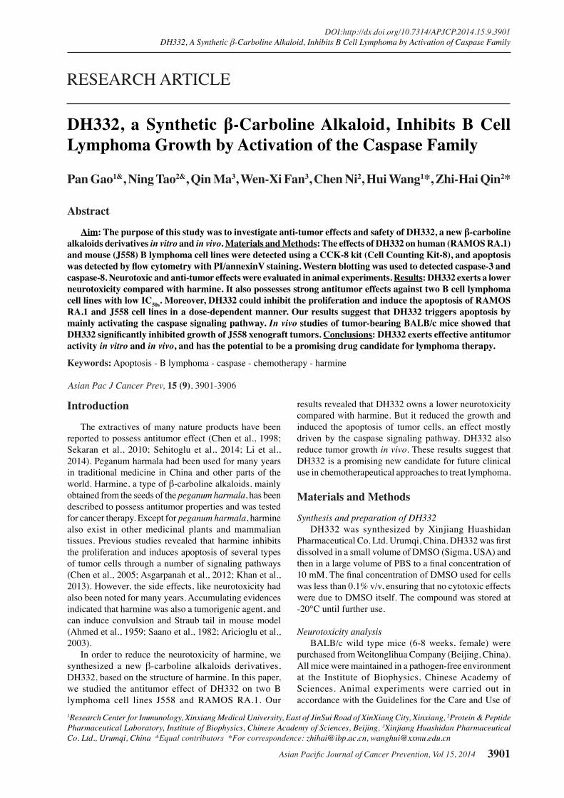
DH332 has lower neurotoxicity in comparison with harmine. Based on the structure of harmine, we synthesized a novel β-carboline alkaloids derivatives DH332 (Figure 1A) and its pharmacodynamic character was then investigated. Wild type (WT) mice were intraperitoneally injected with the same dose of DH332 or harmine and the neural responses of the treated mice were recorded. The results indicated that after treatment with harmine, the mice presented some neurotoxicity symptoms, like convulsion, Straub tail and tremble, which disappeared in 3-15 minutes after injection. However, when we administered DH332, no comparable symptoms could be detected (Figure 1B). Therefore, the neurotoxicity of DH332 is much lower than harmine. DH332 induced growth inhibition and apoptosis in B lymphoma cells. We tested the cytotoxicity of DH332 in vitro to assess the pharmacologic effect. Two B lymphoma cell lines, murine plasmacytoma cell line J558 and human B lymphoma cell line RAMOS RA.1, were used to determine the concentrations of DH332 that induce 50% growth inhibition (IC50) of tumor cells. As shown in Figure 1C, DH332 displayed a significant inhibitory effect on the two cell lines with IC50s (95% confidence intervals) of 8.02 to 11.22 μM and 9.08 to 12.90 μM, respectively. Next, we tested the effect of DH332 on the survival of J558 cells using the Cell Counting Kit-8 (CCK-8) assay. The J558 cells were exposed to various concentrations (0, 5, 10, 20 μM) of DH332 for 24 hrs, and OD450 values were recorded for cell viability. DH332 significantly reduced the growth of J558 cells in a dose-dependent manner (Figure 1D). The inhibition ratio of DH332 was
Asian Pacific Journal of Cancer Prevention, Vol 15, 2014 3903
DOI:http://dx.doi.org/10.7314/APJCP.2014.15.9.3901DH332, A Synthetic β-Carboline Alkaloid, Inhibits B Cell Lymphoma by Activation of Caspase Family
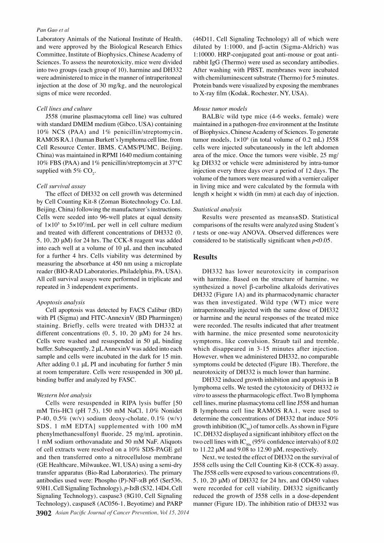
about 29%, 58% and 67% at the concentrations of 5, 10, 20 μM, respectively. Besides inhibition of cell survival, we also tested the impact of DH332 on apoptosis using the J558 cell line. Tumor cells were exposed to concentrations of DH332 identical to those used in the CCK-8 assay. After 24 hrs of DH332 treatment, cells were collected and analyzed by flow cytometry with PI/AnnexinV staining. As shown in Figure 1E, after treatment with DH332, both the percentage of the early (right lower quadrant, PI- AnnexinV+) and later (right upper quadrant, PI+ AnnexinV+) stage of apoptosis were found to increase in a dose-dependent manner. In addition, there is a significant difference of apoptosis index between 12 h and 48 h in 20 μM DH332 group (Figure 1F). DH332 activates the caspase family. The results demonstrated above raise the question about the mechanism behind the effect of DH332 regarding apoptosis induction and decreased growth of tumor cells. To this end, we investigated the changes in expression of caspases and PARP (poly ADP ribose polymerase) known to play essential roles in the apoptotic process. Our results shows the levels of the activated forms of caspase3 and caspase8 were up-regulated and the expression of full-length PARP was down-regulated after treatment with 10 μM DH332 for various time points (0, 4, 12, 24 h) (Figure 2A). z-VAD eliminates the DH332-mediated cell death. Since DH332 induce apoptosis in J558 cells through the
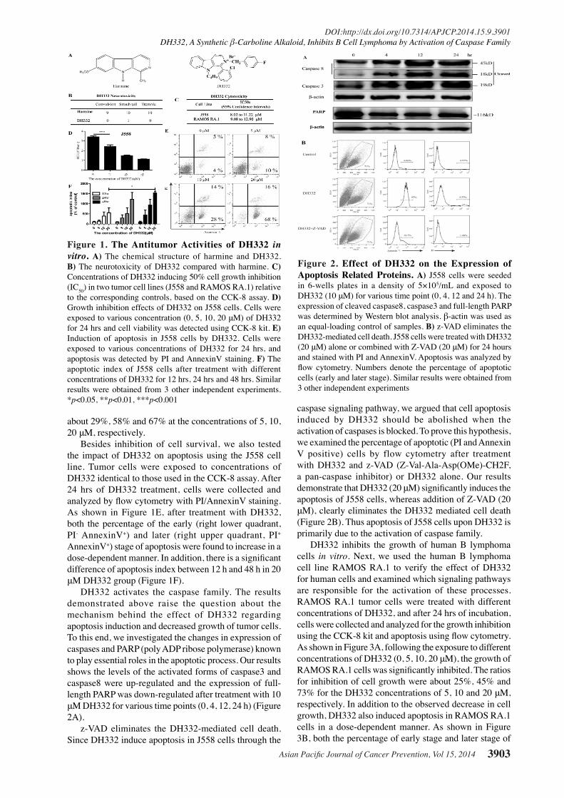
caspase signaling pathway, we argued that cell apoptosis induced by DH332 should be abolished when the activation of caspases is blocked. To prove this hypothesis, we examined the percentage of apoptotic (PI and Annexin V positive) cells by flow cytometry after treatment with DH332 and z-VAD (Z-Val-Ala-Asp(OMe)-CH2F, a pan-caspase inhibitor) or DH332 alone. Our results demonstrate that DH332 (20 μM) significantly induces the apoptosis of J558 cells, whereas addition of Z-VAD (20 μM), clearly eliminates the DH332 mediated cell death (Figure 2B). Thus apoptosis of J558 cells upon DH332 is primarily due to the activation of caspase family. DH332 inhibits the growth of human B lymphoma cells in vitro. Next, we used the human B lymphoma cell line RAMOS RA.1 to verify the effect of DH332 for human cells and examined which signaling pathways are responsible for the activation of these processes. RAMOS RA.1 tumor cells were treated with different concentrations of DH332, and after 24 hrs of incubation, cells were collected and analyzed for the growth inhibition using the CCK-8 kit and apoptosis using flow cytometry. As shown in Figure 3A, following the exposure to different concentrations of DH332 (0, 5, 10, 20 μM), the growth of RAMOS RA.1 cells was significantly inhibited. The ratios for inhibition of cell growth were about 25%, 45% and 73% for the DH332 concentrations of 5, 10 and 20 μM, respectively. In addition to the observed decrease in cell growth, DH332 also induced apoptosis in RAMOS RA.1 cells in a dose-dependent manner. As shown in Figure 3B, both the percentage of early stage and later stage of
Figure 1. The Antitumor Activities of DH332 in vitro. A) The chemical structure of harmine and DH332. B) The neurotoxicity of DH332 compared with harmine. C) Concentrations of DH332 inducing 50% cell growth inhibition (IC50) in two tumor cell lines (J558 and RAMOS RA.1) relative to the corresponding controls, based on the CCK-8 assay. D) Growth inhibition effects of DH332 on J558 cells. Cells were exposed to various concentration (0, 5, 10, 20 μM) of DH332 for 24 hrs and cell viability was detected using CCK-8 kit. E) Induction of apoptosis in J558 cells by DH332. Cells were exposed to various concentrations of DH332 for 24 hrs, and apoptosis was detected by PI and AnnexinV staining. F) The apoptotic index of J558 cells after treatment with different concentrations of DH332 for 12 hrs, 24 hrs and 48 hrs. Similar results were obtained from 3 other independent experiments. *p<0.05, **p<0.01, ***p<0.001
Figure 2. Effect of DH332 on the Expression of Apoptosis Related Proteins. A) J558 cells were seeded in 6-wells plates in a density of 5×10⁵/mL and exposed to DH332 (10 μM) for various time point (0, 4, 12 and 24 h). The expression of cleaved caspase8, caspase3 and full-length PARP was determined by Western blot analysis. β-actin was used as an equal-loading control of samples. B) z-VAD eliminates the DH332-mediated cell death. J558 cells were treated with DH332 (20 μM) alone or combined with Z-VAD (20 μM) for 24 hours and stained with PI and AnnexinV. Apoptosis was analyzed by flow cytometry. Numbers denote the percentage of apoptotic cells (early and later stage). Similar results were obtained from 3 other independent experiments
Pan Gao et al
Asian Pacific Journal of Cancer Prevention, Vol 15, 20143904
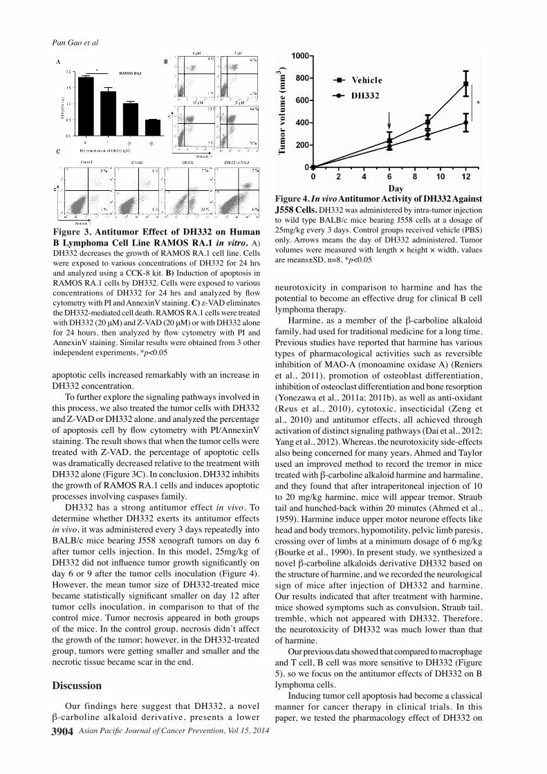
apoptotic cells increased remarkably with an increase in DH332 concentration. To further explore the signaling pathways involved in this process, we also treated the tumor cells with DH332 and Z-VAD or DH332 alone, and analyzed the percentage of apoptosis cell by flow cytometry with PI/AnnexinV staining. The result shows that when the tumor cells were treated with Z-VAD, the percentage of apoptotic cells was dramatically decreased relative to the treatment with DH332 alone (Figure 3C). In conclusion, DH332 inhibits the growth of RAMOS RA.1 cells and induces apoptotic processes involving caspases family. DH332 has a strong antitumor effect in vivo. To determine whether DH332 exerts its antitumor effects in vivo, it was administered every 3 days repeatedly into BALB/c mice bearing J558 xenograft tumors on day 6 after tumor cells injection. In this model, 25mg/kg of DH332 did not influence tumor growth significantly on day 6 or 9 after the tumor cells inoculation (Figure 4). However, the mean tumor size of DH332-treated mice became statistically significant smaller on day 12 after tumor cells inoculation, in comparison to that of the control mice. Tumor necrosis appeared in both groups of the mice. In the control group, necrosis didn’t affect the growth of the tumor; however, in the DH332-treated group, tumors were getting smaller and smaller and the necrotic tissue became scar in the end.
Discussion
Our findings here suggest that DH332, a novel β-carboline alkaloid derivative, presents a lower
neurotoxicity in comparison to harmine and has the potential to become an effective drug for clinical B cell lymphoma therapy.
Harmine, as a member of the β-carboline alkaloid family, had used for traditional medicine for a long time. Previous studies have reported that harmine has various types of pharmacological activities such as reversible inhibition of MAO-A (monoamine oxidase A) (Reniers et al., 2011), promotion of osteoblast differentiation, inhibition of osteoclast differentiation and bone resorption (Yonezawa et al., 2011a; 2011b), as well as anti-oxidant (Reus et al., 2010), cytotoxic, insecticidal (Zeng et al., 2010) and antitumor effects, all achieved through activation of distinct signaling pathways (Dai et al., 2012; Yang et al., 2012). Whereas, the neurotoxicity side-effects also being concerned for many years. Ahmed and Taylor used an improved method to record the tremor in mice treated with β-carboline alkaloid harmine and harmaline, and they found that after intraperitoneal injection of 10 to 20 mg/kg harmine, mice will appear tremor, Straub tail and hunched-back within 20 minutes (Ahmed et al., 1959). Harmine induce upper motor neurone effects like head and body tremors, hypomotility, pelvic limb paresis, crossing over of limbs at a minimum dosage of 6 mg/kg (Bourke et al., 1990). In present study, we synthesized a novel β-carboline alkaloids derivative DH332 based on the structure of harmine, and we recorded the neurological sign of mice after injection of DH332 and harmine. Our results indicated that after treatment with harmine, mice showed symptoms such as convulsion, Straub tail, tremble, which not appeared with DH332. Therefore, the neurotoxicity of DH332 was much lower than that of harmine.
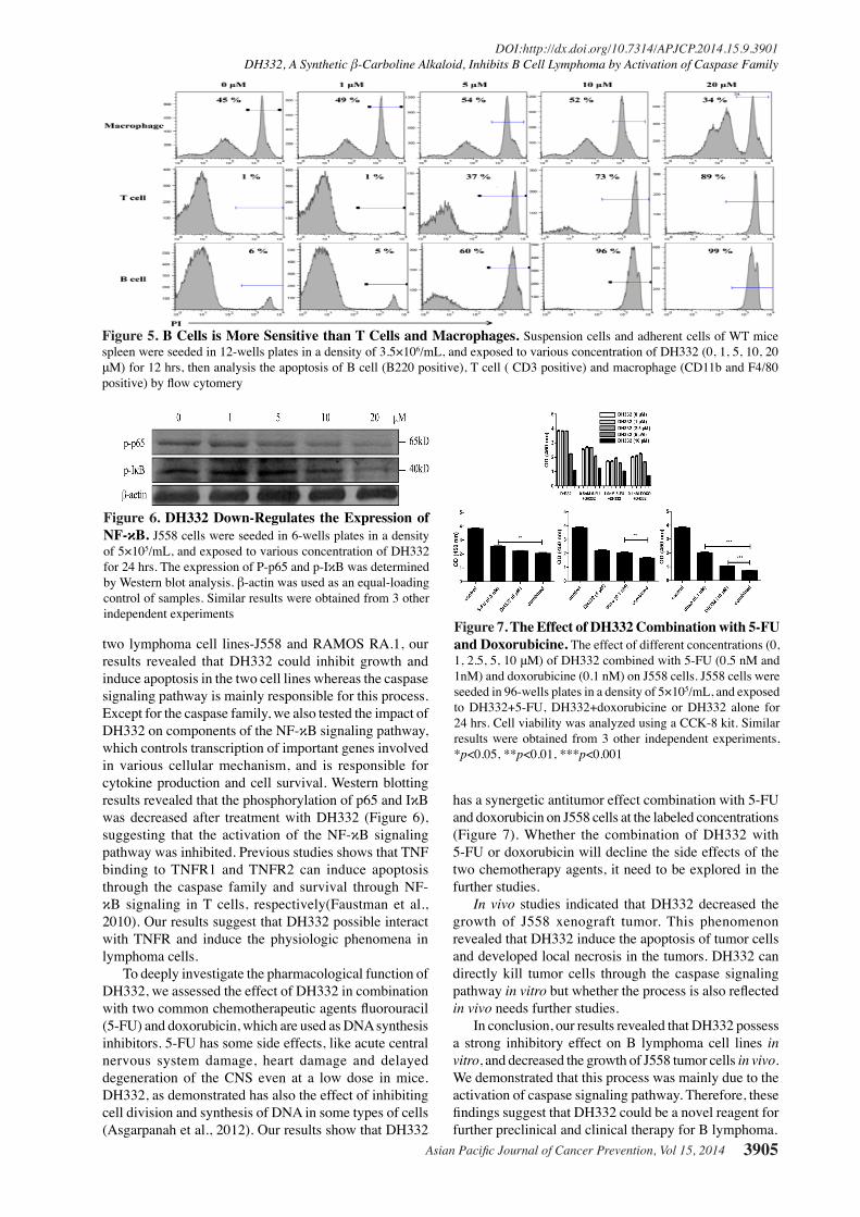
Our previous data showed that compared to macrophage and T cell, B cell was more sensitive to DH332 (Figure 5), so we focus on the antitumor effects of DH332 on B lymphoma cells.
Inducing tumor cell apoptosis had become a classical manner for cancer therapy in clinical trials. In this paper, we tested the pharmacology effect of DH332 on
Figure 3. Antitumor Effect of DH332 on Human B Lymphoma Cell Line RAMOS RA.1 in vitro. A) DH332 decreases the growth of RAMOS RA.1 cell line. Cells were exposed to various concentrations of DH332 for 24 hrs and analyzed using a CCK-8 kit. B) Induction of apoptosis in RAMOS RA.1 cells by DH332. Cells were exposed to various concentrations of DH332 for 24 hrs and analyzed by flow cytometry with PI and AnnexinV staining. C) z-VAD eliminates the DH332-mediated cell death. RAMOS RA.1 cells were treated with DH332 (20 μM) and Z-VAD (20 μM) or with DH332 alone for 24 hours, then analyzed by flow cytometry with PI and AnnexinV staining. Similar results were obtained from 3 other independent experiments, *p<0.05
Figure 4. In vivo Antitumor Activity of DH332 Against J558 Cells. DH332 was administered by intra-tumor injection to wild type BALB/c mice bearing J558 cells at a dosage of 25mg/kg every 3 days. Control groups received vehicle (PBS) only. Arrows means the day of DH332 administered. Tumor volumes were measured with length × height × width, values are means±SD, n=8, *p<0.05
Asian Pacific Journal of Cancer Prevention, Vol 15, 2014 3905
DOI:http://dx.doi.org/10.7314/APJCP.2014.15.9.3901DH332, A Synthetic β-Carboline Alkaloid, Inhibits B Cell Lymphoma by Activation of Caspase Family
has a synergetic antitumor effect combination with 5-FU and doxorubicin on J558 cells at the labeled concentrations (Figure 7). Whether the combination of DH332 with 5-FU or doxorubicin will decline the side effects of the two chemotherapy agents, it need to be explored in the further studies.
In vivo studies indicated that DH332 decreased the growth of J558 xenograft tumor. This phenomenon revealed that DH332 induce the apoptosis of tumor cells and developed local necrosis in the tumors. DH332 can directly kill tumor cells through the caspase signaling pathway in vitro but whether the process is also reflected in vivo needs further studies.
In conclusion, our results revealed that DH332 possess a strong inhibitory effect on B lymphoma cell lines in vitro, and decreased the growth of J558 tumor cells in vivo. We demonstrated that this process was mainly due to the activation of caspase signaling pathway. Therefore, these findings suggest that DH332 could be a novel reagent for further preclinical and clinical therapy for B lymphoma.
Figure 7. The Effect of DH332 Combination with 5-FU and Doxorubicine. The effect of different concentrations (0, 1, 2.5, 5, 10 μM) of DH332 combined with 5-FU (0.5 nM and 1nM) and doxorubicine (0.1 nM) on J558 cells. J558 cells were seeded in 96-wells plates in a density of 5×10⁵/mL, and exposed to DH332+5-FU, DH332+doxorubicine or DH332 alone for 24 hrs. Cell viability was analyzed using a CCK-8 kit. Similar results were obtained from 3 other independent experiments, *p<0.05, **p<0.01, ***p<0.001
Figure 5. B Cells is More Sensitive than T Cells and Macrophages. Suspension cells and adherent cells of WT mice spleen were seeded in 12-wells plates in a density of 3.5×10⁶/mL, and exposed to various concentration of DH332 (0, 1, 5, 10, 20 μM) for 12 hrs, then analysis the apoptosis of B cell (B220 positive), T cell ( CD3 positive) and macrophage (CD11b and F4/80 positive) by flow cytomery
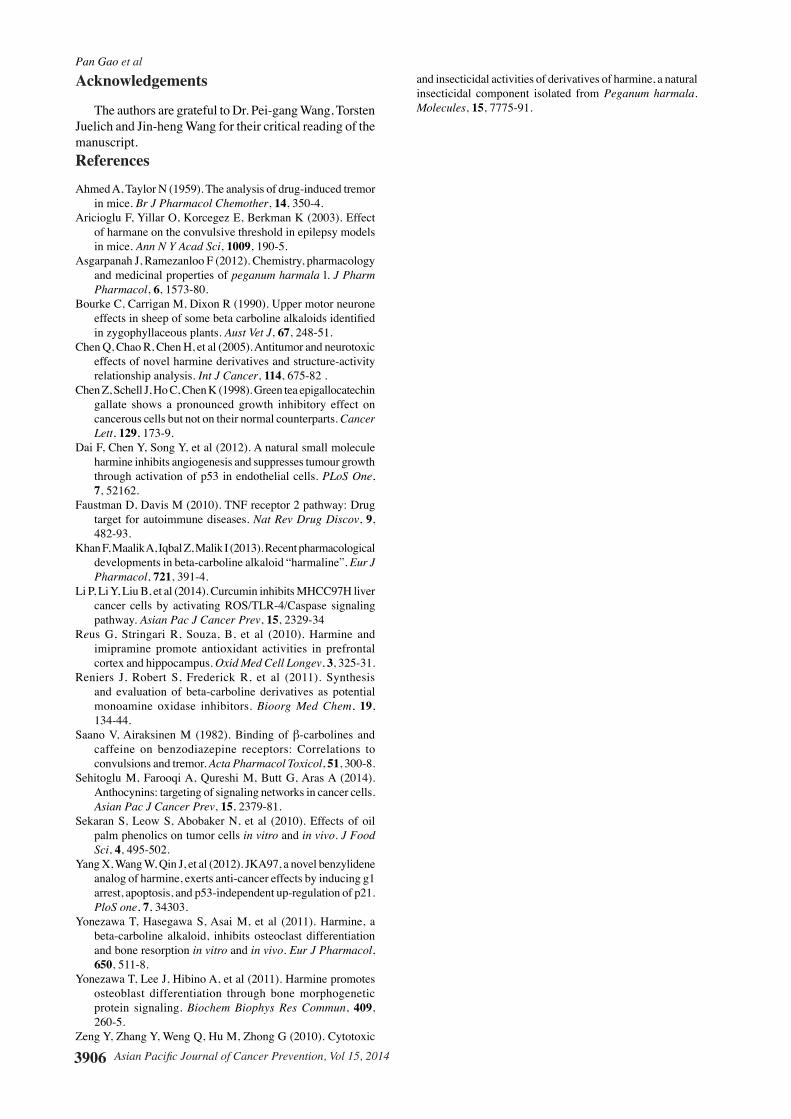
Figure 6. DH332 Down-Regulates the Expression of NF-κB. J558 cells were seeded in 6-wells plates in a density of 5×10⁵/mL, and exposed to various concentration of DH332 for 24 hrs. The expression of P-p65 and p-IκB was determined by Western blot analysis. β-actin was used as an equal-loading control of samples. Similar results were obtained from 3 other independent experiments
two lymphoma cell lines-J558 and RAMOS RA.1, our results revealed that DH332 could inhibit growth and induce apoptosis in the two cell lines whereas the caspase signaling pathway is mainly responsible for this process. Except for the caspase family, we also tested the impact of DH332 on components of the NF-κB signaling pathway, which controls transcription of important genes involved in various cellular mechanism, and is responsible for cytokine production and cell survival. Western blotting results revealed that the phosphorylation of p65 and IκB was decreased after treatment with DH332 (Figure 6), suggesting that the activation of the NF-κB signaling pathway was inhibited. Previous studies shows that TNF binding to TNFR1 and TNFR2 can induce apoptosis through the caspase family and survival through NF-κB signaling in T cells, respectively(Faustman et al., 2010). Our results suggest that DH332 possible interact with TNFR and induce the physiologic phenomena in lymphoma cells.
To deeply investigate the pharmacological function of DH332, we assessed the effect of DH332 in combination with two common chemotherapeutic agents fluorouracil (5-FU) and doxorubicin, which are used as DNA synthesis inhibitors. 5-FU has some side effects, like acute central nervous system damage, heart damage and delayed degeneration of the CNS even at a low dose in mice. DH332, as demonstrated has also the effect of inhibiting cell division and synthesis of DNA in some types of cells (Asgarpanah et al., 2012). Our results show that DH332
Pan Gao et al
Asian Pacific Journal of Cancer Prevention, Vol 15, 20143906
Acknowledgements
The authors are grateful to Dr. Pei-gang Wang, Torsten Juelich and Jin-heng Wang for their critical reading of the manuscript. References
Ahmed A, Taylor N (1959). The analysis of drug-induced tremor in mice. Br J Pharmacol Chemother, 14, 350-4.
Aricioglu F, Yillar O, Korcegez E, Berkman K (2003). Effect of harmane on the convulsive threshold in epilepsy models in mice. Ann N Y Acad Sci, 1009, 190-5.
Asgarpanah J, Ramezanloo F (2012). Chemistry, pharmacology and medicinal properties of peganum harmala l. J Pharm Pharmacol, 6, 1573-80.
Bourke C, Carrigan M, Dixon R (1990). Upper motor neurone effects in sheep of some beta carboline alkaloids identified in zygophyllaceous plants. Aust Vet J, 67, 248-51.
Chen Q, Chao R, Chen H, et al (2005). Antitumor and neurotoxic effects of novel harmine derivatives and structure-activity relationship analysis. Int J Cancer, 114, 675-82 .
Chen Z, Schell J, Ho C, Chen K (1998). Green tea epigallocatechin gallate shows a pronounced growth inhibitory effect on cancerous cells but not on their normal counterparts. Cancer Lett, 129, 173-9.
Dai F, Chen Y, Song Y, et al (2012). A natural small molecule harmine inhibits angiogenesis and suppresses tumour growth through activation of p53 in endothelial cells. PLoS One, 7, 52162.
Faustman D, Davis M (2010). TNF receptor 2 pathway: Drug target for autoimmune diseases. Nat Rev Drug Discov, 9, 482-93.
Khan F, Maalik A, Iqbal Z, Malik I (2013). Recent pharmacological developments in beta-carboline alkaloid “harmaline”. Eur J Pharmacol, 721, 391-4.
Li P, Li Y, Liu B, et al (2014). Curcumin inhibits MHCC97H liver cancer cells by activating ROS/TLR-4/Caspase signaling pathway. Asian Pac J Cancer Prev, 15, 2329-34
Reus G, Stringari R, Souza, B, et al (2010). Harmine and imipramine promote antioxidant activities in prefrontal cortex and hippocampus. Oxid Med Cell Longev, 3, 325-31.
Reniers J, Robert S, Frederick R, et al (2011). Synthesis and evaluation of beta-carboline derivatives as potential monoamine oxidase inhibitors. Bioorg Med Chem, 19, 134-44.
Saano V, Airaksinen M (1982). Binding of β-carbolines and caffeine on benzodiazepine receptors: Correlations to convulsions and tremor. Acta Pharmacol Toxicol, 51, 300-8.
Sehitoglu M, Farooqi A, Qureshi M, Butt G, Aras A (2014). Anthocynins: targeting of signaling networks in cancer cells. Asian Pac J Cancer Prev, 15, 2379-81.
Sekaran S, Leow S, Abobaker N, et al (2010). Effects of oil palm phenolics on tumor cells in vitro and in vivo. J Food Sci, 4, 495-502.
Yang X, Wang W, Qin J, et al (2012). JKA97, a novel benzylidene analog of harmine, exerts anti-cancer effects by inducing g1 arrest, apoptosis, and p53-independent up-regulation of p21. PloS one, 7, 34303.
Yonezawa T, Hasegawa S, Asai M, et al (2011). Harmine, a beta-carboline alkaloid, inhibits osteoclast differentiation and bone resorption in vitro and in vivo. Eur J Pharmacol, 650, 511-8.
Yonezawa T, Lee J, Hibino A, et al (2011). Harmine promotes osteoblast differentiation through bone morphogenetic protein signaling. Biochem Biophys Res Commun, 409, 260-5.
Zeng Y, Zhang Y, Weng Q, Hu M, Zhong G (2010). Cytotoxic
and insecticidal activities of derivatives of harmine, a natural insecticidal component isolated from Peganum harmala. Molecules, 15, 7775-91.


![Volumetric Assessment of Hepatocellular Carcinoma …tumor cell signaling pathways resulting in reduction of tumor neoangiogenesis and stimulation of apoptosis.[2, 3] Macroscopically,](https://img.pdfslide.us/doc/110x75/5e60835231ad4c0442607404/volumetric-assessment-of-hepatocellular-carcinoma-tumor-cell-signaling-pathways.jpg)