Embed Size (px)
Citation preview
1
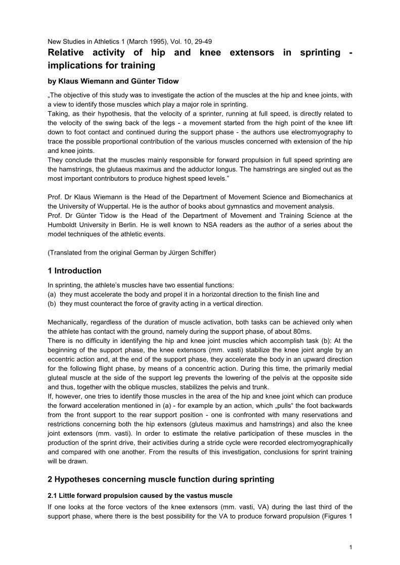
New Studies in Athletics 1 (March 1995), Vol. 10, 29-49
Relative activity of hip and knee extensors in sprinting - implications for training by Klaus Wiemann and Günter Tidow
„The objective of this study was to investigate the action of the muscles at the hip and knee joints, with a view to identify those muscles which play a major role in sprinting. Taking, as their hypothesis, that the velocity of a sprinter, running at full speed, is directly related to the velocity of the swing back of the legs - a movement started from the high point of the knee lift down to foot contact and continued during the support phase - the authors use electromyography to trace the possible proportional contribution of the various muscles concerned with extension of the hip and knee joints. They conclude that the muscles mainly responsible for forward propulsion in full speed sprinting are the hamstrings, the glutaeus maximus and the adductor longus. The hamstrings are singled out as the most important contributors to produce highest speed levels.” Prof. Dr Klaus Wiemann is the Head of the Department of Movement Science and Biomechanics at the University of Wuppertal. He is the author of books about gymnastics and movement analysis. Prof. Dr Günter Tidow is the Head of the Department of Movement and Training Science at the Humboldt University in Berlin. He is well known to NSA readers as the author of a series about the model techniques of the athletic events. (Translated from the original German by Jürgen Schiffer)
1 Introduction In sprinting, the athlete’s muscles have two essential functions: (a) they must accelerate the body and propel it in a horizontal direction to the finish line and (b) they must counteract the force of gravity acting in a vertical direction. Mechanically, regardless of the duration of muscle activation, both tasks can be achieved only when the athlete has contact with the ground, namely during the support phase, of about 80ms. There is no difficulty in identifying the hip and knee joint muscles which accomplish task (b): At the beginning of the support phase, the knee extensors (mm. vasti) stabilize the knee joint angle by an eccentric action and, at the end of the support phase, they accelerate the body in an upward direction for the following flight phase, by means of a concentric action. During this time, the primarily medial gluteal muscle at the side of the support leg prevents the lowering of the pelvis at the opposite side and thus, together with the oblique muscles, stabilizes the pelvis and trunk. If, however, one tries to identify those muscles in the area of the hip and knee joint which can produce the forward acceleration mentioned in (a) - for example by an action, which „pulls“ the foot backwards from the front support to the rear support position - one is confronted with many reservations and restrictions concerning both the hip extensors (gluteus maximus and hamstrings) and also the knee joint extensors (mm. vasti). In order to estimate the relative participation of these muscles in the production of the sprint drive, their activities during a stride cycle were recorded electromyographically and compared with one another. From the results of this investigation, conclusions for sprint training will be drawn.
2 Hypotheses concerning muscle function during sprinting
2.1 Little forward propulsion caused by the vastus muscle If one looks at the force vectors of the knee extensors (mm. vasti, VA) during the last third of the support phase, where there is the best possibility for the VA to produce forward propulsion (Figures 1

2
a and 1 b), the disproportion between the vertical and the horizontal force components is obvious. In the case of maximum activation, the VA would produce an upward rather than a forward propulsion of the body because of the greater proportion of vertical force components. This would lead to too high a flight phase. Therefore, it is to be expected that the VA, if they contract at all during the rear support, do not do so maximally but only to such a degree that the optimization of the height of the flight phase guarantees the maximization of the stride rate. This means that, at least during that phase of the sprint which is characterized by the athlete running with his trunk upright, the VA cannot be considered as a muscle producing forward acceleration. In spite of this, MANN et al. (1986) as well as FARRAR & THORLAND (1987) regard the knee extensors as the main accelerators during the sprint. However, this can - if at all - only be true of that phase after the start, during which the athlete still runs with a pronounced forward body lean (Figure 1 c).
Figure 1: Splitting of the vectors of the force of the knee extension (fv) into a vertical (cv) and a horizontal
component (ch) during two phases of the rear support (a and b) of full sprinting as well as during the rear support of the starting phase (c)
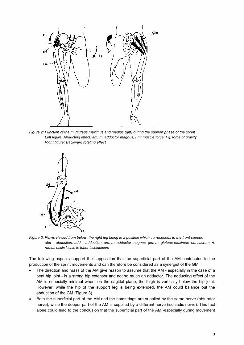
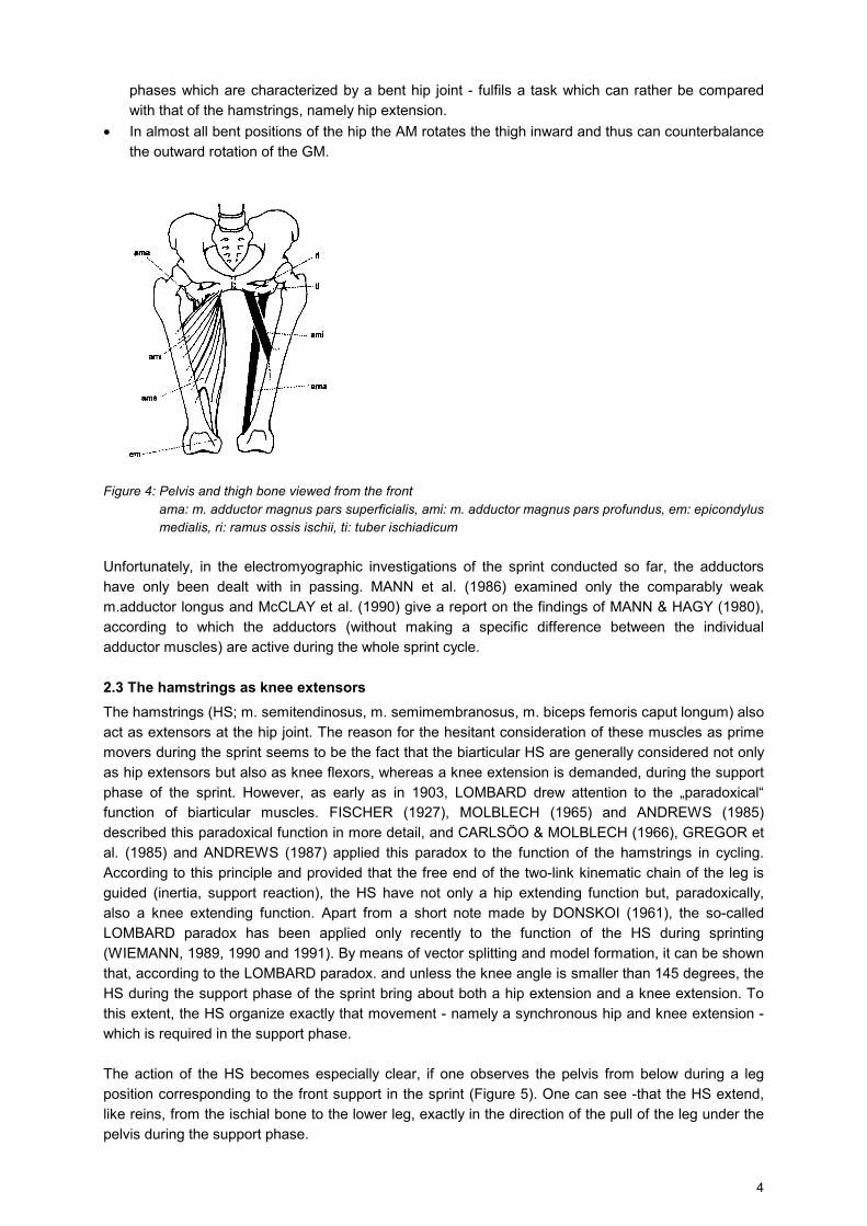
2.2 The search for a synergist of the gluteus maximus muscle As forward acceleration in sprinting is obviously produced by hip extension rather than knee extension (WASER 1985, LEMAIRE, & ROBERTSON 1989, AE et al. 1992), it seems plausible to suppose that the strongest hip extensor, namely the m. gluteus maximus (GM), takes a major part in this extension. However, there are some doubts concerning this supposition, because the GM does not function only as a hip extensor; it also rotates the thigh outward, especially when the hip joint is extended. This could cause a backward rotation of the pelvis on the side of the free leg during the support phase (Figure 2, right side), which might hinder the long forward swing of the free leg. Furthermore, the GM also has an abducting effect on the leg - especially in the case of an angled hip joint (Figure 3). This could have a negative effect on the straight movement of the support leg from front to back. However, outward rotation and abduction would not be of any consequence in the sprint, if another muscle could act together with the GM, both to support the GM during the hip extension and also to neutralize the abducting effect of the GM. This task could be taken over by the m. adductor magnus (AM), especially by its superficial part, which has its origin exactly at the ischial bone, medially beside the origin of the hamstrings and inserts to the medial epicondyle (Figures 3 and 4).
3
Figure 2: Function of the m. gluteus maximus and medius (gm) during the support phase of the sprint
Left figure: Abducting effect. am: m. adductor magnus, Fm: muscle force, Fg: force of gravity Right figure: Backward rotating effect
Figure 3: Pelvis viewed from below, the right leg being in a position which corresponds to the front support
abd = abduction, add = adduction, am: m. adductor magnus, gm: m. gluteus maximus, os: sacrum, ri: ramus ossis ischii, ti: tuber ischiadicum
The following aspects support the supposition that the superficial part of the AM contributes to the production of the sprint movements and can therefore be considered as a synergist of the GM: • The direction and mass of the AM give reason to assume that the AM - especially in the case of a
bent hip joint - is a strong hip extensor and not so much an adductor. The adducting effect of the AM is especially minimal when, on the sagittal plane, the thigh is vertically below the hip joint. However, while the hip of the support leg is being extended, the AM could balance out the abduction of the GM (Figure 3).
• Both the superficial part of the AM and the hamstrings are supplied by the same nerve (obturator nerve), while the deeper part of the AM is supplied by a different nerve (ischiadic nerve). This fact alone could lead to the conclusion that the superficial part of the AM -especially during movement
4
phases which are characterized by a bent hip joint - fulfils a task which can rather be compared with that of the hamstrings, namely hip extension.
• In almost all bent positions of the hip the AM rotates the thigh inward and thus can counterbalance the outward rotation of the GM.
Figure 4: Pelvis and thigh bone viewed from the front
ama: m. adductor magnus pars superficialis, ami: m. adductor magnus pars profundus, em: epicondylus medialis, ri: ramus ossis ischii, ti: tuber ischiadicum
Unfortunately, in the electromyographic investigations of the sprint conducted so far, the adductors have only been dealt with in passing. MANN et al. (1986) examined only the comparably weak m.adductor longus and McCLAY et al. (1990) give a report on the findings of MANN & HAGY (1980), according to which the adductors (without making a specific difference between the individual adductor muscles) are active during the whole sprint cycle.
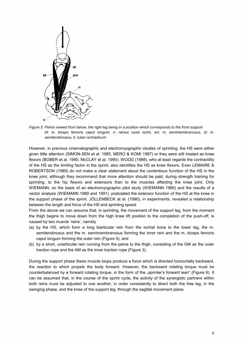
2.3 The hamstrings as knee extensors The hamstrings (HS; m. semitendinosus, m. semimembranosus, m. biceps femoris caput longum) also act as extensors at the hip joint. The reason for the hesitant consideration of these muscles as prime movers during the sprint seems to be the fact that the biarticular HS are generally considered not only as hip extensors but also as knee flexors, whereas a knee extension is demanded, during the support phase of the sprint. However, as early as in 1903, LOMBARD drew attention to the „paradoxical“ function of biarticular muscles. FISCHER (1927), MOLBLECH (1965) and ANDREWS (1985) described this paradoxical function in more detail, and CARLSÖO & MOLBLECH (1966), GREGOR et al. (1985) and ANDREWS (1987) applied this paradox to the function of the hamstrings in cycling. According to this principle and provided that the free end of the two-link kinematic chain of the leg is guided (inertia, support reaction), the HS have not only a hip extending function but, paradoxically, also a knee extending function. Apart from a short note made by DONSKOI (1961), the so-called LOMBARD paradox has been applied only recently to the function of the HS during sprinting (WIEMANN, 1989, 1990 and 1991). By means of vector splitting and model formation, it can be shown that, according to the LOMBARD paradox. and unless the knee angle is smaller than 145 degrees, the HS during the support phase of the sprint bring about both a hip extension and a knee extension. To this extent, the HS organize exactly that movement - namely a synchronous hip and knee extension - which is required in the support phase. The action of the HS becomes especially clear, if one observes the pelvis from below during a leg position corresponding to the front support in the sprint (Figure 5). One can see -that the HS extend, like reins, from the ischial bone to the lower leg, exactly in the direction of the pull of the leg under the pelvis during the support phase.
5
Figure 5: Pelvis viewed from below, the right leg being in a position which corresponds to the front support
bf: m. biceps femoris caput longum, ri: ramus ossis ischii, sm: m. semimembranosus, st: m. semitendinosius, ti: tuber ischiadicum
However, in previous cinematographic and electromyographic studies of sprinting, the HS were either given little attention (SIMON-SEN et al. 1985, MERO & KOMI 1987) or they were still treated as knee flexors (BOBER et al. 1990, McCLAY et al. 1990). WOOD (1988), who at least regards the contractility of the HS as the limiting factor in the sprint, also identifies the HS as knee flexors. Even LEMAIRE & ROBERTSON (1989) do not make a clear statement about the contentious function of the HS in the knee joint, although they recommend that more attention should be paid, during strength training for sprinting, to the hip flexors and extensors than to the muscles affecting the knee joint, Only WIEMANN, on the basis of an electromyographic pilot study (WIEMANN 1986) and the results of a vector analysis (WIEMANN 1989 and 1991), postulated the extensor function of the HS at the knee in the support phase of the sprint. JÖLLENBECK at al. (1990), in experiments, revealed a relationship between the length and force of the HS and sprinting speed. From the above we can assume that, in sprinting, the movement of the support leg, from the moment the thigh begins to move down from the high knee lift position to the completion of the push-off, is caused by two muscle ‘reins’, namely (a) by the HS, which form a long biarticular rein from the ischial bone to the lower leg, the m.
semitendinosus and the m. semimembranosus forming the inner rein and the m. biceps femoris caput longum forming the outer rein (Figure 5), and
(b) by a short, uniarticular rein running from the pelvis to the thigh, consisting of the GM as the outer traction rope and the AM as the inner traction rope (Figure 3).
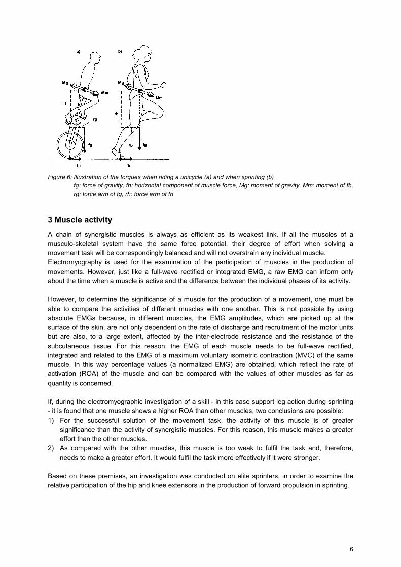
During the support phase these muscle loops produce a force which is directed horizontally backward, the reaction to which propels the body forward. However, the backward rotating torque must be counterbalanced by a forward rotating torque, in the form of the „sprinter’s forward lean“ (Figure 6). It can be assumed that, in the course of the sprint cycle, the activity of the synergistic partners within both reins must be adjusted to one another, in order consistently to direct both the free leg, in the swinging phase, and the knee of the support leg, through the sagittal movement plane.
6
Figure 6: Illustration of the torques when riding a unicycle (a) and when sprinting (b)
fg: force of gravity, fh: horizontal component of muscle force, Mg: moment of gravity, Mm: moment of fh, rg: force arm of fg, rh: force arm of fh
3 Muscle activity A chain of synergistic muscles is always as efficient as its weakest link. If all the muscles of a musculo-skeletal system have the same force potential, their degree of effort when solving a movement task will be correspondingly balanced and will not overstrain any individual muscle. Electromyography is used for the examination of the participation of muscles in the production of movements. However, just like a full-wave rectified or integrated EMG, a raw EMG can inform only about the time when a muscle is active and the difference between the individual phases of its activity. However, to determine the significance of a muscle for the production of a movement, one must be able to compare the activities of different muscles with one another. This is not possible by using absolute EMGs because, in different muscles, the EMG amplitudes, which are picked up at the surface of the skin, are not only dependent on the rate of discharge and recruitment of the motor units but are also, to a large extent, affected by the inter-electrode resistance and the resistance of the subcutaneous tissue. For this reason, the EMG of each muscle needs to be full-wave rectified, integrated and related to the EMG of a maximum voluntary isometric contraction (MVC) of the same muscle. In this way percentage values (a normalized EMG) are obtained, which reflect the rate of activation (ROA) of the muscle and can be compared with the values of other muscles as far as quantity is concerned. If, during the electromyographic investigation of a skill - in this case support leg action during sprinting - it is found that one muscle shows a higher ROA than other muscles, two conclusions are possible: 1) For the successful solution of the movement task, the activity of this muscle is of greater
significance than the activity of synergistic muscles. For this reason, this muscle makes a greater effort than the other muscles.
2) As compared with the other muscles, this muscle is too weak to fulfil the task and, therefore, needs to make a greater effort. It would fulfil the task more effectively if it were stronger.
Based on these premises, an investigation was conducted on elite sprinters, in order to examine the relative participation of the hip and knee extensors in the production of forward propulsion in sprinting.
7
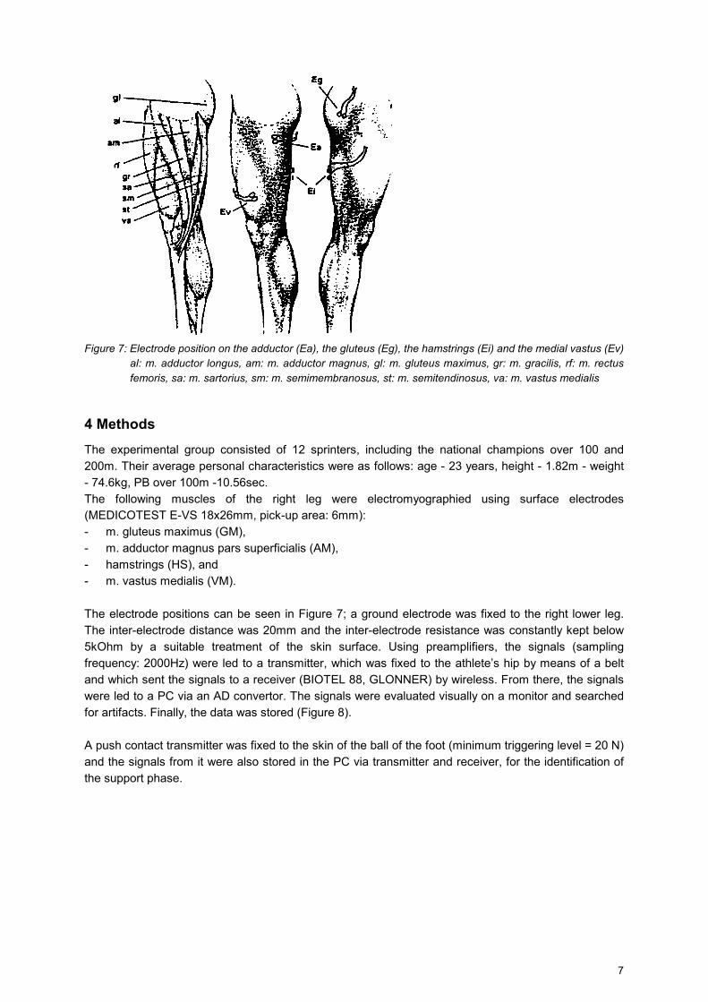
Figure 7: Electrode position on the adductor (Ea), the gluteus (Eg), the hamstrings (Ei) and the medial vastus (Ev)
al: m. adductor longus, am: m. adductor magnus, gl: m. gluteus maximus, gr: m. gracilis, rf: m. rectus femoris, sa: m. sartorius, sm: m. semimembranosus, st: m. semitendinosus, va: m. vastus medialis
4 Methods The experimental group consisted of 12 sprinters, including the national champions over 100 and 200m. Their average personal characteristics were as follows: age - 23 years, height - 1.82m - weight - 74.6kg, PB over 100m -10.56sec. The following muscles of the right leg were electromyographied using surface electrodes (MEDICOTEST E-VS 18x26mm, pick-up area: 6mm): - m. gluteus maximus (GM), - m. adductor magnus pars superficialis (AM), - hamstrings (HS), and - m. vastus medialis (VM). The electrode positions can be seen in Figure 7; a ground electrode was fixed to the right lower leg. The inter-electrode distance was 20mm and the inter-electrode resistance was constantly kept below 5kOhm by a suitable treatment of the skin surface. Using preamplifiers, the signals (sampling frequency: 2000Hz) were led to a transmitter, which was fixed to the athlete’s hip by means of a belt and which sent the signals to a receiver (BIOTEL 88, GLONNER) by wireless. From there, the signals were led to a PC via an AD convertor. The signals were evaluated visually on a monitor and searched for artifacts. Finally, the data was stored (Figure 8). A push contact transmitter was fixed to the skin of the ball of the foot (minimum triggering level = 20 N) and the signals from it were also stored in the PC via transmitter and receiver, for the identification of the support phase.
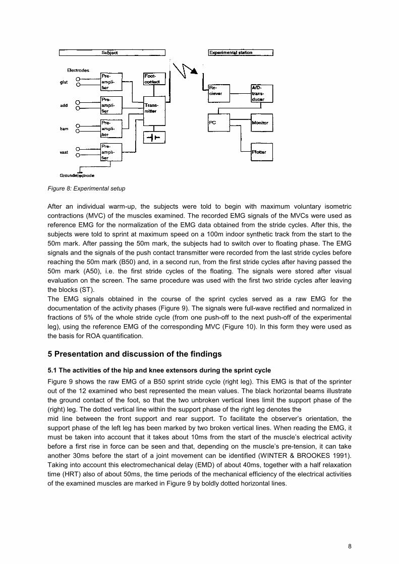
8
Figure 8: Experimental setup After an individual warm-up, the subjects were told to begin with maximum voluntary isometric contractions (MVC) of the muscles examined. The recorded EMG signals of the MVCs were used as reference EMG for the normalization of the EMG data obtained from the stride cycles. After this, the subjects were told to sprint at maximum speed on a 100m indoor synthetic track from the start to the 50m mark. After passing the 50m mark, the subjects had to switch over to floating phase. The EMG signals and the signals of the push contact transmitter were recorded from the last stride cycles before reaching the 50m mark (B50) and, in a second run, from the first stride cycles after having passed the 50m mark (A50), i.e. the first stride cycles of the floating. The signals were stored after visual evaluation on the screen. The same procedure was used with the first two stride cycles after leaving the blocks (ST). The EMG signals obtained in the course of the sprint cycles served as a raw EMG for the documentation of the activity phases (Figure 9). The signals were full-wave rectified and normalized in fractions of 5% of the whole stride cycle (from one push-off to the next push-off of the experimental leg), using the reference EMG of the corresponding MVC (Figure 10). In this form they were used as the basis for ROA quantification.
5 Presentation and discussion of the findings
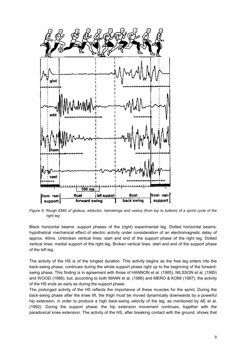
5.1 The activities of the hip and knee extensors during the sprint cycle Figure 9 shows the raw EMG of a B50 sprint stride cycle (right leg). This EMG is that of the sprinter out of the 12 examined who best represented the mean values. The black horizontal beams illustrate the ground contact of the foot, so that the two unbroken vertical lines limit the support phase of the (right) leg. The dotted vertical line within the support phase of the right leg denotes the mid line between the front support and rear support. To facilitate the observer’s orientation, the support phase of the left leg has been marked by two broken vertical lines. When reading the EMG, it must be taken into account that it takes about 10ms from the start of the muscle’s electrical activity before a first rise in force can be seen and that, depending on the muscle’s pre-tension, it can take another 30ms before the start of a joint movement can be identified (WINTER & BROOKES 1991). Taking into account this electromechanical delay (EMD) of about 40ms, together with a half relaxation time (HRT) also of about 50ms, the time periods of the mechanical efficiency of the electrical activities of the examined muscles are marked in Figure 9 by boldly dotted horizontal lines.
9
Figure 9: Rough EMG of gluteus, adductor, hamstrings and vastus (from top to bottom) of a sprint cycle of the
right leg Black horizontal beams: support phases of the (right) experimental leg. Dotted horizontal beams: hypothetical mechanical effect of electric activity under consideration of an electromagnetic delay of approx. 40ms. Unbroken vertical lines: start and end of the support phase of the right leg. Dotted vertical lines: medial support of the right leg. Broken vertical lines: start and end of the support phase of the left leg. The activity of the HS is of the longest duration. This activity begins as the free leg enters into the back-swing phase, continues during the whole support phase right up to the beginning of the forward-swing phase. This finding is in agreement with those of HANNON et al. (1985), NILSSON et al. (1985) and WOOD (1986), but, according to both MANN et al. (1986) and MERO & KOMI (1987), the activity of the HS ends as early as during the support phase. The prolonged activity of the HS reflects the importance of these muscles for the sprint. During the back-swing phase after the knee lift, the thigh must be moved dynamically downwards by a powerful hip extension, in order to produce a high back-swing velocity of the leg, as mentioned by AE et al. (1992). During the support phase, the hip extension movement continues, together with the paradoxical knee extension. The activity of the HS, after breaking contact with the ground, shows that
10
here, during the full speed Sprint, the knee flexion is not brought about passively by inertia but actively through the contraction of the HS.
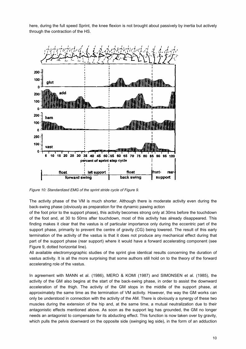
Figure 10: Standardized EMG of the sprint stride cycle of Figure 9. The activity phase of the VM is much shorter. Although there is moderate activity even during the back-swing phase (obviously as preparation for the dynamic pawing action of the foot prior to the support phase), this activity becomes strong only at 30ms before the touchdown of the foot and, at 30 to 50ms after touchdown, most of this activity has already disappeared. This finding makes it clear that the vastus is of particular importance only during the eccentric part of the support phase, primarily to prevent the centre of gravity (CG) being lowered. The result of this early termination of the activity of the vastus is that it does not produce any mechanical effect during that part of the support phase (rear support) where it would have a forward accelerating component (see Figure 9, dotted horizontal line). All available electromyographic studies of the sprint give identical results concerning the duration of vastus activity. It is all the more surprising that some authors still hold on to the theory of the forward accelerating role of the vastus. In agreement with MANN et al. (1986), MERO & KOMI (1987) and SIMONSEN et al. (1985), the activity of the GM also begins at the start of the back-swing phase, in order to assist the downward acceleration of the thigh. The activity of the GM stops in the middle of the support phase, at approximately the same time as the termination of VM activity. However, the way the GM works can only be understood in connection with the activity of the AM. There is obviously a synergy of these two muscles during the extension of the hip and, at the same time, a mutual neutralization due to their antagonistic effects mentioned above. As soon as the support leg has grounded, the GM no longer needs an antagonist to compensate for its abducting effect. This function is now taken over by gravity, which pulls the pelvis downward on the opposite side (swinging leg side), in the form of an adduction
11
of the support leg (Figure 2, left side). To put it another way, the task which the GM must take over during the support phase, namely the stabilization of the pelvis against the effect of gravity, makes the abduction-neutralizing function of the AM superfluous. Thus the AM can stop its activity approx. 30ms prior to the grounding of the foot. This premature cessation of activity is possible only due to muscle stiffness existing during relaxation time. Another reason for this may be the fact that the thigh has meanwhile been moved backwards up to the height of the origin of the AM, so that the AM loses its hip-extending effect. Towards the end of the eccentric part of the support phase, the GM stops its activity, together with the vastus, and leaves further hip extension to the HS alone. The AM is the only one of the muscles examined to show a second activity phase during the sprint stride cycle. This phase extends over the first half of the forwardswing phase. It may be assumed that here the AM works as a hip flexor and, together with other hip flexors, has the task of slowing down the extension movement of the hip joint and transferring it into a hip flexing movement. In the course of this action, the AM will counterbalance other hip flexors, in order to neutralize their abducting (m. tensor fasciae latae) and outward rotating (m. sartorius) effect. However, the activity of the AM has come to an end when the knee is vertically below the hip. As in this position the thigh moves in a forward direction besides the origin of the AM at the pelvis, the hip-flexing effect of the AM is changed into hip extension.
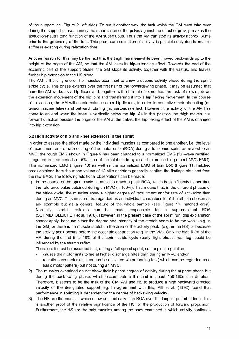
5.2 High activity of hip and knee extensors in the sprint In order to assess the effort made by the individual muscles as compared to one another, i.e. the level of recruitment and of rate coding of the motor units (ROA) during a full-speed sprint as related to an MVC, the rough EMG shown in Figure 9 has been changed to a normalized EMG (full-wave rectified, integrated in time periods of 5% each of the total stride cycle and expressed in percent MVC-EMG). This normalized EMG (Figure 10) as well as the normalized EMG of task B50 (Figure 11, hatched area) obtained from the mean values of 12 elite sprinters generally confirm the findings obtained from the raw EMG. The following additional observations can be made: 1) In the course of the sprint cycle all muscles reach a peak ROA, which is significantly higher than
the reference value obtained during an MVC (= 100%). This means that, in the different phases of the stride cycle, the muscles show a higher degree of recruitment and/or rate of activation than during an MVC. This must not be regarded as an individual characteristic of the athlete chosen as an- example but as a general feature of the whole sample (see Figure 11, hatched area). Normally, stretch reflexes can be made responsible for a supramaximal ROA (SCHMIDTBLEICHER et al. 1978). However, in the present case of the sprint run, this explanation cannot apply, because either the degree and intensity of the stretch seem to be too weak (e.g. in the GM) or there is no muscle stretch in the area of the activity peak, (e.g. in the HS) or because the activity peak occurs before the eccentric contraction (e.g. in the VM). Only the high ROA of the AM during the first 5 to 10% of the sprint stride cycle (early flight phase; rear leg) could be influenced by the stretch reflex. Therefore it must be assumed that, during a full-speed sprint, supraspinal regulation - causes the motor units to fire at higher discharge rates than during an MVC and/or - recruits such motor units as can be activated when running fast( which can be regarded as a
basic motor pattern) but not during an MVC. 2) The muscles examined do not show their highest degree of activity during the support phase but
during the back-swing phase, which occurs before this and is about 150-160ms in duration. Therefore, it seems to be the task of the GM, AM and HS to produce a high backward directed velocity of the designated support leg. In agreement with this, AE et al. (1992) found that performance in sprinting is dependent on the degree of backswing velocity.
3) The HS are the muscles which show an identically high ROA over the longest period of time. This is another proof of the relative significance of the HS for the production of forward propulsion. Furthermore, the HS are the only muscles among the ones examined in which activity continues
12
during the whole support phase. Obviously, their task is to pull the support leg backward at the velocity developed during the back-swing phase.
4) While the peak ROA of the GM, HS and VM is approximately in the area of 120 to 140%, the AM must make a significantly higher effort. From this, it can be concluded that the AM either takes over an outstanding task for the production of the sprinting strides and/or that the AM is too weak for the task it has to fulfil during the sprint and, for this reason, must make a special effort.
5) The fact that even the ROA of the VM reaches values around 120% of the MVC activity is particularly surprising, since one can assume that the VM is not maximally activated during the sprint. If the vastus used its whole force potential during each support phase of the sprint, the flight phases of the sprint strides would become unusually high, the strides would resemble a bounding run (similar to the step of the triple jump) consequently stride rates would be reduced.
Figure 11: Standardized EMG (each EMG is the mean value of 12 elite sprinters) of a sprint cycle of full sprinting
(hatched area) and a stride cycle of the subsequent slow run (black line) *: p<0.05
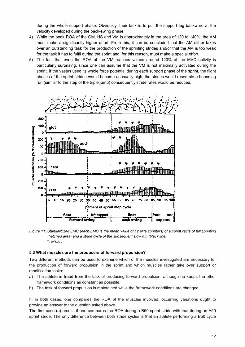
5.3 What muscles are the producers of forward propulsion? Two different methods can be used to examine which of the muscles investigated are necessary for the production of forward propulsion in the sprint and which muscles rather take over support or modification tasks: a) The athlete is freed from the task of producing forward propulsion, although he keeps the other
framework conditions as constant as possible. b) The task of forward propulsion is maintained while the framework conditions are changed. If, in both cases, one compares the ROA of the muscles involved, occurring variations ought to provide an answer to the question asked above. The first case (a) results if one compares the ROA during a B50 sprint stride with that during an A50 sprint stride. The only difference between both stride cycles is that an athlete performing a B50 cycle
13
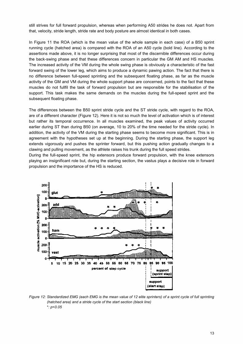
still strives for full forward propulsion, whereas when performing A50 strides he does not. Apart from that, velocity, stride length, stride rate and body posture are almost identical in both cases. In Figure 11 the ROA (which is the mean value of the whole sample in each case) of a B50 sprint running cycle (hatched area) is compared with the ROA of an A50 cycle (bold line). According to the assertions made above, it is no longer surprising that most of the discernible differences occur during the back-swing phase and that these differences concern in particular the GM AM and HS muscles. The increased activity of the VM during the whole swing phase is obviously a characteristic of the fast forward swing of the lower leg, which aims to produce a dynamic pawing action. The fact that there is no difference between full-speed sprinting and the subsequent floating phase, as far as the muscle activity of the GM and VM during the whole support phase are concerned, points to the fact that these muscles do not fullfil the task of forward propulsion but are responsible for the stabilisation of the support. This task makes the same demands on the muscles during the full-speed sprint and the subsequent floating phase. The differences between the B50 sprint stride cycle and the ST stride cycle, with regard to the ROA, are of a different character (Figure 12). Here it is not so much the level of activation which is of interest but rather its temporal occurrence. In all muscles examined, the peak values of activity occurred earlier during ST than during B50 (on average, 10 to 20% of the time needed for the stride cycle). In addition, the activity of the VM during the starting phase seems to become more significant. This is in agreement with the hypotheses set up at the beginning. During the starting phase, the support leg extends vigorously and pushes the sprinter forward, but this pushing action gradually changes to a clawing and pulling movement, as the athlete raises his trunk during the full speed strides. During the full-speed sprint, the hip extensors produce forward propulsion, with the knee extensors playing an insignificant role but, during the starting section, the vastus plays a decisive role in forward propulsion and the importance of the HS is reduced.
Figure 12: Standardized EMG (each EMG is the mean value of 12 elite sprinters) of a sprint cycle of full sprinting
(hatched area) and a stride cycle of the start section (black line) *: p<0.05
14
6 Implications for training The question as to how the findings shown above can be integrated into sprint training in a profitable way will be answered by examining the relevance of these findings to the different methods of improving locomotor speed. In this context, it may be assumed that there are at least seven approaches to improving this ability: • technique training • relaxation training • hypertrophy training • speed endurance training • speed-strength training • variation of loading cycles providing a recovery from the previous stress.
6.1 Technomotor behaviour In the course of evolution, the optimal solution to the task of reaching a maximum locomotor speed on two legs has presumably changed a great deal. In retrospect, one can only speculate upon the flight and hunting movement patterns which were used by the various forms of bipeds and which were particularly successful. The running behaviour of tyrannosaurus rex in Steven Spielberg’s film „Jurassic Park“ can serve as an example in this context. For the time being, the end stage of the motor evolution seems to be the synchronous and two-legged bounding behaviour of the kangaroo, the one-legged alternating running behaviour, without pelvis rotation, of ostriches and the sprinting behaviour of homo Sapiens, with rotation of the pelvis and contralateral arm swing. The latter pattern has perhaps been used for 1 million years. It is, therefore, unlikely that the „ambling“* movements of the ancient Olympic sprinters, as presented on most vases of Greek antiquity, really represent a technique used approx. 2,500 years ago. At any rate, investigations on ‘The Effect of Arm Behaviour on Running Velocity’, conducted by ourselves and as yet unpublished, prove that sprinting with both arms fixed to the trunk results in an increase in the time to run 100m of about 10%. If one arm is kept horizontally extended over a distance of 10m, as in the passing of the baton in a relay race with unavoidable „ambling“ phases, there is a slowing down by 2 to 3%. Even the swinging of straight arms with fixed elbow joints, with a constantly higher moment of inertia than during natural running, greatly changes the running behaviour and the stride pattern in a 35m sprint after a standing start and reduces running velocity significantly by about 3 (sample: male and female sport students and sprinters; N=50). These observations imply that, apart from extreme individual cases, the remaining scope for technomotor improvement is probably very small. Therefore, in the context of sprint co-ordination, there cannot be a completely different solution, but that variant should be looked for which guarantees the highest efficiency. This efficiency is primarily determined functionally and anatomically. As shown in the framework of the present analysis, the ischiocrural muscles are here of decisive importance. The movement pattern corresponding with this is the so-called „running high“ (WASER 1985) with an active „pulling leg or foot touchdown“ (WIEMANN 1986). In 1988, Florence Griffith-Joyner (USA) demonstrated this type of sprint technique in pure form. A special programme devoted to the development of this specific variant of sprinting technique was recently conducted with male and female sport students (N=32). The aim of this programme, which consisted of 20 training sessions and lasted for two months, was the development of the swing-pull with clawing foot action. The training for this led to a significant improvement in speed by about 30ms over a distance of 35m. The improvement of the experimental groups, as compared with the control group, was independent of the emphasis of the training programme. In subgroup A, the training programme included exercises for the stabilisation function of the trunk muscles, especially those used for the stabilisation of the pelvis, as the key element of force transmittance from the legs to the
15
trunk. The members of group B performed only exercises which were aimed at the improvement of sprint co-ordination through the ‘pull-style’ running action. In spite of the relatively small gain in running speed, the ‘swing-pull technique’ did bring about changes in the movement behaviour, as documented by video. In some subjects these changes were very significant. Leaving aside the well known problem of sprinting talent, it may be deduced from this that it is obviously not sufficient to impart ‘ideal’ models of sprinting co-ordination as the desired goal. In adults at least, with a highly automated interaction of the CNS and the skeletal muscles, which has been moulded during the whole of their life, a two month training programme can, at least, lead to an external change of the movement behaviour. However, this does not say anything about the concomitant ‘internal’ stress on the neuron level but an indirect insight is possible, if one considers the following finding: sprinting at full speed, with an arm extended horizontally to the front, over a distance of 10m, following a maximum „normal“ acceleration of 25m from a standing start, leads to an increase of the running time by 120ms over the first 25m (subjects: sport students)! From this it may be concluded that every interference with the natural sprint pattern of movement, which is stored in the subcortex, with muscle-related exactly fixed times, periods and intensities, creates a problem. It is possible that urgently needed relaxation phases are hindered or even eliminated. This shows that it probably takes years of training to replace an existing sprint movement pattern with a new one and to stabilize this new pattern so that no energy is wasted in concentrating on the new movements. Years of target-oriented training with adolescent runners will be needed to decide whether a programme for the improvement of sprint technique, based on running with a clawing foot action, will be effective. Furthermore, it will not be possible for the athletes to master the aimed at technique unless the performance ability of the prime movers producing forward propulsion is increased at the same time (see ch. 5.3). Things might be different when conducting a programme with youth or even children.
6.2 Relaxation training Even the fastest land animals are very rarely injured. The primary cause of this is certainly selective pressure and the fact that quadrupeds have much better balance than bipeds even on rough terrain. However, there are two additional factors to be considered: Firstly, animals presumably are not aware of the execution of fluent and exact cyclic sprint movements and, secondly, they do not perform any strength training. Therefore, there is a decisive contrast to most human sprinters. As already mentioned, even human beings should not become too aware of movement details when sprinting. However, frequent overtension of muscles, together with co-activations of muscles not actually participating in forward propulsion, is typical of all-out sprinting, with the following results: Firstly, a muscle which is activated at the wrong time reduces the contraction velocity of its antagonist and thereby increases its energy consumption. By contracting continuously, the agonist prevents its own microregeneration, which is urgently needed, for two reasons: On the one hand, the intramuscular compression remains so high that the blood flow is stopped. Therefore, there is neither a sufficient supply of nutrients nor a removal of waste products. On the other hand, the α-motor neurons, which activate the motor units of the agonist, are permanently stressed. However, the type II motor neurons, which are responsible for fast contractions, very soon tire (GRIMBY & HANNERZ 1981). Normally, these motor neurons discharge only for a few milliseconds, with intermittent short bursts. A permanent activation causes them to fatigue very quickly, so that they are no longer able to produce the highfrequency discharges necessary to cause the muscle fibres to contract. This results in a considerable reduction of the total impulse of the relevant prime movers. This is all the more true, if the muscle concerned is predominately composed of fast-twitch fibres. Presumably, these considerations led WYSOTSCHIN (1976) to speak of „the art of muscle relaxation during the sprint“ and to develop a corresponding training programme. Considering that sprint races
16
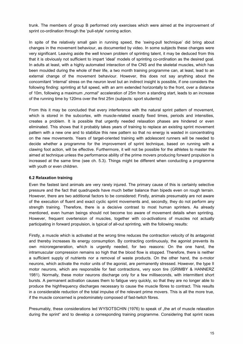
generally require the highest degree of concentration and tension, it is indeed an „art“ to activate only those muscles which are needed for locomotion and then, after an optimal contraction, to relax them as quickly as possible. This fast alternation of tension and relaxation determines the athlete’s stride rate, which, together with stride length, determines the athlete’s running velocity. The ability to relax one’s muscles can be trained. For example, V. Borsov (URS/ Ukraine) used appropriate training for this facility. However, unlike the optimization of intermuscular co-ordination, the capacity for intramuscular relaxation is linked very closely to the existing fibre distribution. Intramuscular relaxation ability, therefore, is mainly a genetically determined indicator of talent. So, ultimately, the dynamics of the reabsorption of calcium ions into the sarcoplasmatic reticulum determines the relaxation time of a previously contracted muscle fibre. This re-uptake capacity is at its highest in type IIb fibres (ALWAY 1992). However, strength training leads to a change in the proportional distribution in a fast or a mixed muscle, in such a way that both cross-section methods (CSM) and neuronal activation methods (NAM) lead to a left transformation (see Figure 13). This means that certain strength training methods unintentionally reduce the relaxation ability of the loaded muscle groups. Consequently, incorrect strength training or full-speed sprinting after or during (macro)cycles of a corresponding training emphasis (especially if cross-section methods are applied) can lead to injuries, which occur most frequently at the back of the thigh. There are two interconnected reasons for this: Firstly, the faulty selection of strength training exercises can result in a muscular imbalance. It is almost a tradition with many coaches and athletes to place the emphasis on strengthening the knee extensors. This leads to a further increase of the natural asymmetry of the contractile force of the thigh muscles, to the detriment of the ischiocrural muscles. Secondly, there is a contractility decrement in the strength-trained muscles. This does not only lead to an increase of contraction time but, more importantly, also to an increase of relaxation time. If the knee extensors need a longer period of relaxation, the possible result will be a brief overloading of the fast but weaker ischiocrural muscles, during the fast alternation between tension and relaxation at the front or, reciprocally, at the back of the thigh.
Figure 13: Fibre spectrum of the skeletal muscles and its changeability through training Of course, this is not to say that faster sprint times cannot be attained through increased strength. The aim is rather to train the sprint-relevant muscles as specifically as possible. Following the findings presented in this article, it is essential to shift the emphasis to the training of the ischiocrural muscles and the muscles which are responsible for lifting the knees. Furthermore, it appears to be especially important to consider carefully the specific effect of strength training methods in the overall training
17
plan. In this regard, the effect on the fibre spectrum is also relevant and will be discussed in more detail in the last chapter of this article. Finally it should be mentioned that so many injuries occur in supramaximal sprint training using ‘towing systems’ (VIITASALO et al. 1982; MERO et al. 1982; BOSCO & VITTORI 1986; MERO & KOMI 1990) because, in such training, the relaxation ability of the sprintrelevant muscles is strained to the limit.
6.3 Hypertrophy training One possibility of increasing the contractile force of a muscle is to induce a hypertrophy of this muscle by loading it mechanically. The increase of the contractile potential up to the exhaustion of its adaptation reserve can be brought about in three ways: • by sustained stretching, • by excluding a synergist, • by a super-threshold contraction series. The first possibility leads to a lengthening of the sarcomer chains. If each sarcomer can produce a certain tension, the result is an increased total contractile force of the respective fibre. Corresponding adaptation mechanisms, after long-term stretching, can be verified in animal experiments (ANTONIO & GONYEA 1993). However, the lengthening of fibres by 20 to 30% (!), which was found in these experiments, would be counterproductive in sprinting. The reason for this is that force is applied primarily during the support phase, when the ischiocrural muscles are in an almost unstretched state. An accentuated stretch would, therefore, according to the length-tension-diagram (HERZOG et al. 1992), shift the optimal degree of actomyosin overlapping from 2.6µm to a more stretched position or more acute angle position in the hip joint; i.e. it would be shifted to the front and thus removed from the support phase. The second possibility, termed „compensatory hypertrophy“, can be observed in weakened synergistic muscles (caused by injury or illness) and can be provoked in animal experiments by the elimination or removal of such a muscle. This leads to the change in the fibre distribution, to the advantage of slower fibre types. This change is especially pronounced in fast muscles (DIFFEE et al. 1993). The third possibility, namely a training induced hypertrophy, is, according to recent findings, also accompanied by a fibre transformation (KLITGAARD et al. 1991; STARON et al. 1991; STARON et al 1994). Thus. very few type IIb fibres can be seen in strength-trained muscles. This leads both to a considerable reduction in contractile velocity and to an increase of relaxation time. However, the former disadvantage is, at least to some extent, counteracted by an increase in contractile strength. Provided that there is a linear increase of tension during the contraction process, the impulse developed in 90ms by a muscle with a contraction time of 180ms (i.e. consisting of type I fibres) will be the same as that of a muscle half as strong but consisting only of type IIb fibres. However, apart from the problem of relaxation, one must also consider the increase of the athlete’s body weight, which is a concomitant of hypertrophy. In this respect even sprinting is a sort of weight-limited event. The EMG results form the basis for the selection of the muscles to be loaded at 70 to 85% of the athlete’s maximum strength, with 10 to 5 repetitions per set and rest intervals of 2 to 3min duration (see above). For the free leg swing and subsequent support-pull movements, which almost overlap each other, the hip extensors need to be strengthened. However, from the point of view of antagonistic action, an effective knee lift is possible, only if the muscles which are activated synergistically for hip flexion are submitted to a superthreshold stress, too. For the time being, it must remain an open question as to whether this action should be emphasised and based on the intraindividual degree of supramaximal EMG activity (as related to the reference value of 100% which has been determined isometrically). It is also conceivable that individual set values of maximum strength might be
18
developed, on the basis of body weight for the hip extensors or leg weight for the hip flexors. Corresponding studies are currently being conducted at the Universities of Wuppertal and Bochum. For the present, one can conclude only that it does not seem to be useful to spend many years maximizing the strength of the sprintspecific prime movers by means of hypertrophy methods.
6.4 Speed endurance training The lactate concentration in the arterial capillary blood of world-class sprinters, just after finishing a 100m race, has been found to have values of between 10 and 13mmol/1. This clearly shows that the creatine phosphate stores, even if filled to the maximum for immediate ATP resynthesis, are not sufficient to provide the energy for approximately 22 sub-ballistic contractions per leg. Considering this, it is hardly surprising that biopsies of the vastus lateralis muscle of sprinters of different levels of performance show the predominance of type IIa fibres. Contrary to the common expectation that there should be a predominance of the fast twitch type lib fibres in the leg muscles of the fastest sprinters, this result implies that, at least in the anti-gravitation muscles, the daily energy balance induces a mixed range of muscle fibres even in the very best sprinters. The fact that the fibre distribution in the vastus of marathoners has been found to be exactly the opposite of that in sprinters (type I fibres = 75%, type II fibres = 25% in endurance athletes) compensates for the `disappointment’ mentioned above. Furthermore, it is still not known whether the ischiocrural muscles, or knee-lifters, which have been identified as performance-determining, show a similar distribution pattern as the vastus group. Corresponding biopsy results for the ischiocrural muscles have not been available but the energetic demands support this point of view. However, a high predominance of type IIb fibres might well be the secret of the success of the top sprinters; i.e. their secret may lie in the possession of natural ability, something they are endowed with genetically and without training. In that case, a partial transformation of the proportion of type IIb fibres to IIa fibres would only be possible through a ‘conditioning training’ that included sprint repetitions and a correspondingly anaerobiclactacid energy provision. However, it is obvious that, even after such a training, the athletes with the highest percentage of IIb fibres would retain the highest percentage of fastest ME. This could suffice to produce high shortlived accelerations of the extremities on an intramuscular synergistic basis or to intervene for a few milliseconds in the contraction processes required for these accelerations,. Furthermore, only athletes with a high „basic repertoire“ of type IIb fibres can possibly achieve a considerable retransformation to the original fibre distribution (to an increased contractility), by interspersing phases of recovery with those containing high training loads. It should now be clear that, for successful 100m sprinting, it is essential to increase the energy flow rate of the anaerobic glycolysis, together with the enzyme concentration needed for this, by means of purposeful training. It is obvious, also, that there is an increased energy demand in the 200m sprint. However, it may not be so easy to understand that the sprint training of long jumpers, triple jumpers and pole vaulters, because of the considerably less energy expenditure in their events, should be accentuated in a different way. In these athletes, who perform approximately 10 muscle actions per leg within a period of maximally 5 seconds, the anaerobic-alactacid ATP resynthesis is predominant. As opposed to other training methods of optimizing sprinting speed, sprint repetitions and the processes of energy provision optimized by this type of training have a special position, with regard to the muscles which must be primarily stressed, because here, through the activation of that part of the nervous system directly concerned with the sprint action, a „natural“ stressing of the ischiocrural muscles is guaranteed.
6.5 Neuronal activation training During the push-off from the starting blocks and the starting strides, it is especially important for the sprinter to produce a maximum impulse in a very short period of time. The „classical extension loop“ (TITTEL 1965) is firstly responsible for this. In order not only to activate the greatest possible proportion of the contractile potential of the hip and knee extension muscles but also to contract these
19
muscles as quickly as possible, high and even maximum innervation rates are necessary. Only thus is it possible to overcome the recruiting threshold, which in fast or mixed muscles is in the area of 80% of the athlete’s maximum strength and to achieve the steepest rates of increase, in terms of force strength curves. It has been found that the best method of increasing maximum strength without an increase of body weight is to use loads of between 90 to 100% of the maximum with explosive efforts. Correspondingly, 1 to 3 repetitions with a complete rest interval of 5min are required. This type of load is typical of the so-called „neuronal activation method“ (NAM). It should be used over a period of approx. three to four weeks - mainly after the use of the hypertrophy method. Athletes should be warned against using NAM loads without appropriate preparation, because there is a high risk of injury. To date there have been no biopsy findings concerning the effect of NAM on the human fibre spectrum. However, the contraction times of 3 to 6sec, during deep squats at 100% of maximum strength, which were measured by HÄKKINEN et al. (1993), are almost identical with those set by CAIOZZO et al. (1992) in animal experiments. When resistances of 90% (concentrically) or 125% (eccentrically) were used, there were highly significant transformations of the fibre spectrum of the trained and fast running muscles after only 4 sets of 8 repetitions per day, with 8sec rest between the repetitions and 15min between the sets. Contraction times of only 2sec per repetition were sufficient, within only four weeks, to lead to a considerable reduction in the proportion of type IIb fibres. If this finding is transferred. with reservations, to human beings, it must be assumed that the use of NAM, especially in sprinters with an originally high percentage of type IIb fibres in the running muscles, firmly establishes the left transformation induced by hypertrophy training, or at least reduces a retransformation, which should be the aim. However, it seems useful to do NAM training just for a strictly limited period of time within the yearly cycle, mainly to remove gradually the strength deficit that can be caused by hypertrophy training. In this regard, the investigation results suggest attention being given in the programme not only to the extension loop but also, and especially, to the most important prime movers of basic speed, i.e. the ischiocrural muscles and kneelifters. As far as the differing function of these muscle groups is concerned, it would appear to be useful to use explosive strength loads, especially for the prime movers responsible for knee-lift - mm. rectus femoris, iliopsoas, tensor fasciae latae and, last but not least, the add. magnus. The action time of these muscles is extremely short as compared to that of the ischiocrural muscles. However - especially after hypertrophy training - the ischiocrural muscles should also be neuronally activated to a maximum degree. 6.6 Speed-strength training In order to create a fluent transition to sprint-specific demands, as for example TABATSCHNIK (1991) did, with his „Speed Chute System“, it seems appropriate, in sprint strength training, to demand explosive efforts, not only against high to maximum resistances but also against medium resistances. Contrary to the orthodox speed strength method (SSM), the „time-controlled speed-strength method“ (TCSSM; TIDOW 1990) controls the duration of the rest intervals between the sets and between the repetitions. The duration of rest intervals between the sets should be 5min, in order to guarantee as complete a regeneration as possible. This rest interval needs to be strictly controlled because even resistances of 30% of maximum strength within a set lead to fatigue-induced velocity decrements. In the case of loads of 50% of the athlete’s maximum strength, there is a reduction in speed of even 15 to 20% during the 5th repetition, provided the athlete works rhythmically. With 10 repetitions, such reductions are progressive and incompatible with the training goal of speed strength. They imply endurance strength loads (TIDOW 1994) and induce left transformations of the fibre spectrum.
6.7 Optimization of the training load: recovery cycles The results of animal experiments, in particular, prove that the often quoted „late transformation“, when examined closely, can be attributed to the timely retransformation of the fibre spectrum
20
previously modified by training. Comparatively great volumes and short rest intervals within a training session or between training sessions cause especially fast muscles to be in an energetic „emergency“. These muscles adapt to such loads by changing their energy provision. The analogy to hypertrophy training cannot be disregarded. There is a reduction of the contractility of the fastest fibres and there is an increase in endurance capability, because of the increased mitochondria) volume and capillarization (RATHER et a1.1991). Only phases of recovery, with significantly reduced amounts of training, make a return to the ‘natural’ fibre spectrum possible. This effect was convincingly shown by STARON et al. (1991) in strength-trained women, by fitting in a break in training of several months duration and by using muscle biopsy. As long as training-induced hypertrophy in the respective muscles can be, at least to some extent, retained over a certain period of time by appropriate measures, the result is a higher speed-strength ability. In this respect it seems essential, with the approach of the season’s highlights, to reduce significantly the amount of strength and conditioning training , to use longer rest intervals and to place the emphasis on quality. This signifies a programme of high intensity. In their investigations, HENNIG & LOMO (1985) traced the natural impulse patterns of the running muscles of freely moving rats and these investigations were continued by EKEN & GUNDERSEN (1988) and recently by AUSONI et al. (1990), by means of electrostimulation of denervated fast muscles. The results of their research indicate that an approximate return to (or retransformation of) the natural spectrum can be achieved only by the use of low resistances, high stimulation frequencies and long rest intervals with a low volume of training. On the one hand, this suggests much more attention being paid to the total load. On the other hand, such findings also imply that there should be a much more pronounced differentiation within the groups of sprinters.
7 Summary When looking for those muscles of the knees and hips which are responsible for the acceleration and horizontal velocity of the body during full-speed sprinting, the gluteus maximus, adductor magnus, hamstrings and the knee extensors have been identified as the most important ones. Electromyographic results concerning the degree of muscle activity (ROA) in 12 elite sprinters show that the hamstrings are active during the whole phase of hip extension (back-swing and support phase) while the gluteus maximus, like the knee extensors, „fire“ only during the backswing phase and the first half of the support phase. However, the adductor magnus has already ceased to be active at the beginning of the support phase. The peak ROA of the muscles examined are clearly above 100% of MVC activity. The adductor magnus shows a conspicuously high peak ROA of 200%. These findings, together with Biomechanical considerations, lead to the suggestion that the hamstrings in particular, together with a muscle rein consisting of gluteus maximus and adductor magnus, supply the energy needed for forward propulsion, by providing a high back-swing velocity of the support leg. In this context the adductor magnus seems to be loaded to an especially high degree. The m. vastus medialis in general and the gluteus maximus, to some extent during the support phase, fulfil only anti-gravitation functions. This leads to corresponding implications for sprint training. * In this context, ambling means moving one’s right (or left) arm forward together with the right (or left) leg, instead of „right arm/left leg“ or „left arm/right leg.“
21
REFERENCES ADAMS, G.R. et al (1993): Skeletal muscle myosin heavy chain composition and resistance training. J. Appl. Physiol. 74 (2): pp. 911-915 AE, M. / ITO, A. / SUZUKI, M. (1992): The men’s 100 meters. New Studies in Athletics 7, 1, pp. 47-52 ANDERSEN, J.L. / KLITGAARD, H. / SALTIN, B. (1994): Myosin heavy chain isoforms in single fibres from m. vastus lateralis of sprinters: influence of exercise. Acta Physiol. Scand.151 (2) pp. 135-142 ANDREWS, J.G. (1985): A general method for determining the functional role of a muscle. Journal of Biomechanical Engineering 107, pp. 348-353 ANDREWS, J.G. (1987): The functional roles of the hamstrings and quadriceps during cycling: Lombards paradox revisited. Journal of Biomechanics 6, pp. 565-575 ANTONIO, J. / GONYEA, W.J. (1993): Progressive stretch overload of skeletal muscle results in hypertrophy befor hyperplasia. J. Appl. Physiol. 74: pp.1893-1898 AUSONI, S. et al (1990): Expression of myosin heavy chain isoforms in stimulated fast and slow rat muscles. In: Journal of Neuroscience 10, pp.153-160 BOBER, T. / MULARCZYK, W. et al (1990): The mechanics of the leg swing in running. Techniques in Athletics, Cologne, 7-9 June 1990 Conference proceedings, Vol. 2, pp. 507-510 BOSCO, C. / VITTORI, C. (1986): Biomechanical characteristics of sprint running during maximal and supra-maximal speed. NSA 1: pp. 39-45 BOSCO, C. / VITTORI, C. (1987): Biomechanische Merkmale des Sprints während maximaler and supramaximaler Geschwindigkeit. Leistungssport 1, pp. 41-43 CAIOZZO V.J. / MA, E. ET AL: A new animal model for modulating myosin isoform expression by altered mechanical activity. Journal of Applied Physiology 73, 4, pp.1432ff CARLSÖÖ, S. / MOLBECH, S. (1966):The functions of certain two-joint muscles in a closed muscular chain. Acta Morphologica Neerlando-Scandinavica 6, pp. 377-386 DIFFEE, G.M. / McCUE, S. / LAROSA, A. / HERRICK, R.E. / BALDWIN, K.M. (1993): Interaction of various mechanical activity models in regulation of myosin heavy chain isoform expression. Journal of Applied Physiology 74, 5, pp. 2517-2523 DIFFEE, G.M. ET AL (1993): Activity-induced regulation of myosin isoform distribution: A comparison of two different contractile activity programs. In: J. Appl. Physiol. 74 (S): pp. 2509-2516 EKEN, T. / GUNDERSEN, K. (1988): Chronic electrical stimulation resembling normal motor unit activity. Effects on denervated fast and slow rat muscles. In: J. Physiol. 402: pp. 651-669 FARRAR, M. / THORLAND, W. (1987): Relationship between isokinetic strength and sprint times in college-age men. Journal of Sports Medicine and Physical Fitness 27, 3, pp. 368-372 FISCHER, K. (1927): Zur geführten Wirkung mehrgelenkiger Muskeln. Zeitschrift fur Anatomie and Entwicklungsgeschichte 83, pp. 7S2ff
22
GREGOR, R.J. / CAVANAGH, P.R. / LAFORTUNE, M.A. (1985): Knee flexor moments during propulsion in cycling - a creative solution to Lombard’s paradox. Journal of Biomechanics 18, 5, pp. 307-316 GRIMBY, L. / HANNERZ, J. (1981): Flexibility of recruitment order of continuously and intermittently discharching motor units in voluntary contraction. In: Desmedt, J.E. (Hrsg.): Progress in Clinical Neurophysiology. Band 9. Basel: pp. 201-211 GRIMBY, L. (1987): Motor unit recruitment during normal locomotion. In: Medicine Sport Science 26: pp.142-151 HÄKKINEN, K. (1993): Neuromuscular fatigue and recovery in male and female athletes during heavy resistance exercise. International Journal of Sports Medicine 14, 2: pp. S3-S9 HANNON, P. R. / RASMUSSEN, S. A. / DEROSA, C. P. (1985): Electromyographic patterns during level and inclined treadmill running and their relationship to step cycle measures. Research Quarterly S6, 4, pp. 334-338 HATHER, B. M. / TESCH, P. A. /BUCHANAN, P. / DUDLEY, G.A. (1991): Influence of eccentric actions on skeletal muscle adaptation to resistance training. Acta Physiologica Scandinavica 143, 2, pp. 177-185 HENNIG, R. / LOMO, T. (1985): Firing patterns of motor units in normal rats. In: Nature 314:164-166 HERZOG, W. et al. (1992): Force-length properties and functional demands of cat gastrocnemius, soleus and plantaris muscles. Journal of Biomechanics 2S, 11: pp. 1329-1336 JÖLLENBECK, T. / HAHN, K. / WIEMANN, K. (1990): Kraft- and Dehnungstraining der ischiocruralen Muskeln zur Verbesserung, der Sprintleistung. Techniques in Athletics, Cologne, 7-9 June 1990 Conference proceedings, Vol. 2, pp. 479-485 KLITGAARD, H. et al (1991): Myosin heavy chain composition of single fibres from m. biceps brachii of male body builders. In: Acta Physiol. Scandinavia 140: pp. 175-180 LEMAIRE, E.D. / ROBERTSON, D.G.E. (1989): Power in sprinting. Track and Field Journal 3S, pp. 1317 MANN, R. (1986): A biomechanical analysis of sprinting. Track Technique 94, pp. 3000-3003 MANN, R.A. / HAGY, J.L. (1980): Running, jogging and walking: A comparative electromyographic and biomechanical study. Bateman, J.E./Trott, A.: The foot and the ankle. New York: Thieme-Stratton, pp. 167-175 McCLAY, LS. / LAKE, M.J. / CAVANAGH, P.R. (1990): Muscle activity in running. Cavanagh, P.R. (Hrsg.): Biomechanics of Distance Running. Chapter 6, pp. 165-186 MERO, A. / KOMI, P. ( 1990): Auswirkungen stimulierter supramaximaler Sprints auf die neuromuskuläre and anaerobe Leistung. Leistungssport 20 (1): pp. 33-35 MERO, A. / KOMI, P.V. (1987): Electromyographic activity in sprinting at speeds ranging from sub-maximal to supra-maximal. Medicine and Science in Sports and Exercise 19, 3, pp. 266-274
23
MERO, A. / KOMI, P.V. (1990): Reaction time and electromyographic activity during a sprint start. European Journal of Applied Physiology and Occupational Physiology 61,1-2, pp. 73ff MERO, A. / LUHTANEN, P. / KOMI, P.V. (1982): Zum Einfluß von Kontaktphasenmerkmalen auf die Schrittfrequenz bei Maximalsprint. Leistungssport 12, 4, pp. 308-313 MOLBECH, S. (1965): On the paradoxical effect of some two-joint muscles. Acta Morphologica Neerlando-Scandinavica 6, pp. 171-178 NILSSON, J. / THORSTENSSON, A. / HALBERTSMA, J. (1985): Changes in leg movements and muscle activity with speed of locomotion and mode of progression in humans. Acta Physiologica Scandinavica 123, 4, pp. 457475 SCHMIDTBLEICHER, D. / DIETZ, V. / NOTH, J. / ANTONI, M. (1978): Auftreten and funktionelle Bedeutung des Muskeldehnungsreflexes bei Lauf- and Sprintbewegungen. Leistungssport 8, 6, pp. 480-490 SIMONSEN, E.B. / THOMSEN, L. / KLAUSEN, K. (1985): Activity of mono- and biarticular leg muscles during sprint running. European Journal of Applied Physiology and Occupational Physiology S4, pp. 524-532 STARON, R.S. et al (1991): Strength and skeletal muscle adaptations in heavyresistance-trained women after detraining and retraining. In: 7. Appl. Physiol. 70 (2): pp. 631-640 STARON, R.S. et al (1994): Skeletal muscle adaptations during early phase of heavy resistance training in men and women. In: J. Appl. Physiol. 76 (3): pp.1247-1255 STARON, R.S. / LEONARDI, M.J. / KARAPONDO, D.L. et al (1991): Strength and skeletal muscle adaptations in heavyresistance-trained women after detraining and retraining. Journal of Applied Physiology 70, 2, pp. 631ff TABACHNIK, B. ( 1991): Neue effektive Mittel für die Entwicklung der Sprintschnelligkeit. Leistungssport 21 (3): pp. 51-54 TIDOW, G. (1990): Aspects of strength training in athletics. In: NSA S (1): pp.93-110 TIDOW, G. (1995): Lösungsansätze zur Optimierung des Schnellkrafttrainings auf der Basis muskelbioptischer Befunde. In: Brack, R./Hohmann, A./Wieland, H. (Hrsg.): Trainingssteuerung - Konzeptionelle and trainingsmethodische Aspekte. Stuttgart: 219-225 VIITASALO, J.T. ET AL (1982): Trainingswirkungen des Schlepptrainings auf die Laufschnelligkeit, die Maximal- and Explosivkraft. Leistungssport 12 (3) pp. 185-189 WASER, J. (1985): Zum Techniktraining beim Laufen. Leistungssport 1, pp. 34-38 WIEMANN, K. (1986): Die Muskelaktivität beim Laufen. Leistungssport 4, pp. 27-31 WINTER, E.M. / BROOKES, F.B.C. (1991): Electromechanical response times and muscle elasticity in men and women. European Journal of Applied Physiology and Occupational Physiology 63, 2, pp. 124ff
24
WOOD, G.A. (1986): Optimal performance criteria and limiting factors in sprint running. New Studies in Athletics 2, pp. SS-63 WYSOTSCHIN, 7. (1976): Die Muskelentspannung von Sprintern. Die Lehre der Leichtathletik 19: pp. 593-596 Acknowledgements This study was supported by the Federal Institute of Sport Science, Cologne, project number: VF 0407/05/15/92.


![Clinical Study Gait Performance and Lower-Limb Muscle ... · knee extensors, and exors [ , ]. Gait asymmetry has also been negatively related to the residual strength of various muscle](https://img.pdfslide.us/doc/110x75/60b6e9cc49138529c25e11aa/clinical-study-gait-performance-and-lower-limb-muscle-knee-extensors-and-exors.jpg)